Embed Size (px)
Citation preview

5/27/2012
1
Evaluation of Invasion in Papillary Lesions
Yunn-Yi Chen, MD, PhDProfessor
“We have struggled with this differential diagnosis (benign from malignant papillary lesions) in our laboratory of surgical pathology for many years, and we still find it to be probably the most difficult diagnostic problem that we face with breast neoplasms……And we check the accuracy of our diagnosis by very long-term follow-up of the patients.”
--Dr. Cushman D. Haagensen, 1986
� Overview of papillary lesions
� Benign alterations in papilloma mimicking invasion
� Invasion arising in papillary DCIS
� What is “invasive papillary carcinoma”?• Intracystic/encapsulated papillary carcinoma• Solid papillary carcinoma
Outline of Talk
� Overview of papillary lesions
� Benign alterations in papilloma mimicking invasion
� Invasion arising in papillary DCIS
� What is “invasive papillary carcinoma”?• Intracystic/encapsulated papillary carcinoma• Solid papillary carcinoma
Outline of Talk

5/27/2012
2
Classification of papillary lesions
� Papilloma� involved by non-atypical proliferative changes� involved by atypical hyperplasia (atypical papillom a)� involved by DCIS (DCIS arising in a papilloma)
� Papillary DCIS
� Intracystic/encapsulated papillary carcinoma
� Solid papillary carcinoma
� Invasive papillary carcinoma
Useful IHC markers for papillary lesions--p63, SMM and CK5/6
Predominantlypapillary
Predominantlysolid
Papilloma vs papillary carcinomaBenign papilloma retains a continuous layer of
ME cells along the fibrovascular cores
P63 stain

5/27/2012
3
Papillary carcinoma lacks or has a paucity of ME cells along the fibrovascular cores
P63 stain
Solid papillary DCIS vs papilloma with florid UDH
Solid papillary DCIS Papilloma with florid hyperplas ia
Solid papillary DCIS vs papilloma with florid UDH
CK5/6 stain� Overview of papillary lesions
� Benign alterations in papilloma mimicking invasion
� Invasion arising in papillary DCIS
� What is “invasive papillary carcinoma”?• Intracystic/encapsulated papillary carcinoma• Solid papillary carcinoma
Outline of Talk

5/27/2012
4
Benign alterations in papillomaPotential diagnostic pitfalls
�Sclerosis
�Infarct / Necrosis Mimic Invas ive Cancer
�Epithelial Displacement
�Squamous metaplasia
Sclerosis in papilloma/sclerosed papilloma
Sclerosis in papillomaPitfall: mimic invasive carcinoma, esp in core bio psy
Sclerosis in papilloma
Myoepithelial markers confirm benign
P63 stain

5/27/2012
5
Sclerosed papillomaSclerosed papilloma--
distorted & angulated tubules in fibrotic stroma
Pitfalls: Mask the papillary process, mimic invasi on
Sclerosed papilloma
p63
CK5/6SMM
Caution: ME markers may be reduced or focally abse nt in sclerosed papillomaInfarct / necrosis in papilloma

5/27/2012
6
Infarct / necrosis in papillomaPitfall: mimic invasive cancer, esp in a core biops y
Pseudo-invasive growth in papilloma
� After infarct� After duct rupture� After biopsy (epithelial displacement)
Settings:
Diagnostic clues favoring benign nature:
� Granulation tissue� Hemosiderin� Cholesterol cleft� Squamous metaplasia� Myoepithelium
Squamous metaplasia in papillomawith pseudo-invasive growth
Pitfall: - Mimic metaplastic carcinoma- ME markers or - in squamous metaplasia
Pseudo-invasive growth in papilloma
� Myoepithelium is not always present
� Caution advised in evaluating epithelium entrappedwithin inflamed granulation tissue
� Caution advised in core biopsiesExcision may be needed to exclude cancer

5/27/2012
7
“In practice, if a papillary tumour in the ductal lumen is judged to be benign, a nubbin of apparent infiltration of the wall can safely be disregarded, and is usually the result of the process described (pseudo-infiltration)”
-- Professor John G. Azzopardi , 1979
� Overview of papillary lesions
� Benign alterations in papilloma mimicking invasion
� Invasion arising in papillary DCIS
� What is “invasive papillary carcinoma”?• Intracystic/encapsulated papillary carcinoma• Solid papillary carcinoma
Outline of Talk
Invasion arising from papillary DCIS
� When papillary intraductal carcinomas invade, they generally assume the pattern of infiltrating ductal carcinoma and lack a paillary architecture
� Papillary DCIS with sclerosis: mimic invasion
� ME marker may be attenuated in papillary DCIS
Avoid overDxof invasion
DCIS involving sclerosed papilloma or sclerosis in papillary DCIS may mimic invasion

5/27/2012
8
Sclerosis in papillary DCIS may mimic invasionME markers rule out invasion
p63
CK5/6SMM
IDC arising in association with papillary DCIS
p63 stain
Note the patchy loss of ME marker in papillary DCIS
� Overview of papillary lesions
� Benign alterations in papilloma mimicking invasion
� Invasion arising in papillary DCIS
� What is “invasive papillary carcinoma”?• Intracystic/encapsulated papillary carcinoma• Solid papillary carcinoma
Outline of Talk Invasive papillary carcinoma (IPC)
� Definition: An invasive carcinoma with predominantly papillary morphology
� What are the pathologic features of IPC?
� Is papillary carcinoma that lacks a peripheral laye r of ME cells an IPC?

5/27/2012
9
� Incidence: 2.2 % (35/1603)
� Pathologic� Circumscribed (2/3 cases)� Intermediate histologic grade� Associated papillary DCIS
� Clinical� Non-Caucasian� Post-menopausal women� Lower incidence of + LN (32%)� Better survival, prognosis similar to pure tubular and
mucinous ca
“ stromal non-intraductalcomponent was self-evident”
“Unique histologic type of invasive mammary cancer with a favorable prognosis”
Pure invasive papillary carcinoma
Examples of invasive papillary carcinoma from NSABP B04
Examples of invasive papillary carcinoma from NSABP B04
? Invasive micropapillary carcinoma

5/27/2012
10
Intracystic papillary carcinoma Intracystic papillary carcinoma
Synonyms:
Definition:
� Encysted papillary carcinoma� Encapsulated papillary carcinoma
� Papillary carcinoma in a large cystic space� Well circumscribed, surrounded by a fibrous wall� Special type of DCIS (WHO 2003)� Histology of papillary DCIS, except:
Lacks peripheral myoepithelium
Intracystic/encapsulated papillary carcinoma
SMM
Intracystic/encapsulated papillary Carcinoma

5/27/2012
11
Is intracystic papillary carcinoma invasive?
Compressed Myoepithelium
Invaded PastMyoepithelium
?In-situ Invasive
Controversial !
Encapsulated PC showing skeletal muscle invasion
Encapsulated PC showing skeletal muscle invasion
(case contributed by Dr. K. Che Prasad)
Encapsulated PC showing vascular invasion

5/27/2012
12
Mammary papillary carcinoma metastatic to lung(h/o DCIS 10 years ago, s/p mastectomy)
(case contributed by Dr. Jeffrey A. McDavit)
AJSP 2011
Intracystic/encapsulated papillary carcinoma: an invasive tumor with circumscribed growth and exc ellent prognosis
Intracystic papillary carcinoma
Synonyms:
Definition:
� Encysted papillary carcinoma� Encapsulated papillary carcinoma
� Papillary carcinoma in a large cystic space� Well circumscribed, surrounded by a fibrous wall� Special type of DCIS (WHO 2003)� Histology of papillary DCIS, except:
Lacks peripheral myoepithelium
Clinical: � 6th-7th decades� Adjacent conventional in situ or invasive ca
may be present�If pure: Behavior mostly similar to DCIS, with
rare cases of nodal metastases, localrecurrence, and skeletal muscle invasion

5/27/2012
13
Encapsulated papillary ca with adjacent IDC, NOS
EPC IDC IDC
EPC
Solid papillary carcinoma
(courtesy of Dr. Dean Fong)
Solid papillary carcinoma
Definition: � Papillary carcinoma with a solid growth pattern� Well-circumscribed, single or multiple nodules� Special histologic and IHC features� Heterogeneous: Myoepithelium may be + or -
Solid papillary carcinoma

5/27/2012
14
Histologic features of solid papillary carcinoma
PlasmacytoidSpindle cellsPseudo-rosette around cores
Histologic features of solid papillary carcinoma
Mucinous production Neuroendocrine differentiation
Chromogranin stain
In situ SPCcalponin
Some solid papillary carcinomashave intact myoepithelial layer
Some solid papillary carcinomaslack peripheral myoepithelium
p63 stain

5/27/2012
15
SPC as one single large nodule
p63 stain
SPC: one single large nodule, no ME layer
SPC with multiple nodules,no ME layer
SMM
� Large tumor size
� Complex, irregular, coalescent papillary nodules/ne sts
� Irregular tumor-stromal interface
� Encircling large blood vessels
� Entrapment of benign ducts and lobules
Features suggestive of invasion in SPC
5/27/2012
16
3.5 cm, well-circumscribed mass
SPC-- large tumor size SPC--complex, irregular, coalescent papillary nests
SPC--irregular tumor-stromal interface
p63 stain
Invasive PC Adjacent papillary DCIS
Tumor-stromal interface in papillary carcinoma
Irregularvs
smooth

5/27/2012
17
SPC encircling large blood vesselsPC showing entrapment of normal ducts and lobules
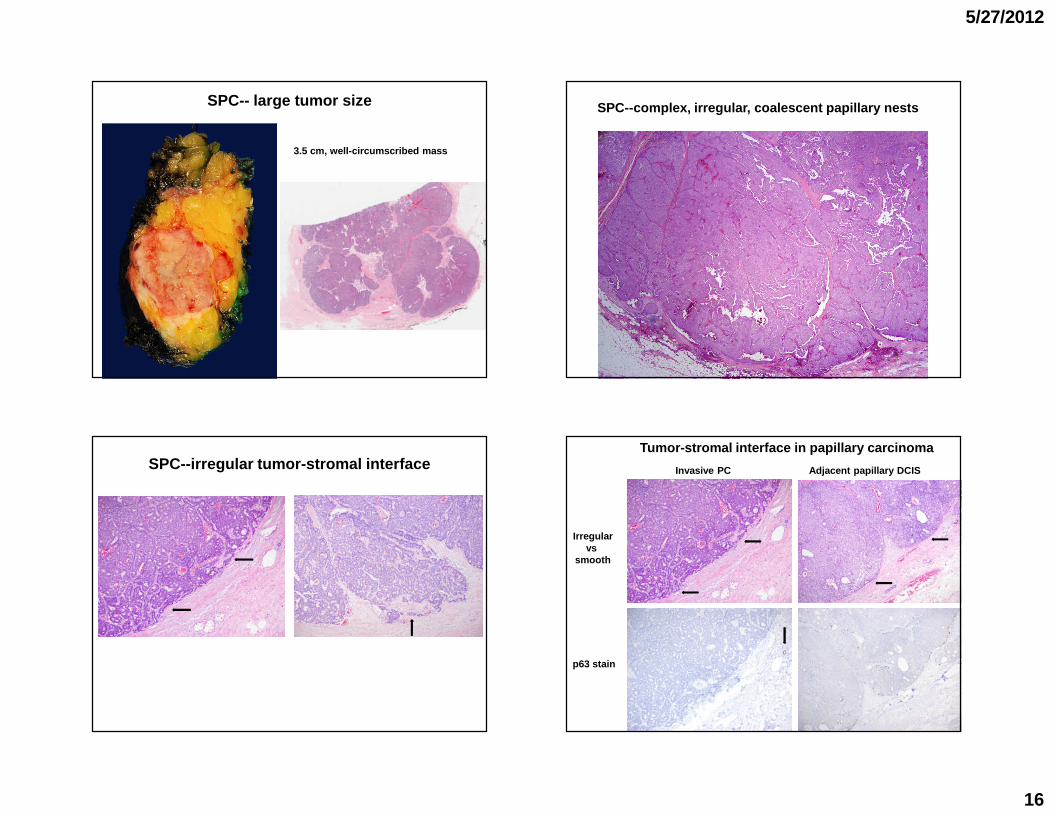
� Large tumor size
� Complex, irregular, coalescent papillary nodules/ne sts
� Irregular tumor-stromal interface
� Encircling large blood vessels
� Entrapment of benign ducts and lobules
� Invading stroma/fat or skeletal muscle
Features suggestive of invasion in SPC SPC showing stromal invasion
5/27/2012
18
� Large tumor size
� Complex, irregular, coalescent papillary nodules/ne sts
� Irregular tumor-stromal interface
� Encircling large blood vessels
� Entrapment of benign ducts and lobules
� Invading stroma/fat and skeletal muscle
� Metastatic disease with same solid papillary growth
Features suggestive of invasion in SPC
SPC in breast
Metastatic ca in LN with SPC pattern
SPC showing LN metastasis
SPC showing LN metastasis
SPC in breast
Metastatic ca in LN with SPC pattern
Solid papillary carcinoma
Definition: � Papillary carcinoma with a solid growth pattern� Well-circumscribed, single or multiple nodules� Special histologic and IHC features� Heterogeneous: Myoepithelium may be + or -
Clinical: � 6th-7th decades� Adjacent conventional invasive ca may be present
often mucinous or neuroendocrine-like�If pure and lack myoepithelium
Indolent behaviorLow rate of LN metastasis

5/27/2012
19
Tumor with fibrovascular cores lined by epithelial cells
IHC markers (p63, SMM, ± CK5/6): evaluate malignancy and invasion
Exclude benign papillary lesions++ ME cells along papillae, + CK5/6
In situ papillary ca- or ME cells along papillae, - CK5/6+ ME cells at periphery of space
- ME cells, - CK5/6
+ ME cells
Encapsulated/circumscribed papillary ca:Circumscribed nodule of PC ina dilated cystic space, with a thick fibrous capsule
Approach for evaluating papillary carcinoma
Tumor with fibrovascular cores lined by epithelial cells
IHC markers (p63, SMM, ± CK5/6): evaluate malignancy and invasion
Exclude benign papillary lesions++ ME cells along papillae, + CK5/6
In situ papillary ca- ME cells along papillae, - CK5/6+ ME cells at periphery of space
- ME cells, - CK5/6
+ ME cells
Circumscribed Solid papillary ca:Circumscribed nodule of PC with a solid papillary growth pattern and a smooth tumor-stromal interface
UCSF approach for evaluating papillary carcinoma
Dx: Encapsulated/circumscribed PC or SPC;see comment.
Comment: The tumor has a well-circumscribed border, but lacks a peripheral myoepithelial layer. These lesions likely represent a very low-grade invasive tumor with an expansile growth pattern and an excellent prognosis. The incidence of local recurrence or nodal metastasis is low and incidence of distant metastasis or cancer-related death is extremely low.
Tumor with fibrovascular cores lined by epithelial cells
IHC markers (p63, SMM, ± CK5/6): evaluate malignancy and invasion
Exclude benign papillary lesions++ ME cells along papillae, + CK5/6
In situ papillary ca- ME cells along papillae, - CK5/6+ ME cells at periphery of space
- ME cells, - CK5/6
+ ME cells
Invasive papillary ca:Carcinoma with predominantly papillary morphology with features of stromal invasion
UCSF approach for evaluating papillary carcinoma

5/27/2012
20
Dx: Invasive PC; see comment.
Comment: Invasive papillary carcinoma is a special type of invasive carcinoma with a favorable prognosis. These tumors are associated with a low risk of LN metastasis and infrequent development of distant recurrence.
Tumor with fibrovascular cores lined by epithelial cells
IHC markers (p63, SMM, ± CK5/6): evaluate malignancy and invasion
Exclude benign papillary lesions++ ME cells along papillae, + CK5/6
In situ papillary ca- ME cells along papillae, - CK5/6+ ME cells at periphery of space
- ME cells, - CK5/6
+ ME cells
Encapsulated/circumscribed papillary ca:Circumscribed nodule of PC ina dilated cystic space, with a thick fibrous capsule
Circumscribed solid papillary ca:Circumscribed nodule of PC with a solid papillary growth pattern and a smooth tumor-stromal interface
Invasive papillary ca:Carcinoma with predominantly papillary morphology with features of stromal invasion
UCSF approach for evaluating papillary carcinoma
�Older women
� ER +, PR +, HER2 –
� Favoralbe prognosis, low rate of LN metastasis
� Less genomic changes than ER & grade-matched IDC
� Genomic profile remarkably similar in the 3 morphologic subtypes of PC
ME-negative papillary ca (EPC, SPC and IPC)
Duprez R et al: J Pathol 2012Eberle C et al: USCAP abstract 2012
When encapsulated/circumscribed PC associated with conventional invasive ca
Dx: 1. IDC, 0.7 cm; see comment2. Encapsulated papillary ca, 2.1 cm3. pT1b
� Tumor type and stage based on nonpapillaryinvasive component
� Report: associated with EPC, size, for clinical and imaging correlation
IDC: 0.7 cm
EPC: 2.1 cm

5/27/2012
21
Pure encapsulated/circumscribed PC without conventional invasive ca
� No consensus in staging
� UCSF: Tx or Tis with a comment
� Avoid overtreatment, but understand potential low rate of recurrence and metastasis
Invasive papillary carcinoma
� Use the size of the lesion for pT
� Comment about special type of invasive carcinoma with a favorable prognosis
� Matter of debate
� Avoid overtreatment
� Overall, manage similar to DCIS
� Adequate local control: appropriate treatment
Management for pure ME-negative papillary ca (I)
Pal SK et al: Breast Cancer Res Treat 2010Rakha E et al: AJSP 2011
� ? SLN sampling � appropriate for those with frankly invasive growth pattern
� ? Hormonal therapy (probably yes)
� ? Radiation therapy
� Chemotherapy not appropriate
Management for pure ME-negative papillary ca (II)
Pal SK et al: Breast Cancer Res Treat 2010Rakha E et al: AJSP 2011

5/27/2012
22
Take home message
� Alterations in benign papilloma may mimic invasion
� ME markers may be in various benign papillary lesi ons and in papillary DCIS
� Avoid overdiagnosis of invasion, especially with a benign papilloma background and in CNB
� ME-negative “intracystic” papillary carcinoma and s olid papillary carcinoma are likely low-grade invasive t umors
� Conservative management for ME-negative papillary carcinoma
Acknowledgement
� UCSF residents/fellows: beautiful gross photos
� Contributing pathologists: challenging cases and wonderful gross photos
Selected references
� Collins LS et al: Papillary lesions of the breast: selected diagnostic and management issues. Histopathology 2008;52:20-29.
� Duprez R et al: Immunophenotypic and genomic characte rization of papillary carcinoma of the breast. J Pathol 2012;226:427-441.
� Koerner F. Papilloma and papillary carcinoma. Semin D iagn Pathol 2010;27:13-30.� Nassar H et al: Clinicopathologic analysis of solid p apillary carcinoma of the breast
and associated invasive carcinomas. Am J Surg Pathol 2006;30:501-507.� Pal SK et al: Papillary carcinoma of the breast: an overview. Breast Cancer Res Treat
2010;122:637-645.� Rakha EA et al: Encapsulated papillary carcinoma of the breast: an invasive tumor
with excellent prognosis. Am J Surg Pathol 2011;35:10 93-1103.� Ueng et al: Papillary neoplasm of the breast. A revi ew. Arch Pathol Lab Med
2009;133:893-907.� Wynveen CA et al: Intracystic papillary carcinoma of the breast. An in situ or invasive
tumor? Results of immunohistochemical analysis and c linical follow-up. Am J SurgPathol 2011;35:1-14.


![Mucinous Neoplasm: A Case Report A Rare Case of Low-grade ... · cell adenocarcinoma, or neuroendocrine carcinoma [3]. Mucinous adenocarcinoma accounts for Mucinous adenocarcinoma](https://img.dokumen.tips/doc/110x75/5d66f73588c993283a8b59a1/mucinous-neoplasm-a-case-report-a-rare-case-of-low-grade-cell-adenocarcinoma.jpg)