Embed Size (px)
Citation preview
Yersinia pestis Targets the Host Endosome Recycling Pathwayduring the Biogenesis of the Yersinia-Containing Vacuole ToAvoid Killing by Macrophages
Michael G. Connor,a* Amanda R. Pulsifer,a Donghoon Chung,a,b Eric C. Rouchka,c Brian K. Ceresa,d
Matthew B. Lawrenza,b
aDepartment of Microbiology & Immunology, University of Louisville School of Medicine, Louisville, Kentucky,USA
bCenter for Predictive Medicine for Biodefense and Emerging Infectious Diseases, University of LouisvilleSchool of Medicine, Louisville, Kentucky, USA
cDepartment of Computer Engineering and Computer Science, University of Louisville, Louisville, Kentucky,USA
dDepartment of Pharmacology & Toxicology, University of Louisville School of Medicine, Louisville, Kentucky,USA
ABSTRACT Yersinia pestis has evolved many strategies to evade the innate immunesystem. One of these strategies is the ability to survive within macrophages. Uponphagocytosis, Y. pestis prevents phagolysosome maturation and establishes a modi-fied compartment termed the Yersinia-containing vacuole (YCV). Y. pestis actively in-hibits the acidification of this compartment, and eventually, the YCV transitions froma tight-fitting vacuole into a spacious replicative vacuole. The mechanisms to gener-ate the YCV have not been defined. However, we hypothesized that YCV biogenesisrequires Y. pestis interactions with specific host factors to subvert normal vesiculartrafficking. In order to identify these factors, we performed a genome-wide RNA in-terference (RNAi) screen to identify host factors required for Y. pestis survival in mac-rophages. This screen revealed that 71 host proteins are required for intracellularsurvival of Y. pestis. Of particular interest was the enrichment for genes involved inendosome recycling. Moreover, we demonstrated that Y. pestis actively recruitsRab4a and Rab11b to the YCV in a type three secretion system-independent man-ner, indicating remodeling of the YCV by Y. pestis to resemble a recycling endo-some. While recruitment of Rab4a was necessary to inhibit YCV acidification and lys-osomal fusion early during infection, Rab11b appeared to contribute to later stagesof YCV biogenesis. We also discovered that Y. pestis disrupts global host endocyticrecycling in macrophages, possibly through sequestration of Rab11b, and this pro-cess is required for bacterial replication. These data provide the first evidence thatY. pestis targets the host endocytic recycling pathway to avoid phagolysosomal mat-uration and generate the YCV.
IMPORTANCE Yersinia pestis can infect and survive within macrophages. However,the mechanisms that the bacterium use to subvert killing by these phagocytes havenot been defined. To provide a better understanding of these mechanisms, we usedan RNAi approach to identify host factors required for intracellular Y. pestis survival.This approach revealed that the host endocytic recycling pathway is essential forY. pestis to avoid clearance by the macrophage. We further demonstrate that Y. pes-tis remodels the phagosome to resemble a recycling endosome, allowing the bacte-rium to avoid the normal phagolysosomal maturation pathway. Moreover, we showthat infection with Y. pestis disrupts normal recycling in the macrophage and thatdisruption is required for bacterial replication. These findings provide the first evi-
Received 28 September 2017 Accepted 17January 2018 Published 20 February 2018
Citation Connor MG, Pulsifer AR, Chung D,Rouchka EC, Ceresa BK, Lawrenz MB. 2018.Yersinia pestis targets the host endosomerecycling pathway during the biogenesis of theYersinia-containing vacuole to avoid killing bymacrophages. mBio 9:e01800-17. https://doi.org/10.1128/mBio.01800-17.
Editor Michele S. Swanson, University ofMichigan—Ann Arbor
Copyright © 2018 Connor et al. This is anopen-access article distributed under the termsof the Creative Commons Attribution 4.0International license.
Address correspondence to Matthew B.Lawrenz, [email protected].
* Present address: Michael G. Connor, InstitutePasteur, Paris, France.
RESEARCH ARTICLE
crossm
January/February 2018 Volume 9 Issue 1 e01800-17 ® mbio.asm.org 1
on March 3, 2019 by guest
http://mbio.asm
.org/D
ownloaded from

dence that Y. pestis targets the host endocytic recycling pathway in order to evadekilling by macrophages.
KEYWORDS intracellular survival, plague, Rab GTPases, Yersinia pestis, endosomerecycling
Yersinia pestis is a facultative intracellular pathogen that causes the human diseaseknown as plague (1, 2). There are three forms of human plague: bubonic, pneu-
monic, and septicemic. Each form of plague results in an acute infection that isdelineated by the tissues primarily colonized by Y. pestis. Bubonic plague is the mostcommon form and arises after a bite from a Y. pestis-infected flea. The bacteria rapidlydisseminate from the inoculation site through the lymphatic system and colonize thedraining lymph node (1, 3, 4). Eventually, the bacteria enter and replicate in thebloodstream, leading to septicemic plague (1, 5). In rare cases, Y. pestis can be directlyinoculated into blood by a flea or from the bite of an infected animal, resulting inprimary septicemic plague without colonization of the lymphatic system (1). From theblood, Y. pestis is distributed throughout the body and colonizes other tissues, such asthe spleen, liver, and lungs. Colonization of the lungs leads to the development ofsecondary pneumonic plague and the potential for patients to aerosolize Y. pestis bycoughing, and potential person-to-person transmission. Inhalation of infected aerosolsby naive individuals can result in colonization of the lungs by Y. pestis and thedevelopment of primary pneumonic plague. All three forms of plague are very rapidinfections with high mortality rates in the absence of early antibiotic treatment (1, 6).Furthermore, the ability for aerosol transmission of Y. pestis raises the potential for thisbacterium to be used as a biological weapon (7).
Y. pestis is maintained through a zoonotic transmission cycle between rodents andfleas (1, 2). The ability of Y. pestis to exist in both the mammalian and flea hosts is aresult of acquiring virulence factors required for the mammalian host and transmissionfactors required for flea colonization (1, 8–10). The bacterium regulates these factors toensure expression of appropriate factors only when required (1, 8–12). Importantly,antiphagocytic factors expressed during mammalian infection, like the Ysc type threesecretion system (T3SS), secreted Yop effectors, and the Caf1 capsule, are not requiredfor flea infection, and thus are repressed in the flea vector (13, 14). Therefore, there isa transition period when Y. pestis is highly susceptible to recognition and phagocytosisby macrophages and neutrophils immediately upon flea transmission (15–17). Thissusceptibility was highlighted by intravital microscopy of the infection site by Shannonet al. (17). Following transmission of Y. pestis via the flea to the dermis of the ear,polymorphonuclear leukocytes (PMNs) are rapidly recruited to the infection site andappear to phagocytose the bacteria. To a lesser degree, host macrophages are alsorecruited and engulf bacteria, and infected macrophages appeared to migrate awayfrom the infection site. Growing evidence suggests that these two phagocytes havevery different abilities to kill Y. pestis (15, 17–22). Specifically, neutrophils appear to bemore efficient at killing phagocytosed Y. pestis than macrophages (20–22). Moreover,several studies suggest that Y. pestis actively inhibits killing by both mouse and humanmacrophages (16, 19, 23–28).
The importance of intracellular Y. pestis survival in virulence has been highlighted byseveral studies. Ye et al. showed that animals depleted for macrophage/dendritic cellpopulations exhibited delayed Y. pestis dissemination and subsequently lower bacterialburdens during plague infection (29). Moreover, a phoPQ mutant, which is defective forsurvival within macrophages (23, 24, 30, 31), has a 75-fold attenuation in subcutaneousinfection of BALB/c mice and a significant delay in the development of lethal disease inSwiss Webster mice (30, 31). While PhoPQ regulates many genes in Y. pestis, and thusmay have pleiotropic effects during infection, many of the genes regulated by thisoperon have been shown to be important for stress response and intracellular survival,indicating that deficiency in intracellular survival of the phoPQ mutant contributes tothe attenuated phenotypes (23). In contrast, Y. pestis is defective in intracellular survival
Connor et al. ®
January/February 2018 Volume 9 Issue 1 e01800-17 mbio.asm.org 2
on March 3, 2019 by guest
http://mbio.asm
.org/D
ownloaded from

in macrophages from canines and Mus spretus SEG mice, species that are relativelyresistant to plague, compared to common laboratory murine macrophages, indicatingthat the ability of macrophages to kill Y. pestis may contribute to susceptibility toplague (32–34). These data, combined with studies showing the sensitivity of Y. pestisto PMN killing (20–22), suggest that Y. pestis infection of macrophages may provide anintracellular niche to avoid killing by PMNs during early stages of bubonic plague.
Upon phagocytosis by macrophages, Y. pestis actively inhibits phagosome-mediatedkilling (16, 23, 25–28). A hallmark of this process is the inhibition of phagosomeacidification by Y. pestis (27). The bacterium remains within this phagosome throughoutthe course of the intracellular infection, eventually remodeling it into a compartmentcalled the Yersinia-containing vacuole (YCV). In addition to maintaining a neutral pH, asubset of the YCVs eventually mature into autophagosome-like compartments, acquir-ing both LC3-II and double membranes (27). During late infection, the YCV expandsfrom a tight-fitting vacuole to a spacious vacuole, coinciding with bacterial replication(23, 27, 33). Recently, we identified the first host factor required by Y. pestis for the YCVbiogenesis process (35). The host GTPase Rab1b, which normally mediates endoplasmicreticulum (ER)-Golgi trafficking (36, 37), is rapidly recruited to the YCV and is requiredfor Y. pestis to inhibit vacuole acidification and phagosome maturation. These datademonstrate that Y. pestis manipulates host factors to subvert phagosomal maturationand to generate a protective replicative niche within the macrophage. Here we describea genome-wide, RNA interference (RNAi)-based high-throughput screen to identifyadditional host factors required for intracellular survival of Y. pestis. Network analysis ofthese genes revealed enrichment for host factors involved in the endocytic recyclingpathway. We further show that Y. pestis actively recruits Rab4 and Rab11 to remodel theYCV to resemble host recycling endosomes in order to avoid phagolysosomal matura-tion in a T3SS-independent manner. Moreover, we demonstrate that Y. pestis infectionalso disrupts global host cell recycling, likely through sequestration of Rab11, and thatdisruption in recycling is important for intracellular replication.
RESULTSY. pestis requires host cell signal transduction, transport, and localization
pathways to survive in macrophages. RNAi has been used to identify host factorsrequired for intracellular survival of several pathogens (38–49). As macrophages arespecifically infected during Y. pestis infection, our first goal was to select a macrophagecell line that was amendable to Lipofectamine-mediated transfection and RNAi neces-sary for high-throughput screening. Toward this end, we tested small interfering RNA(siRNA) transfection and knockdown in several human and mouse macrophage celllines. While robust RNAi was observed in mouse macrophages, we were unable toreproducibly knock down gene expression in human cell lines (data not shown). On thebasis of these results, we chose RAW264.7 mouse macrophages for further optimiza-tion. Using a combination of siRNAs targeting genes of variable expression levels, weoptimized Lipofectamine/siRNA concentrations and transfection time to consistentlyachieve �70% knockdown of targets (Fig. 1A and B). Next, we infected cells transfectedwith Rab2 siRNA and Cop�1 siRNA (both siRNAs are known to inhibit intracellularsurvival of other pathogens [41, 42, 48]) with Y. pestis CO92 pCD1(-) LuxPtolC, a biolu-minescent bioreporter that can differentiate as little as a twofold difference in intra-cellular bacteria (Fig. 1C and D; R2 � 0.89) (50), to demonstrate that this bioreporter canbe used to kinetically monitor changes in intracellular survival (Fig. 1E). Finally, usingCop�1 siRNA as a positive control, we calculated Z= factor values for this assay of 0.61and 0.83 at 2 and 10 h postinfection, respectively (Fig. 1F). Together, these data indicatea highly reproducible assay amenable to high-throughput screening (Fig. 1G).
Using this RNAi assay, 17,370 host genes were screened using a pooled siRNAapproach (three siRNAs for each target were pooled into one well), and we monitoredchanges in intracellular survival of Y. pestis. Each plate also contained control wellstransfected with scrambled or COP�1 siRNAs. Bioluminescence was measured at 2 and10 h postinfection, and Z= factors were calculated from the control wells (the average
Y. pestis Targeting of the Host Recycling Pathway ®
January/February 2018 Volume 9 Issue 1 e01800-17 mbio.asm.org 3
on March 3, 2019 by guest
http://mbio.asm
.org/D
ownloaded from

FIG 1 RNAi-based assay to identify host factors required for intracellular survival of Y. pestis. Todetermine whether reproducible RNAi could be achieved in RAW264.7 macrophages, cells were reversetransfected with siRNAs targeting indicated genes. (A and B) Forty-eight hours posttransfection, cells (n �3) were harvested for RNA isolation and qRT-PCR (data represent the level of gene expression comparedto the level for the scrambled siRNA control) (A) or protein isolation for Western blot analysis (�-actin wasused as a loading control) (B). �-GAPDH, anti-GAPDH antibody; �-�actin, anti-�actin antibody. Todemonstrate that the Y. pestis CO92 pCD1(-) LuxPtolC bioreporter accurately represents intracellularbacterial numbers, RAW264.7 macrophages were infected with Y. pestis CO92 pCD1(-) LuxPtolC at theindicated MOIs (n � 12), and extracellular bacteria were killed with gentamicin. (C) Bioluminescence (inrelative light units [RLU]) of intracellular bacteria was determined at 1, 4, 8, and 18 h postinfection. (D)At 18 h, cells from each MOI (n � 3) were lysed, and bacterial numbers (CFU) were determined andcompared to 18-h bioluminescence (in RLU). (E) To demonstrate that RNAi targeting specific genes couldimpact the intracellular survival of Y. pestis, RAW264.7 macrophages were transfected with siRNAstargeting Rab2A or COP�1. Forty-eight hours posttransfection, macrophages were infected with Y. pestisCO92 pCD1(-) LuxPtolC (MOI of 10). Extracellular bacteria were killed with gentamicin, and intracellularbacterial bioluminescence was monitored over time. Data are represented as percent RLU of scrambled (Scr)siRNA control. (F) To demonstrate the robustness of the assay, RAW264.7 macrophages (n � 48) were reversetransfected with either scrambled siRNA (negative control) or siRNA targeting Cop�1 (positive control).Forty-eight hours posttransfection, macrophages were infected with Y. pestis CO92 pCD1(-) LuxPtolC (MOI of10). Extracellular bacteria were killed with gentamicin, and intracellular bacterial bioluminescence wasdetermined at 2 and 10 h postinfection. The Z’ factors from four independent experiments are shown (thebars are means). (G) Overview of optimized high-throughput assay for RNAi screening.
Connor et al. ®
January/February 2018 Volume 9 Issue 1 e01800-17 mbio.asm.org 4
on March 3, 2019 by guest
http://mbio.asm
.org/D
ownloaded from
Z= factors for the screen at 2 and 10 h were 0.57 and 0.66, respectively). Biolumines-cence was normalized for each plate based on control wells, and changes in intracel-lular survival of Y. pestis were ranked by normalized scores (Fig. 2A; see also Data Set S1in the supplemental material). A total of 325 genes that inhibited bacterial growth and39 genes that promoted bacterial growth were selected for secondary validation. Forthe secondary screen, a single siRNA (siRNA “A”) was used to validate the primaryscreen results. Furthermore, the primary hits were validated against two differentY. pestis strains, one with the Ysc type three secretion system (T3SS) (KIMD19 pCD1(�)
LuxPtolC) and one without it (CO92 pCD1(-) LuxPtolC). A direct correlation was observedbetween the two strains (Fig. 2B; rs � 0.87), supporting previous studies showing thatthe T3SS is dispensable for intracellular survival (16, 24, 35, 50). From the primary hits,135 genes showed �40% inhibition of intracellular survival of Y. pestis and 7 showeda hypervirulent phenotype with �20% more growth than scrambled controls. These142 genes were further screened using a second siRNA (siRNA “B”). Of the 142 genes,RNAi of 71 of these genes continued to show inhibition of intracellular Y. pestis survival,while one retained a hypervirulent phenotype (�10% more growth than scrambledcontrols; Data Set S1 and Table S1).
Gene Ontology (GO) clustering and network analyses of the 71 validated inhibitionhits using all GO evidence codes, a minimum kappa score of 0.4, and a P value thresholdof 0.05 revealed substantial clustering within the validated data set (Fig. 2D). Of the fiveenriched groups, the largest cluster was under vesicle-mediated transport (Fig. 2D andE; P � 0.001). Under the parent GO term clusters, detailed GO terms significantlyfocused on host trafficking networks, with transport and localization as commonthemes. Additional enrichment included small-GTPase-mediated signal transductionand regulation of response to stress (Fig. 2D and E). Within the small GTPase signaltrafficking, six Rab GTPases appeared to be required for intracellular survival of Y. pestis,and two trafficking pathways were highlighted: (i) endocytic recycling (Rab4a andRab20) and (ii) retrograde trafficking (Rab1b and Rab2b) (51–56). Together, these dataindicate targeting of specific host Rab-mediated signaling pathways by Y. pestis duringinfection of macrophages.
Host cell recycling is essential for Y. pestis survival. Rab4a, Rab11b, and Myo5b
are well-characterized contributors to cell recycling (53). While Rab4a was a validatedgene in our screen and interconnected to the largest enriched ontology (vesicle-mediated trafficking), the other two genes did not pass the primary screen criteria. RNAiof Rab11b inhibited Y. pestis survival by only 50% in the primary screen, and Myo5B wascytotoxic (upon subsequent analysis, only one of the three Myo5B siRNAs used in theprimary screen was cytotoxic; this siRNA was not included in further studies). However,because of the importance of these proteins in the recycling pathway, we chose toindependently verify the contributions of Rab4a, Rab11b, and Myo5b to the intracel-lular survival of Y. pestis (Fig. 3 and Fig. S1). RNAi resulted in �50% knockdown of eachgene target (Fig. 3A) with no significant loss in cell viability (Fig. 3B). Subsequentinfection confirmed that knockdown of all three genes impacted the ability of Y. pestisto survive within the macrophage. Knockdown of Rab4a had the largest impact,inhibiting Y. pestis survival by �40% at 2 h (Fig. 3C; P � 0.001) and �80% at 10 h(Fig. 3D; P � 0.001). Interestingly, knockdown of Rab11b and Myo5B had no significantimpact on Y. pestis survival at 2 h (Fig. 3C) but attenuated Y. pestis by �40% at 10 h(Fig. 3D; P � 0.001). Bioluminescence data were confirmed at 10 h by conventionalbacterial enumeration (Fig. 3E). Furthermore, while RNAi of Rab11b had a minor impacton intracellular survival between 2 h and 10 h postinfection, RNAi of Rab4a had agreater impact on the ability of bacteria to survive intracellularly between these twotime points (Fig. 3F). Importantly, knockdown of Rab4a or Rab11b did not alter theexpression of Rab GTPases involved with phagolysosome maturation or impact phago-cytosis of Y. pestis (Fig. S2). Together, these data indicate that Y. pestis requires the hostcell recycling pathway to avoid killing by macrophages.
Y. pestis Targeting of the Host Recycling Pathway ®
January/February 2018 Volume 9 Issue 1 e01800-17 mbio.asm.org 5
on March 3, 2019 by guest
http://mbio.asm
.org/D
ownloaded from

FIG 2 Identification of host factors required for intracellular survival of Y. pestis. RAW264.7 macrophages were reverse transfected with siRNAs for 48 h. (A)Transfected cells were infected with Y. pestis CO92 pCD1(-) LuxPtolC (MOI of 10), and intracellular bacterial bioluminescence (in RLU) was determined at 2 and10 h postinfection. RLU values were normalized to the values for the controls and ranked from lowest to highest. Normalized scores of �0.4 are indicated bylight blue shading, and normalized scores of �1.4 are indicated by yellow shading. (Inset) Average Z factor (Z=) � standard deviation (SD) for all 205 screenedplates. (B and C) For secondary validation, cells transfected with siRNA “A” (B) or siRNA “B” (C) were infected with Y. pestis CO92 pCD1(-) LuxPtolC or KIMD19pCD1(�) LuxPtolC (MOI of 10), and intracellular bacterial bioluminescence (in RLU) was determined at 10 h postinfection. RLU values for each strain werenormalized to the values for the controls and compared. Normalized scores of �0.6 are indicated by blue shading. (D) Cytoscape-generated layout for GeneOntology (GO) term node clusters, with significant genes per cluster highlighted in red. Clusters are color coded by parent ontology, and subgroup ontologyis labeled in black. Lines represent interconnections between detailed terms. Node size denotes significance. (E) Pie chart representing the percent parentontology represented as a whole within validated genes. pV, P value.
Connor et al. ®
January/February 2018 Volume 9 Issue 1 e01800-17 mbio.asm.org 6
on March 3, 2019 by guest
http://mbio.asm
.org/D
ownloaded from

Y. pestis requires Rab4a to avoid YCV acidification and lysosomal fusion. Wepreviously showed that RNAi of Rab1b significantly reduces intracellular survival ofY. pestis at 2 h postinfection, which directly correlated with an increase in the frequencyof YCV acidification (35). Therefore, we next determined the impact of Rab4a, Rab11b,and Myo5b RNAi on YCV acidification using Lysotracker (Fig. 4A to C). As previouslyshown (27, 35), Y. pestis actively inhibited YCV acidification, with only ~20% of YCVscontaining live Y. pestis colocalizing with Lysotracker by 80 min postinfection. Incontrast, YCVs containing paraformaldehyde (PFA)-killed Y. pestis rapidly acidified, with�75% colocalization by 20 min postinfection. As predicted by our relative light unit(RLU) data, Rab4a knockdown resulted in a significant increase in the frequency of YCVacidification (P � 0.001), approaching levels similar to PFA-killed bacteria. Unlike Rab4a,RNAi of Rab11b and Myo5b resulted in only a slight increase in colocalization, and by80 min, colocalization remained significantly lower than both Rab4a siRNA-treated cellsor cells infected with PFA-killed Y. pestis.
Phagosome acidification is directly linked to lysosomal fusion (57), so we next askedwhether Rab4a is required for Y. pestis to avoid YCV fusion with lysosomal compart-
FIG 3 Rab4a, Rab11b, and Myo5b are required for intracellular survival of Y. pestis. RAW 264.7 macrophageswere transfected with siRNAs targeting Rab4a, Rab11b, or Myo5B. (A and B) Forty-eight hours aftertransfection, RNA samples were harvested for qRT-PCR (n � 9) (represented as relative expression ofscrambled-siRNA-treated cells) (A) or cell viability was determined (n � 5) (B). (C and D) To determine theimpact of RNAi on Y. pestis survival, transfected RAW264.7 macrophages (n � 6) were infected withY. pestis CO92 pCD1(-) LuxPtolC (MOI of 10), and intracellular bacterial numbers were determined bybioluminescence (in RLU) at 2 h (C) or 10 h (D) postinfection and compared to the values for scrambled(Scr) controls. (E) At 10 h postinfection, a subset of samples (n � 3) were harvested for conventionalbacterial enumeration. (F) Percent of intracellular bioluminescence at 10 h postinfection compared to 2 hpostinfection. Values that are significantly different by one-way ANOVA with Tukey’s posthoc test areindicated by asterisks as follows: **, P � 0.01; ***, P � 0.001. Values that are not significantly different (ns)are indicated.
Y. pestis Targeting of the Host Recycling Pathway ®
January/February 2018 Volume 9 Issue 1 e01800-17 mbio.asm.org 7
on March 3, 2019 by guest
http://mbio.asm
.org/D
ownloaded from
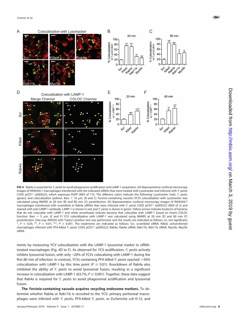
ments by measuring YCV colocalization with the LAMP-1 lysosomal marker in siRNA-treated macrophages (Fig. 4D to F). As observed for YCV acidification, Y. pestis activelyinhibits lysosomal fusion, with only ~20% of YCVs colocalizing with LAMP-1 during thefirst 80 min of infection. In contrast, YCVs containing PFA-killed Y. pestis reached �50%colocalization with LAMP-1 by this time point (P � 0.01). Knockdown of Rab4a alsoinhibited the ability of Y. pestis to avoid lysosomal fusion, resulting in a significantincrease in colocalization with LAMP-1 (63.7%; P � 0.001). Together, these data suggestthat Rab4a is required for Y. pestis to avoid phagosomal acidification and lysosomalfusion.
The Yersinia-containing vacuole acquires recycling endosome markers. To de-termine whether Rab4a or Rab11b is recruited to the YCV, primary peritoneal macro-phages were infected with Y. pestis, PFA-killed Y. pestis, or Escherichia coli K-12, and
FIG 4 Rab4a is essential for Y. pestis to avoid phagosome acidification and LAMP-1 acquisition. (A) Representative confocal microscopyimages of RAW264.7 macrophages transfected with the indicated siRNAs that were treated with Lysotracker and infected with Y. pestisCO92 pCD1(-) pGEN222, which expresses EGFP (MOI of 7.5). The different colors indicate the following: Lysotracker (red), Y. pestis(green), and colocalization (yellow). Bars � 10 �m. (B and C) Yersinia-containing vacuole (YCV) colocalization with Lysotracker wascalculated using IMARIS at 20 min (B) and 80 min (C) postinfection. (D) Representative confocal microscopy images of RAW264.7macrophages transfected with scrambled or Rab4a siRNAs that were infected with Y. pestis CO92 pCD1(-) pGEN222 (MOI of 3) andstained with anti-LAMP-1 antibody. LAMP-1 is shown in red, and Y. pestis is shown in green. Yellow arrows indicate locations of bacteriathat do not colocalize with LAMP-1 and white arrowheads indicate bacteria that colocalize with LAMP-1 based on Imaris COLOCfunction. Bars � 5 �m. (E and F) YCV colocalization with LAMP-1 was calculated using IMARIS at 20 min (E) and 80 min (F)postinfection. One-way ANOVA with Tukey’s posthoc test was performed, and the results are indicated as follows: ns, not significant;*, P � 0.05; **, P � 0.01; ***, P � 0.001. The treatments are indicated as follows: Scr, scrambled siRNA; Killed, untransfectedmacrophages infected with PFA-killed Y. pestis CO92 pCD1(-) pGEN222; Rab4a, Rab4a siRNA; Rab11b, Rab11b siRNA; Myo5b, Myo5bsiRNA.
Connor et al. ®
January/February 2018 Volume 9 Issue 1 e01800-17 mbio.asm.org 8
on March 3, 2019 by guest
http://mbio.asm
.org/D
ownloaded from

recruitment of endogenous Rab4a and Rab11b to the YCV was determined by immu-nofluorescence using anti-Rab antibodies (Fig. 5A to F). At 20 min postinfection, asignificantly higher number of YCVs colocalized with Rab4a and Rab11b than vacuolescontaining E. coli (Fig. 5C and E; P � 0.05 and P � 0.01, respectively). By 80 minpostinfection, YCVs retained significantly higher colocalization with Rab11b than E. coli-containing vacuoles (P � 0.01). However, while YCVs trended toward higher colocal-ization with Rab4A compared to vacuoles containing E. coli or killed Y. pestis, thesedifferences were not statistically different. To confirm these results, RAW264.7 macro-phages were transfected with plasmids expressing Rab4a or Rab11b fused to enhancedgreen fluorescent protein (EGFP) to monitor the localization of Rab proteins indepen-dently of antibodies (58) and 24 h later infected with bacteria. At 20 min postinfection,~50% of PFA-killed Y. pestis and E. coli K-12 containing phagosomes colocalized withRab4a-EGFP (Fig. 5D). However, as observed in primary macrophages, a significantlyhigher number of vacuoles containing live Y. pestis colocalized with Rab4a-EGFP (76%;P � 0.01). By 80 min postinfection, both PFA-killed Y. pestis and E. coli K-12 decreasedin colocalization with Rab4a-EGFP, indicating loss of the GTPase during phagosomematuration, but vacuoles containing live Y. pestis maintained Rab4a-EGFP at a statisti-cally higher frequency (~61%; P � 0.01). As the infection continued, colocalization ofthe YCV with Rab4a-EGFP decreased, and by 2 and 20 h postinfection, it approachedbackground levels (Fig. 5G). In contrast to Rab4a-EGFP, fewer vacuoles containingPFA-killed Y. pestis or E. coli K-12 colocalized with Rab11b-EGFP at 20 and 80 minpostinfection (Fig. 5F). However, the majority of YCVs containing live Y. pestis colocal-ized with Rab11b-EGFP, approaching 85% by 80 min postinfection (P � 0.001). Fur-thermore, Y. pestis continued to colocalize with Rab11b-EGFP at a high frequencythroughout the course of the infection (Fig. 5H).
Finally, to determine whether Rab proteins are specifically recruited to phagosomescontaining Y. pestis and not to all phagosomes of Y. pestis-infected macrophages,RAW264.7 macrophages transfected with Rab4a-EGFP or Rab11b-EGFP were coinfectedwith Y. pestis and E. coli K-12. Colocalization between Rab4a-EGFP and bacterium-containing vacuoles at 20 min postinfection and between Rab11b-EGFP and bacterium-containing vacuoles at 2 h postinfection was determined by confocal microscopy(Fig. 5I to L). In cells infected with both bacteria, Y. pestis-containing vacuoles had ahigher frequency of colocalization with both Rab proteins than E. coli K-12-containingvacuoles. Furthermore, the frequency of E. coli K-12 colocalization was not higherduring coinfection than observed during single infection with just E. coli K-12 (Fig. 5Dand F). Together, these data indicate that Y. pestis actively recruits Rab4a and Rab11bto the YCV during early stages of macrophage infection and that recruitment isspecifically to vacuoles containing live Y. pestis.
Y. pestis infection disrupts host recycling. Recruitment of Rab4a and Rab11b tothe YCV indicated that Y. pestis remodels its phagosome to resemble a recyclingendosome. Because of these links to host cell recycling, we next tested whetherinfection with Y. pestis impacts host cell recycling by monitoring recycling of the hosttransferrin receptor (TfR). RAW264.7 macrophages were infected with Y. pestis CO92,PFA-killed Y. pestis, or E. coli K-12. At 2 and 24 h postinfection, intracellular TfRs weredifferentially labeled from extracellular TfRs, and intracellular TfR intensity was deter-mined by microscopy (Fig. 6A and B). Unlike PFA-killed bacteria, infection with liveY. pestis significantly disrupted recycling of TfR as early as 2 h postinfection, resultingin accumulation of intracellular TfR, and continued to impact recycling for 24 h (Fig. 6Cto F). Furthermore, intracellular TfR intensity also increased as the Y. pestis multiplicityof infection (MOI) increased. Inhibition of TfR recycling was also specific for macro-phages containing intracellular Y. pestis, as uninfected cells from the same cultures didnot show elevated retention of TfR (Fig. S5). Importantly, infection with 10-fold-highernumbers of E. coli K-12 did not result in a significant change in TfR retention comparedto uninfected macrophages (Fig. 6G and H), indicating that recycling disruption is nota general response to macrophage activation.
Y. pestis Targeting of the Host Recycling Pathway ®
January/February 2018 Volume 9 Issue 1 e01800-17 mbio.asm.org 9
on March 3, 2019 by guest
http://mbio.asm
.org/D
ownloaded from

FIG 5 Y. pestis recruits Rab4a and Rab11b to the YCV. (A and B) Representative confocal microscopy images of primary peritonealmacrophages infected with Y. pestis CO92 pCD1(-) pGEN::mCherry (Live) (MOI of 3), PFA-killed Y. pestis CO92 pCD1(-) pGen::mCherry(Killed) (MOI of 3), or E. coli K-12 pGEN::mCherry (E. coli) (MOI of 20) and labeled with anti-Rab4a (A) or anti-Rab11b (B) antibodies.Merged (bacteria [red] and Rab protein [green]) and YCV-Rab colocalization fields generated by Imaris (COLOC) are shown. Bars �5 �m. (C) Frequency of colocalization of bacterium-containing vacuoles with endogenous Rab4a in peritoneal macrophages. (D)
(Continued on next page)
Connor et al. ®
January/February 2018 Volume 9 Issue 1 e01800-17 mbio.asm.org 10
on March 3, 2019 by guest
http://mbio.asm
.org/D
ownloaded from

Next, we confirmed that disruption of recycling occurs during infection of primaryhuman monocyte-derived macrophages (hMDMs). As observed for RAW264.7 macro-phages, infection of hMDMs with Y. pestis resulted in a significant increase in intracel-lular TfR intensity, while infection with PFA-killed Y. pestis or E. coli K-12 had nosignificant impact on recycling (Fig. 6I and J). In contrast to Y. pestis, infection withSalmonella enterica serotype Typhimurium, another intracellular pathogen, had no
FIG 5 Legend (Continued)Frequency of colocalization of bacterium-containing vacuoles with Rab4a-EGFP in RAW264.7 macrophages transfected with pEGFP-Rab4a. Yp, Y. pestis; Ec, E. coli. (E) Frequency of colocalization of bacterium-containing vacuoles with endogenous Rab11b in peritonealmacrophages. (F) Frequency of colocalization of bacterium-containing vacuoles with Rab11b-EGFP in RAW264.7 macrophagestransfected with pEGFP-Rab11b. (G) Frequency of YCV colocalization with Rab4a-EGFP during 10 h of Y. pestis CO92 pCD1(-)
pGEN::mCherry infection of RAW264.7 macrophages transfected with pEGFP-Rab4a. (H) Frequency of YCV colocalization withRab11b-EGFP during 10 h of Y. pestis CO92 pCD1(-) pGEN::mCherry infection of RAW264.7 macrophages transfected with pEGFP-Rab11b. (I and J) Representative images of RAW264.7 macrophages transiently transfected with pEGFP-Rab4a (I) or pEGFP-Rab11b (J)and coinfected with Y. pestis CO92 pCD1(-) (blue) (MOI of 3) or E. coli K-12 pGEN::mCherry (red) (MOI of 20). Bars � 10 �m. (K and L)Frequency of colocalization of Y. pestis- or E. coli-containing vacuoles in coinfected cells withEGFP-Rab4a (K) or EGFP-Rab11b (L).One-way ANOVA with Tukey’s posthoc test was performed, and the results are indicated as follows: ns, not significant; *, P � 0.05; **,P � 0.01; ***, P � 0.001.
FIG 6 Y. pestis infection disrupts host cell recycling. (A) Representative images of uninfected RAW264.7 macro-phages showing total TfR (Unwashed) and intracellular TfR remaining after washing with high-salt, low-pH bufferto remove antibody labeling of extracellular receptors (Washed). (B) Representative images of uninfected RAW264.7macrophages infected with Y. pestis CO92 pCD1(-) (Live Yp) (MOI of 20), PFA-killed Y. pestis CO92 pCD1(-) (Killed Yp)(MOI of 20), or E. coli K-12 (MOI of 100) for 24 h and then washed with high-salt, low-pH buffer to remove antibodylabeling of extracellular receptors. The nuclei are stained with DAPI (blue) and TfR (green). Bars � 10 �m. (C to H)Mean field intensity of TfR signal per cell was calculated by confocal microscopy at 2 and 24 h postinfection withPFA-killed Y. pestis CO92 pCD1(-) (Killed Yp) (C and D), live Y. pestis CO92 pCD1(-) (Live Yp) (E and F), or E. coli K-12(G and H). UI, uninfected. (I and J) Mean field intensity of TfR signal per cell from hMDMs infected with live Y. pestisCO92 pCD1(-) (Yp) (MOI of 10), PFA-killed Y. pestis CO92 pCD1(-) (Killed) (MOI of 10), E. coli K-12 (Ec) (MOI of 100),or S. enterica Typhimurium (Sal) (MOI of 100) for 2 h (I) or 24 h (J). Data from one experiment representative of threeindependent experiments are shown. Each data point represents the mean field intensity (TfR) per cell from anindividual field (n � 25; ~100 cells per field). The bars represent the means. One-way ANOVA with Dunnett’sposthoc test was performed (values compared to uninfected values), and the results are shown as follows: ns, notsignificant; **, P � 0.01; ***, P � 0.001.
Y. pestis Targeting of the Host Recycling Pathway ®
January/February 2018 Volume 9 Issue 1 e01800-17 mbio.asm.org 11
on March 3, 2019 by guest
http://mbio.asm
.org/D
ownloaded from

significant impact on TfR recycling (Fig. 6I and J) (S. enterica Typhimurium infection ofmacrophages was independently monitored to ensure bacterial infection, growth, andhost cell viability [Fig. S3]). Importantly, intracellular growth of Y. pestis was notimpacted by incubation with TfR antibody (Fig. S4). Together, these data demonstratethat Y. pestis actively disrupts host cell recycling, disruption is not a default response bymacrophages to bacteria, and disruption is pathogen specific.
Disruption of host cell recycling is required for Y. pestis replication. SinceRab11b is recruited to and retained on the YCV, we next explored whether Y. pestisinfection disrupts host cell recycling through sequestration of Rab11b and depletion ofavailable cellular Rab11b for vesicular trafficking. If this was occurring, then overex-pression of Rab11b may be able to restore depleted Rab11b levels and host cellrecycling. To test this hypothesis, RAW264.7 macrophages were transfected with aplasmid overexpressing wild-type Rab11b-EGFP (58), infected with Y. pestis, and TfRrecycling was monitored. Importantly, overexpression of Rab11b-EGFP did not alterTfR recycling in uninfected cells (Fig. S6). At both 2 and 24 h postinfection, intracellularTfR intensity and TfR-positive endosomes per cell were significantly lower in cellsoverexpressing Rab11b-EGFP (Fig. 7A and B), demonstrating that overexpression ofRab11b restored host cell recycling during infection. To determine whether restorationof recycling impacted intracellular survival of Y. pestis, we quantified bacterial numbersas a function of bacterial fluorescence (Fig. 7C and D). At 2 h postinfection, there wereno differences in the bacterial numbers of Rab11b-overexpressing and untransfectedcells. However, by 24 h postinfection, bacterial numbers significantly increased in theuntransfected cells, while the signal area did not increase in transfected cells (P �
0.001). Furthermore, overexpression of Rab4a-EGFP, which is not sequestered to theYCV (Fig. 5G), did not alter bacterial replication (Fig. 7E and F). Together, these datasuggest that Y. pestis infection limits Rab11b availability through sequestration to theYCV, resulting in disruption of host cell recycling. Furthermore, disruption of recyclingis required in order for Y. pestis to replicate in macrophages.
DISCUSSION
While it has been known for decades that Y. pestis survives within a vacuolarcompartment within macrophages (27, 28), the mechanisms leading to subversion ofphagolysosome killing by macrophages have not been defined. To better understandthe processes involved in the biogenesis of the protective YCV by Y. pestis, weconducted an RNAi genome-wide screen that identified 71 host proteins necessary forsurvival of Y. pestis inside macrophages. Bioinformatic analysis showed enrichment forthree key cellular processes: vesicular trafficking, vesicular transport, and vesicularlocalization. Refining the interactome generated from this screen suggested that thehost endocytic recycling pathway is key for Y. pestis to survive in macrophages.Expanding on these findings, we demonstrated for the first time that Y. pestis remodelsthe YCV by recruiting endocytic recycling compartment (ERC) markers Rab4a andRab11b. Importantly, trafficking of endosomes to the ERC, which is mediated by Rab4and Rab11, is thought to prevent cargo within these compartments from entry intodegradative compartments such as phagolysosomes (53). Therefore, by remodeling theYCV, Y. pestis may take advantage of this normal, nondegradative pathway to avoidlysosomal fusion and escape killing by the macrophage. A model summarizing addi-tions to the YCV biogenesis process based on these data is outlined in Fig. 8.
While Rab4a and Rab11b are recruited to the YCV, RNAi of these two genes resultedin very distinct phenotypes. Knockdown of Rab4a resulted in rapid killing of Y. pestiswithin 2 h of infection, while changes in bacterial survival in Rab11b siRNA-treatedmacrophages were not apparent until later. These findings suggest that Rab4a isrequired to avoid early steps in phagosome maturation, while Rab11b interactionscontribute to later stages in YCV biogenesis. Supporting this hypothesis, we observedthat Rab4a is required to avoid phagosome acidification and lysosomal fusion, whichbegins within 20 min of phagocytosis. We have also shown that Rab1b is also requiredto avoid acidification (35), highlighting that avoidance of YCV acidification is a key step
Connor et al. ®
January/February 2018 Volume 9 Issue 1 e01800-17 mbio.asm.org 12
on March 3, 2019 by guest
http://mbio.asm
.org/D
ownloaded from

in the intracellular survival of Y. pestis. Future studies to better understand the kineticsof Rab1b, Rab4a, and Rab11b recruitment and retention will help understand thedynamics of these early steps in YCV biogenesis and whether these proteins aredependent on one another for stepwise recruitment. Importantly, direct recruitment ofRab1b and Rab4a to the YCV in order to inhibit acidification could also explain why liveY. pestis did not inhibit the acidification of phagosomes containing PFA-killed Y. pestisin coinfected macrophages previously reported by Pujol et al. (27). This is furthersupported by our data here that live bacteria recruit Rab4a and Rab11b only to the YCVthey are contained within (Fig. 5K and L). Furthermore, these data also suggest thebacterial factors that mediate Rab recruitment are likely located at the YCV and notdistributed throughout the cell.
FIG 7 Overexpression of Rab11b restores recycling and inhibits intracellular growth of Y. pestis. (A to D)RAW264.7 macrophages were transiently transfected with pEGFP-Rab11b prior to infection with Y. pestisCO92 pCD1(-) pGEN::mCherry (MOI of 10), and TfR was differentially labeled and imaged by confocalmicroscopy. Individual Rab11b-overexpressing transfected (OE) and untransfected (Un) cells were man-ually isolated using IMARIS to maintain host cell boundaries and analyzed for intracellular TfR andbacteria (n � 50 cells each). (A) Intracellular TfR intensity per cell. (B) Number of TfR-positive endosomesper cell. (C) Intracellular bacterial numbers as a function of mCherry signal area per cell. (D) Represen-tative image of infected cells overexpressing Rab11b at 24 h postinfection. (E) Intracellular bacterialnumbers as a function of mCherry signal area per cell for cells overexpressing Rab4a (OE) compared tountransfected cells (Un). (F) Representative image of infected cells from Rab4a overexpression studies at24 h postinfection. Data from one experiment representative of three independent experiments areshown. Paired Student’s t test was used to compare samples from the same time point and ANOVA withTukey’s posthoc test was used for comparisons different time points (in panels C and E only), and resultsare indicated as follows: ns, not significant; **, P � 0.01; ***, P � 0.001. Red bars show the mean values.For microscopy images, Y. pestis (red), Rab protein (green), bacteria in untransfected cells (whitearrowheads), and bacteria in Rab-overexpressing cells (yellow arrowheads) are indicated. Cell borders areshown outlined in white.
Y. pestis Targeting of the Host Recycling Pathway ®
January/February 2018 Volume 9 Issue 1 e01800-17 mbio.asm.org 13
on March 3, 2019 by guest
http://mbio.asm
.org/D
ownloaded from

In contrast to Rab1b and Rab4a, knockdown of Rab11b had only a minor impact onYCV acidification, with the majority of YCVs still avoiding acidification (58% versus 22%for Rab4A RNAi at 80 min). This change in YCV acidification frequency correlated withincreased bacterial survival at 2 h postinfection in Rab11b siRNA-treated cells (Fig. 3C).However, we observed significantly lower bacterial numbers in Rab11b siRNA-treatedmacrophages at 10 h postinfection. Notably, while the number of Y. pestis in Rab11bsiRNA-treated macrophages decreased slightly between 2 and 10 h postinfection,bacteria were beginning to replicate in the scrambled-siRNA-treated cells (Fig. 3F).These data indicate that Rab11b knockdown may restrict intracellular replication ofY. pestis as opposed to survival. Anaplasma phagocytophilum (59) and Chlamydiaspecies (60) have also been shown to recruit Rab4 and Rab11 to their vacuoles, andknockdown of Rab11 in cells infected with Chlamydia trachomatis (61) and Coxiellaburnetii (39) inhibits bacterial replication of these species. During infection, these threepathogens develop large vacuoles, which require acquisition of vesicular membranes.It has been suggested that interception of Rab11-positive recycling endosomes couldprovide both membranes for vacuole expansion and nutrients for bacterial replication(59, 61). The YCV also expands into a spacious vacuole after ~8 h of infection, thoughvacuoles do not expand in size to the degree observed for the aforementionedpathogens (16, 27, 28). This time frame coincides with the beginning of intracellularreplication of Y. pestis, suggesting that Y. pestis may also intercept recycling endosomesfor similar purposes. During these studies, we did not observe evidence for theformation of spacious YCVs in Rab11b siRNA-treated macrophages, supporting thathijacking of recycling endosomes may be occurring during Y. pestis infection andsuggesting convergent evolution by multiple intracellular pathogens. However, if directinterception of recycling endosomes is the only function of Rab11b targeting duringY. pestis infection, we would not expect that overexpression of Rab11b would inhibitbacterial growth, as the YCV still acquires Rab11b in overexpressed cells and shouldstill be able to intercept recycling endosomes. Importantly, while Huang et al. andRzomp et al. used overexpression of Rab-GFPs to localize the Rab4 and Rab11 to theAnaplasma- and Chlamydia-containing vacuoles, they did not report inhibition inbacterial growth during overexpression of Rab11 (59, 60). These data indicate thatinhibition of intracellular replication is not a general artifact of Rab11 overexpression,but perhaps a pathogen-specific phenotype. Therefore, while our current data do notexclude interception of recycling endosomes through Rab11b, our overexpression datasuggest that a novel mechanism used by Y. pestis to manipulate the biology of the
FIG 8 Model of the biogenesis of the Yersinia-containing vacuole (YCV). Y. pestis engages the hostendosome recycling pathway by recruiting Rab GTPases to the YCV in order to generate a protectivereplicative niche in a two-step process. First, Rab1b and Rab4a are recruited to the YCV, which is requiredfor the bacterium to inhibit phagosome acidification and fusion with the lysosome. While these two Rabproteins are eventually lost from the YCV, Rab11b is retained on the YCV over the entire course of infection.Retention of Rab11b leads to a global inhibition of host recycling, which is required for Y. pestis to replicatein macrophages. Rab4, Rab11, and Rab1b proteins are shown in the figure as gray circles labeled 4, 11, and1B, respectively.
Connor et al. ®
January/February 2018 Volume 9 Issue 1 e01800-17 mbio.asm.org 14
on March 3, 2019 by guest
http://mbio.asm
.org/D
ownloaded from
macrophage, which is yet to be defined, contributes to intracellular replication ofY. pestis.
We also demonstrated for the first time that macrophage recycling is disruptedduring Y. pestis infection. This disruption does not appear to be a general response ofmacrophages to encountering bacteria, as infection with PFA-killed Y. pestis, E. coli K-12,or S. enterica Typhimurium did not disrupt recycling in macrophages. While otherbacterial pathogens have been shown to recruit recycling markers to their vacuole, toour knowledge, this is the first description of a bacterial infection disrupting globalendosome recycling during infection. Disruption of host recycling by Y. pestis couldalter several different aspects of macrophage biology that could impact infection. Forexample, Longatti et al. have shown that disruption of recycling inhibits starvation-induced autophagy and autophagy induction is dependent on active Rab11 (62, 63).More recently, Szatmári et al. demonstrated that Rab11b interacts with Hook, a nega-tive regulator of endosome maturation, to facilitate cross talk between recyclingendosomes and induction of autophagy (64). These links between Rab11, recycling, andautophagy could be important in the context of Y. pestis infection. Several studies haveshown that both Y. pestis and Yersinia pseudotuberculosis induce autophagy duringinfection, and eventually YCVs take on characteristics of autophagosomes (65). Inter-estingly, autophagy does not seem to be detrimental to Y. pestis (27), but it may berequired for bacterial replication (66). Furthermore, Y. pestis does not appear to escapethe YCV, raising questions of why is autophagy triggered and how is the YCV targetedfor autophagosome formation. Our discovery that Y. pestis interacts with Rab11b andthe host recycling pathway provide potential answers to these questions. For example,disruption of endocytic recycling may induce conditions/signals similar to starvationthat trigger autophagy pathways in the macrophage. Additionally, sequestration ofRab11b by Y. pestis may trigger autophagy nucleation at the YCV, resulting in autopha-gosome formation. Therefore, the consequence of inhibiting host cell recycling byY. pestis may be to specifically induce autophagy to promote replication. Future studiesto address these hypotheses are ongoing and important to define the contribution ofRab11b sequestration and recycling disruption on the pathogenesis of Y. pestis.
In summary, we have demonstrated for the first time that Y. pestis remodels the YCVto resemble a recycling endocytic vacuole in a T3SS-independent manner, and thebacterium disrupts host recycling during infection. These findings suggest that inter-actions with the recycling pathway are important for multiple steps in the YCVbiogenesis process. We also showed that overexpression of Rab11b overcomes theability of Y. pestis to disrupt recycling and this prevents bacterial replication, suggestingthat recruitment of Rab11b to the YCV has trans-acting effects on the cell that arebeneficial to the bacterium. Future studies to further define the impact disruption ofhost cell recycling has on the biology of the macrophage will be important tounderstand how Y. pestis avoids killing by these phagocytes.
MATERIALS AND METHODSEukaryotic cells, bacterial strains, and plasmids. RAW264.7 macrophages were obtained from
ATCC and cultured in Dulbecco modified Eagle medium (DMEM) containing 100 mM glucose plus 10%fetal bovine serum (FBS) (HyClone). Peritoneal macrophages were isolated from C57BL/6 mice aspreviously described (67). Human monocyte-derived macrophages (hMDMs) were isolated with minormodifications as previously described (68–70). Briefly, hMDMs were isolated from peripheral bloodsamples from healthy, antibiotic-free adult donors (institutional review board [IRB] protocol 04.0358)using a Ficoll-Hypaque gradient. Monocytes were suspended in RPMI 1640 plus 20% FBS (Biowest), thenaliquoted into six-well ultralow attachment plates (Greiner Bio One), and incubated for 3 days at 37°C and5% CO2. On day 4, nonadherent cells were aspirated, monolayers were scraped and resuspended in RPMI1640 plus 10% FBS, and 2.5 � 105 cells/well were transferred into the wells of a 24-well tissue cultureplate (Greiner Bio One). On days 6 and 7, the medium was removed, and replaced with RPMI 1640 plus5% FBS and RPMI 1640 plus 1% FBS, respectively. hMDMs were used for studies on day 8.
Y. pestis CO92 pCD1(-) (71) and KIMD-19 (BEI Resources NR-4681) were cultivated at 26°C in Difco brainheart infusion broth (Becton, Dickinson, and Co.). E. coli K-12 DH5� (New England Biolabs) and Salmonellaenterica Typhimurium L2 (ATCC 14028s) were cultivated at 37°C in Luria-Bertani broth (Miller) (Becton,Dickinson, and Co.). Bioluminescent derivatives were generated using the LuxPtolC bioreporter as de-scribed previously (50). To generate fluorescent bacterial strains, bacteria were transformed withpGEN222, pGEN-PEM7::DsRED, or pGEN222::mCherry (72) and maintained with 50 �g/ml carbenicillin
Y. pestis Targeting of the Host Recycling Pathway ®
January/February 2018 Volume 9 Issue 1 e01800-17 mbio.asm.org 15
on March 3, 2019 by guest
http://mbio.asm
.org/D
ownloaded from

(Sigma). Inactivation of Y. pestis was achieved by incubating bacteria with 2.5% paraformaldehyde (PFA)for 30 min at room temperature as previously described (35). During RAW264.7 macrophage infections,extracellular Y. pestis and E. coli were killed with a 1-h treatment of 16 �g/ml gentamicin, followed bymaintenance in 2 �g/ml; extracellular S. Typhimurium was killed with 100 �g/ml gentamicin andmaintained in 10 �g/ml. For peritoneal macrophages and hMDMs, the 1-h gentamicin concentration forY. pestis was lowered to 8 �g/ml, with maintenance in 1 �g/ml or no maintenance, respectively (25).
RNAi and transfection of RAW264.7 macrophages. To confirm RNAi efficiency in RAW264.7macrophages, cells were forward transfected with 20 �l of a solution with a final concentration of 1 �Mof Silencer siRNA (Life Technologies) diluted in Opti-MEM (Life Technologies) mixed with 10 �l of 0.03%(vol/vol) Lipofectamine RNAiMax/Opti-MEM (Life Technologies) for 48 h (35). Total RNA from 1.6 � 106
transfected cells (n � 3) was isolated and converted to cDNA, and quantitative reverse transcription-PCR(qRT-PCR) was performed using Sybr green (Life Technologies) (35). Relative expression was calculatedusing the ΔΔCT method (73). For protein expression, 3 � 105 transfected cells were harvested, and totallysates were analyzed by Western blotting using anti-glyceraldehyde-3-phosphate dehydrogenase (anti-GAPDH) (PA1-988; Pierce), anti-Cop�1 (PA1-061; Pierce), and antiactin (ab8227; Abcam) antibodies aspreviously described (35). For expression of Rab-EGFP GTPases, RAW264.7 macrophages were transientlytransfected with pEGFP-Rab11b and pEGFP-Rab4a (74) using JetPrime as described by the manufacturer(Polyplus).
RNAi primary and validation screens. RAW264.7 macrophages were forward transfected with smallinterfering RNAs (siRNAs) from the Silencer siRNA mouse genome library v3 (Ambion). The librarycontains three siRNAs targeting each gene, which were pooled for the primary screen. siRNAs weresuspended in 20 �l Opti-MEM at a final concentration of 1 �M and then mixed with 10 �l of 0.03%(vol/vol) Lipofectamine RNAiMax/Opti-MEM and added to each well of a white flat-bottom 96-well plate(Greiner Bio One). For each plate, column 12 included wells containing scrambled siRNA (negativecontrol; n � 3) and Cop�1 siRNA (positive control; n � 3) as controls for transfection efficiency andplate-to-plate variation. The plates were incubated at room temperature for 10 min, and then 1 � 104
RAW264.7 macrophages suspended in 80 �l of DMEM plus 10% FBS (HyClone) were added. The cellswere incubated for 48 h and then infected with Y. pestis CO92 LuxPtolC pCD1(-) (multiplicity of infection[MOI] of 10) and synchronized by centrifugation (200 � g) for 5 min. Twenty minutes after infection,extracellular bacteria were killed with gentamicin as described above. Intracellular bacteria were quan-tified as a function of bioluminescence at 20 min and 2 h and 10 h postinfection using a Synergy 4 platereader (BioTek; 1-s read with sensitivity set at 150). After the 10-h bioluminescent read, cell viability wasdetermined using alamarBlue (Life Technologies). Briefly, 10 �l alamarBlue was added directly to eachwell and incubated at 37°C and 5% CO2 for 2 h, and fluorescence was determined using a Synergy 4 platereader (excitation wavelength, 560 nm; emission wavelength, 600 nm) and compared to the average ofthe scrambled-siRNA control wells. For each plate, a Z factor (Z=) was calculated from the control wellsusing the formula: 1 � (3� (SD Cop�1 RLU – SD scrambled RLU)/(AVG scrambled RLU – AVG Cop�1 RLU))where SD is the standard deviation, AVG is the average. Plates with Z= of �0.3 were repeated. Intracellularsurvival was normalized for each plate using the following formula: (siRNA RLU/AVG Cop�1 RLU)/(AVGscrambled RLU/AVG Cop�1 RLU). Primary screen selection criteria was set at �60% inhibition of Y. pestissurvival and �50% cytotoxicity. Primary hits were validated using two single siRNAs in independentvalidation screens. Selection criteria for the validation screen were �40% inhibition of Y. pestis survivaland �50% cytotoxicity.
Bioinformatic analysis. Validated and primary screen hits were stored with both Entrez Gene andMGI identifiers. Interacting partners were identified using all experimental evidence codes from BioGRID(75) and STRING (76) databases using MGI and Entrez Gene identifiers. Interactors for the input data setswere validated and primary hits were characterized by (i) direct interactions within each individual dataset and (ii) direct interactions from validated to primary hits. These interactions were stored and importedinto Cytoscape (v3.30) to generate interaction maps (77). For Gene Ontology (GO) clustering, thevalidated hits were imported by Entrez Gene identifier to Cytoscape plug-ins ClueGO (78) and CluePedia(79). Genes were clustered using all GO evidence codes, a minimum kappa score of 0.4, and a P valuethreshold of 0.05.
Fluorescence confocal microscopy. To monitor vacuole acidification, macrophages were incubatedwith 75 nM Lysotracker red DND-99 for 1 h prior to fixation with 2.5% PFA (35). Endogenous Rab proteinswere labeled using anti-Rab4a (10347-1-AP; Protein Tech) and anti-Rab11b (sc-26591; Santa Cruz)antibodies at concentrations of 1:100 and 1:200, respectively. Y. pestis was labeled with rabbit anti-Y. pestis serum at a 1:10,000 dilution, and the transferrin receptor (TfR) was labeled with anti-TfR antibody(ab84036; Abcam) at 1 �g/ml. All secondary antibodies were obtained from Jackson ImmunoResearchand used at the following concentrations: anti-rabbit antibody conjugated to Alexa Fluor 647 (catalog no.711-605-152), 1:4,000; anti-goat antibody conjugated to Alexa Fluor 647 (catalog no. 705-605-147),1:1,000; anti-rabbit antibody conjugated to Alexa Fluor 488 (catalog no. 111-545-144), 1:2,000. Coverslipswere mounted with Prolong gold with 4=,6=-diamidino-2-phenylindole (DAPI) for 24 h prior to imaging.All cells were imaged using a Zeiss LSM 710 laser confocal microscope. Colocalization was determinedusing the COLOC module in IMARIS 8.0 (Bitplane). TfR intensity and mCherry quantification weredetermined using Fiji (80). To quantify Y. pestis replication within cells, individual infected cells weresegregated, and the corresponding bacterial fluorescent channel was analyzed for signal area (in squaremicrometers) per cell using Fiji (80).
Differential staining of intracellular TfR. To monitor TfR recycling, macrophages were infected withbacteria as described above in the presence of 1 �g/ml of anti-TfR antibody, which was maintainedthroughout the course of infection. At 2 and 24 h postinfection, the cells were gently washed three times
Connor et al. ®
January/February 2018 Volume 9 Issue 1 e01800-17 mbio.asm.org 16
on March 3, 2019 by guest
http://mbio.asm
.org/D
ownloaded from
with ice-cold stripping buffer (Hanks buffered salt solution [HBSS] with 50 mM glycine, 150 mM NaCl, and0.2% bovine serum albumin [BSA] [pH 4]) to remove anti-TfR antibody bound to the cell surface(extracellular receptors) and nonspecific binding antibody. The cells were then fixed with 2.5% PFA for15 min at room temperature and incubated with permeabilization buffer (0.5% Tween 20, 3% BSA)overnight at 4°C. For indirect immunofluorescence labeling of internal TfR, cells were incubated for 1 hwith anti-rabbit antibody labeled with Alexa Fluor 488 diluted in permeabilization buffer. TfR intensityper cell was calculated as follows: (TfR signal � number of endosomes)/number of nuclei.
Statistics. All experiments were repeated three times to ensure reproducibility. Unless noted, dataare shown as the means � standard deviations (SDs) from three independent experiments. Formicroscopy, each experiment analyzed at least 50 YCVs, 50 individual cells, or 25 fields, and poweranalyses were performed posthoc to ensure that appropriate sample sizes were analyzed. P values werecalculated using Student’s t test or one-way analysis of variance (ANOVA), with appropriate posthoctesting, using GraphPad Prism software.
SUPPLEMENTAL MATERIALSupplemental material for this article may be found at https://doi.org/10.1128/mBio
.01800-17.FIG S1, TIF file, 0.2 MB.FIG S2, TIF file, 1.6 MB.FIG S3, TIF file, 1.2 MB.FIG S4, TIF file, 0.7 MB.FIG S5, TIF file, 0.8 MB.FIG S6, TIF file, 0.7 MB.TABLE S1, PDF file, 0.1 MB.DATA SET S1, XLSX file, 1.2 MB.
ACKNOWLEDGMENTSWe thank Jarrod Pennington and Julie Sotsky for help with the siRNA screening,
Chris Price and Yousef Abu Kwaik for hMDMs, and Shintaro Seto (Hamamatsu UniversitySchool of Medicine) for the pEGFP-Rab4a and pEGFP-Rab11b plasmids.
Research reported in this publication was supported in part by the National Instituteof Allergy and Infectious Diseases of the National Institutes of Health (NIH-NIAID) underawards R21AI097608 and R21AI119557 and Bridge Funding from the Department ofMicrobiology and Immunology at the University of Louisville (M.B.L.).
REFERENCES1. Perry RD, Fetherston JD. 1997. Yersinia pestis— etiologic agent of plague.
Clin Microbiol Rev 10:35– 66.2. Butler T. 2013. Plague gives surprises in the first decade of the 21st
century in the United States and worldwide. Am J Trop Med Hyg89:788 –793. https://doi.org/10.4269/ajtmh.13-0191.
3. St John AL, Ang WXG, Huang M-N, Kunder CA, Chan EW, Gunn MD,Abraham SN. 2014. S1P-dependent trafficking of intracellular Yersiniapestis through lymph nodes establishes buboes and systemic infection.Immunity 41:440 – 450. https://doi.org/10.1016/j.immuni.2014.07.013.
4. Gonzalez RJ, Lane MC, Wagner NJ, Weening EH, Miller VL. 2015. Dissem-ination of a highly virulent pathogen: tracking the early events thatdefine infection. PLoS Pathog 11:e1004587. https://doi.org/10.1371/journal.ppat.1004587.
5. Kwit N, Kugeler K, Petersen J, Plante L, Yaglom H, Kramer V, Schwartz B,House J, Colton L, Feldpausch A, Drenzek C, Baumbach J, DiMenna M,Fisher E, Debess E, Buttke D, Weinburke M, Percy C, Schriefer M, Gage K,Mead P. 2015. Human plague - United States, 2015. MMWR Morb MortalWkly Rep 64:918 –919.
6. Kugeler KJ, Staples JE, Hinckley AF, Gage KL, Mead PS. 2015. Epidemi-ology of human plague in the United States, 1900 –2012. Emerg InfectDis 21:16 –22. https://doi.org/10.3201/eid2101.140564.
7. Inglesby TV, Dennis DT, Henderson DA, Bartlett JG, Ascher MS, Eitzen E,Fine AD, Friedlander AM, Hauer J, Koerner JF, Layton M, McDade J,Osterholm MT, O’Toole T, Parker G, Perl TM, Russell PK, Schoch-Spana M,Tonat K. 2000. Plague as a biological weapon: medical and public healthmanagement. Working Group on Civilian Biodefense. JAMA 283:2281–2290. https://doi.org/10.1001/jama.283.17.2281.
8. Hinnebusch BJ. 2005. The evolution of flea-borne transmission in Yer-sinia pestis. Curr Issues Mol Biol 7:197–212.
9. Hinnebusch BJ, Perry RD, Schwan TG. 1996. Role of the Yersinia pestishemin storage (hms) locus in the transmission of plague by fleas.Science 273:367–370. https://doi.org/10.1126/science.273.5273.367.
10. Paskewitz SM. 1997. Transmission factors for insect-vectored microor-ganisms. Trends Microbiol 5:171–173. https://doi.org/10.1016/S0966-842X(97)01026-3.
11. Hinnebusch BJ. 2012. Biofilm-dependent and biofilm-independentmechanisms of transmission of Yersinia pestis by fleas. Adv Exp Med Biol954:237–243. https://doi.org/10.1007/978-1-4614-3561-7_30.
12. Chouikha I, Hinnebusch BJ. 2012. Yersinia–flea interactions and theevolution of the arthropod-borne transmission route of plague. CurrOpin Microbiol 15:239 –246. https://doi.org/10.1016/j.mib.2012.02.003.
13. Vadyvaloo V, Jarrett C, Sturdevant DE, Sebbane F, Hinnebusch BJ. 2010.Transit through the flea vector induces a pretransmission innate immu-nity resistance phenotype in Yersinia pestis. PLoS Pathog 6:e1000783.https://doi.org/10.1371/journal.ppat.1000783.
14. Plano GV, Schesser K. 2013. The Yersinia pestis type III secretion system:expression, assembly and role in the evasion of host defenses. ImmunolRes 57:237–245. https://doi.org/10.1007/s12026-013-8454-3.
15. Spinner JL, Winfree S, Starr T, Shannon JG, Nair V, Steele-Mortimer O,Hinnebusch BJ. 2014. Yersinia pestis survival and replication withinhuman neutrophil phagosomes and uptake of infected neutrophils bymacrophages. J Leukoc Biol 95:389 –398. https://doi.org/10.1189/jlb.1112551.
16. Straley SC, Harmon PA. 1984. Growth in mouse peritoneal macrophagesof Yersinia pestis lacking established virulence determinants. Infect Im-mun 45:649 – 654.
17. Shannon JG, Bosio CF, Hinnebusch BJ. 2015. Dermal neutrophil, macro-phage and dendritic cell responses to Yersinia pestis transmitted by
Y. pestis Targeting of the Host Recycling Pathway ®
January/February 2018 Volume 9 Issue 1 e01800-17 mbio.asm.org 17
on March 3, 2019 by guest
http://mbio.asm
.org/D
ownloaded from
fleas. PLoS Pathog 11:e1004734. https://doi.org/10.1371/journal.ppat.1004734.
18. Spinner JL, Hinnebusch BJ. 2012. The life stage of Yersinia pestis in theflea vector confers increased resistance to phagocytosis and killing bymurine polymorphonuclear leukocytes. Adv Exp Med Biol 954:159 –163.https://doi.org/10.1007/978-1-4614-3561-7_20.
19. Burrows TW, Bacon GA. 1956. The basis of virulence in Pasteurella pestis:the development of resistance to phagocytosis in vitro. Br J Exp Pathol37:286 –299.
20. Vagima Y, Zauberman A, Levy Y, Gur D, Tidhar A, Aftalion M, ShaffermanA, Mamroud E. 2015. Circumventing Y. pestis virulence by early recruit-ment of neutrophils to the lungs during pneumonic plague. PLoS Pat-hog 11:e1004893. https://doi.org/10.1371/journal.ppat.1004893.
21. Lukaszewski RA, Kenny DJ, Taylor R, Rees DG, Hartley MG, Oyston PC.2005. Pathogenesis of Yersinia pestis infection in BALB/c mice: effects onhost macrophages and neutrophils. Infect Immun 73:7142–7150. https://doi.org/10.1128/IAI.73.11.7142-7150.2005.
22. Janssen WA, Surgalla MJ. 1969. Plague bacillus: survival within hostphagocytes. Science 163:950 –952. https://doi.org/10.1126/science.163.3870.950.
23. Grabenstein JP, Fukuto HS, Palmer LE, Bliska JB. 2006. Characterization ofphagosome trafficking and identification of PhoP-regulated genes im-portant for survival of Yersinia pestis in macrophages. Infect Immun74:3727–3741. https://doi.org/10.1128/IAI.00255-06.
24. Grabenstein JP, Marceau M, Pujol C, Simonet M, Bliska JB. 2004. Theresponse regulator PhoP of Yersinia pseudotuberculosis is important forreplication in macrophages and for virulence. Infect Immun 72:4973– 4984. https://doi.org/10.1128/IAI.72.9.4973-4984.2004.
25. Pujol C, Bliska JB. 2003. The ability to replicate in macrophages isconserved between Yersinia pestis and Yersinia pseudotuberculosis. InfectImmun 71:5892–5899. https://doi.org/10.1128/IAI.71.10.5892-5899.2003.
26. Pujol C, Grabenstein JP, Perry RD, Bliska JB. 2005. Replication of Yersiniapestis in interferon gamma-activated macrophages requires ripA, a geneencoded in the pigmentation locus. Proc Natl Acad Sci U S A 102:12909 –12914. https://doi.org/10.1073/pnas.0502849102.
27. Pujol C, Klein KA, Romanov GA, Palmer LE, Cirota C, Zhao Z, Bliska JB.2009. Yersinia pestis can reside in autophagosomes and avoid xe-nophagy in murine macrophages by preventing vacuole acidification.Infect Immun 77:2251–2261. https://doi.org/10.1128/IAI.00068-09.
28. Straley SC, Harmon PA. 1984. Yersinia pestis grows within phagolyso-somes in mouse peritoneal macrophages. Infect Immun 45:655– 659.
29. Ye Z, Kerschen EJ, Cohen DA, Kaplan AM, van Rooijen N, Straley SC. 2009.Gr1� cells control growth of YopM-negative Yersinia pestis during sys-temic plague. Infect Immun 77:3791–3806. https://doi.org/10.1128/IAI.00284-09.
30. Bozue J, Mou S, Moody KL, Cote CK, Trevino S, Fritz D, Worsham P. 2011.The role of the phoPQ operon in the pathogenesis of the fully virulentCO92 strain of Yersinia pestis and the IP32953 strain of Yersinia pseudotu-berculosis. Microb Pathog 50:314–321. https://doi.org/10.1016/j.micpath.2011.02.005.
31. Oyston PC, Dorrell N, Williams K, Li SR, Green M, Titball RW, Wren BW.2000. The response regulator PhoP is important for survival under condi-tions of macrophage-induced stress and virulence in Yersinia pestis.Infect Immun 68:3419 –3425. https://doi.org/10.1128/IAI.68.6.3419-3425.2000.
32. Rust JH, Jr, Cavanaugh DC, O’Shita R, Marshall JD. 1971. The role ofdomestic animals in the epidemiology of plague. I. Experimental infec-tion of dogs and cats. J Infect Dis 124:522–526. https://doi.org/10.1093/infdis/124.5.522.
33. Ponnusamy D, Clinkenbeard KD. 2012. Yersinia pestis intracellular para-sitism of macrophages from hosts exhibiting high and low severityof plague. PLoS One 7:e42211. https://doi.org/10.1371/journal.pone.0042211.
34. Pachulec E, Abdelwahed Bagga RB, Chevallier L, O’Donnell H, Guillas C,Jaubert J, Montagutelli X, Carniel E, Demeure CE. 2017. Enhanced mac-rophage M1 polarization and resistance to apoptosis enable resistanceto plague. J Infect Dis 216:761–770. https://doi.org/10.1093/infdis/jix348.
35. Connor MG, Pulsifer AR, Price CT, Abu Kwaik Y, Lawrenz MB. 2015.Yersinia pestis requires host Rab1b for survival in macrophages. PLoSPathog 11:e1005241. https://doi.org/10.1371/journal.ppat.1005241.
36. Martinez O, Goud B. 1998. Rab proteins. Biochim Biophys Acta 1404:101–112. https://doi.org/10.1016/S0167-4889(98)00050-0.
37. Satoh A, Wang Y, Malsam J, Beard MB, Warren G. 2003. Golgin-84 is a
rab1 binding partner involved in Golgi structure. Traffic 4:153–161.https://doi.org/10.1034/j.1600-0854.2003.00103.x.
38. Kühbacher A, Emmenlauer M, Rämo P, Kafai N, Dehio C, Cossart P, Pizarro-Cerdá J. 2015. Genome-wide siRNA screen identifies complementary sig-naling pathways involved in Listeria infection and reveals different actinnucleation mechanisms during Listeria cell invasion and actin comet tailformation. mBio 6:e00598-15. https://doi.org/10.1128/mBio.00598-15
39. McDonough JA, Newton HJ, Klum S, Swiss R, Agaisse H, Roy CR. 2013.Host pathways important for Coxiella burnetii infection revealed bygenome-wide RNA interference screening. mBio 4:e00606-12. https://doi.org/10.1128/mBio.00606-12.
40. Ooi YS, Stiles KM, Liu CY, Taylor GM, Kielian M. 2013. Genome-wide RNAiscreen identifies novel host proteins required for alphavirus entry. PLoSPathog 9:e1003835. https://doi.org/10.1371/journal.ppat.1003835.
41. Thornbrough JM, Hundley T, Valdivia R, Worley MJ. 2012. Humangenome-wide RNAi screen for host factors that modulate intracellularSalmonella growth. PLoS One 7:e38097. https://doi.org/10.1371/journal.pone.0038097.
42. Akimana C, Al-Khodor S, Abu Kwaik Y. 2010. Host factors required formodulation of phagosome biogenesis and proliferation of Francisellatularensis within the cytosol. PLoS One 5:e11025. https://doi.org/10.1371/journal.pone.0011025.
43. Kumar D, Nath L, Kamal MA, Varshney A, Jain A, Singh S, Rao KV. 2010.Genome-wide analysis of the host intracellular network that regulatessurvival of Mycobacterium tuberculosis. Cell 140:731–743. https://doi.org/10.1016/j.cell.2010.02.012.
44. Jayaswal S, Kamal MA, Dua R, Gupta S, Majumdar T, Das G, Kumar D, RaoKV. 2010. Identification of host-dependent survival factors for intracel-lular Mycobacterium tuberculosis through an siRNA screen. PLoS Pathog6:e1000839. https://doi.org/10.1371/journal.ppat.1000839.
45. Qin QM, Pei J, Ancona V, Shaw BD, Ficht TA, de Figueiredo P. 2008. RNAiscreen of endoplasmic reticulum-associated host factors reveals a role forIRE1alpha in supporting Brucella replication. PLoS Pathog 4:e1000110.https://doi.org/10.1371/journal.ppat.1000110.
46. Krishnan MN, Ng A, Sukumaran B, Gilfoy FD, Uchil PD, Sultana H, BrassAL, Adametz R, Tsui M, Qian F, Montgomery RR, Lev S, Mason PW, KoskiRA, Elledge SJ, Xavier RJ, Agaisse H, Fikrig E. 2008. RNA interferencescreen for human genes associated with West Nile virus infection. Nature455:242–245. https://doi.org/10.1038/nature07207.
47. Derré I, Pypaert M, Dautry-Varsat A, Agaisse H. 2007. RNAi screen inDrosophila cells reveals the involvement of the Tom complex in Chla-mydia infection. PLoS Pathog 3:1446 –1458. https://doi.org/10.1371/journal.ppat.0030155.
48. Agaisse H, Burrack LS, Philips JA, Rubin EJ, Perrimon N, Higgins DE. 2005.Genome-wide RNAi screen for host factors required for intracellularbacterial infection. Science 309:1248 –1251. https://doi.org/10.1126/science.1116008.
49. Philips JA, Rubin EJ, Perrimon N. 2005. Drosophila RNAi screen revealsCD36 family member required for mycobacterial infection. Science 309:1251–1253. https://doi.org/10.1126/science.1116006.
50. Sun Y, Connor MG, Pennington JM, Lawrenz MB. 2012. Developmentof bioluminescent bioreporters for in vitro and in vivo tracking ofYersinia pestis. PLoS One 7:e47123. https://doi.org/10.1371/journal.pone.0047123.
51. Stein MP, Müller MP, Wandinger-Ness A. 2012. Bacterial pathogenscommandeer Rab GTPases to establish intracellular niches. Traffic 13:1565–1588. https://doi.org/10.1111/tra.12000.
52. Bhuin T, Roy JK. 2014. Rab proteins: the key regulators of intracellularvesicle transport. Exp Cell Res 328:1–19. https://doi.org/10.1016/j.yexcr.2014.07.027.
53. Grant BD, Donaldson JG. 2009. Pathways and mechanisms of endocyticrecycling. Nat Rev Mol Cell Biol 10:597– 608. https://doi.org/10.1038/nrm2755.
54. Li F, Vierstra RD. 2012. Autophagy: a multifaceted intracellular system forbulk and selective recycling. Trends Plant Sci 17:526 –537. https://doi.org/10.1016/j.tplants.2012.05.006.
55. Luzio JP, Piper SC, Bowers K, Parkinson MD, Lehner PJ, Bright NA. 2009.ESCRT proteins and the regulation of endocytic delivery to lysosomes.Biochem Soc Trans 37:178 –180. https://doi.org/10.1042/BST0370178.
56. Luzio JP, Pryor PR, Gray SR, Gratian MJ, Piper RC, Bright NA. 2005.Membrane traffic to and from lysosomes. Biochem Soc Symp 72:77– 86.https://doi.org/10.1042/bss0720077.
57. Kinchen JM, Ravichandran KS. 2008. Phagosome maturation: going
Connor et al. ®
January/February 2018 Volume 9 Issue 1 e01800-17 mbio.asm.org 18
on March 3, 2019 by guest
http://mbio.asm
.org/D
ownloaded from

through the acid test. Nat Rev Mol Cell Biol 9:781–795. https://doi.org/10.1038/nrm2515.
58. Seto S, Tsujimura K, Koide Y. 2011. Rab GTPases regulating phagosomematuration are differentially recruited to mycobacterial phagosomes.Traffic 12:407– 420. https://doi.org/10.1111/j.1600-0854.2011.01165.x.
59. Huang B, Hubber A, McDonough JA, Roy CR, Scidmore MA, Carlyon JA.2010. The Anaplasma phagocytophilum-occupied vacuole selectively re-cruits Rab-GTPases that are predominantly associated with recyclingendosomes. Cell Microbiol 12:1292–1307. https://doi.org/10.1111/j.1462-5822.2010.01468.x.
60. Rzomp KA, Scholtes LD, Briggs BJ, Whittaker GR, Scidmore MA. 2003. RabGTPases are recruited to chlamydial inclusions in both a species-dependent and species-independent manner. Infect Immun 71:5855–5870. https://doi.org/10.1128/IAI.71.10.5855-5870.2003.
61. Rejman Lipinski A, Heymann J, Meissner C, Karlas A, Brinkmann V, MeyerTF, Heuer D. 2009. Rab6 and Rab11 regulate Chlamydia trachomatisdevelopment and golgin-84-dependent Golgi fragmentation. PLoS Pat-hog 5:e1000615. https://doi.org/10.1371/journal.ppat.1000615.
62. Longatti A, Lamb CA, Razi M, Yoshimura S, Barr FA, Tooze SA. 2012.TBC1D14 regulates autophagosome formation via Rab11- and ULK1-positive recycling endosomes. J Cell Biol 197:659 – 675. https://doi.org/10.1083/jcb.201111079.
63. Longatti A, Tooze SA. 2012. Recycling endosomes contribute to autopha-gosome formation. Autophagy 8:1682–1683. https://doi.org/10.4161/auto.21486.
64. Szatmári Z, Kis V, Lippai M, Hegedus K, Faragó T, Lorincz P, Tanaka T,Juhász G, Sass M. 2014. Rab11 facilitates cross-talk between autophagyand endosomal pathway through regulation of Hook localization. MolBiol Cell 25:522–531. https://doi.org/10.1091/mbc.E13-10-0574.
65. Ligeon LA, Moreau K, Barois N, Bongiovanni A, Lacorre DA, WerkmeisterE, Proux-Gillardeaux V, Galli T, Lafont F. 2014. Role of VAMP3 and VAMP7in the commitment of Yersinia pseudotuberculosis to LC3-associatedpathways involving single- or double-membrane vacuoles. Autophagy10:1588 –1602. https://doi.org/10.4161/auto.29411.
66. Moreau K, Lacas-Gervais S, Fujita N, Sebbane F, Yoshimori T, Simonet M,Lafont F. 2010. Autophagosomes can support Yersinia pseudotuberculo-sis replication in macrophages. Cell Microbiol 12:1108 –1123. https://doi.org/10.1111/j.1462-5822.2010.01456.x.
67. Ray A, Dittel BN. 2010. Isolation of mouse peritoneal cavity cells. J Vis Exp35 https://doi.org/10.3791/1488.
68. Welsh CT, Summersgill JT, Miller RD. 2004. Increases in c-Jun N-terminalkinase/stress-activated protein kinase and p38 activity in monocyte-derived macrophages following the uptake of Legionella pneumophila.Infect Immun 72:1512–1518. https://doi.org/10.1128/IAI.72.3.1512-1518.2004.
69. Olakanmi O, Britigan BE, Schlesinger LS. 2000. Gallium disrupts ironmetabolism of mycobacteria residing within human macrophages. In-
fect Immun 68:5619 –5627. https://doi.org/10.1128/IAI.68.10.5619-5627.2000.
70. Santic M, Molmeret M, Abu Kwaik Y. 2005. Maturation of the Legionellapneumophila-containing phagosome into a phagolysosome withingamma interferon-activated macrophages. Infect Immun 73:3166 –3171.https://doi.org/10.1128/IAI.73.5.3166-3171.2005.
71. Doll JM, Zeitz PS, Ettestad P, Bucholtz AL, Davis T, Gage K. 1994. Cat-transmitted fatal pneumonic plague in a person who traveled fromColorado to Arizona. Am J Trop Med Hyg 51:109 –114. https://doi.org/10.4269/ajtmh.1994.51.109.
72. Galen JE, Nair J, Wang JY, Wasserman SS, Tanner MK, Sztein MB, LevineMM. 1999. Optimization of plasmid maintenance in the attenuated livevector vaccine strain Salmonella typhi CVD 908-htrA. Infect Immun 67:6424 – 6433.
73. Livak KJ, Schmittgen TD. 2001. Analysis of relative gene expression datausing real-time quantitative PCR and the 2(�Delta Delta C(T)) method.Methods 25:402– 408. https://doi.org/10.1006/meth.2001.1262.
74. Seto S, Matsumoto S, Ohta I, Tsujimura K, Koide Y. 2009. Dissection ofRab7 localization on Mycobacterium tuberculosis phagosome. BiochemBiophys Res Commun 387:272–277. https://doi.org/10.1016/j.bbrc.2009.06.152.
75. Stark C, Breitkreutz BJ, Reguly T, Boucher L, Breitkreutz A, Tyers M. 2006.BioGRID: a general repository for interaction datasets. Nucleic Acids Res34:D535–D539. https://doi.org/10.1093/nar/gkj109.
76. Szklarczyk D, Franceschini A, Wyder S, Forslund K, Heller D, Huerta-CepasJ, Simonovic M, Roth A, Santos A, Tsafou KP, Kuhn M, Bork P, Jensen LJ,von Mering C. 2015. STRING v10: protein-protein interaction networks,integrated over the tree of life. Nucleic Acids Res 43:D447–D452. https://doi.org/10.1093/nar/gku1003.
77. Shannon P, Markiel A, Ozier O, Baliga NS, Wang JT, Ramage D, Amin N,Schwikowski B, Ideker T. 2003. Cytoscape: a software environment forintegrated models of biomolecular interaction networks. Genome Res13:2498 –2504. https://doi.org/10.1101/gr.1239303.
78. Bindea G, Mlecnik B, Hackl H, Charoentong P, Tosolini M, Kirilovsky A,Fridman WH, Pagès F, Trajanoski Z, Galon J. 2009. ClueGO: a Cytoscapeplug-in to decipher functionally grouped gene ontology and pathwayannotation networks. Bioinformatics 25:1091–1093. https://doi.org/10.1093/bioinformatics/btp101.
79. Bindea G, Galon J, Mlecnik B. 2013. CluePedia Cytoscape plugin: pathwayinsights using integrated experimental and in silico data. Bioinformatics29:661– 663. https://doi.org/10.1093/bioinformatics/btt019.
80. Schindelin J, Arganda-Carreras I, Frise E, Kaynig V, Longair M, Pietzsch T,Preibisch S, Rueden C, Saalfeld S, Schmid B, Tinevez JY, White DJ,Hartenstein V, Eliceiri K, Tomancak P, Cardona A. 2012. Fiji: an open-source platform for biological-image analysis. Nat Methods 9:676 – 682.https://doi.org/10.1038/nmeth.2019.
Y. pestis Targeting of the Host Recycling Pathway ®
January/February 2018 Volume 9 Issue 1 e01800-17 mbio.asm.org 19
on March 3, 2019 by guest
http://mbio.asm
.org/D
ownloaded from