Embed Size (px)
Citation preview

X Protein of Hepatitis B Virus Inhibits Fas-Mediated Apoptosis and Is Associated
With Upregulation of the SAPK/JNK Pathway
Jingyu Diao1, Aye Aye Khine
2,3, Farida Sarangi
3, Eric Hsu
2,3, Caterina Iorio
3, Lee Anne Tibbles
1, James R.
Woodgett1,2
, Josef Penninger1,2,3
, and Christopher D. Richardson1,2,3,*
1Department of Medical Biophysics, University of Toronto, 610 University St., Toronto, Ontario, Canada
M5G 2M9
2Ontario Cancer Institute, Princess Margaret Hospital, 610 University St., Toronto, Ontario, Canada M5G
2M9
3Amgen Research Institute, 620 University Ave., Suite 706, Toronto, Ontario, Canada M5G 2C1
*Corresponding Author: Dr. Chris Richardson, Amgen Research Institute, 620 University Ave., Suite
706, Toronto, Ontario, Canada M5G 2C1.
Phone: 1-416-204-2280; Fax: 1-416-204-2278; E-mail: [email protected]
Running Title: HBx Inhibits Fas-Mediated Apoptosis
Key Words : hepatitis B; X protein; SAPK; apoptosis; Fas
Copyright 2000 by The American Society for Biochemistry and Molecular Biology, Inc.
JBC Papers in Press. Published on November 30, 2000 as Manuscript M006026200 by guest on February 21, 2020
http://ww
w.jbc.org/
Dow
nloaded from

2
Summary:
The X protein from a chronic strain of hepatitis B virus (HBx) was determined to inhibit Fas-mediated
apoptosis and promote cell survival. Fas-mediated apoptosis is the major cause of hepatocyte damage
during liver disease. Experiments demonstrated that cell death caused by anti-Fas antibodies was blocked
by the expression of HBx in human primary hepatocytes and mouse embryo fibroblasts. This effect was
also observed in mouse erythroleukemia cells that lacked p53, indicating that protection against Fas-
mediated apoptosis was independent of p53. Components of the signal transduction pathways involved in
this protection were studied. The SAPK/JNK pathway has previously been suggested to be a survival
pathway for some cells undergoing Fas-mediated apoptosis, and kinase assays showed that SAPK activity
was highly upregulated in cells expressing the HBx protein. Normal mouse fibroblasts expressing HBx
were protected from death, while identical fibroblasts lacking the SEK1 component from the SAPK
pathway succumbed to Fas-mediated apoptosis, whether HBx was present or not. Assays showed that
caspase 3 and 8 activities and the release of cytochrome c from mitochondria were inhibited, in the presence
of HBx, following stimulation with anti-Fas antibodies. Co-precipitation and confocal
immunofluorescence microscopy experiments demonstrated that HBx localizes with a cytoplasmic complex
containing MEKK1, SEK1, SAPK and 14-3-3 proteins. Finally, mutational analysis of HBx
demonstrated that a potential binding region for 14-3-3 proteins was essential for induction of SAPK/JNK
activity and protection from Fas-mediated apoptosis.
by guest on February 21, 2020http://w
ww
.jbc.org/D
ownloaded from
3
Introduction:
Hepatitis B virus (HBV) is a leading cause of cirrhosis and hepatocellular carcinoma (reviewed in
1-3) and currently more than 400 million people are chronically infected with this virus worldwide.
Multiple factors including damage caused by inflammatory cytokines, mutations incurred during liver
regeneration, defects in DNA repair, integration of viral DNA into the host cell genome, host genomic
instability, activation of cellular oncogenes, and induction of cell survival pathways have been implicated
as causes leading to liver cancer. However, the exact molecular events progressing to liver carcinogenesis
remain to be elucidated. The genome of HBV consists of a partially double stranded circular DNA
spanning 3200 nucleotides. Mammalian hepatitis viruses (human, woodchuck, ground squirrel) encode 4
overlapping reading frames which code for surface glycoproteins (PreS1, PreS2, S), core proteins (C and
PreC), polymerase (P), and the X protein (HBx). The X gene encodes a 17 kDa (154 amino acid) protein
which has been attributed a number of functions (reviewed in 2,4-8) including transcriptional
transactivation of viral and cellular genes, binding to p53, inhibition of nucleotide excision DNA repair,
stimulation of signal transduction pathways, and interference with proteosome activity.
There appears to be a close correlation between expression of HBx and the development of chronic
liver disease and hepatocellular carcinoma (9-11). Removal of the X gene from woodchuck hepatitis viral
DNA prevents the virus from establishing chronic infections and tumors in experimental animals (12,13).
HBx is clearly a multifunctional protein but its mechanism of action has been controversial (2). It was
initially described to be a promiscuous transactivator capable of stimulating a variety of viral and host
gene promoters indirectly through its interaction with transcription factors. These included the proto-
oncogenes (c-myc, N-myc and c-Jun), transcription factors (AP-1, NF-κB, ATF/CREB, ERCC3, RPB5 of
RNA polymerase), HBV enhancers and the human immunodeficiency virus long terminal repeat (see
references in (6-8,14). The X gene product has also been suggested to be a protease inhibitor due to the
presence of serpin-like protease domains in its sequence (15). To support this hypothesis, HBx has been
found to associate with proteosomes (16-18). More recently it has been proposed that HBx inhibits
caspase 3 activity (19). However, the role of HBx as a protease inhibitor is still controversial since the
existence of true serpin-like domains in the protein has been disputed (2). Another intriguing property of
HBx is its association with the DNA repair protein DDBP1/XPE which could account for an accumulation
of mutations in the liver over the long course of chronic hepatitis (20).
by guest on February 21, 2020http://w
ww
.jbc.org/D
ownloaded from

4
Most DNA viruses such as simian virus 40, polyomavirus, papillomaviruses, poxviruses,
retroviruses, baculoviruses, and herpesviruses contain gene products which block and reduce apoptosis
(reviewed in 21-23). These viral proteins can block cell death in a variety of ways including interaction
with p53, degradation of p53, subversion of components from the TNF or Fas pathways, inhibition of
cytochrome c release from the mitochondria, or reduction of caspase activity. Several laboratories have
demonstrated that HBx is able to interact with p53 and can interfere with p53-mediated transcriptional
activation of other genes (24-27). However, both inhibition and activation of p53-mediated apoptosis by
HBx has been reported, causing many investigators to question the significance of this interaction in vivo
(2,28). In addition, many of the viral anti-apototic proteins are multifunctional and can bind p53, but are
also able to block apoptosis in other ways. For example, the simian virus 40 large T antigen and the
adenovirus E1B/19K protein were recently shown to protect cells from Fas-mediated apoptosis (29-31).
With these precedents, we were intrigued about the possible effects of HBx upon apoptotic pathways,
particularly Fas-mediated apoptosis, within the infected cell.
Many viral oncogenes have been shown to upregulate the SAPK/JNK signal transduction pathway
(reviewed in 32). These include the E1B/19K protein of adenovirus (33), the Tat protein of HIV (34), the
LMP1 protein of Epstein-Barr virus, the Tax protein of HTLV-1, the angiogenic G protein receptor of
Kaposi sarcoma virus, as well as the HBx protein of HBV (35,36). Cellular oncogenes such as TPL2
(tumour progression locus 2 protein), Bcr-Abl tyrosine kinase, Her2/Neu gene product associated with
many breast tumours, EGF stimulated proliferation, the Ret oncoprotein, the mas gene product, and the
Met protein, increase SAPK levels (reviewed in 32). Clearly upregulation of SAPK activity, often in
parallel with increased MAPK, appears to be associated with cellular transformation. Higher levels of
SAPK have also been implicated in hepatocyte growth and regeneration (37,38) and deletion of the
upstream kinase, SEK1, has been shown to reduce SAPK activity and impair liver development in mice
(39).
Recently HBx protein has been implicated in the activation of signal transduction pathways which
support the survival of the cell. Several reports have demonstrated that HBx in liver and fibroblast cell
lines stimulates the receptor tyrosine kinase-Ras-Raf-MAPK pathway, activates c-fos/c-jun mediated
transcription, favors Go/S cell cycle transition, and protects cells from serum starvation (40-43). It has
been reported that Src kinases, but not Ras, are upregulated in cells containing HBx and WHBx and that
by guest on February 21, 2020http://w
ww
.jbc.org/D
ownloaded from

5
HBV genome replication is stimulated by the enhanced kinase activity (43,44). Two other reports indicate
that the SAPK/JNK pathway is also upregulated by HBx (35,36). Still other findings suggest that HBx
stimulates NFκB signal transduction pathways (45-48), another group suggests it activates Jak1-STAT
signaling (49), and more recently HBx has been shown to activate the phosphatidylinositol 3-kinase
pathway (50). Clearly HBx can modulate a number of signal pathways in the cytoplasm.
In the present study, we explored the ability of HBx protein to inhibit apoptosis and dissected the
signal transduction pathways involved in this protection. We were primarily interested in Fas-mediated
apoptosis and cell death induced by serum starvation, since both Fas ligand and growth factors play
important roles in liver damage and the regeneration of heptaocytes during hepatitis (51). We first
observed that HBx blocked Fas-mediated apoptosis in primary human hepatocytes and that it supported
their survival in culture. The same effects were demonstrated in mouse erythroleukemia cells that lacked
p53. Mouse fibroblasts that lacked the SEK1 component of the stress kinase pathway were also much
more sensitive to Fas-mediated apoptosis than wild type cells. Normal fibroblasts that contained HBx
were protected against cell death while the SEK1 deficient cells underwent apoptosis whether the viral
protein was present or not. The SAPK/JNK pathway has previously been suggested as a survival pathway
for cells undergoing Fas-mediated apoptosis, and assays show that SAPK is highly upregulated in cells
containing HBx. Immunopreciptation and immunofluorescence confocal microscopy showed that HBx
localizes with the SAPK/JNK complex. Upregulation of SAPK by HBx may help rescue cells from Fas-
mediated apoptosis.
by guest on February 21, 2020http://w
ww
.jbc.org/D
ownloaded from

6
Experimental Procedures:
Cell lines:
Chang liver cells were purchased from the American Type Culture Collection (Manassas, Va). Human
primary hepatocytes were supplied by Clonetics Corporation (San Diego, Ca.) and were grown in
hepatocyte culture medium provided by the company. DP16-1 mouse erythroleukemia cells were provided
by Dr. S. Benchimol (Ontario Cancer Institute, Toronto, Canada) and the cells were propagated in MEM
alpha (GIBCO/BRL, Gaithersburg, Md.) supplemented with 10% fetal calf serum. SEK1 -/- and SEK1+/+
mouse fibroblasts came from Dr. J. Woodgett and the cells were cultured in DMEM containing 10% fetal
calf serum.
Plasmids:
The DNA coding regions of HBx and HBx-deletion mutant flanked by engineered restriction enzyme sites
were generated by polymerase chain reactions from the plasmid pAM6 (#45020D) (American Type Culture
Collection, Manassas, Va) harboring the wild-type HBV adw HBx open reading frame (GeneBank
X51970). The expression vector pRBK-HBx was constructed by inserting HBx DNA fragments
downstream of the RSV promoter between the NheI and XhoI cloning sites of the pRBK vector
(Invitrogen, Carlsbad, Ca). Expression vectors containing N-terminal deletions in HBx were also made
using the same insert sites. The bicistronic expression vector pIRES-EGFP-HBx was constructed by
ligation of the HBx DNA fragments between the EcoRV-EcoRI cloning sites of vector pIRES-EGFP
(Clontech, Palo Alto, Ca.). Through use of the CMV promoter a bicistronic mRNA was transcribed which
directs the synthesis of the HBx protein from its own AUG and enhanced green fluorescent protein (EGFP)
under control of the internal ribosome entry site (IRES). The pBMN retroviral expression vector was
obtained from G. Nolan (Stanford University, Ca) and modified to contain an IRES-GFP element (J.
Ruland, Amgen Institute, Toronto, Canada). Mutations in the HBx gene were generated with the
QuickChangeTM
XL site-directed mutagenesis kit (Stratagene, La Jolla, Ca).
by guest on February 21, 2020http://w
ww
.jbc.org/D
ownloaded from

7
Transfections and generation of HBx cell lines:
Erythroleukeima cells and Chang liver cells at 50-70% confluency were transfected or cotransfected with
plasmids, using Lipofectamine (GIBCO-BRL). SuperFect (Qiagen, Valencia, Ca.) was used in
transfections of the primary human hepatocytes and mouse fibroblast cell lines. DNA transfections were
performed with protocols supplied with the reagents. Transfection efficiencies ranged from 2-4% for
primary hepatocytes, 60% for Chang liver cells, but were as high as 80% for mouse fibroblast lines.
Stable cell lines were selected by culturing DP-16 erythroleukemia cells, Chang liver cells, or mouse
fibroblasts containing pRBK-HBx in the presence of hygromycin (270 µg/ml). Expression of HBx in the
various cell lines was verified by RT-PCR, immunoprecipitation followed by immunoblot analysis, and
immunofluorescence microscopy.
Antibodies:
Monoclonal antibodies directed against HBx peptide (amino acids 50-88) were supplied by Chemicon
International Inc. (Temecula, Ca). Polyclonal antiserum directed against HBx protein was produced in our
laboratory by immunizing rabbits with a maltose binding protein (MBP)-HBx fusion generated in E.coli.
Protein was purified by affinity chromatography. Antibodies from this antiserum were purified using
affinity chromatography with HBx-(His6) coupled to nickel sepharose. Rabbit polyclonal antisera directed
against HBx peptide (aa2-21) coupled to keyhole limpet hemocyanin (KLH) was also generated in our
laboratory. The antibodies specific for this HBx peptide were purified from these antisera by affinity
chromatography using peptide-conjugated to Affi-Gel 10 (Bio-Rad, Hercules, Ca). Other antibodies came
from commercial sources: rabbit polyclonal antibody against 14-3-3β protein (Santa Cruz Biotechnology),
rabbit polyclonal antibody against MKK4/SEK1 (Upstate Biotechnology), monoclonal antibody against
MEKK1 (Santa Cruz Biotechnology), rabbit polyclonal antibody against SAPK1/JNK (Upstate
Biotechnology), monoclonal antibody against the phospho-14-3-3 binding motif (New England Biolabs),
anti-mouse Fas (PharMingen, San Diego), goat-anti-mouse-TRITC (Sigma, St. Louis, Mo), goat anti-
rabbit-FITC (Sigma, St. Louis, Mo.), and anti-hamster Ig (PharMingen).
by guest on February 21, 2020http://w
ww
.jbc.org/D
ownloaded from

8
Immunofluorescence microscopy:
Chang liver cells were grown to confluency in 8-well chamber slides (Nalge Nunc International). Medium
was removed and the cells were washed once with PBS prior to fixation. Cells were fixed with 4%
paraformaldehyde in PBS for 15 min and washed once with PBS. Subsequently the cells were
permeabilized with 0.2% Triton X-100 in PBS at room temperature for 10 min. Non-specific protein
binding to the cells was blocked by incubating the cells with blocking solution (1% BSA and 0.1% Triton
X-100 in PBS) at 37o C for 30 min. Primary antibody was diluted 1:50 in blocking solution, incubated
with fixed cells for 3 hours, and washed 3 times with PBS at room temperature. Cells were subsequently
incubated with secondary antibody consisting of either goat-anti-mouse-TRITC (Sigma, St. Louis, Mo) or
goat anti-rabbit-FITC (Sigma) which had been diluted 1:50 in blocking buffer. Incubations were performed
at 37o for 1 hour and washed 4 times with PBS. Finally, the chambers were removed and the slides were
mounted with coverslips using Fluorescent Mounting Solution (DAKO, Hiroshima Japan). Fluorescently
labeled cells were viewed with a Zeiss LSM510 confocal microscope and the images were analyzed by the
LSM510 image browser software.
Analysis of DNA ladders due to apoptosis using gel electrophoresis:
Following induction of apoptosis, 104
cells were lysed in Tris-EDTA buffer containing 1% Triton-X100
and RNase (10 µg/ml) for 30 minutes at 37°C. Lysates were subsequently treated with proteinase K (0.5
mg/ml) at 37°C for about 8 hrs. The entire lysate was loaded onto a 2% agarose gel and subjected to
electrophoresis for 2 hours at 65 V. The DNA ladder was visualized by staining DNA fragments with
ethidium bromide and examining the gel under UV light.
Cell viability and apoptosis analysis by fluorescence cytometry (FACS):
Cells were plated and grown overnight until they were 80% confluent at which time they were treated as
indicated with Fas antibodies. Subsequently, cell media containing detached cells was collected and the
remaining adherant cells were released by trypsinization (1 min) and combined with the detached cells.
Collected cells were centrifuged and washed twice with cold PBS and resuspended in 100 µl of binding
buffer (PharMingen). Subsequently, 5 µl of Annexin V-PE and 5 µl of 7-AAD (PharMingen) were added
by guest on February 21, 2020http://w
ww
.jbc.org/D
ownloaded from

9
to the cell suspension and mixed gently. The cells were stained at room temperature in the dark for 15 min
and analyzed by on a Becton Dickinson fluorescence cytometer using CellQuest software (52).
SAPKs/JNKs Kinase assay:
A nonradioactive method of measuring SAPK/JNK activity was employed based on the SAPK/JNK Assay
Kit provided by New England BioLabs (Beverly, Ma.). Briefly, 5 x 10⁶ cells were lysed and 250 µg total
protein was used in each reaction. An N terminal c-jun fusion protein bound to sepharose beads was used
to pull down SAPK/JNK from cell lysates. The kinase reaction was carried out in the presence of ATP and
c-jun phosphorylation was selectively measured using a phospho-c-jun antibody and immunoblot analysis.
Proteins associated with the c-jun beads were also detected on the immunoblots using specific antibodies.
For the radioactive kinase assay, specific antibodies to SAPK/JNK (New England BioLabs) were used to
selectively immunoprecipitate SAPK/JNK from cell lysates overnight at 4o C. Protein G was used to bind
the immunocomplexes. The resulting immunoprecipitate was then incubated with c-Jun protein (1-89)
fusion protein (New England BioLabs) in the presence of 1 mM of cold ATP and 10 mCi of (γ-32
P) ATP,
and supplemented with kinase buffer as used for the non-radioactive kinase assay; immunoprecipitated
active SAPK phosphorylated c-Jun at 30oC for 30 minutes. Phosphorylated c-Jun was separated by SDS-
PAGE and detected and quantitated by phosphoimager analysis. The c-Jun bands were also cut out from
the gel and quantitated by liquid scintillation assays.
Caspase assays:
6-well microtiter plates containing 1 x 10⁶ cells per chamber were grown to 80% confluency and treated
with Fas antibodies. Cells were suspended by trypsin treatment and collected by centrifugation at 400xg
for 10 min. Pellets were resuspended in 100 µl of cold cell lysis buffer provided in ApoAlert caspase
fluorescent assay kits (Clontech, Palo Alto, Ca). Cell lysates were centrifuged at 18,000xg for 3 min at 4o
C and 50 µl of the supernatants were transferred to 96-well microtiter plates for detection of caspase 3 or
caspase 8 activity. The remaining portions of the supernatants were assayed for protein concentration.
Caspase 3 and caspase 8 activities were measured using fluorescent peptide substrates (DEVD-AFC and
IETD-AFC, respectively) using a Wallach fluorimeter according to the ApoAlert kit instructions.
by guest on February 21, 2020http://w
ww
.jbc.org/D
ownloaded from

10
Cytochrome c release assay:
Adherant cells were suspended by treatment with trypsin (GIBCO/BRL). 5 x 10⁶ cells were suspended in
0.5 ml of serum-free medium and incubated with 5 µg/ml of protein G (Sigma, St. Louis, Mo) and either
5µg/ml of hamster antibodies directed against Fas or hamster control IgG. The cells were gently shaken at
37o C for 4 hrs and collected by centrifugation at 600xg for 2 min. Mitochondria and cytosol fractions
were prepared as previously described (53) with the following modifications. Cells were suspended in cold
buffer consisting of 250 mM mannitol, 0.5 mM EGTA, 0.1%(w/v) BSA, leupeptin (1µg/ml), pepstatin A
(1µg/ml), antipain (50µg/ml), PMSF (0.1mM), 5mM HEPES pH 7.2, and disrupted using a Dounce
homogenizer. Unbroken cells and nuclei were sedimented and removed by centrifugation at 600xg for 5
min at 4o
C. The supernatants were further centrifuged at 12,000xg for 10 min at 4o C to sediment the
mitochondria. Proteins in the supernatants were denatured with 4X SDS sample buffer and mitochondrial
pellets were solubilized in 1X SDS sample buffer. Samples were subjected to electrophoresis on 10-20%
acrylamide tricine buffered gels, proteins were transferred to nitrocellulose membranes, and immunoblot
analysis was performed with antibodies directed against cytochrome c.
Generation of recombinant retroviruses expressing HBx:
HBx was inserted between the EcoRI and XhoI of the retroviral expression vector, pBMN-IRES-GFP.
Recombinant retrovirus was generated by introducing 10 µg of pBMN-HBx-IRES-GFP into 5 x 10⁶ cells
of the ecotropic packaging Phoenix cell line using the calcium phosphate transfection method (54).
Generation of high titre, helper-free retroviruses occurred following the transient transfection. Recombinant
retrovirus was harvested at 48 hrs post-transfection. Mouse embryo fibroblasts and DP16 mouse
erythroleukemia cells were infected at a m.o.i. of 10 in 6-well microtiter plates (5 x 10⁵ cells/chamber).
Cells were incubated for 24 hrs prior to conducting assays.
Immunoprecipitation and Western blotting:
Cell lysates were prepared by using a Dounce homogenizer and ice-cold buffer containing 1 mM EGTA, 5
mM MgCl2, 142.5 mM KCl, 10 mM HEPES (pH7.5) with 0.5% (w/v) NP-40 supplemented with the
protease inhibitors of leupeptin (1 µg/ml), pepstatin A (1 µg/ml), antipain (50 µg/ml) and PMSF (0.1
mM). The lysates were further disrupted by sonication at a maximum frequency output for 6 sec.
by guest on February 21, 2020http://w
ww
.jbc.org/D
ownloaded from

11
Unbroken cells and nuclei were sedimented and removed by centrifugation at 600g for 5 min. Cell lysates
were further centrifuged at 10,000g for 15 minutes at 4°C and the pellets were discarded. For
immunoprecipitation studies, antibodies were added to the cell lysates (10 µg/ml) and incubations were
performed at 4°C overnight. Protein G beads (Pharmacia) were then added to the reaction for additional 3
hours incubation. The beads were washed at least three times using the cell lysis buffer. Proteins
associated with the beads were solubilized in electrophoresis sample buffer and resolved by SDS-PAGE,
transferred to PVDF membranes (Boehringer Mannheim), incubated with 5% skim milk and 0.05% Tween-
200 in PBS, and probed with specific primary antibodies. Bound antibodies were detected with goat anti-
rabbit or anti-mouse antibodies which had been conjugated to horseradish peroxidase and ECL (Boehringer
Mannheim).
by guest on February 21, 2020http://w
ww
.jbc.org/D
ownloaded from

12
Results:
Expression of HBx protein in human primary hepatocytes protects cells from Fas-mediated
apoptosis
In order to study the effects of HBx in human liver, we transfected expression vectors containing the X
gene into primary human hepatocytes (Figure 1). The hepatocytes, were cotransfected with two plasmids
containing the genes for HBx and enhanced green fluorescent protein (EGFP), respectively. EGFP
expression verified the efficiency of transfection with the HBx gene. Recombinant HBx protein expression
was also checked by immunoprecipitation followed by immunoblot (data not shown). These cells were
subsequently treated with Fas antibodies at 48 hrs post-transfection and apoptosis was monitored by
fluorescence microscopy. Transfection efficiencies for primary hepatocytes are typically low (2-5 %), but
over 80% of these cells expressed both HBx and EGFP and survived over 2 days following this treatment
(Figure 1A). Those cells expressing control plasmid and EGFP died within 16 hrs of adding the Fas
antibodies (Figure 1B). In the course of observing the transfected cells under the microscope for 26 days,
we also found that the primary hepatocytes transfected with HBx and EGFP (Figure 1C) survived much
longer than control cells co-transfected with EGFP alone (Figure 1D). Similar results were obtained with
the p(HBx)IRES-GFP duo-expression vector, but the fluorescent signal was less intense (data not shown).
Chang liver cells were also transfected with an expression vector pRBK-HBx and 6 stable cell lines were
isolated by hygromycin selection. These cell lines were also resistant to cell death induced by Fas
stimulation or serum starvation and were also partially protected against TNFα-mediated apoptosis. The
presence of HBx in the cell also rendered the primary hepatocytes and liver cell lines resistant to serum
starvation but, on the other hand, appeared to make cells more sensitive to chemical apoptotic stimuli such
as actinomycin D, anisomycin, cisplatin, cycloheximide, dexamethasone, doxorubicin, mytomycin C,
okadaic acid, staurosporine, sorbitol, G418, and wortmannin. The analysis of stable cell lines expressing
HBx was approached with caution, since additional cell mutations could cooperate with the viral
oncoprotein during the course of transformation and hepatocarcinogenesis. Transient expression of HBx in
primary cell lines could more closely reflect the situation occurring within the cell during early stages of
HBV infection. However, in our experiments transient and stable expression of HBx yielded similar
by guest on February 21, 2020http://w
ww
.jbc.org/D
ownloaded from

13
findings. We concluded that HBx could prevent Fas-mediated apoptosis in liver cells and also promote the
survival of primary hepatocytes in culture in the absence of antibiotic selection.
Mouse embyro fibroblasts infected with an HBx recombinant retrovirus are viable and are protected
against Fas-mediated apoptosis
To increase the efficiency of transfection, the HBx gene was cloned into a mouse mammary tumor virus
vector that contained the HBx and EGFP reporter genes under control of a CMV promoter and IRES
element, respectively. Greater than 90% of mouse embryo fibroblasts (MEFs) and mouse erythroleukemia
cells (DP1-6) could be infected with recombinant virus that expressed both HBx and EGFP, as shown by
immunoblot analysis and fluorescence microscopy. The DP-16 cells are a mouse erythroblastoid cell line
from mice infected with Friend leukemia virus that was previously reported to be deficient in p53
production (55). Synthesis of HBx in MEFs and DP-16 cells had no adverse effects and the cells
remained viable in the presence of HBx and EGFP (Figure 2A). Cell viability was quantitated by annexin
V/7-AAD staining of the transiently infected mouse cells. In addition, expression of HBx protected both
MEFs and DP16 cells against Fas-mediated apoptosis. Cell death was monitored by flow cytometry
(Figure 2B) and fluorescence microscopy (Figure 3G, Figure 3H). Expression of HBx was verified by
immunoblot using a specific monoclonal antibody (Figure 2C). In these experiments, apoptosis was
inhibited by at least 75% of the levels found in cells infected with control retrovirus that lacked the HBx
gene. Similar results were found with mouse 3T3 fibroblasts and in experiments using a transient
expression plasmid, pHBx-IRES-EGFP. FACS analysis also indicated that the level of Fas on the cell
surface was unaffected by HBx expression (data not shown). Although a few reports recently suggested
that HBx could induce cell death when expressed at high levels (28,56-59), we found that the HBx of virus
derived from a chronic carrier had no toxic effects and instead protected cells from Fas-mediated apoptosis.
Most other laboratories support the role of HBx as a survival and growth stimulating factor.
HBx prevents apoptosis in cells lacking p53
Several laboratories have demonstrated that HBx is able to interact with p53 and it subsequently interferes
with the p53-mediated transcriptional activation of other genes (24-27). However, both inhibition and
activation of p53-mediated apoptosis by HBx-p53 interaction have been reported, causing many
by guest on February 21, 2020http://w
ww
.jbc.org/D
ownloaded from

14
investigators to question the significance of this interaction in vivo (28). To test whether the presence of
p53 is a factor during the inhibition of Fas-mediated apoptosis by HBx protein, we used a murine
erythroleukemia cell line, DP16, which does not synthesize this endogenous tumor suppressor (55). Stable
cell lines expressing HBx were generated by transfecting pRBK-HBx into DP16 cells and culturing them
in the presence of hygromycin. Expression of HBx was verified by RT-PCR (Figure 4B) and
immunoprecipitation followed by immunoblot analysis. These cell lines expressed HBx at relatively low
levels which were comparable to the amounts of viral protein that are found in the livers of patients with
hepatitis. We subsequently tested the effect of HBx on Fas-mediated apoptosis in the DP16-HBx cell
lines. Cells were treated with antibody directed against the Fas receptor, which mimics the effect of Fas
ligand (FasL). Viability of the treated cells and apoptosis were measured by MTT assay, flow cytometry,
and DNA fragmentation analysis. In Figure 4A, DNA fragmentation analysis showed that Fas-mediated
apoptosis was blocked in stable DP16 cell lines that expressed HBx. Analysis of Fas surface expression
by flow cytometry (FACS) again indicated that HBx did not alter the levels of Fas on the cell surface (data
not shown). A cell line derived from the DP-16 parental cells, ts5.207.3, which overexpresses a
temperature sensitive form of p53 (tsp53Val-135
) that is active at 33oC, was also used to test the effect of
HBx on Fas-mediated apoptosis. Regardless of whether p53 was present or not in the DP-16 cell line,
they were protected from Fas-mediated apoptosis (data not shown). Therefore, in the erythroleukemia cell
lines, HBx appears to block apoptosis induced by Fas antibodies, irrespective of whether p53 is present in
the cells.
HBx prevents Fas-mediated apoptosis in normal mouse fibroblasts but not in the same cells lacking
SEK1 expression
NFκB, SAPK, and PI3K/PKB kinase pathways help to overcome the apoptotic signal (4,39,60-64)
associated with TNFα signalling. A survival pathway that rescues the cell from Fas-mediated apoptosis,
has yet to be described with certainty. However, it was recently shown that the SAPK/JNK signaling
pathway may contribute to protection against Fas-mediated apoptosis and stimulating regeneration of the
liver following damage (65,66). Other laboratories generally support this observation (37,67-69). Still
others have shown that SEK1/MKK4 provides a growth signal for hepatocytes during organogenesis (39).
The protein kinase SEK1 (JNKK/MKK4) is a direct upstream activator of SAPK. Thymocytes from sek1-
by guest on February 21, 2020http://w
ww
.jbc.org/D
ownloaded from

15
/- mice are significantly more susceptible to Fas-mediated apoptosis than similar cells from sek+/+ mice,
although the expression of Fas on the surface of sek-/- and sek+/+ cells is the same (65). We found this
observation was also true for mouse fibroblasts derived from sek1-/- mouse embryos (Figure 5A). In order
to test whether HBx protein can inhibit Fas-mediated apoptosis through activation of the SAPK/JNK
pathway, sek1-/- and sek1+/+ fibroblast cell lines were transfected with pRBK-HBx and stable cell lines
were generated. Mouse fibroblasts, in which HBx protein was either present or absent, were incubated with
Fas antibodies and assayed for apoptosis using annexin V/7AAD staining and flow cytometry. We
observed that HBx-sek1-/- cells succumbed to Fas antibody treatment while the HBx-sek1+/+ became
resistant to the Fas-mediated apoptosis (Figure 5B). These results were in good agreement with those
using mouse fibroblasts that were transiently infected with recombinant retrovirus expressing HBx. We
concluded that the SEK1/SAPK pathway cooperates with HBx to protect cells from Fas-mediated
apoptosis.
HBx inhibits caspase 8 and caspase 3 activities that are induced during Fas-mediated apoptosis
Caspase 8 and caspase 3 are the effectors of Fas-mediated apoptosis and their activities arise following the
cleavage of inactive proenzymes after Fas is stimulated. Using fluorescent peptide substrates, we were able
to show caspase 3 and 8 activities were inhibited by at least 80% in sek1+/+ MEFs that stably expressed
the HBx protein (Figure 6). Inhibition of the caspases 8 and 3 was much less in sek1-/- MEFs, with only
30-35% reduction in activity, when HBx was present. This result was consistent with the inability of HBx
to block Fas-mediated apoptosis in sek1-/- cells. However, the general effect of HBx on apoptosis probably
arises not from the inhibition of caspase activity directly, but by the stimulation of survival signal
transduction pathways which overide the effects of the death pathway. Inhibition of caspase 8 activity was
also greatly inhibited in DP-16 erythroleukemia cells in the presence of HBx (Figure 6A). Interestingly,
caspase 3 activity could not be detected even in control DP-16 cells, indicating that other caspases were
probably involved in executing cell death in these cells. Inhibition of caspase 3 activity by HBx was
previously reported (19). This group suggested that cleavage of procaspase 3 was not inhibited, but that the
downstream effects of caspase 3 on PARP and lamin degradation were blocked by HBx. Inhibition of
caspase 3 did not appear to be due to direct interaction of HBx with the protease. However, this effect of
by guest on February 21, 2020http://w
ww
.jbc.org/D
ownloaded from

16
HBx on caspase 3 activity requires further documentation. Since caspase 3 lies near the end of the apoptotic
cascade, its inhibition confirms that HBx inhibits Fas-mediated apoptosis.
HBx inhibits the release of cytochrome c from mitochondria following induction of Fas-mediated
apoptosis
The release of cytochrome c from mitochondria is an intermediate event in one arm of the Fas-mediated
apoptotic pathway that promotes the activation of apoptotic caspases (70). Interaction of cytochrome c
with Apaf-1 activates procaspase-9 which in turn cleaves and activates the precursors of caspases 3, 6, and
7. However an alternative Fas-mediated pathway bypasses the mitochondrion in many cell types and is
mediated by the activation of procaspase 8 by FADD, which in turn stimulates caspase 3 activity directly
(70,71). The Fas stimulus is actually amplified by the mitochondrial pathway through action of BID on
the mitochondrion, which triggers the release of cytochrome c into the cytosol. We measured the effect of
HBx on cytochrome c release from mitochondria in MEFs (Figure 7A) and DP-16 cells (Figure 7B)
following stimulation of the Fas pathway. Our results indicated that expression of HBx in sek1+/+ and
DP-16 cells prevented the release of cytochrome c during Fas-mediated apoptosis, but had no effect in
sek1-/- cells (Figure 7A). A modified form of HBx which lacked its first 50 amino acids (X∆2-50) was
ineffective in blocking the release of cytochrome c (Figure 7A). Thus, inhibition of cytochrome c release
correlates with diminished caspase 8 and caspase 3 activities that we previously observed. It remains to be
determined whether HBx interacts directly with the mitochondrion to prevent cytochrome c release or
whether HBx interferes with the activation of caspase 8 or cleavage of BID. However, preliminary data in
our laboratory indicate that HBx does not interact directly with procaspase 8, FADD, or BID. The
presence of HBx in MEFs and DP-16 cells correlates with reduced cytochrome c release, which again
supports the role of this viral protein in inhibiting Fas-mediated apoptosis.
HBx protein upregulates SAPK/JNK activity in mouse fibroblast and erythroleukemia cell lines
Previous studies have shown that HBx protein induces a 20 to 25-fold increase in phosphorylation of the
N-terminus of c-jun due to upregulation of SAPK/JNK (35,36). In order to determine whether HBx
protein can modify the SAPK/JNK function in DP16 erythroleukemia cells and MEFs, the specific protein
by guest on February 21, 2020http://w
ww
.jbc.org/D
ownloaded from

17
kinase activity was measured using either a nonradioactive solid phase method or 32
P incorporation assay
(Figure 8). The substrate for SAPK/JNK consisted of the N-terminus of c-jun (amino acids1-89) fused to
GST, which was in turn linked to glutathione sepharose beads. In the non-radioactive solid phase method
for measuring kinase activity, an antibody specific for phosphorylated c-jun was used to probe
immunoblots prepared from cells containing HBx. Our results showed that the presence of HBx protein
did correlate with increased SAPK/JNK activity in the mouse fibroblasts following stimulation of the cells
with Fas antibody (Figure 8A, lanes 2) or TNFα (Figure 8A, lanes 3). Activation of SAPK/JNK was also
observed in Chang liver cell lines expressing HBx. These results were confirmed with a quantitative assay
that measured incorporation 32
P on to the same substrate. Expression of HBx correlated with a 30-fold
increase in SAPK activity in DP16 erythroleukeumia cells and mouse fibroblasts following activation of
the Fas signal transduction pathway (Figures 8B, 8C). Cells without HBx only had a 6-fold increase
following stimulation. The presence of HBx in Chang liver cells also increased the activity of SAPK 20-
fold following stimulation of the stress kinase pathway with either anisomycin or heat shock (data not
shown). Northern blot analysis indicated that the amounts of SEK1 and SAPK mRNA were not increased
due to the presence of HBx, suggesting that either the viral protein increased transcription of upstream
kinases (eg. MLK1-4) or acted directly on SEK1 or SAPK. We concluded that the presence of HBx
correlated with increased SAPK activity in a variety of different cell types.
HBx protein is associated with the MEKK1/SEK1/SAPK kinase complex
Based upon our preceding results, we asked whether HBx protein could interact directly with components
of the SAPK/JNK signaling complex and activate this pathway. A c-jun (1-89) GST fusion protein
conjugated to sepharose beads was used as bait to precipitate associated kinase complexes. This situation
is somewhat artificial since c-jun is normally found in the nucleus. However, both activated and non-
activated forms of SAPK/JNK can interact with c-jun beads. The precipitates from Chang liver cells
(Figure 9A, 9B) and MEFs (Figure 9C) were subsequently examined by immunoblot analysis to determine
whether HBx protein was associated with c-jun beads, SAPK/JNK, or SEK1. Results from both types of
cells showed that MEKK1, SEK1, SAPK/JNK, 14-3-3, and HBx were associated with c-jun beads,
suggesting that HBx protein might be physically associated with the SAPK/JNK kinase complex. A
deleted form of HBx, which lacked the N-terminal 50 amino acids, did not associate with this complex
by guest on February 21, 2020http://w
ww
.jbc.org/D
ownloaded from

18
(Figure 9B, lane 3). GST-sepharose beads that were not linked to the c-jun amino acids did not precipitate
HBx (data not shown). In addition the amount of HBx which associated with c-jun in the sek1(-/-)
fibroblasts was almost negligible (Figure 9C) suggesting that an intact SEK1/SAPK complex was required
for HBx interaction. In addition, antibodies directed against HBx precipitated 14-3-3 proteins (30 kDa),
but SEK1 and SAPK protein bands were obscured by the heavy chain of IgG (Figure 9D). Further studies
to dissect the role of HBx and its role in the kinase complex are underway in our laboratory, but it is
intriguing to speculate that this viral polypeptide may function as an adaptor which upregulates kinase
activity.
Confocal immunofluorescence microscopy confirms that HBx colocalizes with SEK1, SAPK, and 14-
3-3 proteins in the cytoplasm of Chang liver cells
In order to ascertain the cellular location of HBx and also check whether the protein actually colocalized
with components of the SAPK pathway, Chang liver cell lines which stably expressed HBx were fixed and
stained with either TRITC conjugated rabbit polyclonal or mouse monoclonal antibodies directed against
HBx. When viewed by immunofluorescence confocal microscopy, cells containing HBx exhibited a
punctate cytoplasmic labeling with an increased intensity surrounding the nucleus (Figure 10). The
staining is somewhat similar to that found previously by another group (17) who concluded that HBx was
localized to the proteosomes. Another laboratory indicated that HBx is associated with mitochondria and
causes them to aggregate (72). Polyclonal or monoclonal antibodies directed against HBx gave similar
results and the background staining of Chang control cells was negligible (data not shown). FITC
conjugated antibodies directed against SAPK (Figure 10B) and SEK1 (Figure 10C) colocalized exactly
with the TRITC conjugated antibodies that recognized HBx. Overlapping staining is represented by a
yellow color. We concluded that HBx did indeed colocalize as a complex with several components of the
SAPK pathway. As one would expect in the intact cell, HBx did not associate with c-jun transcription
factor, which is found predominantly in the nucleus (Figure 10E). Some members of the 14-3-3
scaffolding protein family have previously been shown to colocalize with MEKK1 (73). However, HBx
did not co-localize as strongly with MEKK1 in our immunofluorescence experiments compared to what we
would have predicted from our co-immunoprecipitation experiments (Figure 10D). This could be due to
non-specificity of the MEKK1 antibody staining or a weaker interaction with HBx when compared to
by guest on February 21, 2020http://w
ww
.jbc.org/D
ownloaded from

19
SEK1 and SAPK/JNK. HBx did appear to interact with some members of the 14-3-3 family of proteins
(Figure 10A). The significance of the interaction of protein kinases with 14-3-3 proteins is being studied
in a number of laboratories (74). It is interesting to note that HBx contains an RXRXXpS
phosphorylation motif (amino acids 26-33) which is found in many phosphoproteins that bind to these
scaffolding proteins (75,76). The association of HBx with this complex of stress kinases may upregulate
the SAPK/JNK activity and help alleviate the apoptotic effects of Fas antibodies on the 4 different types of
cells that were tested in our laboratory.
Mutation of the 26RXRXXS motif of HBx confirms that this region is essential for SAPK
upregulation and inhibition of Fas-mediated apoptosis
To identify regions critical for enhancing SAPK activity and maintaining the viral protein s ability to
suppress Fas-mediated apoptosis, point mutations and deletions in HBx were generated (Fig. 11A). Since
we discovered that there was an interaction between HBx, MEKK1, SEK1, SAPK, and 14-3-3 proteins
(Fig. 9), we focused on the role of 14-3-3 protein binding motif in upregulating SAPK activity and
inhibiting Fas-mediated apoptosis. Human liver cell lines (Huh7 and Chang cells) were transfected with
mutated versions of HBx inserted into pRBK or pIRES-EGFP expression vectors. Mouse fibroblast cells
were also infected with retroviral vectors expressing mutated HBx. Immunoprecipitation with an HBx-
specific antibody followed by Western blot analysis with an antibody specific for the phosphorylated 14-3-
3 binding motif (New England Biolabs) revealed that the 26RXRXXS was indeed phosphorylated and
constituted a 14-3-3 recognition domain. Mutation of the serine residue at position 31 to an alanine, and
deletion of the 26RXRXXS, 25XRXRXXSX, or amino acids 2-50, abolished the interaction of this
antibody with the phosphorylated serine in the 14-3-3 binding motif (Fig. 11B). The effect of specific
mutations and deletions on SAPK activity was also evaluated in liver cells and mouse fibroblasts
following stimulation of the SEK1/SAPK pathway with Fas antibodies, heat shock, or ansomycin
treatment. Anisoymycin treatment was previously shown to strongly stimulate the SEK1 dependent
pathway (65). HBx was shown to dramatically enhance SAPK/JNK activity and the phosphorylation of c-
jun following stimulation with anisomycin (Fig. 11C). Longer ECL exposures of the immunoblots
showed that SAPK/JNK was also stimulated by anisomycin in the control cells containing vector alone,
but not nearly to the degree as when HBx was present. On the other hand, the point mutation HBx31S-A
by guest on February 21, 2020http://w
ww
.jbc.org/D
ownloaded from
20
or deletion of the 26RXRXXS motif greatly reduced or abolished the enhanced SAPK activity due to
anisomycin in cells containing the mutated HBx (Fig. 11C). The effect of these HBx mutations on Fas-
mediated apoptosis was further assessed in mouse embryonic fibroblasts infected with retroviral expression
vectors. Infected cells that expressed both the mutated version of HBx and GFP were sorted by
fluorescence flow cytometry. GFP expression correlated with levels of HBx in the cells. Both the
HBx31S-A and HBx∆26-31 mutants were sensitive to Fas-mediated apoptosis, while MEFs infected with
normal HBx were protected from cell death (Fig. 11D). Thus, the 26RXRXXS motif appears to play an
important role in the protective effect of HBx against Fas-mediated apoptosis and the upregulation of
SAPK. The effect of these mutations on the overall structure of HBx will require further investigation.
by guest on February 21, 2020http://w
ww
.jbc.org/D
ownloaded from

21
Discussion:
A role for HBx in the generation of hepatocellular carcinoma is well-documented, but the
mechanism of action of this protein has remained elusive. In this study, we investigated the effect of HBx
protein on Fas-mediated apoptosis in hepatocytes, erythroleukemia cells lacking p53, normal fibroblasts,
and fibroblasts deficient in SEK1. Our results indicate that HBx protein from the virus of a chronic carrier
is a strong survival factor that is able to protect cells from death induced by anti-Fas antibody, both in
transient and constitutive expression systems. Unlike some previous studies, the anti-apoptotic action of
HBx is independent of p53. Our data provides the first evidence that the SEK1-dependent SAPKs/JNKs
pathway is required for the inhibitiory effect of HBx protein on Fas-mediated apoptosis. We also showed
that HBx either directly or indirectly inhibits caspase 8, caspase 3, and the release of cytochrome c from the
mitochondria. Subsequent studies showed that HBx associated with a protein kinase complex in the
cytoplasm that contained MEKK1, SEK1, SAPK, along with the c-jun-sepharose beads, and that the
SAPK activity in these cells was upregulated 30-fold. These experiments suggest that the presence of HBx
increases the kinase activity associated with this complex without affecting the levels of SAPK or SEK1
mRNA or proteins in the cell.
Our investigation indicates that overexpression of the HBx protein of virus isolated from the
blood of a chronic carrier inhibits Fas-mediated apoptosis. We have also confirmed that HBx favors
survival of the cell under low serum conditions, but it does not appear to protect the cell from chemical
apoptotic stimuli. Our data is in good agreement with other reports that HBx favors cell cycle progression
(41) and inhibits apoptosis during serum starvation (19,26,77). HBx is known to stimulate NFκB (45-
48), SAPK (35,36), and PI3K/PKB cell survival pathways ((50). However, there are some reports that
HBx over-expression in G418 selected cells or induction of the protein with cre-lox, tetracycline, or
dexamethazone controlled promoters sensitized cells to apoptosis due to chemical stimuli (56,58,78-80).
Over-stimulation of stress kinases could account for this effect since these molecules have been shown to
favor either cell survival or cell death depending upon the cell type and apoptotic stimulus (32). Other
investigators have shown that HBx, either by itself or in the presence of E1A, Ha-ras, and v-myc, could
inhibit transformed cell focus formation in the presence of G418, (56,59,80,81). These observations could
result from increased sensitivity of cells to the chemical agent G418 used in the selection of foci. Other
by guest on February 21, 2020http://w
ww
.jbc.org/D
ownloaded from

22
results show that HBx induces transformation of murine hepatocytes (81-84), murine fibroblasts (82), and
rat fibroblasts (19). Transgenic mice which express high levels of HBx under control of the viral promoter
in their livers (85-87) often go on to develop hepatocellular carcinoma. Other laboratories were not able to
reproduce this effect with different promoters (88). However, low levels of expression of HBx in the
presence of c-myc favors transformation in immortalized cell lines and transgenic mice (89). All in all, it
appears that expression of HBx alone cannot lead to cancer, other changes within the cell must also occur.
HBx expression levels, effects on other signal transduction pathways, the specific apoptotic stimulus, and
the type of cells assayed could account for these discrepencies. We have also observed that the sequences
of individual X proteins differ from acute fulminant, chronic, and hepatocellular carcinoma patients and
HBx sequences can be grouped into these catogories. The protein sequence of HBx used in our studies
aligns best with homologous proteins from virus isolated from the blood of chronic and hepatocellular
carcinoma patients. Our laboratory is currently looking at the effects of these sequence differences on HBx
function.
Upregulation of the SAPK pathway is associated with a variety of effects that are largely
determined by the cell type and situation. The effects of stress activated kinases can range from induction
of apoptosis in neurons, increased survival in other cell types, transformation of cells expressing
oncogenes, stimulation of angiogenesis, activation of T cells, proliferation of B cells, production of
cytokines, inflammation, liver regeneration, or responses to cardiovascular and renal damage (reviewed in
32). Deletion of MEKK1 can also prevent activation of SAPK and favours apoptosis in embryonic stem
cells (90). The deletion of c-jun and SEK1 has severe consequences on hepatogenesis and liver
development in mice (39,66,91,92). Interestingly, the Ras/Rac1/Cdc42/SEK/JNK/c-jun signaling pathway
is important in the early proliferative response of hepatocytes after partial hepatectomy in vivo and in the
stimulation of DNA synthesis in primary cultures of rat hepatocytes (37). Cellular oncogenes such as Bcr-
Abl, TPL2, Met, HER2/Neu, Ret, and mas can activate SAPK (32). In many cases both the MAPK/ERK
and SAPK pathways are upregulated, but inhibition of SAPK activity is usually associated with loss of the
transformed phenotype. In addition, mitogens and growth factors such as epidermal growth factor (EGF)
also activate the SAPK pathway. Many viruses act to upregulate SAPK activity in order to increase their
survival (reviewed in 32). For example, adenovirus acrivates SAPK activity and c-jun transcription via its
E1B 19K protein (33). The LMP1 protein of Epstein-Barr virus also activates SAPK, which may
by guest on February 21, 2020http://w
ww
.jbc.org/D
ownloaded from

23
contribute to the transforming properties of this viral protein. HIV-1 tat protein, HTLV-1 tax protein, and
Kaposi sarcoma virus also activate the SAPK pathway. Therefore, it is most likely that activation of
SAPK/JNK by HBx protein provides a survival or anti-apoptotic signal for non-transformed hepatocytes
and contributes at least in part to the transformation of hepatocytes and the development of hepatocellular
carcinoma.
The molecular mechanism by which HBx stimulates the SAPK pathway and inhibits Fas-
mediated apoptotis and cell death through serum starvation is unkown. However, the situation bears some
resemblance to the inhibition of apoptosis and SAPK activation associated with the E1B 19K protein of
adenovirus (33). This viral protein upregulates SAPK activity but also inhibits Fas-mediated apoptosis by
binding to Bax, Apaf1, and FADD/caspase 8 in the signal transduction pathway (31,93). It is not known
whether HBx also interacts directly with components in the Fas pathway, with mitochondrial membrane
proteins, or indirectly influences the apoptotic signal transduction pathway by activating SAPK.
Inhibition of apoptosis by HBx may involve crosstalk between the Fas apoptosis and SAPK pathway,
but a simpler, less elegant explanation might be that the MAPK and SAPK survival/proliferative pathways
just overpower the death pathways due to Fas activation or serum starvation.
Our results indicate that MEKK1, SEK1, SAPK, 14-3-3 protein, and HBx form a complex in the
cytoplasm. It was recently demonstrated that 14-3-3 proteins interact with MEKK1, 2, 3, but not MEKK4
(73). The 14-3-3 proteins associate with a number of different signaling proteins through phosphoserine
and have been proposed to be important in controlling mitogenic signaling pathways and inhibiting
apoptosis (74,94). 14-3-3 proteins also interact with Raf-1, polyoma middle tumor antigen, PKB, ASK1,
and the Bcl family member BAD. It has been suggested that 14-3-3 proteins behave as a scaffolds or
anchors to localize protein kinase activity. It is known that proteins which bind to 14-3-3 usually
contain RSxpSxP or RxRxxpS domains. Analysis of the HBx protein sequence reveals that the HBx
protein has a potential 14-3-3 binding domain (26RxRxxS) and preliminary findings indicate that 14-3-3 is
also in the SAPK complex. It is also interesting to note that 14-3-3 proteins have a punctate cytoplasmic
distribution which colocalizes with MEKK1. Using confocal microscopy we observed that the majority of
HBx in the cell is also present in the cytoplasm as punctate structures which colocalize with 14-3-3
protein. However, we have not yet determined which of the 7 isoforms of 14-3-3 is present in the SAPK
complex.
by guest on February 21, 2020http://w
ww
.jbc.org/D
ownloaded from
24
Although there is no unifying hypothesis as to how HBx initiates hepatocarcinogenesis, numerous
publications seem to indicate that this protein is multifunctional. Many viral oncogenes (SV40 T antigen,
adenovirus E1B19K, adenovirus E1B55K, HTLV tax, and LMP1 of Epstein-Barr virus) have similar
functions in the process of cellular transformation. Our results show for the first time that HBx inhibits
some apoptotic processes that are independent of the effects of p53. We also confirm that upregulation of
SAPK activity and the presence of the SEK1 upstream kinase are associated and required for protection
from Fas-mediated apoptosis. HBx may stimulate SAPK activity through its presence in a complex
consisting of MEKK1, SEK1, SAPK, and 14-3-3 proteins. Further dissection of this interaction is in
progress. It has previously been observed that the so-called transcription acitvation domains of HBx
(amino acids 67-69 and 110-139) are neither required nor sufficient for cell transformation (95). Point
mutations in these 2 regions have no effect upon the transformation process. Instead, the first 50 amino
acids of HBx have been implicated in dimerization, repression of transcriptional activation, and
transformation (8, 95). The signficance of the 14-3-3 protein binding motif requires further investigation
and the kinase that phosphorylates this region remains to be identified. Naturally occurring mutations in
HBx from fulminant, chronic, and hepatocellular carcinoma strains of HBV also affect this region. Our
studies suggest that the 14-3-3 binding motif may play a very important role in transformation process.
Since HBx has been shown to interact with and stimulate other kinases such as PKC (96), Jak 1 (49), src-
like kinases (43), IκBK (46,48), PI-3-K (50), and PKB/AKT (J. Diao, unpublished result), it is interesting
to speculate that HBx might act as an adaptor or kinase activator that enhances the phosphorylation of
HBx-associated proteins. By analogy, other investigators have shown that the Tax protein of HTLV-1
increases SAPK/JNK activity (97,98). Tax protein has recently been shown to bind to and upregulate
MEKK1, accounting for both the upregulation of SAPK and and NFκB activity (99). HBx has also been
reported to upregulate NFκB activity and it may do so through a similar mechanism. In addition, whether
HBx and SAPK interact directly with the mitochondria and other components of the Fas pathway, as was
recently suggested by two laboratories (72,100), is also being explored. Clearly HBx is a multifunctional
protein that has the capacity to interact with components of several signal transduction pathways within the
cell, resulting in deregulation of growth and proliferation. Its effects appear to be a first step in the process
of hepatocarcinogenesis.
by guest on February 21, 2020http://w
ww
.jbc.org/D
ownloaded from

25
Acknowledgments:
The authors would like to thank Ms. Marees Harris-Brandts and Ms. Suda Arya for assistance and
preparation of polyclonal and monoclonal antibodies directed against the X protein of hepatitis B virus.
We would also like to extend our appreciation to Dr. Garry Nolan (Stanford University) for making the
mouse mammary tumor virus expression vector pBMN and the Phoenix packaging cell line available to us.
This work was supported by Medical Research Council of Canada Operating Grant MT-10638 and an
Ontario Graduate Scholarship to J. Diao.
by guest on February 21, 2020http://w
ww
.jbc.org/D
ownloaded from
26
Figure Legends
Figure 1. HBx protein expression in primary human hepatocytes protects cells from Fas-mediated
apoptosis and promotes cell survival. Cells were cotransfected with coding sequences for HBx and EGFP
(Panels A and C) or EGFP alone (Panels B and D) using Superfect transfection reagent. After 48 hours,
the cells were incubated with anti-Fas antibodies (5 µg/ml) at 37oC and the cells were examined by
fluorescent microscopy at 0, 16 h and 24 hours following stimulation of the Fas signal transduction
pathway (Panels A and B). Other cells in which HBx was present (Panel C) or absent (Panel D) were
incubated for 26 days and were viewed by fluorescent microscopy at 1, 14, 26 days. HBx+ indicates that
the coding region for HBx was present in the transfected cells while HBx- indicates that it was absent.
Figure 2. Quantitation of viability and apoptosis in mouse embryo fibroblasts (MEFs) and DP16 mouse
erythroleukemia cells treated with Fas antibodies in the presence and absence of HBx expression. MEFs
and DP16 cells were infected with recombinant mouse retroviruses expressing HBx and EGFP. Viability
was measured with an annexin V/7AAD assay and cells infected with the retrovirus vector alone (Retro-
vector) were compared to cells infected with retrovirus expressing HBx (Retro-HBx) (Panel A). Apoptosis
induced by treatment with Fas antibodies was quantitated by annexin V/7AAD assays and death of cells
expressing HBX (Retro-HBx) was compared to control cells infected with the retrovirus vector alone
(Retro-vector) (Panel B). Expression of HBx expression with the recombinant retrovirus vector (Retro-
HBx) was confirmed by Western immunoblot using monoclonal antibodies directed against HBx followed
by ECL detection. Control cells infected with vector alone (Retro-vector) did not contain HBx (Panel C).
Figure 3. Photomicrographs of mouse embryo fibroblasts (MEFs) infected with recombinant retrovirus
vectors expressing HBx and green fluorescent protein (EGFP). At 24 hours post-infection, cells were
treated with Fas antibodies (10 µg/ml) for a further 24 hrs. Cells were viewed by illuminating the cells
with polarized light (Panels A, C, E, and G) or by UV-induced fluorescence (Panels B, D, F, and H).
Untreated controls were infected with recombinant retrovirus that expressed just EGFP alone (Panels A and
B) or retrovirus that expressed both HBx and EGFP (Panels E and F). MEFs that were treated with Fas
antibodies in the absence of HBx (Panels C and D) and the presence of HBx (Panels G and H) are shown.
by guest on February 21, 2020http://w
ww
.jbc.org/D
ownloaded from

27
HBx expression showed no cytotoxicity (Panels E and F) and protected cells from Fas-mediated apoptosis
(Panels G and H). Cells were photographed at 250 X magnification.
Figure 4. HBx protects p53(-) mouse erythroleukemia cells (DP16) from Fas-mediated apoptosis. Panel
A: Cell lines were generated by transfecting DP16 cells with the expression plasmid pRBK-HBx or the
empty vector pRBK and 3 cellular clones with HBx and 1 clone without HBx were selected in the presence
of hygromycin B. DP16 cells (105) were treated with anti-Fas (5 µg/ml) for 0, 3/4, 4, and 6 hrs. Total
DNA was isolated from these cells and DNA fragments produced by apoptosis were resolved on 1.8%
agarose gels. Panel B: The expression of HBx mRNA in DP16 cells (lanes 1-3) was verified by RT-PCR
and compared to cell lines (lanes 4-6) transfected with the expression vector alone (DPpRBK). RT-PCR
analysis of actin mRNA was performed as an internal control. PCR analysis of the HBx vector was
included as a positive control (lane 7).
Figure 5. Mouse fibroblasts deficient in Sek1 are much more sensitive to Fas-mediated apoptosis, and cell
death cannot be reversed by the presence of HBx. Embryonic fibroblasts were derived from Sek1 deficient
mice (Sek1-/-). The susceptibility to Fas-mediated apoptosis in Sek1-/- deficient fibroblasts was compared
to those from wild type mice (Sek+/+) in Panel A. Cell lines which expressed HBx were prepared with
the expression vector pRBK-HBx and control cells were made with the empty vector pRBK. Normal
fibroblasts were protected from cell death by HBx, while Sek1 deficient fibroblasts (Sek1-/-) were equally
senitive to Fas antibodies in the presence or absence of HBx (Panel B). Apoptosis was evaluated by
annexin V/7AAD assays and flow cytometry. DP16 cells with and without HBx served as a positive
control in our experiments and HBx inhibited apoptosis as shown in Figure 4.
Figure 6. Caspase activity is inhibited in mouse fibroblasts and DP16 cells containing HBx. Mouse
embryo fibroblasts, with and without the sek1 gene, and DP16 erythroleukemia cells were transfected with
expression vector alone (pRBK) or vector expressing HBx (pRBK-X). Cells were treated with Fas
antibodies (10 µg/ml) for 2 hrs to induce apoptosis and were quickly lysed. Cytosolic fractions were
obtained by centrifuging the lysates at high speed and caspase 8 (Panel A) and caspase 3 (Panel B)
activities were measured using fluorescent substrates that were specific for each protease as specified in the
by guest on February 21, 2020http://w
ww
.jbc.org/D
ownloaded from

28
ApoAlert kit from Clontech. Relative fluorescence due to caspase activity was measured with a
fluorimeter.
Figure 7. Expression of HBx in mouse embryo fibroblasts and DP16 cells inhibits the release of
cytochrome c from mitochondria. Sek1+/+ and Sek-/- fibroblasts (Panel A) and DP16 cells (Panel B)
were transfected with an HBx expression vector (pRBK-HBx), an HBx deletion mutant (pRBK-X ∆2-50),
or the vector alone, were stimulated with Fas antibodies (10 µg/ml) for 4 hrs. Non-specific IgG1 was used
in place of Fas antibodies in the controls. Cytochrome c released into the cytosol was measured by SDS
polyacrylamide gel electrophoresis followed by immunoblot analysis.
Figure 8. HBx expression is associated with increased SAPK/JNK activity. Panel A: Mouse embryo
fibroblasts (MEF) expressing HBx were prepared using the expression plasmid pRBK-HBx, while control
cells were made with the empty vector pRBK. MEFs were stimulated with with anti-Fas (lane 2), TNFα
(lane 3) and lysed according to instructions for the SAPK assay from New England Biolabs. The cell
lysates were incubated with c-jun coupled to sepharose beads in the presence of ATP. Proteins associated
with the beads were resolved by SDS PAGE, transferred to nitrocellulose, and detected by ECL. In these
experiments, antibodies specific for phosphorylated c-jun (anti-P-c-jun) or antibodies which recognized the
non-phospyorylated form of c-jun (anti-c-jun) were used to monitor phosphorylation.. The intensity of the
P-c-jun band was determined and the increases of SAPK/JNK activity over unstimulated background (lanes
1) were noted. Panels B and C: Quantitative determination of increased SAPK/JNK activity in DP16
cells (DP) and mouse fibroblasts (MEF) in the presence (solid lines) or absence (dotted lines) of HBx.
Cell lines were prepared using the expression plasmid pRBK-HBx (solid lines) or the empty vector,
pRBK, as a control (dotted lines). Specific antibodies to SAPK/JNK were used to recognize the kinase in
cell lysates. Protein G coupled to sepharose was used to precipitate the SAPK/JNK which was
subsequently incubated with c-jun in the presence of (γ-32
P)ATP. Phosphorylated c-jun was resolved by
SDS-PAGE, detected by phosphoimage analysis, and quantitated by liquid scintillation counting.
Samples of SAPK were isolated and activity was quantitated at the indicated times following anti-Fas
stimulation.
by guest on February 21, 2020http://w
ww
.jbc.org/D
ownloaded from

29
Figure 9. HBx coprecipitates with MEKK1, SAPK, SEK1, and 14-3-3 as a complex using c-jun sepharose
beads as bait. Panel A: Chang liver cells in which HBx was absent (lanes 1) or present (lanes 2) were
lysed and incubated with c-jun sepharose beads. The beads were washed at least 3 times and associated
proteins were solubilized in electrophoresis sample buffer and resolved by SDS-PAGE along with samples
of the initial cell lysates. The proteins on the gels were transferred to nitrocellulose membranes and probed
with primary antibodies directed against MEKK1, SAPK, SEK1, HBx, and 14-3-3. Detection was
performed with secondary antibodies and ECL. Panel B: Chang liver cells expressing full-length HBx
(lane 2), N-terminally deleted HBx (lane 3), or empty pRBK expression vector (lane 1) were lysed and
incubated with c-jun sepharose beads. Proteins were resolved by SDS-PAGE and probed with primary
antibodies directed against SAPK and polyclonal antibodies directed against HBx . Detection was
performed with ECL. Panel C: Sek1 deficient mouse fibroblasts (Sek1-/-) and normal fibroblasts
(Sek1+/+) in which HBx was either present (lane 1) or absent (lane 2) were lysed and inucbated with c-jun
sepharose beads. Protein complexes were disrupted in sample buffer, resolved by SDS-PAGE and
subjected to immunoblot analysis with primary antibodies directed against MEKK1, SAPK, SEK1, 14-3-
3, and HBx. In all experiments, non-c-jun-conjugated sepharose beads did not form complexes with HBx
or components of the stress kinase pathway. Panel D: Normal Chang liver cells (lane 1) and Chang liver
cells containing HBx (lane 2) were lysed and incubated with polyclonal antibodies directed against HBx.
Immunoprecipitated proteins were resolved on immunoblots and probed with antibodies against 14-3-3 and
HBx. The secondary antibody also detects the rabbit IgG heavy chain and obscures SAPK and SEK1
protein bands.
Figure 10. Confocal immunofluorescent microscopy confirms that HBx colocalizes with SEK1, SAPK,
and 14-3-3β in Chang liver cells. Cells were fixed with paraformaldehyde and permeabilized with Triton
X-100 detergent. A mouse monoclonal antibody specific for HBx, and rabbit polyclonal antibodies
specific for 14-3-3.β, SAPK, and SEK1 were incubated with the liver cells. Binding of primary antibodies
was detected with goat anti-mouse antibodies conjugated to TRITC (red) or goat anti-rabbit conjugated to
FITC (green). Fluorescently labeled cells were viewed with a Zeiss LSM510 confocal microscope (800 X
magnification) and the images were analyzed with LSM510 image browser software. Colocalization of the
by guest on February 21, 2020http://w
ww
.jbc.org/D
ownloaded from

30
2 fluorescent dyes produces a yellow color. Cells containing HBx (Chang-pRBK-HBx) and cells without
HBx (Chang-pRBK) were also analyzed.
Figure 11. The 14-3-3 binding motif of HBx protein is required for induction of SAPK/JNK activity and
the suppression of Fas-mediated apoptosis. The open reading frame of HBx (GeneBank X51970) was used
to generate truncated, deletion, and mutant constructs (Panel A). Mutant HBx proteins were expressed in
human liver cells (Huh7) and mouse embryo fibroblasts (MEFs). Panel B: Cells were transfected or
infected with expression vectors containing mutant HBx coding sequences. At 48 hrs post-transfection
cells were stimulated with anisomycin (10ng/ml) for 30 min. Cell lysates were prepared and HBx was
immunoprecipitated with specific polyclonal antibodies, proteins were resolved by PAGE and transferred to
immunoblots, and probed with an antibody specific for the phosphorylated 14-3-3 binding motif (New
England Biolabs) in the upper panels or a monoclonal antibody directed against HBx in the lower panels.
Huh7 cells were transfected with pRBK vector (1), pRBK-HBx (2), pRBK-HBx31S-A (3), pRBK-HBx∆2-
50 (4), pRBK-HBx∆104-153 (5), pRBK-HBx∆114-153 (6), pRBK-HBx∆124-153 (7) and stimulated with
anisomycin. MEFs were infected with retrovirus vector (1*,1), retrovirus-HBx (2*,2), retrovirus-HBx31S-
A (3*,3), retrovirus-HBx∆26-31 (4*,4), and retrovirus-HBx∆25-32 (5*,5). The (*) indicates treatment
with anisomycin. Panel C: Cells were transfected with vectors expressing wild type and mutant forms of
HBx. At 48 hrs post-transfection, cells were stimulated with anisomycin (10 ng/ml) for 30 min, cell
lysates were prepared, and SAPK/JNK activity was measured as described in Figure 8A. Phosphorylated c-
jun was detected with a specific monoclonal antibody. On longer ECL exposures, SAPK/JNK activity
could be detected in cells containing the vectors alone. Huh7 cells were transfected with pRBK vector
alone (1), pRBK-HBx (2), pRBK-HBx31S-A (3), pRBK-HBx∆2-50 (4), pRBK-HBx∆104-153 (5),
pRBK-HBx∆114-153 (6), pRBK-HBx∆124-153 (7). MEFs were infected with retrovirus vector (1),
retrovirus-HBx∆26-31 (2), retrovirus-HBx31S-A (3), or retrovirus-HBx (4). Panel D: MEF cells were
infected with recombinant mouse retroviruses expressing HBx, HBx31S-A, and HBx∆26-31. Cells were
sorted by flow cytometry for the GFP and the corresponding mutant HBx expression. The cells were then
treated with Fas antibodies for 24 hours and apoptosis was measured by annexin V/7AAD analysis. The %
of cell death was measured.
by guest on February 21, 2020http://w
ww
.jbc.org/D
ownloaded from

31
References:
1. Ganem, D. (1996) in Fields Virology (B.N. Fields, D. M. K., P.M. Howley, ed) Vol. 2, pp.
6881-6886, 2 vols., Lippincott-Raven, Philadelphia
2. Hildt, E., Hofschneider, P.H., Urban, P. (1996) Sem. Virology 7, 333-347
3. Koshy, R., and Caselmann, W. H. (1998) Hepatitis B virus : molecular mechanisms in disease
and novel strategies for therapy, Imperial College Press, River Edge, N.J.
4. Arbuthnot, P., Capovilla, A., and Kew, M. (2000) J Gastroenterol Hepatol 15(4), 357-68
5. Koike, K. (1995) Intervirology 38(3-4), 134-42
6. Feitelson, M. A., and Duan, L. X. (1997) Am J Pathol 150(4), 1141-57
7. Feitelson, M. A. (1999) J Cell Physiol 181(2), 188-202
8. Murakami, S. (1999) Intervirology 42(2-3), 81-99
9. Diamantis, I. D., McGandy, C. E., Chen, T. J., Liaw, Y. F., Gudat, F., and Bianchi, L. (1992) J
Hepatol 15(3), 400-3
10. Paterlini, P., Poussin, K., Kew, M., Franco, D., and Brechot, C. (1995) Hepatology 21(2), 313-
21
11. Wang, W. L., London, W. T., and Feitelson, M. A. (1991) Cancer Res 51(18), 4971-7
12. Chen, H. S., Kaneko, S., Girones, R., Anderson, R. W., Hornbuckle, W. E., Tennant, B. C.,
Cote, P. J., Gerin, J. L., Purcell, R. H., and Miller, R. H. (1993) J Virol 67(3), 1218-26
13. Zoulim, F., Saputelli, J., and Seeger, C. (1994) J Virol 68(3), 2026-30
14. Rossner, M. T. (1992) J Med Virol 36(2), 101-17
15. Takada, S., Kido, H., Fukutomi, A., Mori, T., and Koike, K. (1994) Oncogene 9(2), 341-8
16. Huang, J., Kwong, J., Sun, E. C., and Liang, T. J. (1996) J Virol 70(8), 5582-91
17. Sirma, H., Weil, R., Rosmorduc, O., Urban, S., Israel, A., Kremsdorf, D., and Brechot, C.
(1998) Oncogene 16(16), 2051-63
18. Hu, Z., Zhang, Z., Doo, E., Coux, O., Goldberg, A. L., and Liang, T. J. (1999) J Virol 73(9),
7231-40
19. Gottlob, K., Fulco, M., Levrero, M., and Graessmann, A. (1998) J Biol Chem 273(50), 33347-53
20. Becker, S. A., Lee, T. H., Butel, J. S., and Slagle, B. L. (1998) J Virol 72(1), 266-72
by guest on February 21, 2020http://w
ww
.jbc.org/D
ownloaded from

32
21. Miller, L. K. a. E. W. (ed) (1998) Apoptosis in Virus Infection Vol. 8. Seminars in Virology
22. Hardwick, J. M. (1998) Sem. Cell Devel. Biol. 9, 339-349
23. Barry, M., and McFadden, G. (1998) Curr Opin Immunol 10(4), 422-30
24. Feitelson, M. A., Zhu, M., Duan, L. X., and London, W. T. (1993) Oncogene 8(5), 1109-17
25. Truant, R., Antunovic, J., Greenblatt, J., Prives, C., and Cromlish, J. A. (1995) J Virol 69(3),
1851-9
26. Wang, X. W., Gibson, M. K., Vermeulen, W., Yeh, H., Forrester, K., Sturzbecher, H. W.,
Hoeijmakers, J. H., and Harris, C. C. (1995) Cancer Res 55(24), 6012-6
27. Takada, S., Kaneniwa, N., Tsuchida, N., and Koike, K. (1997) Oncogene 15(16), 1895-901
28. Chirillo, P., Pagano, S., Natoli, G., Puri, P. L., Burgio, V. L., Balsano, C., and Levrero, M.
(1997) Proc Natl Acad Sci U S A 94(15), 8162-7
29. Rouquet, N., Allemand, I., Molina, T., Bennoun, M., Briand, P., and Joulin, V. (1995)
Oncogene 11(6), 1061-7
30. Rouquet, N. A., I.; Grimber, G.; Molina, T.; Briand, P.; and Joulin, V. (1996) Cell Death Diff.
3, 91-96
31. Perez, D., and White, E. (1998) J Cell Biol 141(5), 1255-66
32. Tibbles, L. A., and Woodgett, J. R. (1999) Cell Mol Life Sci 55(10), 1230-54
33. See, R. H., and Shi, Y. (1998) Mol Cell Biol 18(7), 4012-22
34. Kumar, A., Manna, S. K., Dhawan, S., and Aggarwal, B. B. (1998) J Immunol 161(2), 776-81
35. Benn, J., Su, F., Doria, M., and Schneider, R. J. (1996) J Virol 70(8), 4978-85
36. Henkler, F., Lopes, A. R., Jones, M., and Koshy, R. (1998) J Gen Virol 79(Pt 11), 2737-42
37. Auer, K. L., Contessa, J., Brenz-Verca, S., Pirola, L., Rusconi, S., Cooper, G., Abo, A.,
Wymann, M. P., Davis, R. J., Birrer, M., and Dent, P. (1998) Mol Biol Cell 9(3), 561-73
38. Mitsui, H., Maruyama, T., Kimura, S., and Takuwa, Y. (1998) Hepatology 27(5), 1362-7
39. Ganiatsas, S., Kwee, L., Fujiwara, Y., Perkins, A., Ikeda, T., Labow, M. A., and Zon, L. I.
(1998) Proc Natl Acad Sci U S A 95(12), 6881-6
40. Benn, J., and Schneider, R. J. (1994) Proc Natl Acad Sci U S A 91(22), 10350-4
41. Benn, J., and Schneider, R. J. (1995) Proc Natl Acad Sci U S A 92(24), 11215-9
42. Doria, M., Klein, N., Lucito, R., and Schneider, R. J. (1995) Embo J 14(19), 4747-57
by guest on February 21, 2020http://w
ww
.jbc.org/D
ownloaded from

33
43. Klein, N. P., and Schneider, R. J. (1997) Mol Cell Biol 17(11), 6427-36
44. Klein, N. P., Bouchard, M. J., Wang, L. H., Kobarg, C., and Schneider, R. J. (1999) Embo J
18(18), 5019-5027
45. Kekule, A. S., Lauer, U., Weiss, L., Luber, B., and Hofschneider, P. H. (1993) Nature
361(6414), 742-5
46. Su, F., and Schneider, R. J. (1996) J Virol 70(7), 4558-66
47. Chirillo, P., Falco, M., Puri, P. L., Artini, M., Balsano, C., Levrero, M., and Natoli, G. (1996)
J Virol 70(1), 641-6
48. Weil, R., Sirma, H., Giannini, C., Kremsdorf, D., Bessia, C., Dargemont, C., Brechot, C., and
Israel, A. (1999) Mol Cell Biol 19(9), 6345-54
49. Lee, Y. H., and Yun, Y. (1998) J Biol Chem 273(39), 25510-5
50. Shih, W. L., Kuo, M. L., Chuang, S. E., Cheng, A. L., and Doong, S. L. (2000) J Biol Chem
51. Galle, P. R., Hofmann, W. J., Walczak, H., Schaller, H., Otto, G., Stremmel, W., Krammer, P.
H., and Runkel, L. (1995) J Exp Med 182(5), 1223-30
52. Philpott, N. J., Turner, A. J., Scopes, J., Westby, M., Marsh, J. C., Gordon-Smith, E. C.,
Dalgleish, A. G., and Gibson, F. M. (1996) Blood 87(6), 2244-51
53. Yang, J., Liu, X., Bhalla, K., Kim, C. N., Ibrado, A. M., Cai, J., Peng, T. I., Jones, D. P., and
Wang, X. (1997) Science 275(5303), 1129-32
54. Pear, W. S., M.; and Nolan, G.P. (1997) in Methods in Molecular Medicine: Gene Therapy
Protocols (P., R., ed), pp. 41-57, Human Press, Totowa, NJ
55. Hicks, G. G., and Mowat, M. (1988) J Virol 62(12), 4752-5
56. Kim, H., Lee, H., and Yun, Y. (1998) J Biol Chem 273(1), 381-5
57. Terradillos, O., Pollicino, T., Lecoeur, H., Tripodi, M., Gougeon, M. L., Tiollais, P., and
Buendia, M. A. (1998) Oncogene 17(16), 2115-23
58. Shintani, Y., Yotsuyanagi, H., Moriya, K., Fujie, H., Tsutsumi, T., Kanegae, Y., Kimura, S.,
Saito, I., and Koike, K. (1999) J Gen Virol 80(Pt 12), 3257-65
59. Sirma, H., Giannini, C., Poussin, K., Paterlini, P., Kremsdorf, D., and Brechot, C. (1999)
Oncogene 18(34), 4848-59
by guest on February 21, 2020http://w
ww
.jbc.org/D
ownloaded from

34
60. Natoli, G., Costanzo, A., Ianni, A., Templeton, D. J., Woodgett, J. R., Balsano, C., and
Levrero, M. (1997) Science 275(5297), 200-3
61. Liu, Z. G., Hsu, H., Goeddel, D. V., and Karin, M. (1996) Cell 87(3), 565-76
62. Roulston, A., Reinhard, C., Amiri, P., and Williams, L. T. (1998) J Biol Chem 273(17), 10232-
9
63. Van Antwerp, D. J., Martin, S. J., Kafri, T., Green, D. R., and Verma, I. M. (1996) Science
274(5288), 787-9
64. Brunet, A., Bonni, A., Zigmond, M. J., Lin, M. Z., Juo, P., Hu, L. S., Anderson, M. J., Arden,
K. C., Blenis, J., and Greenberg, M. E. (1999) Cell 96(6), 857-68
65. Nishina, H., Fischer, K. D., Radvanyi, L., Shahinian, A., Hakem, R., Rubie, E. A., Bernstein,
A., Mak, T. W., Woodgett, J. R., and Penninger, J. M. (1997) Nature 385(6614), 350-3
66. Nishina, H., Vaz, C., Billia, P., Nghiem, M., Sasaki, T., De la Pompa, J. L., Furlonger, K.,
Paige, C., Hui, C., Fischer, K. D., Kishimoto, H., Iwatsubo, T., Katada, T., Woodgett, J. R.,
and Penninger, J. M. (1999) Development 126(3), 505-16
67. Assefa, Z., Vantieghem, A., Declercq, W., Vandenabeele, P., Vandenheede, J. R., Merlevede, W.,
de Witte, P., and Agostinis, P. (1999) J Biol Chem 274(13), 8788-96
68. Lenczowski, J. M., Dominguez, L., Eder, A. M., King, L. B., Zacharchuk, C. M., and Ashwell,
J. D. (1997) Mol Cell Biol 17(1), 170-81
69. Toyoshima, F., Moriguchi, T., and Nishida, E. (1997) J Cell Biol 139(4), 1005-15
70. Green, D. R., and Reed, J. C. (1998) Science 281(5381), 1309-12
71. Vaux, D. L., and Korsmeyer, S. J. (1999) Cell 96(2), 245-54
72. Takada, S., Shirakata, Y., Kaneniwa, N., and Koike, K. (1999) Oncogene 18(50), 6965-73
73. Fanger, G. R., Widmann, C., Porter, A. C., Sather, S., Johnson, G. L., and Vaillancourt, R. R.
(1998) J Biol Chem 273(6), 3476-83
74. Rittinger, K., Budman, J., Xu, J., Volinia, S., Cantley, L. C., Smerdon, S. J., Gamblin, S. J.,
and Yaffe, M. B. (1999) Mol Cell 4(2), 153-66
75. Ku, N. O., Liao, J., and Omary, M. B. (1998) Embo J 17(7), 1892-906
76. Yaffe, M. B., Rittinger, K., Volinia, S., Caron, P. R., Aitken, A., Leffers, H., Gamblin, S. J.,
Smerdon, S. J., and Cantley, L. C. (1997) Cell 91(7), 961-71
by guest on February 21, 2020http://w
ww
.jbc.org/D
ownloaded from

35
77. Elmore, L. W., Hancock, A. R., Chang, S. F., Wang, X. W., Chang, S., Callahan, C. P., Geller,
D. A., Will, H., and Harris, C. C. (1997) Proc Natl Acad Sci U S A 94(26), 14707-12
78. Bergametti, F., Prigent, S., Luber, B., Benoit, A., Tiollais, P., Sarasin, A., and Transy, C.
(1999) Oncogene 18(18), 2860-71
79. Lee, Y., Bong, Y., Poo, H., Park, J., Oh, S., Sohn, M., Lee, S., Park, U., Kim, N., and Hyun,
S. (1998) Gene 207(2), 111-8
80. Schuster, R., Gerlich, W. H., and Schaefer, S. (2000) Oncogene 19(9), 1173-80
81. Oguey, D., Dumenco, L. L., Pierce, R. H., and Fausto, N. (1996) Hepatology 24(5), 1024-33
82. Seifer, M., Hohne, M., Schaefer, S., and Gerlich, W. H. (1991) J Hepatol 13(Suppl 4), S61-5
83. Schaefer, S., Seifer, M., Grimmsmann, T., Fink, L., Wenderhold, S., Hohne, M. W., and
Gerlich, W. H. (1998) J Gen Virol 79(Pt 4), 767-77
84. Tarn, C., Bilodeau, M. L., Hullinger, R. L., and Andrisani, O. M. (1999) J Biol Chem 274(4),
2327-36
85. Kim, C. M., Koike, K., Saito, I., Miyamura, T., and Jay, G. (1991) Nature 351(6324), 317-20
86. Koike, K., Moriya, K., Iino, S., Yotsuyanagi, H., Endo, Y., Miyamura, T., and Kurokawa, K.
(1994) Hepatology 19(4), 810-9
87. Yu, D. Y., Moon, H. B., Son, J. K., Jeong, S., Yu, S. L., Yoon, H., Han, Y. M., Lee, C. S.,
Park, J. S., Lee, C. H., Hyun, B. H., Murakami, S., and Lee, K. K. (1999) J Hepatol 31(1), 123-
32
88. Lee, T. H., Finegold, M. J., Shen, R. F., DeMayo, J. L., Woo, S. L., and Butel, J. S. (1990) J
Virol 64(12), 5939-47
89. Terradillos, O., Billet, O., Renard, C. A., Levy, R., Molina, T., Briand, P., and Buendia, M. A.
(1997) Oncogene 14(4), 395-404
90. Yujiri, T., Sather, S., Fanger, G. R., and Johnson, G. L. (1998) Science 282(5395), 1911-4
91. Hilberg, F., Aguzzi, A., Howells, N., and Wagner, E. F. (1993) Nature 365(6442), 179-81
92. Eferl, R., Sibilia, M., Hilberg, F., Fuchsbichler, A., Kufferath, I., Guertl, B., Zenz, R., Wagner,
E. F., and Zatloukal, K. (1999) J Cell Biol 145(5), 1049-61
93. Han, J., Modha, D., and White, E. (1998) Oncogene 17(23), 2993-3005
by guest on February 21, 2020http://w
ww
.jbc.org/D
ownloaded from

36
94. Xing, H., Zhang, S., Weinheimer, C., Kovacs, A., and Muslin, A. J. (2000) Embo J 19(3), 349-
58
95. Gottlob, K., Pagano, S., Levrero, M., and Graessmann, A. (1998) Cancer Res 58, 3566-70
96. Cong, Y. S., Yao, Y. L., Yang, W. M., Kuzhandaivelu, N., and Seto, E. (1997) J Biol Chem
272(26), 16482-9
97. Jin, D.Y., Teramoto, H., Giam, C.Z., Chun, R.F., Gutkind, J.S., and Jeang, K.T. (1997) J Biol
Chem 272 (41), 25816-23
98. Xu, X., Heidenreich, O., Kitajima, I., McGuire, K., Li, Q., Su, B., and Nerenberg, M. (1996)
Oncogene 13 (1), 135-42
99. Yin, M.J., Christerson, L.B., Yamamoto, Y., Kwak, Y.T., Xu, S., Mercurio, F., Barbosa, M.,
Cobb, M.H., and Gaynor, R.B. (1998) Cell 93 (5), 875-84
100. Kharbanda, S., Saxena, S., Yoshida, K., Pandey, P., Kaneki, M., Wang, Q., Cheng, K., Chen,
Y. N., Campbell, A., Sudha, T., Yuan, Z. M., Narula, J., Weichselbaum, R., Nalin, C., and
Kufe, D. (2000) J Biol Chem 275(1), 322-327
by guest on February 21, 2020http://w
ww
.jbc.org/D
ownloaded from

C
D
HBx +
HBx -
Day 1 Day 14 Day 26
Day 1 Day 14 Day 26
16h
B
A
0h 40h
0h 16h 40h
HBx +
HBx -
Fig.1
by guest on February 21, 2020http://w
ww
.jbc.org/D
ownloaded from

0
20
40
60
80
100
MEF DP16
Retro-vector
Retro-HBx
Via
bili
ty (
%)
A
0
20
40
60
80
100
MEF DP16
Retro-vector
Retro-HBx
Ap
op
tosi
s (%
)
B
C Retro-vector
Retro-HBx
MEF DP16 MEF DP16
HBx
Fig.2
by guest on February 21, 2020http://w
ww
.jbc.org/D
ownloaded from

A
DP
pR
BK
DP16-X
1 2 3 DP
pR
BK
DP16-X
1 2 3 DP
pR
BK
DP16-X
1 2 3 DP
pR
BK
DP16-X
1 2 3
0 hr 3/4 hr 4 hr 6 hr
B1 2 3 4 5 6 7
HBx
actin
Fig.4
by guest on February 21, 2020http://w
ww
.jbc.org/D
ownloaded from
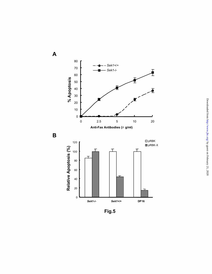
B
0
20
40
60
80
100
120
Sek1-/- Sek1+/+ DP16
pRBK
pRBK-X
Rel
ativ
e A
po
pto
sis
(%)
A
0
10
20
30
40
50
60
70
80
0 2.5 5 10 20
Anti-Fas Antibodies (µ g/ml)
Sek1+/+
Sek1-/-%
Ap
op
tosi
s
Fig.5
by guest on February 21, 2020http://w
ww
.jbc.org/D
ownloaded from

0
20
40
60
80
100
SEK1+/+ SEK1-/- DP16
pRBK
pRBK-XC
asp
ase
8 A
ctiv
ity
(% o
f co
ntr
ol)
A
0
20
40
60
80
100
SEK1+/+ SEK1-/-
pRBK
pRBK-X
Cas
pas
e 3
Act
ivit
y (%
of
con
tro
l)
B
Fig.6
by guest on February 21, 2020http://w
ww
.jbc.org/D
ownloaded from

Fig.7
Cyt c
control Ig
pRBK-HBx pRBK
Ig
anti-Fas+ + ++ ++
3631
21.5
14.5
kDa
Ig
Cyt c
31
21.5
14.5
Sek1+/+
Sek1-/-
A
B
control Iganti-Fas
+ ++ +
DP-pRBKDP16-X
Ig
Cyt c
31
21.5
14.5
kDa
pRBK-X 2-50
by guest on February 21, 2020http://w
ww
.jbc.org/D
ownloaded from

C
32P
Inco
rpo
ratio
n (c
pm
x 1
0-3)
0
5
1 0
1 5
2 0
2 5
3 0
0 3 0 6 0 120 240
Time (min)
MEF-pRBK
MEF-pRBK- X
0
1 0
2 0
3 0
4 0
5 0
6 0
0 2 4 6 8Time (hour)
DPpRBK
DP16- X1DP16- X2
32P
Inco
rpo
ratio
n (c
pm
x 1
0-3)
B
A
1 2 3 1 2 3
anti-c-jun
anti- -c-junP
pRBKpRBKHBx
MEF
Fig.8
by guest on February 21, 2020http://w
ww
.jbc.org/D
ownloaded from

A
B
1 2 1 2
c-Junbinding
cell lysate
SEK1
HBx
SAPK
14-3-3
MEKK1
1 2 3 1 2 3
cell lysate
c-Junbinding
SAPK
HBx
HBx 2-50
Fig.9
by guest on February 21, 2020http://w
ww
.jbc.org/D
ownloaded from

C 1 2 1 2
Sek1 -/- Sek1+/+
HBx
SEK1
14-3-3
SAPK
MEKK1
D
14-3-3
1 2Ig
HBx
Fig.9
by guest on February 21, 2020http://w
ww
.jbc.org/D
ownloaded from

B
C
D
Fig.10
A
by guest on February 21, 2020http://w
ww
.jbc.org/D
ownloaded from

A
B
Fig.11
Huh7
1 2 3 4 5 6 7
Anti-14-3-3 binding motif
Anti-HBx
MEF
1 * 1 2 * 2 3 * 3 4 * 4 5 * 5
1 2 3 4 5
*
HBx
HBx ∆124-153
HBx ∆114-153
HBx ∆104-153
HBx ∆26-31
HBx ∆25-32
HBx ∆2-50
HBx 31S-A
1 2 3 4 5 6 7
HB
x
Vec
tor
HB
x ∆1
24-1
53
HB
x ∆1
04-1
53
HB
x ∆1
14-1
53
HB
x ∆2
-50
HB
x 31
S-A
HB
x ∆2
6-31
HB
x ∆2
5-32
HB
x 31
S-A
HB
x
Vec
tor
by guest on February 21, 2020http://w
ww
.jbc.org/D
ownloaded from

C
Fig.11
D
% F
as-M
edia
ted
Ap
op
tosi
s
0
20
40
60
80
100
120
Ret
ro-v
ecto
r
Ret
ro-H
Bx
Ret
ro-H
Bx
31S
-A
Ret
ro-H
Bx
∆26-
31
MEFHuh7
P-c-jun
c-jun
1 2 3 4 5 6 7
HB
x
Vec
tor
HB
x ∆1
24-1
53
HB
x ∆1
04-1
53
HB
x ∆1
14-1
53
HB
x ∆2
-50
HB
x 31
S-A
1 2 3 4
HB
x ∆2
6-31
HB
x 31
S-A
HB
x
Vec
tor
by guest on February 21, 2020http://w
ww
.jbc.org/D
ownloaded from

James R. Woodgett, Josef Penninger and Christopher D. RichardsonJingyu Diao, Aye Aye Khine, Farida Sarangi, Eric Hsu, Caterina Iorio, Lee Anne Tibbles,
With Upregulation of the SAPK/JNK PathwayX Protein of Hepatitis B Virus Inhibits Fas-Mediated Apoptosis and is Associated
published online November 30, 2000J. Biol. Chem.
10.1074/jbc.M006026200Access the most updated version of this article at doi:
Alerts:
When a correction for this article is posted•
When this article is cited•
to choose from all of JBC's e-mail alertsClick here
by guest on February 21, 2020http://w
ww
.jbc.org/D
ownloaded from