Embed Size (px)
Citation preview

www.le.ac.uk
Analysis
www.le.ac.uk/genie
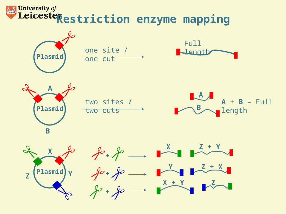
Restriction enzyme mapping
Plasmidone site / one cut
Full length
Plasmid Y
X
Z
A + B = Full length
B
Plasmidtwo sites / two cuts
AA
B
X Z + Y+
Y Z + X+
Z X + Y+
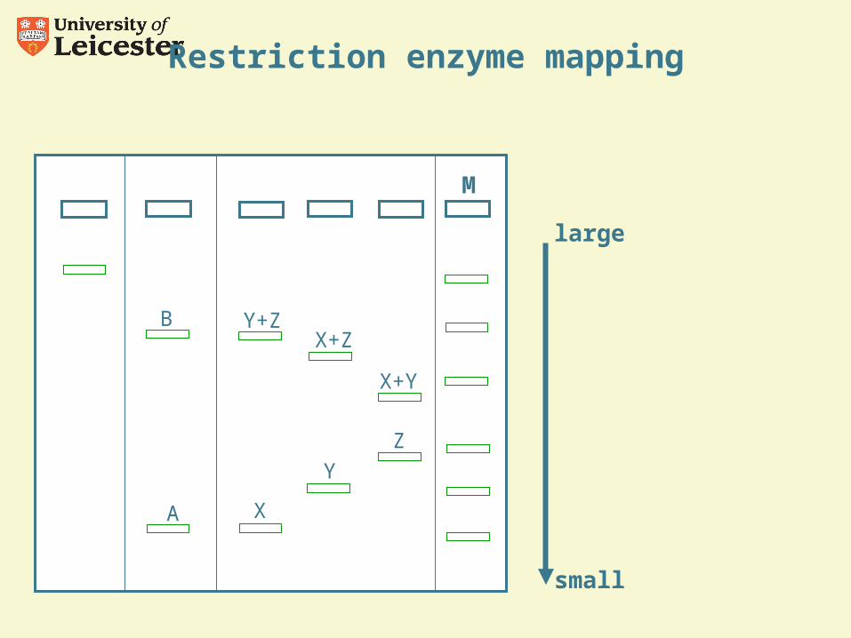
small
large
M
A
B
X
Y
Z
Y+ZX+Z
X+Y
Restriction enzyme mapping
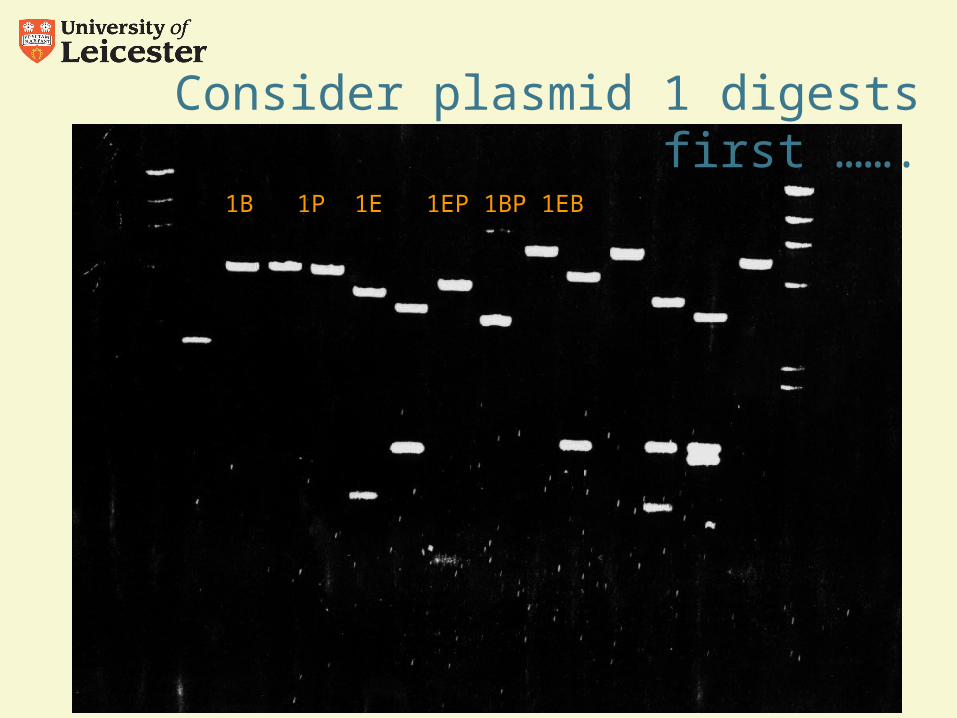
1B 1P 1E 1EP 1BP 1EB
Consider plasmid 1 digests first …….
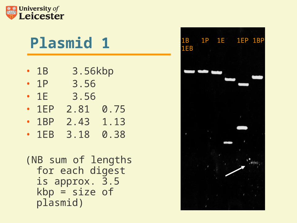
Plasmid 1
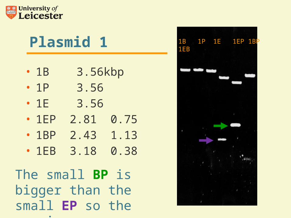
• 1B 3.56kbp• 1P 3.56• 1E 3.56• 1EP 2.81 0.75• 1BP 2.43 1.13• 1EB 3.18 0.38
(NB sum of lengths for each digest is approx. 3.5 kbp = size of plasmid)
1B 1P 1E 1EP 1BP 1EB
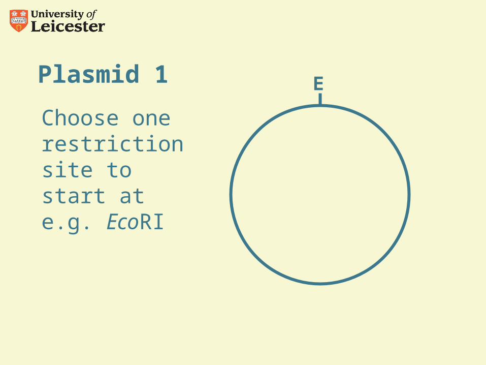
Plasmid 1
Choose one restriction site to start at e.g. EcoRI
E
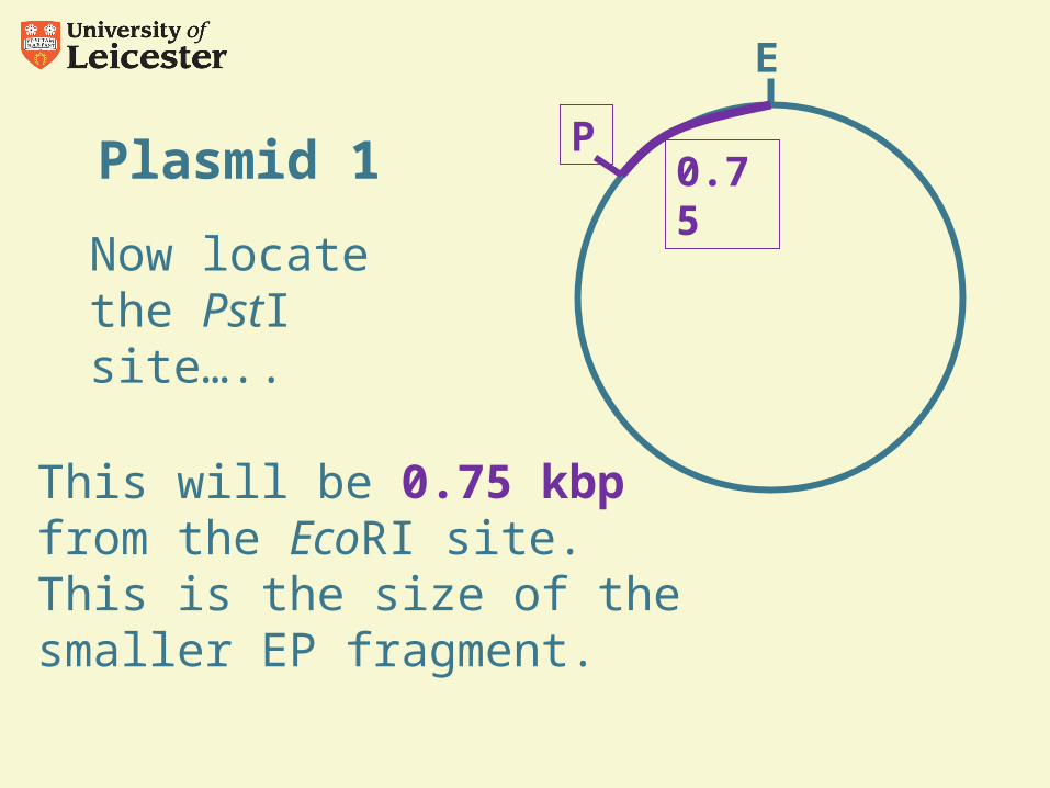
Plasmid 1
Now locate the PstI site…..
E
This will be 0.75 kbp from the EcoRI site. This is the size of the smaller EP fragment.
P0.75
Plasmid 1
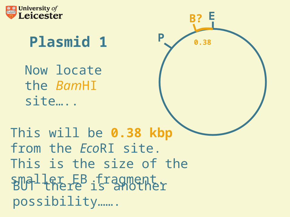
Now locate the BamHI site…..
E
This will be 0.38 kbp from the EcoRI site. This is the size of the smaller EB fragment.
BUT there is another possibility…….
P
B?
0.38
Plasmid 1E
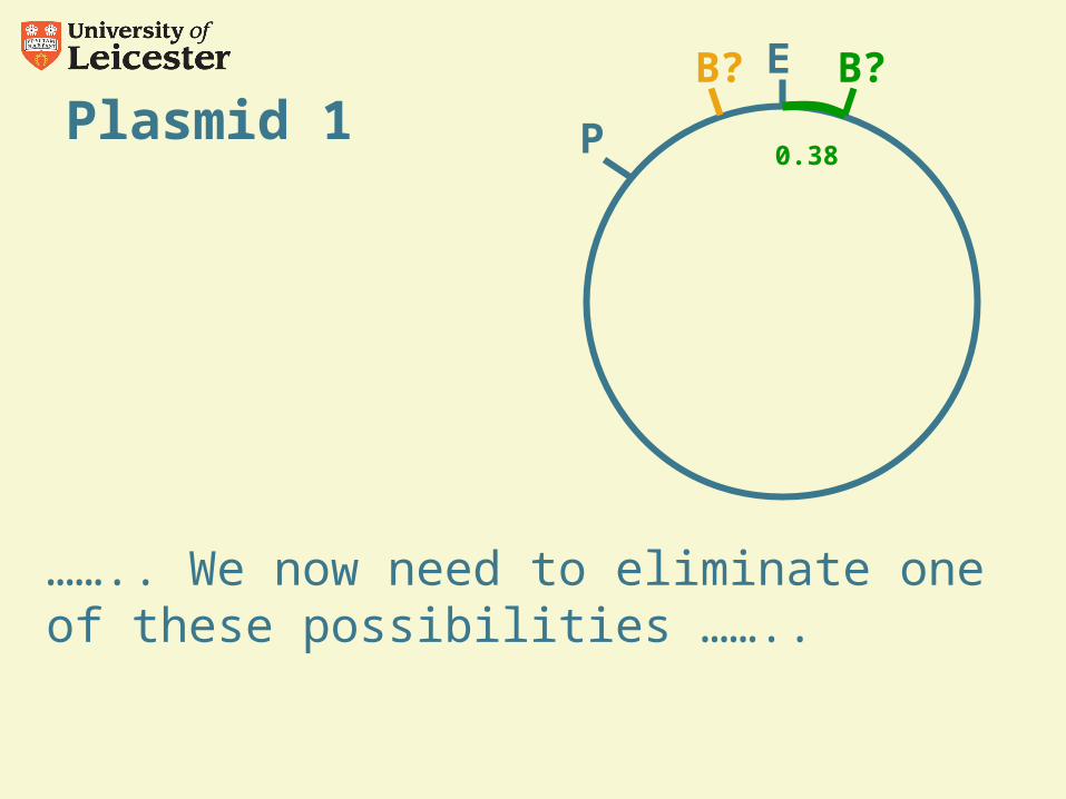
…….. We now need to eliminate one of these possibilities ……..
P
B? B?
0.38
Plasmid 1E
P
B? B?
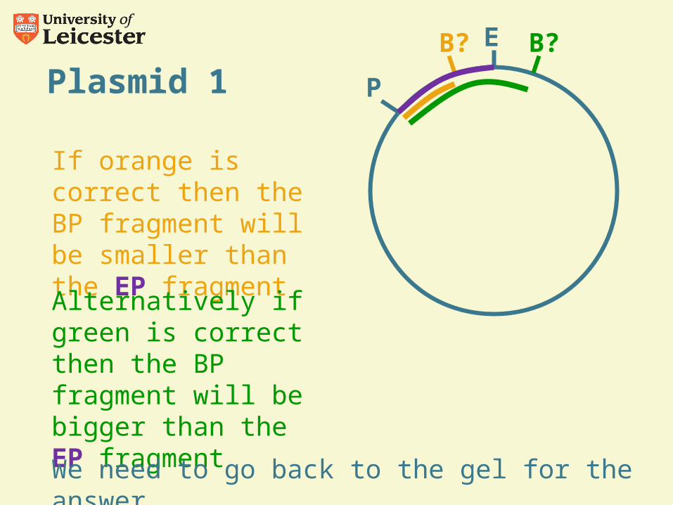
If orange is correct then the BP fragment will be smaller than the EP fragment
Alternatively if green is correct then the BP fragment will be bigger than the EP fragment
We need to go back to the gel for the answer …..
Plasmid 1
• 1B 3.56kbp• 1P 3.56• 1E 3.56• 1EP 2.81 0.75• 1BP 2.43 1.13• 1EB 3.18 0.38
1B 1P 1E 1EP 1BP 1EB
The small BP is bigger than the small EP so the map is ……
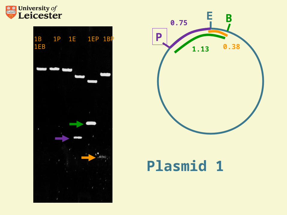
Plasmid 1
E
P
B0.75
1.13
1B 1P 1E 1EP 1BP 1EB0.38
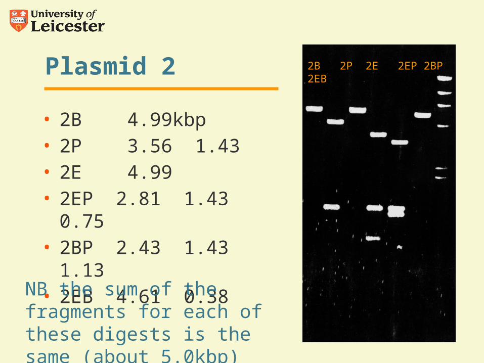
Plasmid 2
• 2B 4.99kbp• 2P 3.56 1.43• 2E 4.99• 2EP 2.81 1.43 0.75• 2BP 2.43 1.43 1.13• 2EB 4.61 0.38
NB the sum of the fragments for each of these digests is the same (about 5.0kbp)
2B 2P 2E 2EP 2BP 2EB
Plasmid 2
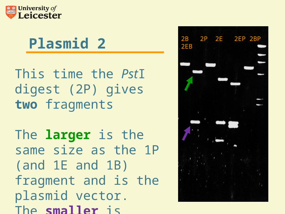
This time the PstI digest (2P) gives two fragments
The larger is the same size as the 1P (and 1E and 1B) fragment and is the plasmid vector.The smaller is 1.43kbp and is the insert DNA
2B 2P 2E 2EP 2BP 2EB
Plasmid 2
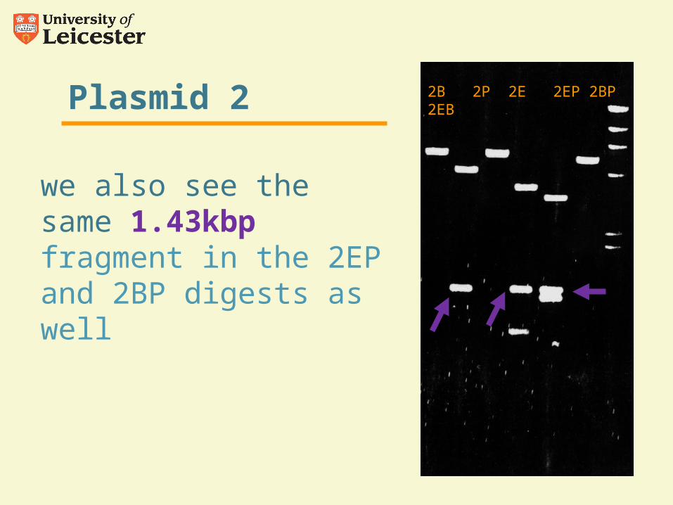
we also see the same 1.43kbp fragment in the 2EP and 2BP digests as well
2B 2P 2E 2EP 2BP 2EB
1B 1P 1E 1EP 1BP 1EB
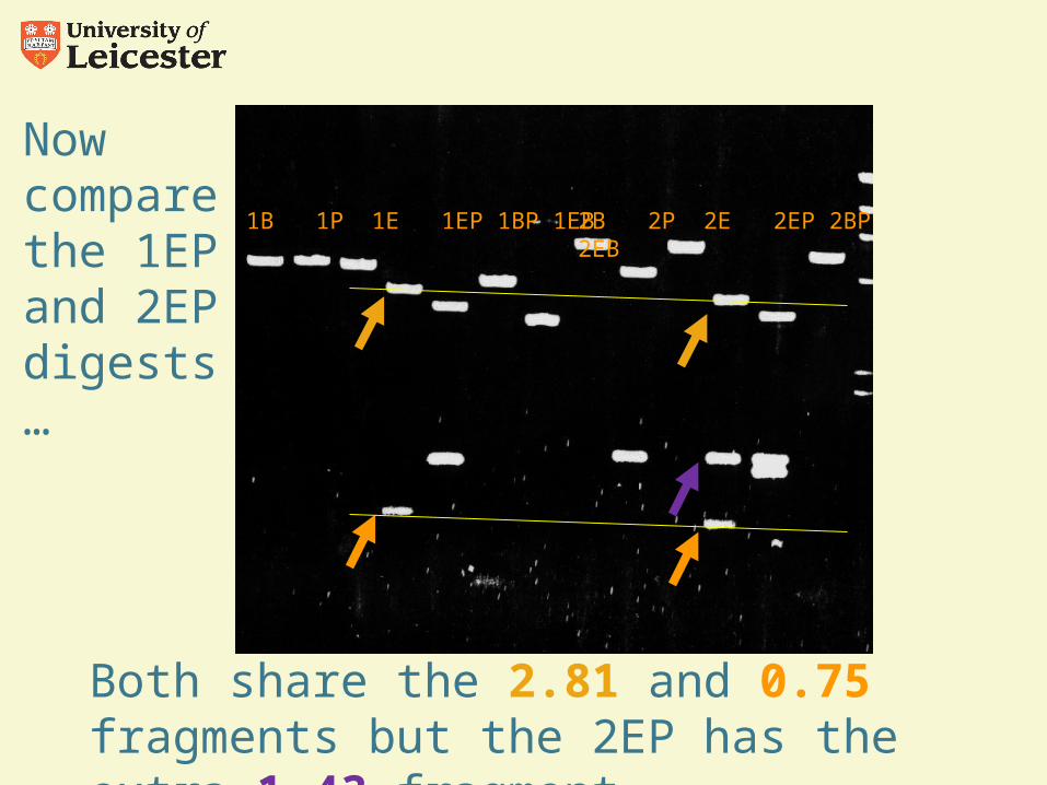
Now compare the 1EP and 2EP digests …
2B 2P 2E 2EP 2BP 2EB
Both share the 2.81 and 0.75 fragments but the 2EP has the extra 1.43 fragment
1B 1P 1E 1EP 1BP 1EB
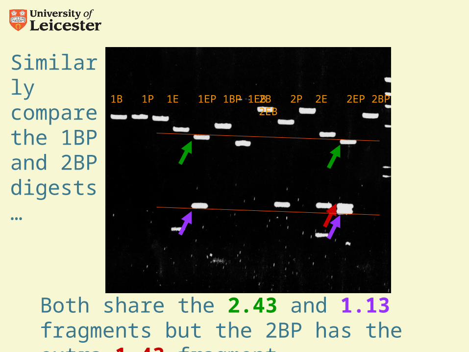
Similarly compare the 1BP and 2BP digests …
2B 2P 2E 2EP 2BP 2EB
Both share the 2.43 and 1.13 fragments but the 2BP has the extra 1.43 fragment
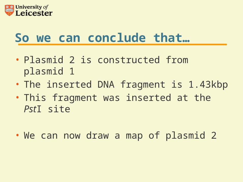
So we can conclude that…
• Plasmid 2 is constructed from plasmid 1• The inserted DNA fragment is 1.43kbp• This fragment was inserted at the PstI site
• We can now draw a map of plasmid 2
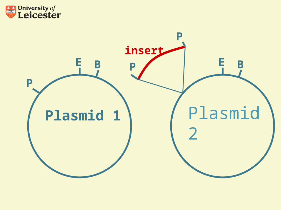
Plasmid 1
E
P
B
Plasmid 2
EP B
Pinsert
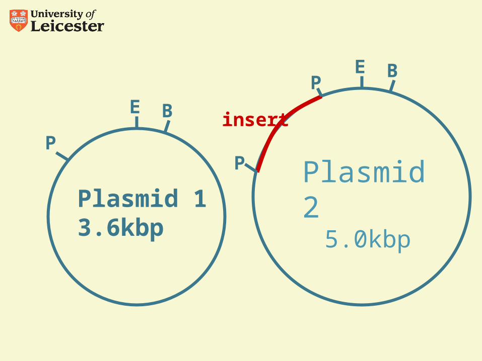
Plasmid 13.6kbp
E
P
B
Plasmid 25.0kbp
E
P
BP
insert
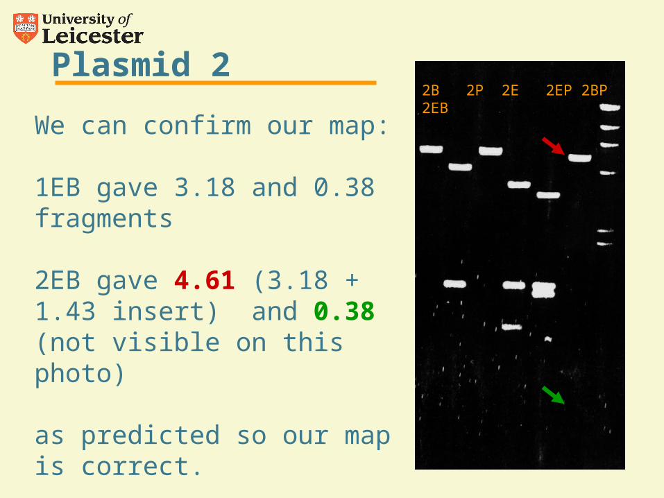
Plasmid 2
We can confirm our map:
1EB gave 3.18 and 0.38 fragments
2EB gave 4.61 (3.18 + 1.43 insert) and 0.38 (not visible on this photo)
as predicted so our map is correct.
2B 2P 2E 2EP 2BP 2EB
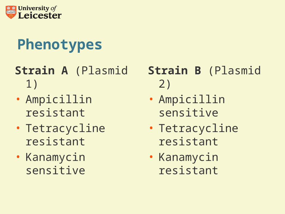
Phenotypes
Strain A (Plasmid 1) • Ampicillin resistant• Tetracycline resistant• Kanamycin sensitive
Strain B (Plasmid 2)• Ampicillin sensitive• Tetracycline resistant• Kanamycin resistant
Conclusions
Inserted fragment carries the Kanamycin resistance gene which is inserted into the PstI site of plasmid 1
This fragment has been inserted into the Ampicillin resistance gene which is disrupted in plasmid 2
We cannot draw many conclusions about the position of the Tetracycline resistance gene except that it is NOT at the PstI site (in fact it spans the BamHI site)