Embed Size (px)
Citation preview
EUKARYOTIC CELL, Apr. 2011, p. 521–529 Vol. 10, No. 41535-9778/11/$12.00 doi:10.1128/EC.00274-10Copyright © 2011, American Society for Microbiology. All Rights Reserved.
Wsp1, a GBD/CRIB Domain-Containing WASP Homolog, IsRequired for Growth, Morphogenesis, and Virulence of
Cryptococcus neoformans�†Gui Shen,1 Amy Whittington,2 and Ping Wang1,2,3*
The Research Institute for Children1 and Departments of Microbiology, Immunology and Parasitology2 andPediatrics,3 Louisiana State University Health Sciences Center, New Orleans, Louisiana 70118
Received 28 October 2010/Accepted 16 February 2011
Human endocytic protein ITSN1 regulates actin reorganization by activating Rho family GTPases, such asCdc42. The process is enhanced by ITSN binding of WASP, an effector of Cdc42 and a potent activator of actinpolymerization. In the human pathogen Cryptococcus neoformans, endocytic protein Cin1 also interacts withCdc42 and Wsp1, an uncharacterized WASP homolog, but the significance of these interactions remainsunknown. Wsp1 contains several conserved domains, including a WASP homology 1 domain (WH1), a GTPasebinding/Cdc42 and Rac interactive binding domain (GBD/CRIB), and a C-terminal domain composed ofverprolin-like, central, and acidic motifs (VCA). Thus, Wsp1 exhibits domain compositions more similar tohuman WASP proteins than Saccharomyces cerevisiae Las17/Bee1, a WASP homolog lacking the GDB/CRIBdomain. Wsp1 is not an essential protein; however, the wsp1 mutant exhibited defects in growth, cytokinesis,chitin distribution, and endocytosis and exocytosis. The wsp1 mutant was also unable to undergo genetic cross,produce the polysaccharide capsule, or secrete the enzyme urease. An in vitro phagocytosis assay showed ahigher phagocytic index for the wsp1 mutant, whose ability to cause lethal infection in a murine model ofcryptococcosis was also attenuated. Our studies reveal divergent evolution of WASP proteins in the fungalphylum and suggest that the conserved function of WASP proteins in the actin cytoskeleton may also impactfungal virulence.
Cryptococcus neoformans is a basidiomycetous fungal patho-gen that infects primarily immunocompromised individuals,causing meningoencephalitis, which is often fatal if left un-treated (3, 19). This haploid organism has a bipolar matingsystem and undergoes genetic cross under laboratory condi-tions. In the environment, C. neoformans propagates primarilythrough budding, and yeast cells, probably in a desiccatedform, and are thought to enter the host through the respiratorytract and cause infection (3, 19). Studies by Kopecka andcolleagues indicated that C. neoformans has a fully developedactin cytoskeleton and it maintains actin dynamics similar toSaccharomyces cerevisiae during mitotic division and asexualreproduction (14).
In unicellular fungi, budding and the establishment of cellpolarity are accomplished through dynamic changes of theactin cytoskeleton, which are regulated by an integrated sig-naling cascade of the Rho family of small GTPases, such asRho, Rac, and Cdc42 (4, 25). Cdc42 is highly conserved amongthe eukaryotic organisms in both sequence and function (12).Cdc42 functions through binding of effector molecules thatcontain the GTPase-binding domain (GBD; also known as theCdc42/Rac interactive binding [CRIB] domain). Among theeffector proteins of Cdc42 that contain the CRIB/GBD do-
main, Wiskott-Aldrich syndrome (WAS) proteins (WASP) aremost known for their ability to promote actin assembly (4).
WASP and similar but neuron-specific WASP (N-WASP)proteins are a family of human proteins (both are referred toas WASPs) whose mutations result in WAS, a rare X-chromo-some-linked hereditary disease characterized by thrombocyto-penia, eczema, and immunodeficiency (5). These disease symp-toms are the result of abnormalities in cytoskeletal structures,polarization, and motility at the cellular level, and most studiesof WASPs have focused on their roles in regulating branchedfilamentous actin through the Arp2/3 complex (see the reviewsin references 27, 39, and 40). The WASPs are multimodularproteins that contain an N-terminal WASP homology 1 do-main (WH1), a basic amino acid domain (B), a CRIB/GBDmotif, a proline-rich domain (PR), and a C-terminal verprolin(V), central connecting (C), and acid amino acid (A) domain(collectively termed VCA) (27, 31). In its resting state, WASPis in an autoinhibitory conformation through the binding of theVCA domain to its B and CRIB/GBD domains (A to B and Vto CRIB/GBD, respectively). Binding of GTP-Cdc42 to WASPthrough the CRIB/GBD domain, phosphatidylinositol bisphos-phate (PIP2) to the B domain, or the SH3 domain to the PRdomain all result in opening of the masked and autoinhibitedVCA domain, leading to its activation of the Arp2/3 protein(15, 27, 31).
Despite the conserved function of WASPs, distinctions existamong various paralogs/homologs, resulting in a complexity intheir filamentous actin activation mechanisms. In the plantmodel Arabidopsis thaliana, no WASP homolog was found, butinstead a family of WAVE (verprolin-homologous) proteins
* Corresponding author. Mailing address: The Research Institutefor Children, LSU Health Sciences Center, Research & EducationBldg., Rm. 3123, New Orleans, LA 70118. Phone: (504) 896-2739. Fax:(504) 894-5379. E-mail: [email protected].
† Supplemental material for this article may be found at http://ec.asm.org/.
� Published ahead of print on 25 February 2011.
521
on July 7, 2018 by guesthttp://ec.asm
.org/D
ownloaded from
that function to promote actin nucleation were found (15). TheWAVE proteins also exist in mammals and, in contrast toWASPs that are activated by Cdc42, WAVE proteins are reg-ulated by Rac (23). In fungi, S. cerevisiae Las17/Bee1, Schizo-saccharomyces pombe Wsp1, and Candida albicans Wal1 areWASP homologs that lack the GBD/CRIB domain (9, 17, 18,32). This indicates that a distinct activation mechanism is likelyto be involved in the functions of these proteins (32). Consis-tent with this proposition, a study suggested that the activationof actin polymerization by Las17/Bee1 is indeed indirect (6).Not surprisingly, human WASP failed to complement an S.cerevisiae las17/bee1 mutant and restored functions (18), de-spite the finding that Las7 exhibited a role in the activation ofactin filaments and some of the las17/bee1 yeast mutant phe-notypes mimicked the cellular defects of Wiskott-Aldrich syn-drome (18, 21).
C. albicans Wal1 exhibits a certain degree of domain simi-larities to S. cerevisiae Las17/Bee1 and S. pombe Wsp1, sharingup to 38% and 28%, respectively, in amino acid sequenceidentity (32). Similar to Las17/Bee1, Wal1 is required for po-larity establishment, intracellular traffic, and vacuolar biogen-esis in C. albicans and for the polarized growth of C. albicanshyphae (32).
Studies of C. neoformans have suggested that a number ofproteins have a role in actin function. The Rho GTPases Cdc42and Cdc420 are required for actin polarization under stressconditions and have also been mostly noted for functioningwith Ras proteins to mediate thermal resistance, a role previ-ously established for the Rac homolog of the fungus (1, 22, 30).GTPase Sav1, an S. cerevisiae Sec4/Rab8 homolog, is involvedin post-Golgi complex secretion (38), a process that involvesthe rearrangement of the actin cytoskeleton (see the review inreference 35). Recently, we identified a novel cryptococcalintersectin protein, Cin1, and characterized its functions, in-cluding those in the intracellular transport and regulation ofthe actin cytoskeleton (28). As a part of the ongoing effort toelucidate the unique Cin1 pathway, we identified a WASPhomolog, Wsp1, and showed that Wsp1 associated with Cin1through an interaction between the Wsp1-PR and Cin1-SH3domains (28). This implies that a functional mechanism similarto WASP of mammalian cells exists in C. neoformans.
Here, we further characterized Wsp1 and examined its func-tion in the growth, morphology, and virulence of the fungus.Through whole-gene and domain-specific gene disruption, weshow that Wsp1 has multiple functions in maintaining cellularmorphology, cytokinesis and chitin distribution, and endocyto-sis and exocytosis. We also show that Wsp1 is less resistant tophagocytosis in an in vitro model and avirulent in a murinemodel of cryptococcosis. Our study illustrates the importantrole of the first fungal WASP homolog containing the GBD/CRIB domain and provides evidence of divergence betweendifferent groups of fungi in the evolution of WASP/Las17 pro-teins.
MATERIALS AND METHODS
Strains, plasmids, and media. C. neoformans var. neoformans (serotype D)strains were used in this study, with strains JEC21 and JEC20 as the standardMAT� and MATa strains (16). All strains are listed in Table S1 of the supple-mental material. Oligonucleotide primers for PCR amplification are listed inTable S2 of the supplemental material. The CDC42(Q61L) allele was obtained
by overlapping PCR. The 5� partial sequence was amplified by PCR with M13reverse primer and primer PW1498 with pGS950 as the template, while the 3�end was amplified with primer PW1497 and M13 forward primer, and also withpGS950 as the template. Overlap PCR was performed with primers PW1495 andPW1497, and the PCR product was then ligated into a TA vector and thesequence was verified. DNA plasmids used in this study are listed in Table S3 ofthe supplemental material. All culture media and reagents were prepared asdescribed previously (28).
Mutant strain construction and complementation. The wsp1::NEO andwsp1::NAT alleles were obtained by the split-marker approach, and the mutantstrains were obtained by biolistic transformation, as described previously (28). Awsp1 mutant was complemented with the full-length WSP1 gene, including thepromoter and terminator sequences, which were amplified through a two-stepprocess. First, the 5� sequence was amplified with primers PW1185 and PW1186and the fragment ligated into plasmid pGS199, resulting in plasmid pGS764.Second, the 3� sequence was amplified with primers PW1187 and PW1193 andinserted into pGS764, resulting in plasmid pGS781. The DNA sequence wasverified by sequencing and the WSP1 gene linked to the NAT marker wasreintroduced into the wsp1::NEO mutant by a second round of biolistic trans-formation.
To construct the WSP1-GBD allele, in which the sequence corresponding toamino acids 160 to 224 was deleted, overlap PCR was performed using twotemplates. One template was amplified with primer PW1188 and M13 forwardprimer from pGS462, and another with primer PW1189 and M13 reverse primerfrom pGS780. Overlap PCR was carried out with primers PW1187 and PW968,and the resulting PCR product was cloned (pGS783). The product was then cutwith BamHI and XbaI and inserted into pGS324 to get the final constructpGS794. In this plasmid, the Wsp1-GBD allele is driven by a constitutively activeglyceraldehyde phosphate dehydrogenase Gpd1 promoter (28). The plasmid wastransformed into the wsp1 mutant strain, and transformants were verified byPCR. Expression levels were estimated by semiquantitative reverse transcription-PCR (see Fig. S1 in the supplemental material).
To generate the wsp1 mutant in DsRed-Sec4 (Sav1) strains, plasmid pGS1139was first introduced into wild-type strain JEC21 by biolistic transformation. Oneof the transformants, GYS506, that exhibited robust red fluorescent signal underthe microscope was transformed with the wsp1::NEO split-marker products toobtain GYS563. This transformant strain was used to examine the role of Wsp1in Sec4/Sav1 localization.
Yeast two-hybrid assay. Full-length WSP1 cDNA was released from plasmidpGS462 with EcoRI and BglII and inserted into pGBKT7, resulting inpGBKT7::WSP1 (pGS849). cDNA for CDC42 was synthesized with primerPW1495 and adaptor primer AUAP and cloned in the TA vector (Agilent),resulting in pGS950. Once verified by DNA sequencing, cDNA was cut withEcoRI and BglII and inserted into pGADT7, resulting in pGS1074. PlasmidDNA was transformed into the yeast AH109 strain, and interactions were as-sessed according to the method previously described (10, 24).
Fluorescence fusion protein construction and detection. To construct theDsRed-Sec4 fusion protein, SEC4/SAV1 cDNA was amplified with PW1510 andAUAP and cloned (pGS1119). The SEC4/SAV1 fragment cut with BamHI andSpeI was inserted into pGS988 (28) to obtain the final construct, pGS1139. TheDsRed-SEC4 fusion construct also has the GPD1 promoter.
Nuclear staining with 4�,6-diamino-2-phenylindole (DAPI) was performed asdescribed previously using fresh cells grown overnight (28). Distributions ofchitin and actin were observed by staining cells with calcofluor white (CFW) andrhodamine-conjugated phalloidin, respectively, as described previously (28). Thevesicles and endosomes were observed by incubating appropriately preparedcells with FM4-64 [N-(3-triethylammoniumpropyl)-4-(p-diethylaminophenyl-hexatrienyl) pyridinium dibromide] (28). Cells were mounted on poly-L-lysine-coated glass slides and observed for fluorescence under a Zeiss Axio Imager 2microscope (Carl Zeiss, Inc., Thornwood, NY).
Mutant phenotype characterization. Assays for cell fusion, conjugation tubeformation, and mating, as well as melanin and capsule production, were per-formed as described previously (28, 33). For growth, cells of an approximateoptical density at (OD600) of 0.1 were serially diluted, and 5 �l each was spottedonto yeast extract-peptone-dextrose (YPD) and yeast nitrogen base plates forgrowth at 25, 30, and 37°C. To assay for urease, aliquots from saturated YPDbroth cultures of wild-type JEC21, wsp1, and wsp1 WSP1 were grown in liquidYPD overnight with shaking at 30°C. Cells were collected by centrifugation andwashed twice with sterile water. Cells were suspended in sterile water andcounted by using a hemacytometer, followed by plating on YPD to ensureaccuracy and viability of counts. A total of 6 � 105 cells were inoculated into 1.5ml of Christensen’s urea broth with or without urea. Cells were incubated up to3 days at 30°C, and absorbance at 550 nm and CFU were estimated daily.
522 SHEN ET AL. EUKARYOT. CELL
on July 7, 2018 by guesthttp://ec.asm
.org/D
ownloaded from
Phagocytosis assay. J774A.1 macrophages of four to five passages were col-lected and counted, and viability was assessed with trypan blue stain (28). Mac-rophages were seeded on coverglasses in 12-well plates and stimulated with 50units/ml mouse recombinant gamma interferon (R&D Systems), 0.3 �g/ml Esch-erichia coli 0111:B4 lipopolysaccharide (Sigma), and 25 �l/ml pooled mouseserum (JEC21 infected) before exposure to yeast cells at a 2:1 multiplicity ofinfection. Mixed cells were incubated at 5% CO2, 37°C for 4 h and observedusing an inverted microscope (Olympus CV41). Noningested yeast cells wereremoved by washing three times in 37°C sterile phosphate-buffered saline (FisherScientific). For phagocytic index determination, coverglasses were fixed in meth-anol and stained with Giemsa stain (Sigma), and three to five fields per cover-glass were photographed under a microscope (Zeiss Axio Imager 2) outfittedwith a Zeiss digital camera. At least 120 macrophages were counted for ingestedyeast cells per infecting strain, and the phagocytic index is reported as thenumber of ingested yeast cells per 100 macrophages.
Virulence assessment. BALB/c mice were inoculated via the lateral tail veinwith 2.5 � 105 fresh-grown cells as determined by counting with a hemacytom-eter and viability plating on YPD medium (�95% viability). For wsp1 mutantsthat exhibited morphological defects, individual chains or clusters of cells wereconsidered a single cell. Mice were monitored twice daily and sacrificed whenmoribund or at 90 days postinfection. Brains, kidneys, and lungs were dissectedfor CFU determinations. Organs were weighed and homogenized in 1 ml ofsterile water containing 50 �g/ml chloramphenicol and plated on YPD followingserial dilution. CFU were determined after 3 to 5 days of growth at 30°C.Homogenates were also stained with India ink and observed in a blinded manner.
For histological studies, brains were preserved in 4% buffered formalin, pro-cessed, embedded, and sectioned at 5-�m thickness. Sections were allowed toadhere to glass slides, stained with periodic acid-Schiff stain (PAS; Sigma), andobserved in a blinded manner. Representative areas were photographed under amicroscope (Olympus BX51) equipped with a digital camera (Olympus).
RESULTS
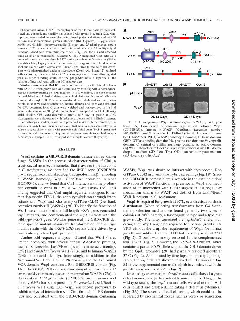
Wsp1 contains a GBD/CRIB domain unique among knownfungal WASPs. In the process of characterization of Cin1, acryptococcal intersectin homolog that plays multiple functionsin C. neoformans, we identified the WSP1 gene (CNBE5050[www-sequence.stanford.edu/cgi-bin/cneoformans]) encodinga WASP homolog, Wsp1 (GenBank accession numberHQ399545), and we found that Cin1 interacts with the proline-rich domain of Wsp1 in a yeast two-hybrid assay (28). Thisfinding suggested that Cin1 might regulate, analogous to hu-man intersectin ITSN1, the actin cytoskeleton through inter-actions with Wsp1 and Rho family GTPase Cdc42 (GenBankaccession number HQ645962) (28). To identify the function ofWsp1, we characterized the full-length WSP1 gene, generatedwsp1 mutants, and complemented the wsp1 mutants with thewild-type WSP1 gene. We also generated the GBD/CRIB do-main-specific mutant strains by transformation of the wsp1mutant strain with the WSP1-GBD mutant allele driven by aconstitutively active Gpd1 promoter.
Amino acid sequence analysis indicated that Wsp1 shareslimited homology with several fungal WASP-like proteins,such as S. cerevisiae Las17/Bee1 (overall amino acid identity,32%) and Candida albicans Wal1 (29%) and to human WASPs(29% amino acid identity). Interestingly, in addition to theN-terminal WH1 domain, the PR domain, and the C-terminalVCA domain, Wsp1 contains a Rho GBD/CRIB domain (Fig.1A). The GBD/CRIB domain, consisting of approximately 17amino acids, commonly occurs in mammalian WASPs (27a). Italso exists in Ustilago maydis (UM03687; overall amino acididentity, 62%) but is not present in S. cerevisiae Las17/Bee1 orC. albicans Wal1 (Fig. 1A). Wsp1 was shown previously toexhibit a physical interaction with Cin1 through the PR domain(28) and, consistent with the GBD/CRIB domain containing
WASPs, Wsp1 was shown to interact with cryptococcal RhoGTPase Cdc42 in a yeast two-hybrid screening (Fig. 1B). Sincethe GBD/CRIB domain plays a key role in the autoinhibition/activation of WASP function, its presence in Wsp1 and medi-ation of an interaction with Cdc42 suggest that a regulatorymechanism similar to WASP but distinct from Las17/Bee1/Wal1 is present in C. neoformans.
Wsp1 is required for growth at 37°C, cytokinesis, and chitindistribution. When selecting transformants from G418-con-taining selection medium, we found generally two types ofcolonies at 30°C, namely, a faster-growing type and a type thatgrew slowly. The latter contained the wsp1::NEO allele, indi-cating that Wsp1 might be required for normal growth. OnYPD without the drug, the requirement of Wsp1 for normalgrowth was subtle at 25 and 30°C but most apparent at 37°C(Fig. 2). Growth was mostly restored in the complementedwsp1 WSP1 (Fig. 2). However, the WSP1-GBD mutant, whichcontains a partial WSP1 allele without the GBD domain drivenby the Gpd1 promoter (28) had partially restored growth at37°C (Fig. 2). As indicated by time-lapse microscopic photog-raphy, the wsp1 mutant showed delayed cell division (see Fig.S2 in the supplemental material), which is consistent with thegrowth assay results at 25°C (Fig. 2).
Microscopy examination of wsp1 mutant cells showed a grossdefect in morphology. In contrast to unicellular budding of thewild-type strain, the wsp1 mutant cells were abnormal, withcells jointed and clustered, indicating a defect in cytokinesis(Fig. 3A). The severity of cell clustering, which could not beseparated by mechanical forces such as vortex or sonication,
FIG. 1. C. neoformans Wsp1 is homologous to WASP/Las17 pro-teins. (A) Comparison of domain organization between Wsp1(CNBE5050), human n-WASP (GenBank accession numberNP_003932), and S. cerevisiae Las17/Bee1 (GenBank accession num-ber CAA99390). WH1, WASP homology 1 domain; B, basic domain;GBD, GTPase binding domain; PR, proline-rich domain; V: verprolindomain; C, central or cofilin homology domain; A, acidic domain.(B) Wsp1 interacts with Cdc42 in a yeast two-hybrid assay. DD, doubledropout medium (SD -Leu -Trp); QD, quadruple dropout medium(SD -Leu -Trp -His -Ade).
VOL. 10, 2011 C. NEOFORMANS GBD/CRIB DOMAIN-CONTAINING WASP HOMOLOG 523
on July 7, 2018 by guesthttp://ec.asm
.org/D
ownloaded from
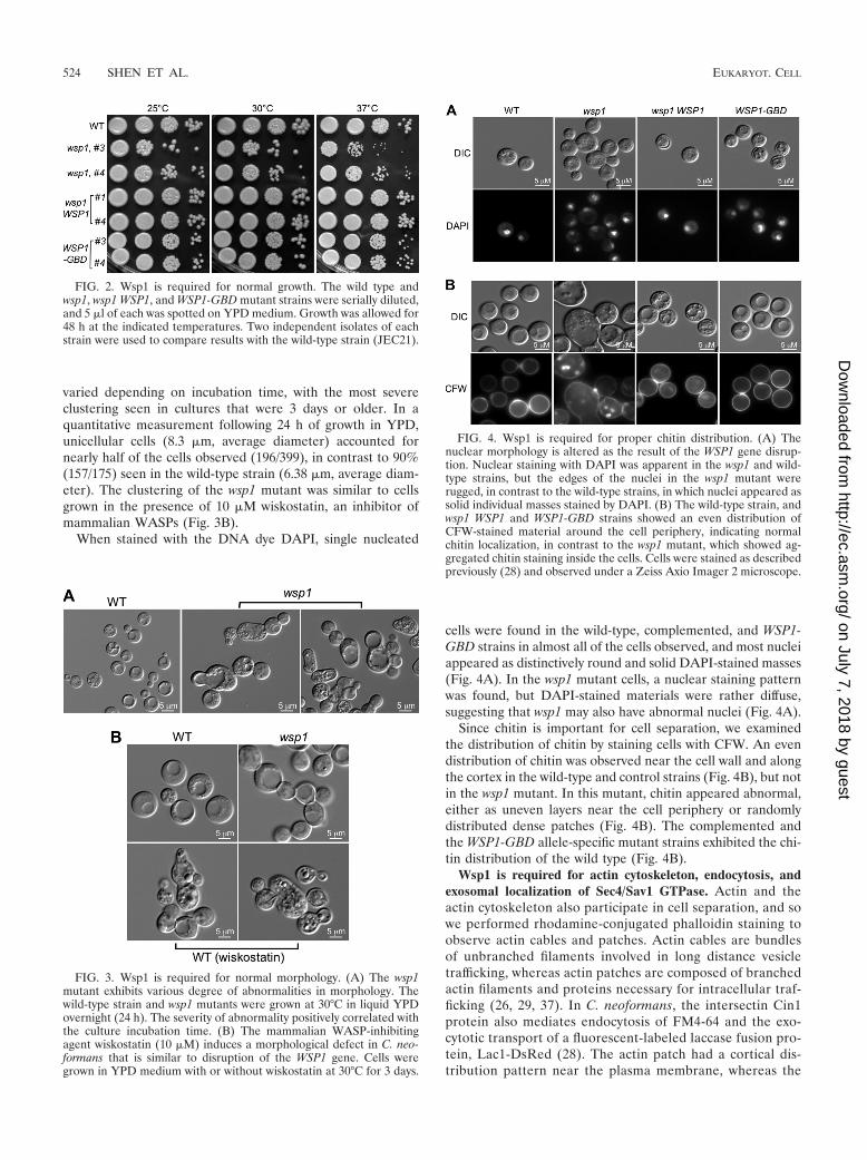
varied depending on incubation time, with the most severeclustering seen in cultures that were 3 days or older. In aquantitative measurement following 24 h of growth in YPD,unicellular cells (8.3 �m, average diameter) accounted fornearly half of the cells observed (196/399), in contrast to 90%(157/175) seen in the wild-type strain (6.38 �m, average diam-eter). The clustering of the wsp1 mutant was similar to cellsgrown in the presence of 10 �M wiskostatin, an inhibitor ofmammalian WASPs (Fig. 3B).
When stained with the DNA dye DAPI, single nucleated
cells were found in the wild-type, complemented, and WSP1-GBD strains in almost all of the cells observed, and most nucleiappeared as distinctively round and solid DAPI-stained masses(Fig. 4A). In the wsp1 mutant cells, a nuclear staining patternwas found, but DAPI-stained materials were rather diffuse,suggesting that wsp1 may also have abnormal nuclei (Fig. 4A).
Since chitin is important for cell separation, we examinedthe distribution of chitin by staining cells with CFW. An evendistribution of chitin was observed near the cell wall and alongthe cortex in the wild-type and control strains (Fig. 4B), but notin the wsp1 mutant. In this mutant, chitin appeared abnormal,either as uneven layers near the cell periphery or randomlydistributed dense patches (Fig. 4B). The complemented andthe WSP1-GBD allele-specific mutant strains exhibited the chi-tin distribution of the wild type (Fig. 4B).
Wsp1 is required for actin cytoskeleton, endocytosis, andexosomal localization of Sec4/Sav1 GTPase. Actin and theactin cytoskeleton also participate in cell separation, and sowe performed rhodamine-conjugated phalloidin staining toobserve actin cables and patches. Actin cables are bundlesof unbranched filaments involved in long distance vesicletrafficking, whereas actin patches are composed of branchedactin filaments and proteins necessary for intracellular traf-ficking (26, 29, 37). In C. neoformans, the intersectin Cin1protein also mediates endocytosis of FM4-64 and the exo-cytotic transport of a fluorescent-labeled laccase fusion pro-tein, Lac1-DsRed (28). The actin patch had a cortical dis-tribution pattern near the plasma membrane, whereas the
FIG. 2. Wsp1 is required for normal growth. The wild type andwsp1, wsp1 WSP1, and WSP1-GBD mutant strains were serially diluted,and 5 �l of each was spotted on YPD medium. Growth was allowed for48 h at the indicated temperatures. Two independent isolates of eachstrain were used to compare results with the wild-type strain (JEC21).
FIG. 3. Wsp1 is required for normal morphology. (A) The wsp1mutant exhibits various degree of abnormalities in morphology. Thewild-type strain and wsp1 mutants were grown at 30°C in liquid YPDovernight (24 h). The severity of abnormality positively correlated withthe culture incubation time. (B) The mammalian WASP-inhibitingagent wiskostatin (10 �M) induces a morphological defect in C. neo-formans that is similar to disruption of the WSP1 gene. Cells weregrown in YPD medium with or without wiskostatin at 30°C for 3 days.
FIG. 4. Wsp1 is required for proper chitin distribution. (A) Thenuclear morphology is altered as the result of the WSP1 gene disrup-tion. Nuclear staining with DAPI was apparent in the wsp1 and wild-type strains, but the edges of the nuclei in the wsp1 mutant wererugged, in contrast to the wild-type strains, in which nuclei appeared assolid individual masses stained by DAPI. (B) The wild-type strain, andwsp1 WSP1 and WSP1-GBD strains showed an even distribution ofCFW-stained material around the cell periphery, indicating normalchitin localization, in contrast to the wsp1 mutant, which showed ag-gregated chitin staining inside the cells. Cells were stained as describedpreviously (28) and observed under a Zeiss Axio Imager 2 microscope.
524 SHEN ET AL. EUKARYOT. CELL
on July 7, 2018 by guesthttp://ec.asm
.org/D
ownloaded from
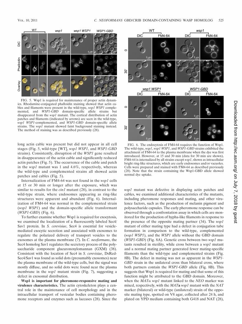
long actin cable was present but did not appear in all cellstages (Fig. 5, wild-type [WT], wsp1 WSP1, and WSP1-GBDstrains). Consistently, disruption of the WSP1 gene resultedin disappearance of the actin cable and significantly reducedactin patches (Fig. 5). The occurrence of the cable and patchin the wsp1 mutant was 1 and 4.6%, respectively, whereasthe wild-type and complemented strains all showed actinpatches and cables (Fig. 5).
Internalization of FM4-64 was not found in the wsp1 cellsat 15 or 30 min or longer after the exposure, which wassimilar to results for the cin1 mutant (28), in contrast to thewild-type strain, where endosomes appearing as ring-likestructures were apparent and abundant (Fig. 6). Internal-ization of FM4-64 was normal in the complemented strain(wsp1 WSP1) and the domain-specific allele transformant(WSP1-GBD) (Fig. 6).
To further examine whether Wsp1 is required for exocytosis,we examined the localization of a fluorescently labeled Sec4/Sav1 protein. In S. cerevisiae, Sec4 is essential for vesicle-mediated exocytic secretion and associated with exosomes toregulate the polarized delivery of transport vesicles to theexosomes at the plasma membrane (7). In C. neoformans, theSec4 homolog Sav1 regulates the secretory process of the poly-saccharide component glucuronoxylomannan (GXM) (38).Consistent with the location of Sec4 in S. cerevisiae, DsRed-Sec4/Sav1 was found as solid dots (presumably exosomes) nearthe plasma membrane of the wild-type cells, but the signal wasmostly diffuse, and no solid dots were found near the plasmamembrane in the wsp1 mutant strain (Fig. 7), suggesting adefect in exosomal distribution.
Wsp1 is important for pheromone-responsive mating andvirulence characteristics. The actin cytoskeleton plays a cen-tral role in the maintenance of cell morphology and in theintracellular transport of vesicular bodies containing phero-mone receptors and enzymes such as laccases (28). Since the
wsp1 mutant was defective in displaying actin patches andcables, we examined additional characteristics of the mutants,including pheromone responses and mating, and other viru-lence factors, such as the production of melanin pigment andpolysaccharide capsules. The early pheromone response can beobserved through a confrontation assay in which cells are mon-itored for the production of hypha-like filaments in response tothe presence of the opposite mating partner (34). The wsp1mutant of either mating type had a defect in conjugation tubeformation in comparison to the wild-type, complemented(wsp1 WSP1), and the WSP1 allele without the GBD domain(WSP1-GBD) (Fig. 8A). Genetic cross between two wsp1 mu-tants resulted in sterility, while cross between a wsp1 mutantand a normal mating partner generated fewer mating-specificfilaments than the wild-type and complemented strains (Fig.8B). The defect in mating was not as apparent in the WSP1-GBD strain in the unilateral cross than bilateral cross, whereboth partners contain the WSP1-GBD allele (Fig. 8B). Thissuggests that Wsp1 is required for mating and that some of thisfunction might be attributed to the GBD domain. Moreover,when the MAT� wsp1 mutant linked to the NEO marker wasmixed, respectively, with the MATa wsp1 mutant with the NATmarker (bilateral) or wild-type (unilateral) strain of the oppo-site mating type, spotted on V8 agar, collected after 24 h, andplated on YPD medium containing both G418 and NAT (28),
FIG. 5. Wsp1 is required for maintenance of proper actin dynam-ics. Rhodamine-conjugated phalloidin staining showed that actin ca-bles and filaments were present in the wild-type, wsp1 WSP1 comple-mented, and WSP1-GBD domain-specific allele strains butdisappeared from the wsp1 mutant. The cortical distribution of actinpatches and filaments (indicated by arrows) are seen in the wild-type,wsp1 WSP1-complemented, and WSP1-GBD domain-specific allelestrains. The wsp1 mutant showed faint background staining instead.The method of staining was as described previously (28).
FIG. 6. The endocytosis of FM4-64 requires the function of Wsp1.The wild-type, wsp1, wsp1 WSP1, and WSP1-GBD strains exhibited theattachment of FM4-64 to the plasma membrane when the dye was firstintroduced. However, at 15 and 30 min (data for 30 min are shown),FM4-64 is internalized by all strains except wsp1, shown as intracellularbright ring-like structures, which are early endosomes and/or vacuoles.Cells were prepared and stained with FM4-64 as described previously(28). Note that the strain containing the Wsp1-GBD allele showednormal dye uptake.
VOL. 10, 2011 C. NEOFORMANS GBD/CRIB DOMAIN-CONTAINING WASP HOMOLOG 525
on July 7, 2018 by guesthttp://ec.asm
.org/D
ownloaded from
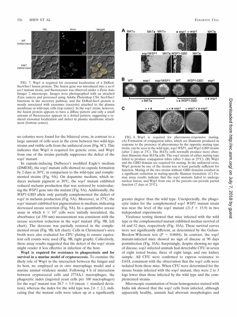
no colonies were found for the bilateral cross, in contrast to alarge amount of cells seen in the cross between two wild-typestrains and visible cells from the unilateral cross (Fig. 8C). Thisindicates that Wsp1 is required for genetic cross, and Wsp1from one of the strains partially suppresses the defect of thewsp1 mutant.
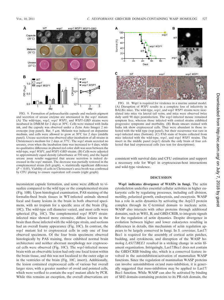
In capsule-inducing Dulbecco’s modified Eagle’s medium(DMEM), the wsp1 mutant cells showed no capsule formationby 2 days at 30°C, in comparison to the wild-type and comple-mented strains (Fig. 9A). On dopamine medium, which in-duces melanin pigment at 30°C, the wsp1 mutant exhibitedreduced melanin production that was restored by reintroduc-ing the WSP1 gene into the mutant (Fig. 9A). Additionally, theWSP1-GBD allele only partially complemented the defect ofwsp1 in melanin production (Fig. 9A). Moreover, at 37°C, thewsp1 mutant exhibited less pigmentation in medium, indicatingdecreased urease secretion (Fig. 9A). In a quantitative ureaseassay in which 6 � 105 cells were initially inoculated, theabsorbance (at 550 nm) measurement was consistent with theurease secretion reduction in the wsp1 mutant (Fig. 9B, leftchart). The decrease was partially restored in the comple-mented strain (Fig. 9B, left chart). Cells in Christensen’s ureabroth were also evaluated for CFU plating to ensure equiva-lent cell counts were used (Fig. 9B, right graph). Collectively,these assay results suggested that the defect of the wsp1 strainmight render it less effective in infection of the host.
Wsp1 is required for resistance to phagocytosis and forsurvival in a murine model of cryptococcosis. To examine thelikely role of Wsp1 in the interaction between the fungus andits host, we employed an in vitro macrophage model and amurine animal virulence model. Following 4 h of interactionbetween cryptococcal cells and J774A.1 macrophages, thephagocytic index (ingested yeast cells per 100 macrophages)for the wsp1 mutant was 20.7 � 5.9 (mean � standard devia-tion), whereas the index for the wild type was 2.6 � 2.2, indi-cating that the mutant cells were taken up at a significantly
greater degree than the wild type. Unexpectedly, the phago-cytic index for the complemented wsp1 WSP1 mutant strainwas near the level of the wsp1 mutant (21.5 � 15.9) in twoindependent experiments.
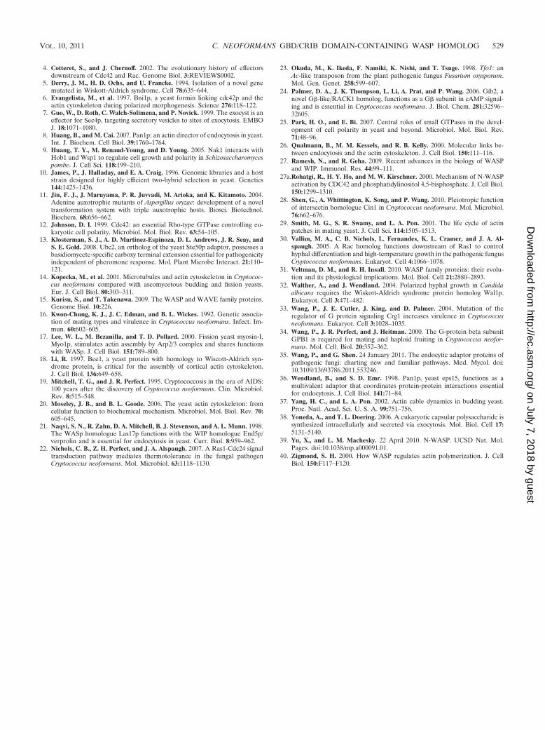
Virulence testing showed that mice infected with the wildtype or the complemented mutant exhibited median survival of44 and 52 days, respectively (Fig. 10A). These survival curveswere not significantly different, as determined by the Gehan-Breslow-Wilcoxon test (P � 0.0684). In contrast, the wsp1mutant-infected mice showed no sign of disease at 90 dayspostinfection (Fig. 10A). Surprisingly, despite showing no signof disease, wsp1-infected animals had detectable CFU in sevenof eight tested brains, three of eight lungs, and one kidneysample. All CFU were confirmed to express resistance toG418, consistent with the observation that the wsp1 cells wereisolated from these mice. When CFU were determined for themouse brains infected with the wsp1 mutant, they were 2 to 3logs lower than those infected by the wild type and the com-plemented strains.
Microscopic examination of brain homogenates stained withIndia ink showed that the wsp1 cells from infected, althoughapparently healthy, animals had aberrant morphologies and
FIG. 7. Wsp1 is required for exosomal localization of a DsRed-Sec4/Sav1 fusion protein. The fusion gene was introduced into a sec4/sav1 mutant strain, and fluorescence was observed under a Zeiss AxioImager 2 microscope. Images were photographed with an attachedZeiss camera and processed using Adobe Photoshop CS4. Sec4/Sav1functions in the secretory pathway, and the DsRed-Sec4 protein ismostly associated with exosomes (exocysts) attached to the plasmamembrane in wild-type cells (top center). In the wsp1 strain, however,the fusion protein appears to have a diffuse pattern and only a smallamount of fluorescence appears in a dotted pattern, suggesting a re-duced exosomal localization and defect in plasma membrane attach-ment (bottom center).
FIG. 8. Wsp1 is required for pheromone-responsive mating.(A) Formation of conjugation tubes, which are filaments produced inresponse to the presence of pheromones by the opposite mating typestrain, can be seen in the wild-type, wsp1 WSP1, and Wsp1-GBD strains(after 2 days at 2°C). The MAT� cells normally produce more abun-dant filaments than MATa cells. The wsp1 strains of either mating typefailed to produce conjugation tubes (after 3 days at 25°C). (B) Wsp1and the GBD domain are required for mating. In the unilateral cross,Wsp1 protein by one of the strains was at least partially sufficient forfunction. Mating of the two strains without GBD domains resulted ina significant reduction in mating-specific filament formation. (C) Fu-sion assay results indicate that the wsp1 mutants failed to undergonuclear fusion, and Wsp1 from one of the parents can provide partialfunction (7 days at 25°C).
526 SHEN ET AL. EUKARYOT. CELL
on July 7, 2018 by guesthttp://ec.asm
.org/D
ownloaded from
inconsistent capsule formation, and some were difficult to vi-sualize compared to the wild type or the complemented strains(Fig. 10B). Upon histological examination, PAS staining of theformalin-fixed brain tissues in WT-infected animals showedfocal and foamy lesions in the brain in both observed speci-mens, with no tropism for a specific area of the brain (Fig.10C). The wild-type cell diameter varied, and most cells werespherical (Fig. 10C). The complemented wsp1 WSP1 strain-infected mice showed more extensive, diffuse lesions in thebrain than those infected with the wild-type strain, and sectionshad an overall foamy appearance (Fig. 10C). In contrast, thewsp1 mutant led to cryptococcal cells in only one of fourobserved specimens. Of the four brain specimens observedhistologically by PAS staining, three exhibited normal brainarchitecture and neither aberrant morphology nor cryptococ-cal cells were observed (Fig. 10C). The wsp1-infected mousebrain with an observable lesion had only one focal lesion withinthe brain tissue, and this was not localized to the outer edge orin the ventricles of the brain (Fig. 10C, insert). Additionally,the lesion contained cryptococcal cells of various but overalllarger sizes, with a greater number of ovoid and pointed cells,which were verified to contain the wsp1 mutant allele by PCR.While this remains an exception, the overall observations are
consistent with survival data and CFU estimation and supporta necessary role for Wsp1 in cryptococcus-host interactionsand wild-type virulence.
DISCUSSION
Wsp1 indicates divergence of WASPs in fungi. The actincytoskeleton underlies essential cellular activities in higher eu-karyotic cells by regulating processes, including cell division,motility, polarized growth, endocytosis, and exocytosis. WASPhas a role in actin dynamics by activating the Arp2/3 proteincomplex through its C-terminal domain to nucleate actin.WASP also interacts with other proteins through additionaldomains, such as WH1, B, and GBD/CRIB, to integrate signalsfor the regulation of actin dynamics. Despite divergence inevolution between higher and lower eukaryotes resulting indifferences in details, this mechanism of actin regulation ap-pears to be largely conserved in fungi. In S. cerevisiae, Las17/Bee1 is required for the assembly of cortical actin patches,budding, and cytokinesis, and disruption of the WASP ho-molog LAS17/BEE1 resulted in a striking change in actin fil-ament organization. Intriguingly, Las17/Bee1 does not containthe GBD/CRIB binding site, which is a conserved feature in-volved in the autoinhibition/activation of mammalian WASPfunctions. Since the regulation of mammalian WASP proteinscan involve autoinhibition or/and trans-inhibition, it is gener-ally suggested that trans-inhibition may be applied to Las17/Bee1 function. While WASP can also be activated by bindingof SH3 domain-containing proteins to its PR-rich domain, the
FIG. 9. Formation of polysaccharide capsule and melanin pigmentand secretion of urease enzyme are attenuated in the wsp1 mutant.(A) The wild-type, wsp1, wsp1 WSP1, and WSP1-GBD strains wereincubated in DMEM for 2 days at 30°C. Cells were stained with Indiaink, and the capsule was observed under a Zeiss Axio Imager 2 mi-croscope (top panel). Bar, 5 �m. Melanin was induced on dopaminemedium, and cells were allowed to grow at 30°C for 2 days (middlepanel). Urease secretion was observed after incubation of all strains inChristensen’s medium for 2 days at 37°C. The wsp1 strain secreted noureases, even when the incubation time was increased to 4 days, whileno qualitative difference in phenol red color shift was seen between thewild-type, wsp1 WSP1, and WSP1-GBD strains. (B) Cells were adjustedto approximately equal density (absorbance at 550 nm), and the liquidurease assay results suggested that urease secretion is indeed de-creased in the wsp1 mutant. The decrease was partially restored in thecomplemented strain (left graph). *, statistically significant difference(P � 0.05). Viability of cells in Christensen’s urea broth was confirmedby CFU plating to ensure equivalent cell counts (right graph).
FIG. 10. Wsp1 is required for virulence in a murine animal model.(A) Disruption of WSP1 results in a complete loss of infectivity inBALB/c mice. The wild-type, wsp1, and wsp1 WSP1 strains were inoc-ulated into mice via lateral tail veins, and mice were observed twicedaily until 90 days postinfection. The wsp1-infected mouse remainedsymptom free, whereas those infected with control strains exhibitedprogressive symptoms and morbidity. (B) Brain smears stained withIndia ink show cryptococcal cells. They were abundant in those in-fected with the wild type (top panel), but their occurrence was rare inwsp1-infected mice (bottom). (C) PAS stain of brains collected frommice infected with the wild-type, wsp1, and wsp1 WSP1 strains. Theinsert in the middle panel (wsp1) details the only brain of four col-lected that had cryptococcal cells (see text for descriptions).
VOL. 10, 2011 C. NEOFORMANS GBD/CRIB DOMAIN-CONTAINING WASP HOMOLOG 527
on July 7, 2018 by guesthttp://ec.asm
.org/D
ownloaded from

activity of Las17/Bee1 can be inhibited by SH3 proteins, suchas SlaI and Bbc1 in S. cerevisiae (for a review see reference 20).Studies of S. pombe Wsp1 and C. albicans Wal1, which areLas17/WASP homologs, demonstrated that these WASP ho-mologs are conserved in function and indicated that fungalWASPs may not contain the GBD/CRIB binding site (17, 32).
We previously identified a multifunctional endocytic pro-tein, Cin1, from the basidiomycetous fungus Cryptococcus neo-formans and established a role of Cin1 in the regulation ofintracellular transport, actin dynamics, growth, and virulenceof the fungus (28). Cin1 shares domain architecture and similarfunctions with human intersectin ITSN1, and homologs ofCin1 cannot be identified in S. cerevisiae or C. albicans, whichhas Pan1, an archetypal endocytic protein involved in endocy-tosis, actin cytoskeleton, and signaling (8, 36). Wsp1 was shownto interact with Cin1 and Cdc42, indicating these three pro-teins likely participate in a complex pathway integrating vari-ous signals to regulate multiple processes, including endocyto-sis and exocytosis, actin dynamics, and signaling (35). While adetailed Cin1-mediated endocytic pathway(s) remains to beillustrated, the unique compositions of Cin1 and Wsp1 pro-teins, as well as the recent identification of SH3 domains spe-cific to basidiomycetous Ste50 (13), all suggest divergent evo-lution of multidomain adaptor proteins within the fungalphylum.
Wsp1 has conserved functions in intracellular transport,actin dynamics, and signaling of C. neoformans. The presenceof a GBD/CRIB domain in Wsp1 suggests that Wsp1 might besubjected to regulation by autoinhibition/activation. A Wsp1-GBD allele-specific mutant was thus included in the generalcharacterization. In short, with the exception of growth assayand bilateral cross results, where reduction of growth (at 37°C)and reduction of mating-specific filaments were apparent, therole of the GBD/CRIB domain appeared to be subtle. This wasrather unexpected, in comparison to mammalian WASPs,where an increase in actin polymerization has been reported(2). We reasoned that there are likely two explanations for this.First, GBD/CRIB is not the only region responsible for auto-inhibition/activation, and the B domain preceding the GBD/CRIB domain may also be required. The B domain interactswith phosphatidylinositol 4,5-bisphosphate to potentiate theeffect of the binding between Cdc42 and the GBD/CRIB do-main (for a review, see reference 39). A more noticeable effectof Wsp1 on actin might be more apparent if both B andGBD/CRIB domains were deleted. Alternately, since Wsp1 isa conserved effector of Cdc42 in actin regulation, as demon-strated in many other systems (4), the effect of a Wsp1-GBD/CRIB domain-specific mutant allele may be more apparent ifCdc42 is rendered in a constitutively inactivated state. Never-theless, further characterization of functions by Wsp1 andCdc42 and their interactions are warranted.
We have identified Wsp1, based on the assumption thatCin1, like ITSN1, functions by interacting with Wsp1/WASPand Rho GTPase Cdc42 to regulate the actin cytoskeleton.Wsp1 was found to interact with Cin1 in our previous study(28) and to bind with Cdc42 in a yeast two-hybrid assay (Fig.1B). We reasoned that Wsp1 likely functions in a conservedfashion analogous to WASP, and we proceeded to examine itscellular function. As expected, the wsp1 mutant displayed de-fects in growth, cytokinesis, chitin distribution, and endocyto-
sis. These functions are in line with those of WASP and Las17/Bee1 protein. Moreover, since the actin cytoskeleton andintracellular transport determine the display of factors contrib-uting to fungal interactions with macrophages and survival inhosts, the wsp1 mutant was unable to cause infection in amurine model. Many of these phenotypes of the wsp1 mutantare quite similar to those of the cin1 mutant, albeit less severe.Further genetic epistasis and biochemical analyses are war-ranted to explore the relationships between the two in theregulation of actin cytoskeleton and other functions.
Wsp1 underscores the importance of actin cytoskeleton ingrowth, differentiation, and virulence. In humans, WAS is aprimary immunodeficiency disease involving both T and B lym-phocytes, which are cells responsible for providing protectionagainst certain viral and fungal infections (T cells) and precur-sors to antibody-producing cells (B cells) in normal individuals.T cells from WAS patients fail to proliferate and to secreteinterleukin-2 after anti-CD3 stimulation, and lymphocytes ofWAS patients also show cytoskeletal abnormalities (27). Thedisease is caused by missense mutations in the WASP genes,especially in the binding sites for the WASP-interacting pro-teins (WIP) that regulate WASP function (11).
Our examination of a C. neoformans mutant strain in whichthe entire WSP1 gene encoding Wsp1 was disrupted provideda direct demonstration for the importance of such proteins.Our phenotypic characterization implicates a role for Wsp1 ingrowth, cytokinesis, and actin distribution of the fungus. Theloss of Wsp1 function resulted in the reduced ability for thefungus to infiltrate macrophages and infect a murine modelanimal. Clearly, while the human WAS diseases were the re-sults of altered or attenuated protein functions, phenotypesexhibited by the wsp1 mutant of C. neoformans provided adirect assessment on the effect of the loss of protein function.It is therefore feasible to propose that study of Wsp1 functioncan provide an alternate but direct approach to explore humanWASP function. True to the “simpler” model of C. neoformansversus much more complex mammalian systems, no WIP pro-teins have been identified from the C. neoformans genome,suggesting that Wsp1 may be subject to a level of regulation farless complicated than that of the mammalian systems. Thus,regulation by Cdc42, a putative upstream activator, and Cin1,whose SH3 and RhoGEF domains bind and potentially acti-vate Wsp1, all point out the importance of exploring the ge-netic relationship among these three proteins. Such explora-tions are no doubt imperative in the study of C. neoformansand in identifying novel anticryptococcal agents.
ACKNOWLEDGMENTS
We thank J. E. Cutler for comments and Sarah Martin for technicalassistance.
This study was supported in part by NIH grants (AI054958 andAI074001) and funding from the Research Institute for Children, NewOrleans, LA.
REFERENCES
1. Ballou, E. R., C. B. Nichols, K. J. Miglia, L. Kozubowski, and J. A. Alspaugh.2010. Two CDC42 paralogues modulate Cryptococcus neoformans thermo-tolerance and morphogenesis under host physiological conditions. Mol. Mi-crobiol. 75:763–780.
2. Cai, L., and J. E. Bear. 2008. Peering deeply inside the branch. J. Cell Biol.180:853–855.
3. Casadevall, A., and J. R. Perfect. 1998. Cryptococcus neoformans. ASMPress, Washington, DC.
528 SHEN ET AL. EUKARYOT. CELL
on July 7, 2018 by guesthttp://ec.asm
.org/D
ownloaded from
4. Cotteret, S., and J. Chernoff. 2002. The evolutionary history of effectorsdownstream of Cdc42 and Rac. Genome Biol. 3:REVIEWS0002.
5. Derry, J. M., H. D. Ochs, and U. Francke. 1994. Isolation of a novel genemutated in Wiskott-Aldrich syndrome. Cell 78:635–644.
6. Evangelista, M., et al. 1997. Bni1p, a yeast formin linking cdc42p and theactin cytoskeleton during polarized morphogenesis. Science 276:118–122.
7. Guo, W., D. Roth, C. Walch-Solimena, and P. Novick. 1999. The exocyst is aneffector for Sec4p, targeting secretory vesicles to sites of exocytosis. EMBOJ. 18:1071–1080.
8. Huang, B., and M. Cai. 2007. Pan1p: an actin director of endocytosis in yeast.Int. J. Biochem. Cell Biol. 39:1760–1764.
9. Huang, T. Y., M. Renaud-Young, and D. Young. 2005. Nak1 interacts withHob1 and Wsp1 to regulate cell growth and polarity in Schizosaccharomycespombe. J. Cell Sci. 118:199–210.
10. James, P., J. Halladay, and E. A. Craig. 1996. Genomic libraries and a hoststrain designed for highly efficient two-hybrid selection in yeast. Genetics144:1425–1436.
11. Jin, F. J., J. Maruyama, P. R. Juvvadi, M. Arioka, and K. Kitamoto. 2004.Adenine auxotrophic mutants of Aspergillus oryzae: development of a noveltransformation system with triple auxotrophic hosts. Biosci. Biotechnol.Biochem. 68:656–662.
12. Johnson, D. I. 1999. Cdc42: an essential Rho-type GTPase controlling eu-karyotic cell polarity. Microbiol. Mol. Biol. Rev. 63:54–105.
13. Klosterman, S. J., A. D. Martinez-Espinoza, D. L. Andrews, J. R. Seay, andS. E. Gold. 2008. Ubc2, an ortholog of the yeast Ste50p adaptor, possesses abasidiomycete-specific carboxy terminal extension essential for pathogenicityindependent of pheromone response. Mol. Plant Microbe Interact. 21:110–121.
14. Kopecka, M., et al. 2001. Microtubules and actin cytoskeleton in Cryptococ-cus neoformans compared with ascomycetous budding and fission yeasts.Eur. J. Cell Biol. 80:303–311.
15. Kurisu, S., and T. Takenawa. 2009. The WASP and WAVE family proteins.Genome Biol. 10:226.
16. Kwon-Chung, K. J., J. C. Edman, and B. L. Wickes. 1992. Genetic associa-tion of mating types and virulence in Cryptococcus neoformans. Infect. Im-mun. 60:602–605.
17. Lee, W. L., M. Bezanilla, and T. D. Pollard. 2000. Fission yeast myosin-I,Myo1p, stimulates actin assembly by Arp2/3 complex and shares functionswith WASp. J. Cell Biol. 151:789–800.
18. Li, R. 1997. Bee1, a yeast protein with homology to Wiscott-Aldrich syn-drome protein, is critical for the assembly of cortical actin cytoskeleton.J. Cell Biol. 136:649–658.
19. Mitchell, T. G., and J. R. Perfect. 1995. Cryptococcosis in the era of AIDS:100 years after the discovery of Cryptococcus neoformans. Clin. Microbiol.Rev. 8:515–548.
20. Moseley, J. B., and B. L. Goode. 2006. The yeast actin cytoskeleton: fromcellular function to biochemical mechanism. Microbiol. Mol. Biol. Rev. 70:605–645.
21. Naqvi, S. N., R. Zahn, D. A. Mitchell, B. J. Stevenson, and A. L. Munn. 1998.The WASp homologue Las17p functions with the WIP homologue End5p/verprolin and is essential for endocytosis in yeast. Curr. Biol. 8:959–962.
22. Nichols, C. B., Z. H. Perfect, and J. A. Alspaugh. 2007. A Ras1-Cdc24 signaltransduction pathway mediates thermotolerance in the fungal pathogenCryptococcus neoformans. Mol. Microbiol. 63:1118–1130.
23. Okuda, M., K. Ikeda, F. Namiki, K. Nishi, and T. Tsuge. 1998. Tfo1: anAc-like transposon from the plant pathogenic fungus Fusarium oxysporum.Mol. Gen. Genet. 258:599–607.
24. Palmer, D. A., J. K. Thompson, L. Li, A. Prat, and P. Wang. 2006. Gib2, anovel G-like/RACK1 homolog, functions as a G subunit in cAMP signal-ing and is essential in Cryptococcus neoformans. J. Biol. Chem. 281:32596–32605.
25. Park, H. O., and E. Bi. 2007. Central roles of small GTPases in the devel-opment of cell polarity in yeast and beyond. Microbiol. Mol. Biol. Rev.71:48–96.
26. Qualmann, B., M. M. Kessels, and R. B. Kelly. 2000. Molecular links be-tween endocytosis and the actin cytoskeleton. J. Cell Biol. 150:111–116.
27. Ramesh, N., and R. Geha. 2009. Recent advances in the biology of WASPand WIP. Immunol. Res. 44:99–111.
27a.Rohatgi, R., H. Y. Ho, and M. W. Kirschner. 2000. Mechanism of N-WASPactivation by CDC42 and phosphatidylinositol 4,5-bisphosphate. J. Cell Biol.150:1299–1310.
28. Shen, G., A. Whittington, K. Song, and P. Wang. 2010. Pleiotropic functionof intersectin homologue Cin1 in Cryptococcus neoformans. Mol. Microbiol.76:662–676.
29. Smith, M. G., S. R. Swamy, and L. A. Pon. 2001. The life cycle of actinpatches in mating yeast. J. Cell Sci. 114:1505–1513.
30. Vallim, M. A., C. B. Nichols, L. Fernandes, K. L. Cramer, and J. A. Al-spaugh. 2005. A Rac homolog functions downstream of Ras1 to controlhyphal differentiation and high-temperature growth in the pathogenic fungusCryptococcus neoformans. Eukaryot. Cell 4:1066–1078.
31. Veltman, D. M., and R. H. Insall. 2010. WASP family proteins: their evolu-tion and its physiological implications. Mol. Biol. Cell 21:2880–2893.
32. Walther, A., and J. Wendland. 2004. Polarized hyphal growth in Candidaalbicans requires the Wiskott-Aldrich syndrome protein homolog Wal1p.Eukaryot. Cell 3:471–482.
33. Wang, P., J. E. Cutler, J. King, and D. Palmer. 2004. Mutation of theregulator of G protein signaling Crg1 increases virulence in Cryptococcusneoformans. Eukaryot. Cell 3:1028–1035.
34. Wang, P., J. R. Perfect, and J. Heitman. 2000. The G-protein beta subunitGPB1 is required for mating and haploid fruiting in Cryptococcus neofor-mans. Mol. Cell. Biol. 20:352–362.
35. Wang, P., and G. Shen. 24 January 2011. The endocytic adaptor proteins ofpathogenic fungi: charting new and familiar pathways. Med. Mycol. doi:10.3109/13693786.2011.553246.
36. Wendland, B., and S. D. Emr. 1998. Pan1p, yeast eps15, functions as amultivalent adaptor that coordinates protein-protein interactions essentialfor endocytosis. J. Cell Biol. 141:71–84.
37. Yang, H. C., and L. A. Pon. 2002. Actin cable dynamics in budding yeast.Proc. Natl. Acad. Sci. U. S. A. 99:751–756.
38. Yoneda, A., and T. L. Doering. 2006. A eukaryotic capsular polysaccharide issynthesized intracellularly and secreted via exocytosis. Mol. Biol. Cell 17:5131–5140.
39. Yu, X., and L. M. Machesky. 22 April 2010. N-WASP. UCSD Nat. Mol.Pages. doi:10.1038/mp.a000091.01.
40. Zigmond, S. H. 2000. How WASP regulates actin polymerization. J. CellBiol. 150:F117–F120.
VOL. 10, 2011 C. NEOFORMANS GBD/CRIB DOMAIN-CONTAINING WASP HOMOLOG 529
on July 7, 2018 by guesthttp://ec.asm
.org/D
ownloaded from


![THE ENZYMES IN PHAGOCYTIC CELLS OF INFLAMMA-€¦ · THE ENZYMES IN PHAGOCYTIC CELLS OF INFLAMMA- TORY EXUDATES. BY EUGENE L. OPIE, M. D. (From the Rocke]eller Institute ]or Medical](https://img.dokumen.tips/doc/110x75/5eae76318e603c29fe31460e/the-enzymes-in-phagocytic-cells-of-inflamma-the-enzymes-in-phagocytic-cells-of.jpg)