Embed Size (px)
Citation preview
9Oxygen and Nitrogen Reactive Metabolitesand Phagocytic Cells
J. Paul RobinsonPurdue University, West Lafayette, Indiana
The goal of this chapter is to clarify the role of phagocytic cells (mainly neutrophils)as mediators of both protection and destruction. Specifically, the linkage between theoxidative metabolic pathways and protection against bacterial invasion is contrastedwith the subsequent tissue damage that ensues when these processes are not proper-ly regulated. Historically, tissue injury, microbial killing, and many pathogenic mech-anisms have been ascribed to reactive oxygen intermediates (ROI). However, withthe realization of the importance of reactive nitrogen intermediates (RNI), attentionhas shifted somewhat from ROI. This recent understanding of the role of nitrogen rad-icals such as nitric oxide and peroxinitrite is compared to the oxygen radical path-ways.
Following the discovery of antioxidants such as superoxide dismutase (SOD) byMcCord and Fridovich in 1969 (McCord and Fridovich, 1969b), most studies pub-lished in neutrophil physiology have been related to the destructive nature of super-oxide (O;), hydrogen peroxide (H,O,), or hydroxyl radical (OH) with the reason-able understanding that these reactive molecules were also the causative agents intissue damage involving phagocytic cells. Neutrophils and macrophages have atremendous capacity for production of these molecules.
Sixty million neutrophils per minute are released into the circulation through the
Phagocyte Function: A Guide for Research and Clinical Evaluation, Edited by J. Paul Robinsonand George F. Babcock. ISBN O-47 1- 12364- 1 Copyright 0 1998 by Wiley-Liss, Inc.
217
218 Reactive Oxygen and Nitrogen Species
normal surveillance mechanism of the reticuloendothelial system. Only erythrocytes(RBC) are produced more prolifically in the body, about 1.5 times the rate of the neu-trophil (Erslev and Weiss, 1977). Thanks to their longer life-span, however, RBC out-number neutrophils in the peripheral circulation by a factor of 103. Neutrophils arereplaced at such a high rate because of their very short half life (several hours) andtotal life-span (3-4 days). A number of important factors affect the final dispositionof these neutrophils, one being a substantial increase-as much as six-to eightfold-in phagocyte production due to stress (Boggs, 1967; Robinson and Mangalik, 1975),and consequently a significant number of immature neutrophils may be present in thecirculation. Because some cytokines can delay apoptosis and thereby increase the life-span of neutrophils, vast quantities of these cells can accumulate at inflammatorysites. The mechanism for removal of neutrophils from inflammatory sites requires theneutrophils to progress toward an apoptotic demise rather than becoming necrotic andreleasing vast quantities of granule enzymes into surrounding tissue. The potential fortissue damage is enormous, particularly if the respiratory mechanism of these cellshas been activated. A delicate balance between manufacture of reactive oxygen rad-icals and their removal or detoxification must be maintained. Understanding the na-ture of this balance mechanism is the key to discerning the difference between pro-tection and destruction in phagocytic cell function.
In this chapter the pathways of oxygen metabolism in phagocytic cells are unrav-eled, as are those of the parallel nitrogen metabolism, and an attempt is made to showthe relationship between protective and destructive mechanisms. Of particular im-portance is our current understanding of the role of peroxynitrite (ONOO-), a mole-cule formed by the union of superoxide and nitric oxide.
OXYGEN-RELATED METABOLISM
A key element in the production of ROI by phagocytes is the NADPH oxidase en-zyme system, which was originally described in neutrophils (Babior et al., 1973). Thisenzyme system is known to consist of several components based upon the b-558 cy-tochrome, a heterodimer consisting of two subunits, gp22-phox ((x unit) and gp91-phox (B unit), as well as a heme moiety and a flavin binding site. It is these (x and l3units that are missing in neutrophils of most patients with chronic granulomatous dis-ease, in which the neutrophils fail to activate the respiratory burst. Additionally, sev-eral other crucial components of the oxidase system must be combined to make theenzyme system active. These include the cytosolic components known as gp47-@ox(a 47 kDa protein) and gp67-phox (a 67 kDa protein), Rat-2 (a GTP-binding protein),and an NADPH-binding protein (Cumutte et al., 1989; Parkos et al., 1987). Phago-cytic cell oxidation pathways are thought to be quite different from those traditional-ly associated with mitochondrial respiration, thus the terminology “phox” for oxi-dase-related proteins-&agocyte aidase, implying that they are somewhat uniqueto phagocytic cells.
In contrast to mitochondrial respiration, where the entire pathway and all compo-nents for respiration are contained within the mitochondria, the phagocyte oxidase
Oxygen-Related Metabolism 219
[gi+rIG T P
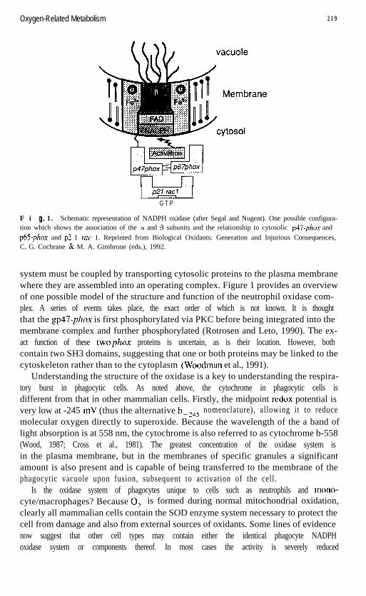
F i 9. 1. Schematic representation of NADPH oxidase (after Segal and Nugent). One possible configura-tion which shows the association of the 01 and p subunits and the relationship to cytosolic p47-phox andp65-phox and p2 1 rut 1. Reprinted from Biological Oxidants: Generation and Injurious Consequences,C. G. Cochrane & M. A. Gimbrone (eds.), 1992.
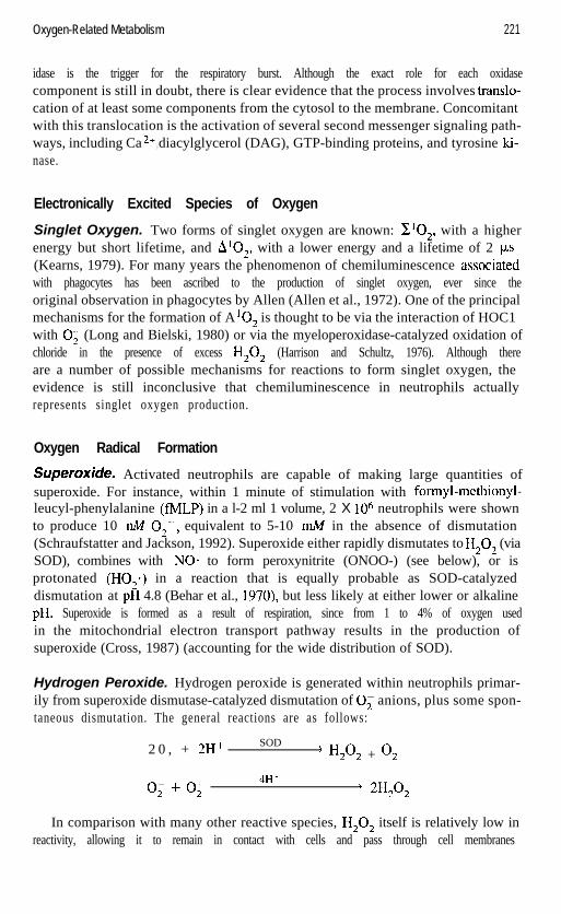
system must be coupled by transporting cytosolic proteins to the plasma membranewhere they are assembled into an operating complex. Figure 1 provides an overviewof one possible model of the structure and function of the neutrophil oxidase com-plex. A series of events takes place, the exact order of which is not known. It is thoughtthat the gp47-phox is first phosphorylated via PKC before being integrated into themembrane complex and further phosphorylated (Rotrosen and Leto, 1990). The ex-act function of these twophox proteins is uncertain, as is their location. However, bothcontain two SH3 domains, suggesting that one or both proteins may be linked to thecytoskeleton rather than to the cytoplasm (Woodman et al., 1991).
Understanding the structure of the oxidase is a key to understanding the respira-tory burst in phagocytic cells. As noted above, the cytochrome in phagocytic cells isdifferent from that in other mammalian cells. Firstly, the midpoint redox potential isvery low at -245 mV (thus the alternative b-,,, nomenclature), allowing it to reducemolecular oxygen directly to superoxide. Because the wavelength of the a band oflight absorption is at 558 nm, the cytochrome is also referred to as cytochrome b-558(Wood, 1987; Cross et al., 1981). The greatest concentration of the oxidase system isin the plasma membrane, but in the membranes of specific granules a significantamount is also present and is capable of being transferred to the membrane of thephagocytic vacuole upon fusion, subsequent to activation of the cell.
Is the oxidase system of phagocytes unique to cells such as neutrophils and mono-cyte/macrophages? Because 0; is formed during normal mitochondrial oxidation,clearly all mammalian cells contain the SOD enzyme system necessary to protect thecell from damage and also from external sources of oxidants. Some lines of evidencenow suggest that other cell types may contain either the identical phagocyte NADPHoxidase system or components thereof. In most cases the activity is severely reduced
Oxygen-Related Metabolism 221
idase is the trigger for the respiratory burst. Although the exact role for each oxidasecomponent is still in doubt, there is clear evidence that the process involves translo-cation of at least some components from the cytosol to the membrane. Concomitantwith this translocation is the activation of several second messenger signaling path-ways, including Ca 2+ diacylglycerol (DAG), GTP-binding proteins, and tyrosine ki-,nase.
Electronically Excited Species of Oxygen
Singlet Oxygen. Two forms of singlet oxygen are known: z102, with a higherenergy but short lifetime, and AlO,, with a lower energy and a lifetime of 2 ps(Keams, 1979). For many years the phenomenon of chemiluminescence .associatedwith phagocytes has been ascribed to the production of singlet oxygen, ever since theoriginal observation in phagocytes by Allen (Allen et al., 1972). One of the principalmechanisms for the formation of AlO, is thought to be via the interaction of HOC1with 0; (Long and Bielski, 1980) or via the myeloperoxidase-catalyzed oxidation ofchloride in the presence of excess H202 (Harrison and Schultz, 1976). Although thereare a number of possible mechanisms for reactions to form singlet oxygen, theevidence is still inconclusive that chemiluminescence in neutrophils actuallyrepresents singlet oxygen production.
Oxygen Radical Formation
Superoxide. Activated neutrophils are capable of making large quantities ofsuperoxide. For instance, within 1 minute of stimulation with formyl-methionyl-leucyl-phenylalanine (fMLP) in a l-2 ml 1 volume, 2 X lo6 neutrophils were shownto produce 10 nM O,-, equivalent to 5-10 m.M in the absence of dismutation(Schraufstatter and Jackson, 1992). Superoxide either rapidly dismutates to H,O, (viaSOD), combines with NO* to form peroxynitrite (ONOO-) (see below), or isprotonated (H02) in a reaction that is equally probable as SOD-catalyzeddismutation at pH 4.8 (Behar et al., 1970), but less likely at either lower or alkalinepH. Superoxide is formed as a result of respiration, since from 1 to 4% of oxygen usedin the mitochondrial electron transport pathway results in the production ofsuperoxide (Cross, 1987) (accounting for the wide distribution of SOD).
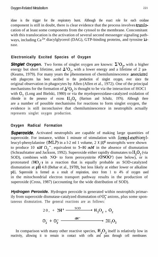
Hydrogen Peroxide. Hydrogen peroxide is generated within neutrophils primar-ily from superoxide dismutase-catalyzed dismutation of 0; anions, plus some spon-taneous dismutation. The general reactions are as follows:
2 0 , + 2H+SOD
) H,O, + 02
0, + 0,4H+
+ 2H202
In comparison with many other reactive species, H,O, itself is relatively low inreactivity, allowing it to remain in contact with cells and pass through cell membranes
Oxygen-Related Metabolism 221
idase is the trigger for the respiratory burst. Although the exact role for each oxidasecomponent is still in doubt, there is clear evidence that the process involves translo-cation of at least some components from the cytosol to the membrane. Concomitantwith this translocation is the activation of several second messenger signaling path-ways, including Ca 2+ diacylglycerol (DAG), GTP-binding proteins, and tyrosine ki-,nase.
Electronically Excited Species of Oxygen
Singlet Oxygen. Two forms of singlet oxygen are known: x10,, with a higherenergy but short lifetime, and AlO,, with a lower energy and a lifetime of 2 ps(Kearns, 1979). For many years the phenomenon of chemiluminescence .associatedwith phagocytes has been ascribed to the production of singlet oxygen, ever since theoriginal observation in phagocytes by Allen (Allen et al., 1972). One of the principalmechanisms for the formation of A lo, is thought to be via the interaction of HOC1with 0, (Long and Bielski, 1980) or via the myeloperoxidase-catalyzed oxidation ofchloride in the presence of excess H,O, (Harrison and Schultz, 1976). Although thereare a number of possible mechanisms for reactions to form singlet oxygen, theevidence is still inconclusive that chemiluminescence in neutrophils actuallyrepresents singlet oxygen production.
Oxygen Radical Formation
&peroxide. Activated neutrophils are capable of making large quantities ofsuperoxide. For instance, within 1 minute of stimulation with formyl-methionyl-leucyl-phenylalanine (fMLP) in a l-2 ml 1 volume, 2 X lo6 neutrophils were shownto produce 10 n.M O,-, equivalent to 5-10 mM in the absence of dismutation(Schraufstatter and Jackson, 1992). Superoxide either rapidly dismutates to H,O, (viaSOD), combines with NO* to form peroxynitrite (ONOO-) (see below), or isprotonated (H02) in a reaction that is equally probable as SOD-catalyzeddismutation at pH 4.8 (Behar et al., 1970), but less likely at either lower or alkalinepH. Superoxide is formed as a result of respiration, since from 1 to 4% of oxygen usedin the mitochondrial electron transport pathway results in the production ofsuperoxide (Cross, 1987) (accounting for the wide distribution of SOD).
Hydrogen Peroxide. Hydrogen peroxide is generated within neutrophils primar-ily from superoxide dismutase-catalyzed dismutation of 0; anions, plus some spon-taneous dismutation. The general reactions are as follows:
2 0 , + 2H+ SOD’ H,O, + 02
0, + 0,4H+
* =I2O2
In comparison with many other reactive species, H202 itself is relatively low inreactivity, allowing it to remain in contact with cells and pass through cell membranes
222 Reactive Oxygen and Nitrogen Species
(Frisch et al., 1983). Catalase breaks down H,O, to oxygen and water, thereby re-moving any potential consequential damage. Interaction with halides such as Cl- viamyeloperoxidase (MPO) and detoxification via the glutathione cycle are the primarymechanisms for removal of H,O, within phagocytes. The glutathione cycle is cou-pled to hexose monophosphate shunt activity because glucose-6-phosphate dehydro-genase and 6-phosphogluconate dehydrogenase reduce NADP+ to NADPH whichmust be reoxidized, a task performed by the glutathione cycle (see Fig. 2). Reducedglutathione (GSH), a vital component in phagocytic cells, can be easily measured us-ing a monobromobimane fluorescent probe (Hedley and Chow, 1994). Reduced glu-tathione is converted to the oxidized form (GSSG) by glutathione peroxidase, andsubsequently reduced back to GSH by NADPH. This cycle is an important “detoxi-fication” system for excess H,O, within the cytosolic environment (Voetman andRoos, 1980).
HY~OC~/O~'OUS Add. The MPO-catalyzed reaction of H,O, with chloride pro-duces a particularly dangerous molecule, hypochlorous acid (HOCl), which acts di-rectly on membrane protein by inactivating sulfhydryl-dependent transporter systems(Schraufstatter et al., 1990). Hypochlorous acid is the predominant species at acidicpH, such as found in activated phagocytes. It also reacts with primary amines to pro-duce monochloramine and taurine monochloramine, ZV-chloramines recognized asvery reactive oxidants. The general reaction proceeds as follows:
H*O*
MPO + 2Cl-) 2HOCl + 2e-
Neutrophils contain a very significant amount of MPO, estimated to be at least 5% ofdry cell weight (Schultz and Kaminker, 1962). This enzyme, found in the azurophilic(primary) granules of neutrophils, has been well characterized biochemically (John-son and Nauseef, 199 1). Other reactions of importance include those with nitrogen-containing compounds to form chloramines. Reactions involving MPO are both nu-merous and complex and beyond the scope of this chapter.
HY&OXJ// R&id. For many years it has been thought that superoxide, while ex-ercising some damaging effects on biological systems, is less important in terms oftissue injury than the more dangerous hydroxyl radical (OH’), whose production fromH,O, is thought to occur by the iron-catalyzed Haber-Weiss reaction:
H*?*
Fe2+) Fe3+ + OH* + OH-
Although this reaction has, to date, provided the most acceptable explanation for tis-sue damage in oxidative systems, there are some troubling points, such as the fact thatthe rate constant for this reaction is somewhat lower than that for the competitive re-action of ascorbic acid with iron. It has therefore been proposed that a more realisticmolecule for tissue damage is ONOO- rather than OH- (Beckman et al., 1990) (seebelow).
Oxygen-Related Metabolism 223
Fluorescent Indicators of Intracellular Oxidation
Many assay systems for measuring neutrophil function have been proposed in the lit-erature. Some recently developed methods allow simultaneous measurement of 0;and H,O,, and NO* as well. These methods are often based upon the use of fluores-cent probes. Five specific probes are discussed here: dichlorofluorescin diacetate, di-hydrorhodamine 123, hydroethidine, parinaric acid, and monobromobimane. Table 1summarizes their commonly used excitation and emission wavelengths and targetmolecules.
Dichlorofluorescin Diacetate. Dichlorofluorescin diacetate (DCFH-DA) hasbeen utilized for H,O, measurement ever since the first application by Keston andBrandt ( 1965) in a bulk cell assay. The probe was later recognized as a useful one fordetermination of H,O, in neutrophils (Gubitz et al., 1976; Homan-Muller et al., 1975)and the technique extended to flow cytometry by Bass et al. (1983). The list of sub-sequent publications, particularly in flow cytometry, is substantial. Dichlorofluo-rescin diacetate has been used to study H,O, production in human neutrophils(Robinson et al., 1994a; Himmelfarb et al., 1992; Epling et al., 1992; Stelzer andRobinson, 1988a; Wolber et al., 1987; Seeds et al., 1985; Smith and Weidemann,1993; Vowells et al., 1995), monocytes/macrophages (Holter et al., 1987; Lepoivre etal., 1986), cultured neurons (Saez et al., 1987), renal epithelial cells (Scott et al.,1988), melanocytes (Boissy et al., 1989), chondrocytes (Tiku et al., 1990), rat en-dothelial cells (Carter et al., 1994b), human umbilical vein endothelial cells(HUVECs) (Niu et al., 1994; Royal1 and Ischiropoulos, 1993), and bovine aorta en-dothelial cells (Royal1 and Ischiropoulos, 1993).
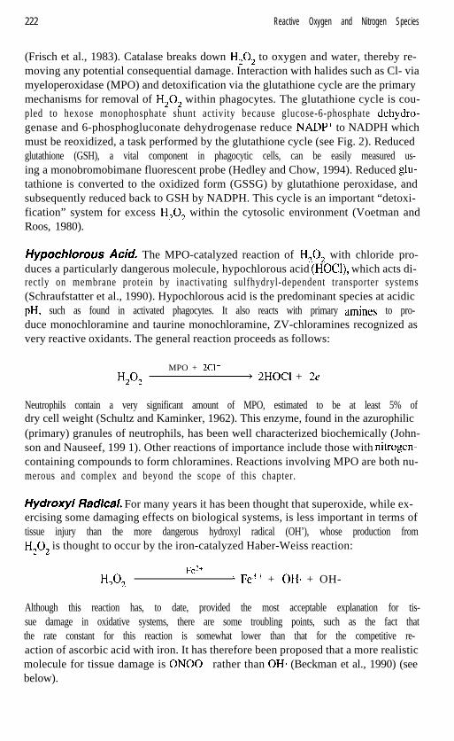
The probe works in the following manner. Dichlorofluorescin diacetate is an es-terified molecule that can freely pass through the cell membrane. Once inside the cell,DCFH-DA is deacetylated by cellular esterases to the nonfluorescent dichlorofluo-rescin (DCFH), which is trapped within the cell by its polar nature. Dichlorofluo-rescin is converted by intracellular oxidants such as H,O, to the green fluorescentmolecule dichlorofluorescein (DCF), a reaction significantly enhanced in the pres-ence of peroxidase. The major difficulties with this probe are its tendency to leak fromthe cell and its lower sensitivity to H,O, as compared to DHR 123. Many studies haveused this probe successfully for intracellular H,O, determination, but careful controlsmust be made to account for cell leakage.
TA B L E 1. Useful Functional Fluorescent Probes
Dye Excitation (nm) Emission (nm) Indicator
DCFH-DA 4 8 8DHR- 123 4 8 8H E 4 8 8Parinaric acid 325-360Monobromobimane 350-360
5 2 0 H2o25 2 0 H,O*6 0 0 0,420-450 Lipid peroxidation420-450 Reduced glutathione
224 Reactive Oxygen and Nitrogen Species
Diacetate
F i Q. 3. Dichlorofluorescin diacetate is hydrolyzed by cellular esterases to the nonfluorescent dichloro-fluorescin, which is readily oxidized to the fluorescent dichlorofluorescein.
Dihydrorhodamine 723. Dihydrorhodamine (DHR 123) is by far the most-usedprobe for measurement of intracellular H,O,. DHR 123 is oxidized directly to rho-damine 123, which is excitable at 488 nm and emits at 5 15 nm in the same emissionrange as DCF and FITC (Rothe et al., 1988). Publications describe its use in humanneutrophils (Wenisch et al., 1996; Emmendorffer et al., 1994; Tanigaki et al., 1993;Waddell et al., 1994; Cao et al., 1993; Demaurex et al., 1996; Smith and Weidemann,1993; Vowells et al., 1995; Rothe et al., 1988; van Pelt et al., 1996; Wenisch et al.,1995), human eosinophils (Elsner et al., 1995), HL60 cells (Kaffenberger and vanBeuningen, 1994), rat mast cells (Tsinkalovsky and Laerum, 1994), guinea pig neu-trophils (Tanigaki et al., 1993), cultured chondrocytes (Hayem et al., 1994), rat braincells (LeBel et al., 1992), rat renal proximal tubular cells (van de Water et al., 1995),mesangial cells (Zent et al., 1995), and L929 cells (Goossens et al., 1995). Combina-tions of DHR123 with surface receptor analysis (Elsner et al., 1994), cell viability us-ing propidium iodide (Clancy et al., 1995), and calcium indicators (Bueb et al., 1995)demonstrate how the probe can be used for simultaneous measurements.
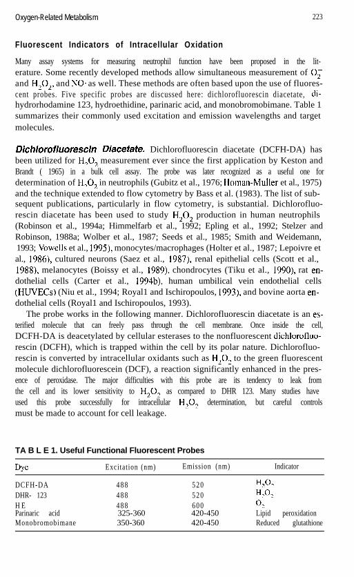
DHR 123 enters the cells as a freely permeable dye that is converted to rhodamine123 and subsequently localized in the mitochondria. The conversion from the non-fluorescent to the fluorescent molecule depends entirely on oxidation products anddoes not require enzymatic catalysis. Once oxidized, the probe is identical to rho-damine 123, a common laser dye. An example of the use of rhodamine 123 directlyin endothelial cells is shown in Figure 5. One significant advantage of the DHR probe
Dihydrorhodoamine 123 Rhodamine 123
F i Q. 4. Dihydrorhodamine enters the cell and is oxidized to rhodamine 123, a fluorescent molecule whichemits at 520 nm, the same as FITC. The oxidation is a result of H,O, production.
Oxygen-Related Metabolism 225
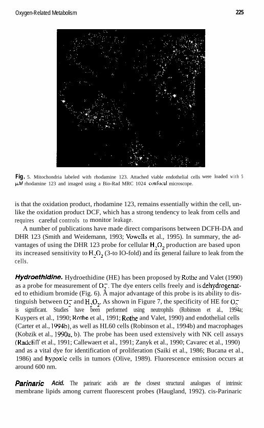
Fig . 5. Mitochondria labeled with rhodamine 123. Attached viable endothelial cells
PM rhodamine 123 and imaged using a Bio-Rad MRC 1024 confocal microscope.were loaded w i t h 5
is that the oxidation product, rhodamine 123, remains essentially within the cell, un-like the oxidation product DCF, which has a strong tendency to leak from cells andrequires careful controls to monitor leakage.
A number of publications have made direct comparisons between DCFH-DA andDHR 123 (Smith and Weidemann, 1993; Vowells et al., 1995). In summary, the ad-vantages of using the DHR 123 probe for cellular H,O, production are based uponits increased sensitivity to H,O, (3-to IO-fold) and its general failure to leak from thecells.
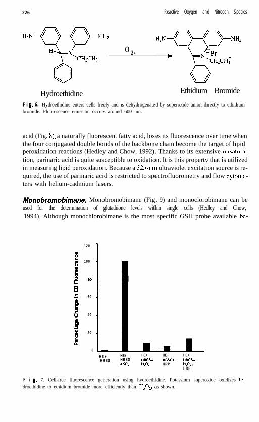
Hydroefhidine. Hydroethidine (HE) has been proposed by Rothe and Valet (1990)as a probe for measurement of 0;. The dye enters cells freely and is dehydrogenat-ed to ethidium bromide (Fig. 6). A major advantage of this probe is its ability to dis-tinguish between 0; and H,O,. As shown in Figure 7, the specificity of HE for 0;is significant. Studies have been performed using neutrophils (Robinson et al., 1994a;Kuypers et al., 1990; Rothe et al., 1991; Rothe and Valet, 1990) and endothelial cells(Carter et al., 1994b), as well as HL60 cells (Robinson et al., 1994b) and macrophages(Kobzik et al., 1990a, b). The probe has been used extensively with NK cell assays(Radcliff et al., 1991; Callewaert et al., 1991; Zanyk et al., 1990; Cavarec et al., 1990)and as a vital dye for identification of proliferation (Saiki et al., 1986; Bucana et al.,1986) and hypoxic cells in tumors (Olive, 1989). Fluorescence emission occurs ataround 600 nm.
Parinaric Acid. The parinaric acids are the closest structural analogues of intrinsicmembrane lipids among current fluorescent probes (Haugland, 1992). cis-Parinaric
226 Reactive Oxygen and Nitrogen Species
N
0 2 -
NH2
Hydroethidine Ethidium Bromide
F i 9. 6. Hydroethidine enters cells freely and is dehydrogenated by superoxide anion directly to ethidiumbromide. Fluorescence emission occurs around 600 nm.
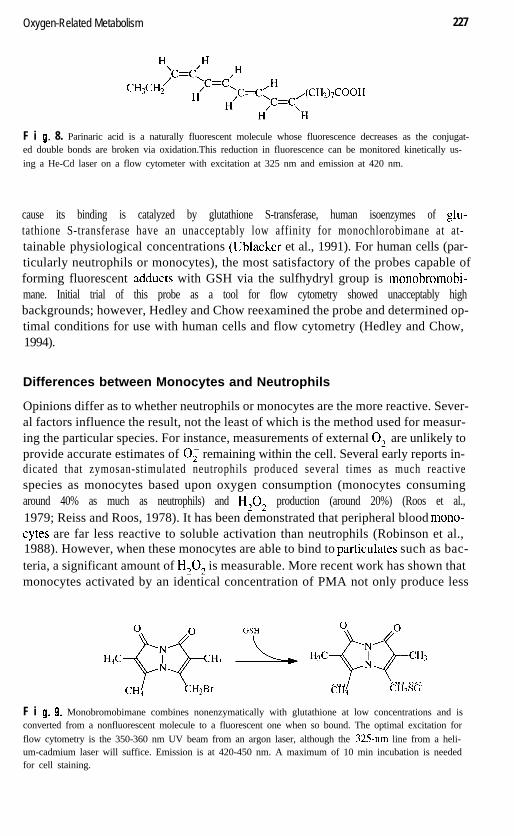
acid (Fig. S), a naturally fluorescent fatty acid, loses its fluorescence over time whenthe four conjugated double bonds of the backbone chain become the target of lipidperoxidation reactions (Hedley and Chow, 1992). Thanks to its extensive unsatura-tion, parinaric acid is quite susceptible to oxidation. It is this property that is utilizedin measuring lipid peroxidation. Because a 325nm ultraviolet excitation source is re-quired, the use of parinaric acid is restricted to spectrofluorometry and flow cytome-ters with helium-cadmium lasers.
Monobromobimane. Monobromobimane (Fig. 9) and monoclorobimane can beused for the determination of glutathione levels within single cells (Hedley and Chow,1994). Although monochlorobimane is the most specific GSH probe available be-
120
100
60
40
20
0
H E +HBSS
HE+ HE+ HE+HBSS HBSS+ HBSS++KO, HA HRP
HE+HBSS+HA +HRP
F i 9. 7. Cell-free fluorescence generation using hydroethidine. Potassium superoxide oxidizes hy-droethidine to ethidium bromide more efficiently than H,O,, as shown.
Oxygen-Related Metabolism 227
F i 9. 8. Parinaric acid is a naturally fluorescent molecule whose fluorescence decreases as the conjugat-ed double bonds are broken via oxidation.This reduction in fluorescence can be monitored kinetically us-ing a He-Cd laser on a flow cytometer with excitation at 325 nm and emission at 420 nm.
cause its binding is catalyzed by glutathione S-transferase, human isoenzymes of glu-tathione S-transferase have an unacceptably low affinity for monochlorobimane at at-tainable physiological concentrations (Ublacker et al., 1991). For human cells (par-ticularly neutrophils or monocytes), the most satisfactory of the probes capable offorming fluorescent adducts with GSH via the sulfhydryl group is monobromobi-mane. Initial trial of this probe as a tool for flow cytometry showed unacceptably highbackgrounds; however, Hedley and Chow reexamined the probe and determined op-timal conditions for use with human cells and flow cytometry (Hedley and Chow,1994).
Differences between Monocytes and Neutrophils
Opinions differ as to whether neutrophils or monocytes are the more reactive. Sever-al factors influence the result, not the least of which is the method used for measur-ing the particular species. For instance, measurements of external 0; are unlikely toprovide accurate estimates of 0; remaining within the cell. Several early reports in-dicated that zymosan-stimulated neutrophils produced several times as much reactivespecies as monocytes based upon oxygen consumption (monocytes consumingaround 40% as much as neutrophils) and H,O, production (around 20%) (Roos et al.,1979; Reiss and Roos, 1978). It has been demonstrated that peripheral blood mono-cytes are far less reactive to soluble activation than neutrophils (Robinson et al.,1988). However, when these monocytes are able to bind to particulates such as bac-teria, a significant amount of H,O, is measurable. More recent work has shown thatmonocytes activated by an identical concentration of PMA not only produce less
H++; ,xb H3c++H3
2 CH3 CH2SG
F i 0. 9. Monobromobimane combines nonenzymatically with glutathione at low concentrations and isconverted from a nonfluorescent molecule to a fluorescent one when so bound. The optimal excitation forflow cytometry is the 350-360 nm UV beam from an argon laser, although the 325-nm line from a heli-um-cadmium laser will suffice. Emission is at 420-450 nm. A maximum of 10 min incubation is neededfor cell staining.
228 Reactive Oxygen and Nitrogen Species
Superoxide HydrogenAnion Peroxide
Neutrophils Monocytes Neutrophils Monocytes
A A A A
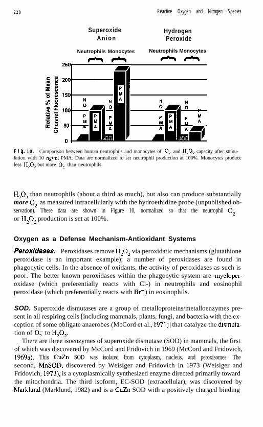
F i g. 10. Comparison between human neutrophils and monocytes of 0; and H,O, capacity after stimu-lation with 10 rig/ml PMA. Data are normalized to set neutrophil production at 100%. Monocytes produceless H,O, but more 0; than neutrophils.
H,O, than neutrophils (about a third as much), but also can produce substantiallymore 0, as measured intracellularly with the hydroethidine probe (unpublished ob-servation). These data are shown in Figure 10, normalized so that the neutrophil O,-or H,O, production is set at 100%.
Oxygen as a Defense Mechanism-Antioxidant Systems
Peroxicfases. Peroxidases remove H,O, via peroxidatic mechanisms (glutathioneperoxidase is an important example); a number of peroxidases are found inphagocytic cells. In the absence of oxidants, the activity of peroxidases as such ispoor. The better known peroxidases within the phagocytic system are myeloper-oxidase (which preferentially reacts with Cl-) in neutrophils and eosinophilperoxidase (which preferentially reacts with Br-) in eosinophils.
SOD. Superoxide dismutases are a group of metalloproteins/metalloenzymes pre-sent in all respiring cells [including mammals, plants, fungi, and bacteria with the ex-ception of some obligate anaerobes (McCord et al., 1971)] that catalyze the dismuta-tion of 0; to H,O,.
There are three isoenzymes of superoxide dismutase (SOD) in mammals, the firstof which was discovered by McCord and Fridovich in 1969 (McCord and Fridovich,1969a). This CuZn SOD was isolated from cytoplasm, nucleus, and peroxisomes. Thesecond, MnSOD, discovered by Weisiger and Fridovich in 1973 (Weisiger andFridovich, 1973), is a cytoplasmically synthesized enzyme directed primarily towardthe mitochondria. The third isoform, EC-SOD (extracellular), was discovered byMarklund (Marklund, 1982) and is a CuZn SOD with a positively charged binding
Oxygen-Related Metabolism 229
domain optimized for localization in the extracellular matrix. This isoenzyme hasbeen shown to have particularly high expression in vascular tissue (Oury et al., 1994)and umbilical cord tissue (Sandstrom et al., 1993).
The structure of CuZn SOD in bovine erythrocytes has been determined as a ho-modimer of 16 kDa with the active site located within a cylinder p structure (Richard-son et al., 1975), where it is well protected and is known to retain catalytic activityduring isolation procedures (Forman and Fridovich, 1973).
The mechanism of action of SOD is that the copper ion at the active site is reducedby one 0; molecule, then reoxidized by another in a continuing cycle (Fridovich,1981). Thus, copper oscillates between the monovalent and divalent states. Copper-containing SOD is inhibitable by cyanide but the Mn form is not (Haffner and Cole-man, 1973). Azide (Misra and Fridovich, 1978) and diethyldithiocarbamate (Heikki-la and Cohen, 1977) (which removes the Cu) are also able to inactivate SOD activityin cell preparations, thus rendering the cells susceptible to autodestruction. RecentlyNaranayan et al. (1998) showed that environmental pollutants such as PCBs reducethe effectiveness of antioxidant systems in human neutrophils, possibly by inactivat-ing SOD activity. If this mechanism is confirmed, it represents the possibility thatchronic low levels of antioxidants may be capable of causing more damage than pre-viously understood, because depletion of SOD and/or glutathione may be responsi-ble for a subsequent increase in apoptosis.
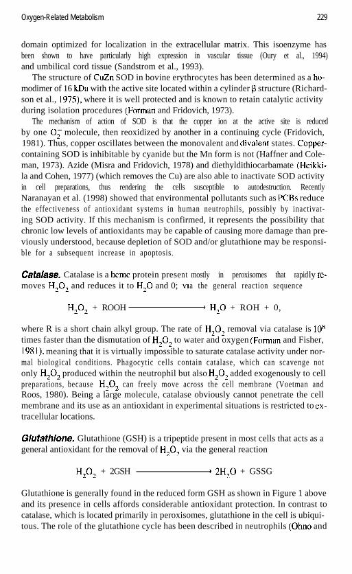
Catalase. Catalase is a heme protein presemoves H,O, and reduces it to H,O and 0;
ntVl
mostly in peroxisomes that rapida the general reaction sequence
ly re-
H,O, + ROOH ’ H,O + ROH + 0,
where R is a short chain alkyl group. The rate of H,O, removal via catalase is lo*times faster than the dismutation of H,O, to water and oxygen (Forman and Fisher,1981), meaning that it is virtually impossible to saturate catalase activity under nor-mal biological conditions. Phagocytic cells contain catalase, which can scavenge notonly H,O, produced within the neutrophil but also H,O, added exogenously to cellpreparations, because H,O, can freely move across the cell membrane (Voetman andRoos, 1980). Being a large molecule, catalase obviously cannot penetrate the cellmembrane and its use as an antioxidant in experimental situations is restricted to ex-tracellular locations.
Ghtathione. Glutathione (GSH) is a tripeptide present in most cells that acts as ageneral antioxidant for the removal of H,O, via the general reaction
H,O, + 2GSH + 2H,O + GSSG
Glutathione is generally found in the reduced form GSH as shown in Figure 1 aboveand its presence in cells affords considerable antioxidant protection. In contrast tocatalase, which is located primarily in peroxisomes, glutathione in the cell is ubiqui-tous. The role of the glutathione cycle has been described in neutrophils (Ohno and
230 Reactive Oxygen and Nitrogen Species
Gallin, 1985) and neutrophil apoptosis (Robinson and Narayanan, 1996), endothelialcells (Andreoli et al., 1986; Tsan et al., 1985), hepatocytes (Keller et al., 1985),platelets (Freedman et al., 1996), and tumor cells (Goossens et al., 1995). Evidencethat nitric oxide may play a regulatory role in neutrophils (and perhaps other cells)has recently been demonstrated (Forslund and Sundqvist, 1995a; Nikitovic andHolmgren, 1996; Clancy et al., 1994).
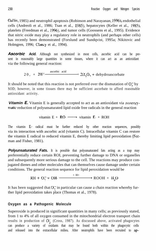
ASCORBIC Acid. Although not synthesized in most cells, ascorbic acid can be pre-sent in reasonably large quantities in some tissues, where it can act as an antioxidantvia the following general reaction:
ascorbic acid2 0 , + 2H+ ) 2H,O, + dehydroascorbate
It should be noted that this reaction is not preferred over the dismutation of 0; bySOD; however, in some tissues there may be sufficient ascorbate to afford reasonableantioxidant activity.
Vitamin E. Vitamin E is generally accepted to act as an antioxidant via nonenzy-matic reduction of polyunsaturated lipid oxide free radicals in the general reaction
vitamin E + RO* -----+ vitamin E* + ROH
The vitamin E* radical must be further reduced by other reaction sequences, possiblyvia its interaction with ascorbic acid (vitamin C). Intracellular vitamin C can restorethe vitamin E radical to reduced vitamin E, thereby limiting lipid peroxidation (For-man and Fisher, 1981).
Polyunsaturated Fats. It is possible that polyunsaturated fats acting as a trap maypreferentially reduce certain ROI, preventing further damage to DNA or organellesand subsequently more serious damage to the cell. The reactions may produce con-jugated dienes and other molecules that can themselves cause damage under certainconditions. The general reaction sequence for lipid peroxidation would be
+ H+RH+O,+OH* ’ ROOH + H,O
It has been suggested that 0; in particular can cause a chain reaction whereby fur-ther lipid peroxidation takes place (Thomas et al., 1978).
Oxygen as a Pathogenic Molecule
Superoxide is produced in significant quantities in many cells; as previously stated,from 1 to 4% of all oxygen consumed in the mitochondrial electron transport chainresults in production of 0; (Cross, 1987). As discussed above, activated phagocytescan produce a variety of oxidants that may be found both within the phagocytic cellsand released into the extracellular milieu. After neutrophils have been recruited to ap-
Nitrogen-Related Metabolism 231
preach, roll, and attach to the vascular endothelium, they transmigrate to the sourceof inflammation. Once at the site, they usually become further activated, producingadditional oxidants. Any neutrophil breakdown releases large quantities of enzymes,with the potential for considerable tissue damage. There are several clear-cut situa-tions in which activated neutrophils cause classic tissue damage, predominantlythrough the production of reactive oxygen species.
One such case is that of complement-mediated lung injury; invasive neutrophilshave been shown to be present in high numbers where severe tissue damage occurs(Till et al., 1982). Similar damage has been observed in a model of immune complexdamage to the lung. In this model, a large neutrophil infiltrate subsequent to intratra-cheal instillation of IgG antibody to bovine serum albumin is thought to be the causeof significant tissue injury (Johnson and Ward, 1974). In terms of free radical gener-ation, it has been shown that alveolar macrophages from patients with asthma have ahigher rate of production of superoxide (Jarjour and Calhoun, 1994).
NITROGEN-RELATED METABOLISM
The discovery of nitric oxide is attributed to Joseph Priestley, a clergyman and part-time chemist, who called his find “nitrous air.” When exposed to iron, this gas wasconverted to nitrous oxide (laughing gas, reviewed by Gilbert, 198 1). The year of thisdiscovery was 1772, but more than 200 years were to pass before the realization thatnitric oxide has a crucial role to play in physiological systems.
The chemistry of nitrogen monoxide (NO) has been elucidated over the past sev-eral years. The most studied molecule has been the radical nitric oxide (NO); how-ever, there are several redox states of NO, including nitrosonium cation (NO+), nitricoxide (NO), and nitroxyl anion (NO-), not dissimilar to the well-known states ofoxygen (dioxygen) (O,), namely, superoxide (0;) and hydrogen peroxide (H202).NO* itself has the lowest molecular weight of any known mammalian secretory prod-uct (Nathan, 1992). The seminal discovery that L-arginine was converted to nitric ox-ide by macrophages and that this was involved in tumoricidal activity (Hibbs et al.,1987) created an entirely new area of research. Literally tens of thousands of reportshave surfaced in the past 10 years associating nitrogen-related metabolites with whatwas previously an exclusive domain of oxygen metabolism.
Production and Properties of Nitric Oxide
Production. Nitric oxide is produced by oxidation of one of the terminal nitrogenson arginine via nitric oxide synthase, producing N-hydroxyarginine, then citrulline,and finally nitric oxide. Each molecule of nitric oxide produced requires one arginine,two oxygens, and 1.5 NADPHs, involving an overall reduction of five electrons.
There are three distinct isoforms of the synthase enzyme, two of which have beentermed constitutive: one from endothelial cells (ECnos-Type III) and one from neu-ronal cells (NCnos -Type I) (Bredt et al., 1991). The constitutive form (cNOS) isCa*+-dependent, appears to be mediated by calmodulin (Pollock et al., 199 l), requires
232 Reactive Oxygen and Nitrogen Species
the presence of co-factors such as tetrahydrobiopterin for activity (Forstermann et al.,1991), and is believed to be located on the cytoplasmic face of the cell membrane(Knowles and Moncada, 1992). The third isoform, calcium-independent and in-ducible (iNOS-Type II), is tightly bound to calmodulin (Nathan and Xie, 1994) andis found in most cells in the body (Stuehr and Griffith, 1992; Nussler and Billiar,1993).
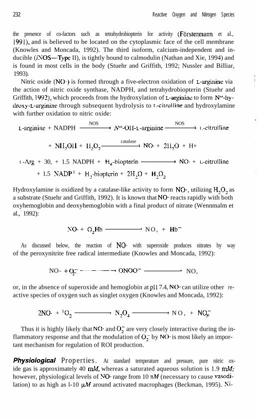
Nitric oxide (NO) is formed through a five-electron oxidation of L-arginine viathe action of nitric oxide synthase, NADPH, and tetrahydrobiopterin (Stuehr andGriffith, 1992), which proceeds from the hydroxylation of L-arginine to form N”-hy-droxy-L-arginine through subsequent hydrolysis to L-citrulline and hydroxylaminewith further oxidation to nitric oxide:
NOS NOSL-arginine + NADPH -N”-OH-L-arginine- L-citrulline
catalase+ NH,OH + H,O, ’ NO* + 2H,O + H+
L-Arg + 30, + 1.5 NADPH + H,-biopterin- NO* + L-citrulline
+ 1.5 NADP+ + H,-biopterin + 2H,O + H,O,
Hydroxylamine is oxidized by a catalase-like activity to form NO*, utilizing H,O, asa substrate (Stuehr and Griffith, 1992). It is known that NO= reacts rapidly with bothoxyhemoglobin and deoxyhemoglobin with a final product of nitrate (Wennmalm etal., 1992):
NO* + 0,Hb ’ N O , + Hb+
As discussed below, the reaction of NO* with superoxide produces nitrates by wayof the peroxynitrite free radical intermediate (Knowles and Moncada, 1992):
NO- +O- - ONOO-2 9 NO,
or, in the absence of superoxide and hemoglobin at pH 7.4, NO. can utilize otheractive species of oxygen such as singlet oxygen (Knowles and Moncada, 1992):
re-
2NOe + lo, ’ N2°4 ’ N O , + NO-3
Thus it is highly likely that NO* and 0; are very closely interactive during the in-flammatory response and that the modulation of 0; by NO* is most likely an impor-tant mechanism for regulation of ROI production.
physio/~~ic~/ Properties. At standard temperature and pressure, pure nitric ox-ide gas is approximately 40 mA4, whereas a saturated aqueous solution is 1.9 mM;however, physiological levels of NO- range from 10 nlM (necessary to cause vasodi-lation) to as high as l-10 ILM around activated macrophages (Beckman, 1995). Ni-
Nitrogen-Related Metabolism 233
tric oxides have potentially extensive half-lives in tissue, as long as 17 h for a 10 nA4concentration and around 11 min for a 1 FM concentration of NO* (Beckman andTsai, 1994).
One of the earlier effects observed for NO- was that it prevented neutrophil ag-gregation (Kubes et al., 1991; McCall et al., 1988). Thus nitric oxide could regulatethe buildup of inflammatory cells and reduce potential tissue damage. The mecha-nism of leukocyte recruitment is well understood. Leukocyte adhesion to endothelialcells is primarily mediated by the P-integrin glycoprotein complex on neutrophils(Tonnesen, 1989). NO- plays a role in P-selectin- dependent leukocyte rolling, forN”-nitro-L-arginine methyl ester pretreatment of rats with L-NAME caused increasedleukocyte rolling (Davenpeck et al., 1994). It is known that once an inflammatorystate is signaled (via a chemoattractant, for instance), neutrophils slow down and gen-tly adhere to the wall of the blood vessel via L-selectin. Because the adherence is oflow affinity, the effect is a rolling along the endothelial layer. The L-selectin (on neu-trophils) binding via E(ELAM- l)- and P(CD62)-selectins (on endothelial cells) is es-sentially intermittent until the neutrophil approaches sufficiently close to the sourceof the inflammatory mediator. At this point, the neutrophil slows and then adheres tothe endothelial surface via adhesion glycoproteins (CD 1 l/CD 18) on neutrophils andtheir ligands (ICAM- 1) on endothelial cells. Now the neutrophil migrates through themicrovessel wall, a process that takes several minutes. Thus the regulatory role thatNO* appears to play in expression of P-selectin is physiologically very significant,with the implication that NO* acts as a homeostatic regulator of neutrophil-endothe-lial interactions (Beckman, 1995).
Other physiological effects of nitric oxide on neutrophils include direct inhibitionof superoxide production, possibly by direct action on the NADPH oxidase (Clancyet al., 1992), providing additional evidence of protection against oxidative injury. Ina similar manner, nitric oxide has been shown to act against mast cells, demonstrat-ing a diminution of mast cell reactivity. It has been shown that exvenous addition ofNO* via the NO- producer nitroprusside decreased the amount of histamine releasedby mast cells (Salvemini et al., 199 1) as well as the amount of platelet activating fac-tor produced by them (Hogaboam et al., 1993).
Peroxynitrite
Peroxynitrite (ONOO-) is formed from a radical-radical reaction between 0; andNO* at a diffusion-limited rate (k = 6.7 X 10⁹M- Is- ‘) (Huie and Padmaja, 1993).It has been known for many years that superoxide and nitric oxide could combine toform peroxynitrite, as well as the conditions under which this will occur (Blough andZafiriou, 1985). Making accurate measurements of ONOO- in biological systems,however, proved more difficult. Beckman et al. (1990) have determined many of theproperties of peroxynitrite. The pK, is 7.49 at 37OC, and once protonated it rapidlydecomposes with a half-life of 1.9 s. Macrophages are capable of producing at leastlmmol/min of peroxynitrite; and the effects on bacterial survival are not altered bymannitol, ethanol, or benzoate, but are enhanced by dimethyl sulfoxide or deferox-amine (Zhu et al., 1992).
234 Reactive Oxygen and Nitrogen Species
When ONOO-is protonated, the resultant peroxynitrous acid (ONOOH) decom-poses to several toxic species with reactivity similar to hydroxyl radical and nitrogendioxide (Volk et al., 1995; Radi et al., 1991; Beckman et al., 1990; Ischiropoulos etal., 1992a,b). Peroxynitrite has also been shown to have high bactericidal capacity inmacrophages (Zhu et al., 1992; Ischiropoulos et al., 1992a,b) and to replace the ac-tive site of CuZn superoxide dismutase, removing its dismutation capacity for super-oxide anion by blocking the 0; binding site (Beckman et al., 1992; Ischiropoulos etal., 1992a,b). DNA strand breakage (Inoue and Kawanishi, 1995; Szabo et al., 1996)demonstrated in macrophages (Szabo et al., 1996), smooth muscle (McCauley andHartmann, 1984; Begley et al., 1985), and thymocytes (Salgo et al., 1995) has beenattributed to ONOO-. Furthermore, ONOO- can directly oxidize sulkydryl groupssuch as cysteine and BSA at physiological pH (7.4) at rates 1000 times faster thanH,O, (Radi et al., 1991a).
The situation is further complicated by the fact that transition metals can catalyzeONOO- to form OH- (hydroxyl anion) plus nitronium ion (NO,+), which can sub-sequently nitrate protein tyrosine residues to produce 3-nitrotyrosine in a reaction cat-alyzed by SOD (Beckman et al., 1992), whose normal role is to dismutate 0;.
It has been suggested that the toxicity of either the 0; or NO* radical is signifi-cantly enhanced when the two combine to form ONOO- (Beckman et al., 1990). Per-oxynitrite has been shown to exhibit strong activity with a number of biologicalmolecules, and a substantial amount of evidence demonstrates that ONOO- is con-siderably more reactive than NO*. The strong oxidizing capability has been shown toact on peroxidation of lipids in the absence of iron (Rubbo et al., 1994; Radi et al.,199 1 a), cause formation of malondialdehyde and conjugated dienes during lipid per-oxidation (Radi et al., 199 lb), and also act directly on carbohydrates (Beckman et al.,1990).
Peroxynitrite inhibits pulmonary ~11 -proteinase inhibitor and therefore oxidizescritical methionine residues in the active site of the enzyme (Moreno and Pryor,1992). Peroxynitrite is also responsible for the oxidation of arachidonic acid and for-mation of F2-isoprostanes via oxidation of low-density lipoproteins (Moore et al.,1995) and causes inhibition of mitochondrial respiratory chain enzymes, includingcytosolic aconitase (Radi et al., 1994; Hausladen and Fridovich, 1994).
Deferoxamine and glutathione are scavengers of peroxynitrite, whereas H,O, hasbeen shown to prolong its half-life (Alvarez et al., 1995). It is likely that most of thedamaging effects against mitochondria result from the activity of peroxynitrite, notOH* or other radicals, for antioxidants against 0; or OH- provide little protection.
Under certain conditions the NO*/O; interaction may behave as a significant an-tioxidant mechanism for the detoxification of 0;. What is clear is that the most im-portant criteria for the role of the NO* and 0, combination may be the relative flux-es of each molecule and the nature of the tissue in which they are formed.
Fluorescent Indicators of Excited Nitrogen Species
Some confusion exists as to what particular fluorescent probes actually measure. Anumber of reports in the literature claim to measure one or another oxidant, when in
Nitrogen-Related Metabolism 235
fact there is no clear evidence as to what molecule is actually being measured. Thisproblem is witnessed by the vast quantity of studies described in the early 1990s pur-porting to measure nitric oxide, but in reality measuring an accumulation of nitrites.This has led many investigators to the conclusion that NO* was indeed produced andwas the key biological species. Many publications have assumed that the species con-tributing the biological effect was NO* when it could well have been, and most prob-ably was, a combination of excited molecules. Regardless, the conclusion has beendrawn on many occasions that NO. was the key to the biological mystery being un-raveled. In a similar manner, NO* and ONOO- measurements using fluorescentprobes may have resulted in the same problem of crossreactivity already observedwith probes for 0; and H,O,.
Dihydrorhodamine 723. DHR 123 has recently become a popular probe for mea-suring peroxynitrite. Haddad et al. (1994) have used DHR 123 to measure peroxyni-trite production, claiming that the simultaneous generation of 0; and NO* resultedin the oxidation of DHR 123 to rhodamine 123. They claim further that DHR 123 isnot oxidized by NO*, O;, or H,O, alone (Kim et al., 1996; Haddad et al., 1994).Clearly there are contradictory publications on the nature and use of certain fluores-cent probes, and the choice of such probes depends entirely on the biological systemin question. Many studies have used DHR 123 as a direct measure of peroxynitrite(Szabo et al., 1995); for cases involving noncellular systems where highly defined re-actions can be studied, the efficacy of the probe is acceptable. In one such controlledsystem the authors concluded that in the presence of oxygen, nitric oxide induces arelatively slow oxidation of dihydrorhodamine owing to the formation of nitrogendioxide and consequently that dihydrorhodamine was a sensitive and efficient trap forperoxynitrite (Miles et al., 1996; Kooy et al., 1994).
When using these probes for more complex biological systems such as cell sus-pensions or for in vivo use, care must be exercised in drawing and interpreting con-clusions from the data. Experiments such as injecting DHR 123 directly into rats andmeasuring subsequent plasma levels of rhodamine 123 spectrofluorometrically arenot straightforward cases of cause and effect. In that instance, the conclusions that theresulting measurement was a direct measure of peroxynitrite (Szabo et al., 1995)must be interpreted with great caution, and in the case of peroxynitrite measurements,are only relative to NOS-inhibitable changes in fluorescence as discussed by Szabo(Szabo et al., 1995).
Dichlorofluorescin Diacetafe. Evidence exists that DCFH-DA under some cir-cumstances may actually measure NO* in addition to H,O,. Such evidence includesthe observation for human neutrophils that a calmodulin inhibitor (W-13) inhibits0, production as measured by cytochrome c and nitroblue tetrazolium assays but en-hances the formation of fluorescent DCF (Rao et al., 1992). Furthermore, this reac-tion was inhibitable by NMMA (N”-monomethyl-L-arginine), an L-arginine analoguethat inhibits production of NO*. Additionally, it was demonstrated that pure NO* gascould directly oxidize DCFH to the fluorescent DCF (Rao et al., 1992).
It has also been observed that DCFH-DA-loaded neutrophils incubated with a
236 Reactive Oxygen and Nitrogen Species
Release of Caged Nitric Oxide inAtta%hed PMN
E 250 CONTROL STUDY
c2 200sg 150
l d
z
. 8 100
88 50x2s OE 0 20 40 60 80 100120 1401bu
Time (seconds) after UV FLASH E 0 1 0 0 2 0 0 3 0 0 4 0 0Time (seconds) CONTROL
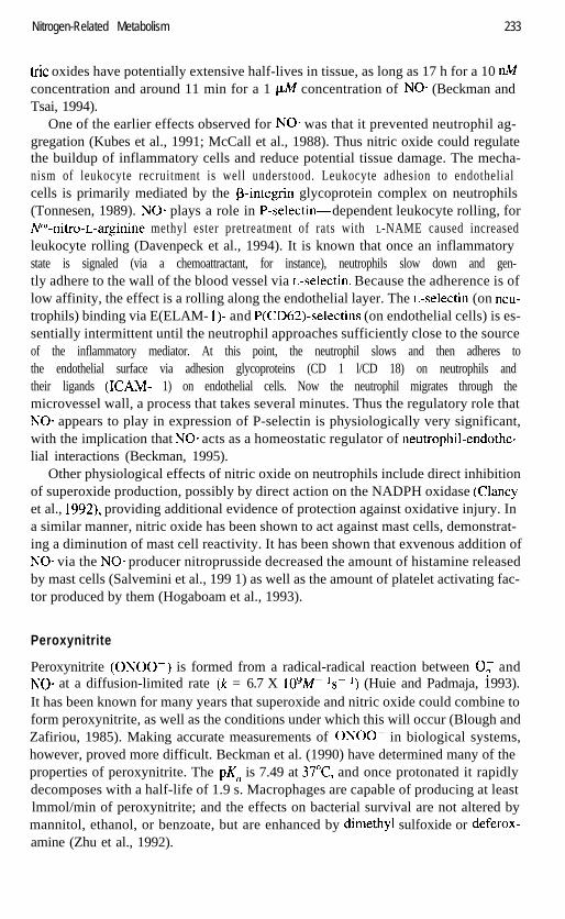
F i Q. 11. The change in fluorescence over time after a controlled UV flash (360 nm excitation) on a smallpopulation of neutrophils previously loaded with DCFH-DA. Neutrophils attached to coverslips coatedwith polylysine were loaded with DCFH-DA (20 @I) and incubated at 37°C on the heated stage of a Bio-Rad MRC 1024 confocal microscope for 20 min to hydrolyze the probe. Cells were then incubated withpotassium nitrosylpentachlororuthenate (PNPCR, a “caged nitric oxide” compound). Control cells notflashed with UV light but given equal exposure to 48%nm excitation demonstrated no fluorescence shift.Neither did cells incubated with DCFH-DA alone and given the same UV flash.
caged nitric oxide showed an increase in DCF fluorescence after the cage was re-leased by ultraviolet light. In these (unpublished) studies it appears that the DCFH-DA probe was measuring the released NO*, for the cells were otherwise not stimu-lated to produce reactive oxygen species (Fig. 11). Ultraviolet light alone did notappear to change the fluorescent nature of the DCFH probe.
Nitric Oxide Production in Neutrophils
Although there is no doubt that neutrophils can produce staggering quantities of oxy-gen radicals, their ability to produce nitric oxide has been disputed. Before the bio-logical properties of nitric oxide had been elucidated, reports of the impact of rat neu-trophils on relaxation of smooth muscle (Rimele et al., 1988), elevated cGMP levels(Lee et al., 1988), and platelet antiagreggation factor (McCall et al., 1989) would sug-gest involvement of NO* based upon present-day knowledge. Subsequently, the “fac-tor” present in these neutrophils was shown to be nitric oxide (Schmidt et al., 1989;Wright et al., 1989). The controversy, however, has continued.
The question as to whether the presence of nitrites indicates intracellular NO* pro-duction or results from alternative metabolic pathways has been addressed by a num-ber of groups. Klebanoff and Nathan (1993) measured nitrite production in humanneutrophils in the presence of azide and catalase; nitrite production was unaffectedby addition of either SOD or monomethylarginine. When stimulated neutrophils werereplaced with the H,O,-generating system glucose-glucose oxidase, nitrite was alsoproduced, leading these authors to conclude that nitrite production did not reflect ni-tric oxide synthase activity, but rather the catalase-catalyzed conversion of azide in
Nitrogen-Related Metabolism 237
the presence of H,O, generated by stimulated neutrophils. Another study by Padgettand Pruett (1995) found that rat, mouse, and human neutrophils were able to pro-duce very small amounts of nitrite, far less than would be required for antimicrobialactivity. They conclude that the small amount of NO* produced by these cells maybe related to intercellular signaling rather than playing a role as a defense mecha-nism.
These data are contrasted with a significant literature confirming the presence ofboth iNOS and subsequent production of significant amounts of NO-. Identificationof iNOS expression has been confirmed in human neutrophils by measuring reversetranscription polymerase chain reaction (RT-PCR) products (Kolls et al., 1994; Cooket al., 1994; Evans et al., 1996). In one neutrophil sudy, PCR (polymerase chain re-action) products to both the endothelial constitutive (756 bp) and neuronal constitu-tive (629 bp) NOS isoforms were identified (Chen and Mehta, 1996). Confirmationof iNOS or NO- production has been by Southern blot (Chen and Mehta, 1996), spec-trophotometric measurement of nitric oxide-dependent methemoglobin formationfrom oxyhemoglobin (Larfars and Gyllenhamrnar, 1995), and antibodies againstiNOS in rat neutrophils (Clark et al., 1996). Detailed immunohistochemistry with aspecific anti-nitrotyrosine antibody showed intense staining in both macrophages andneutrophils in mouse lung (Akaike et al., 1996) and rat macrophages and neutrophils(Goldman et al., 1996), and reports of the conversion of “H-arginine to 3H-citrullinein human monocytes and neutrophils (Laffi et al., 1995) have been published.
Secondary evidence exists for the effect of NOS inhibitors on neutrophil function.For example, NOS inhibition attenuated chemotaxis of both unstimulated and primedneutrophils, suggesting a role for NO- synthesis in neutrophil emigration (Wildhirt etal., 1995) as well as in monocyte chemotaxis (Belenky et al., 1993), and exogenousNO* has been shown to induce chemotactic locomotion in human neutrophils (Beau-vais et al., 1995). Other evidence of NO* production is based upon nitrite productionalone (Ahmed and Weidemann, 1996; Dias-Da-Motta et al., 1996; Carreras et al.,1994a; Biswas et al., 1993) and chemiluminescence (Forslund and Sundqvist, 1995a;Catz et al., 1995; Carreras et al., 1994b).
Wright et al. (1989) showed that human neutrophils were able to generate nitricoxide at a rate of 1.8 nmol/2 X lo6 cells/30 min. Direct evidence of NO* through de-tection in the gas phase of the specific chemiluminescence resulting from the reac-tion of nitric oxide with ozone showed categorically that neutrophils produce NO*.
It is becoming increasingly clear that NO*, present either exogenously or intracel-lularly, exerts serious influence on the physiology of neutrophil function. Productionof 0; is reduced by the direct effects of extracellular NO* on the neutrophil mem-brane-bound oxidase (Forslund and Sundqvist, 1995b). Augmentation of bacterialphagocytosis by human neutrophils in the presence of L-arginine (but not D-arginineor glycine) has been demonstrated (Moffat et al., 1996). NO* has been shown to re-duce neutrophil adherence (Clancy et al., 1995; Egdell et al., 1994; McCall et al.,1988) and cause depletion in intracellular glutathione (Clancy et al., 1994), but not toaffect neutrophil-mediated killing by fMLP-activated rat neutrophils (Wagner et al.,1996). Anesthetics such as lidocaine stimulate NO. production in human neutrophils(Mamiya et al., 1995).
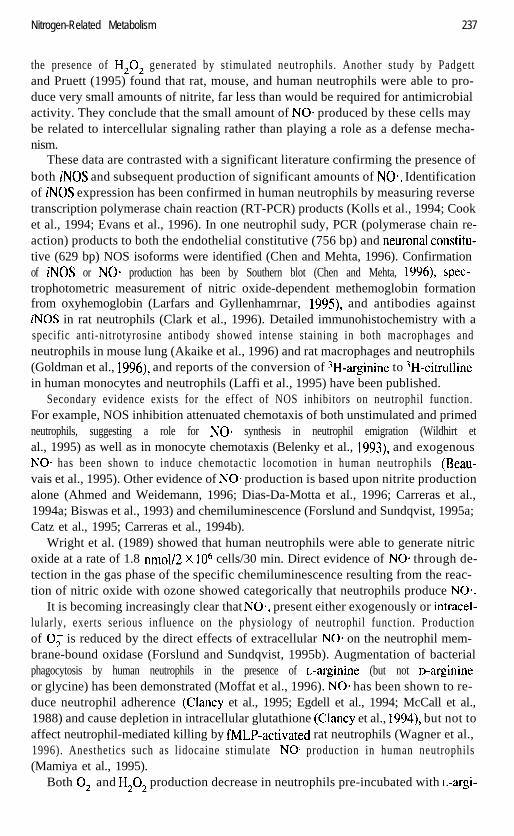
Both 0; and H,O, production decrease in neutrophils pre-incubated with L-argi-
238 Reactive Oxygen and Nitrogen Species
Superoxide HydrogenAnion Peroxide
Control L-arg in ine Control L-arg in ine
F i 9. 12. Relative percent of 0; and H,O, in human neutrophils with and without added L-arginine. 2rig/ml PMA was used for cell stimulation. Both basal and stimulated 0, and H,O, levels decreased afterpreincubation of PMN with 1 mM L-arginine.
nine and stimulated with low concentrations of PMA (2 rig/ml) (Fig. 12). In one setof experiments, reductions were 35 and 43% for 0, and H,O,, respectively. In un-stimulated cells, L-arginine also caused a reduction in the basal O;- and H,O,-in-duced fluorescence by approximately 15% each. These data provide evidence that L-
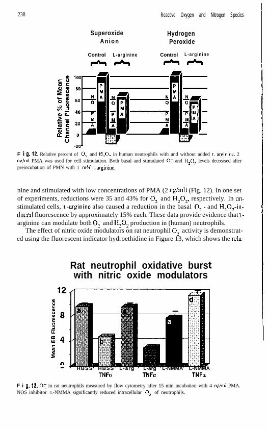
arginine can modulate both 0; and H,O, production in (human) neutrophils.The effect of nitric oxide modulators on rat neutrophil 0; activity is demonstrat-
ed using the fluorescent indicator hydroethidine in Figure 13, which shows the rela-
Rat neutrophil oxidative burswith nitric oxide modulators
HBSS HBSS L-arg L-arg L-NMMA L-NMMATNFa TNFa TNFa
F i 9. 13. 0; in rat neutrophils measured by flow cytometry after 15 min incubation with 4 rig/ml PMA.NOS inhibitor L-NMMA significantly reduced intracellular 0; of neutrophils.
Nitrogen-Related Metabolism 239
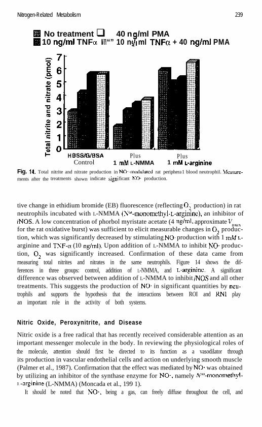
No treatment q 40 n /ml PMA10 ng/mI TNFa :!j;y:I”“” 10 ng ml TNFa + 40 ng!mI PMA7
Fig. 14. Total nitrite and nitrate production in NO*-modulated rat peripheraments after the treatments shown indicate signi ficant NO. production.
H BSS/G/BSA Plus PlusControl 1 mM L-NMMA 1 mM L-arginine
1 blood neutrophil. Measure-
tive change in ethidium bromide (EB) fluorescence (reflecting 0; production) in ratneutrophils incubated with L-NMMA (NW-monomethyl-L-arginine), an inhibitor ofiNOS. A low concentration of phorbol myristate acetate (4 rig/ml, approximate Vmaxfor the rat oxidative burst) was sufficient to elicit measurable changes in 0; produc-tion, which was significantly decreased by stimulating NO* production with 1 W L-arginine and TNF-(x (10 rig/ml). Upon addition of L-NMMA to inhibit NO* produc-tion, 0; was significantly increased. Confirmation of these data came frommeasuring total nitrites and nitrates in the same neutrophils. Figure 14 shows the dif-ferences in three groups: control, addition of L-NMMA, and L-arginine. A significantdifference was observed between addition of L-NMMA to inhibit iNOS and all othertreatments. This suggests the production of NO* in significant quantities by neu-trophils and supports the hypothesis that the interactions between ROI and RN1 playan important role in the activity of both systems.
Nitric Oxide, Peroxynitrite, and Disease
Nitric oxide is a free radical that has recently received considerable attention as animportant messenger molecule in the body. In reviewing the physiological roles ofthe molecule, attention should first be directed to its function as a vasodilator throughits production in vascular endothelial cells and action on underlying smooth muscle(Palmer et al., 1987). Confirmation that the effect was mediated by NO* was obtainedby utilizing an inhibitor of the synthase enzyme for NO*, namely W-monomethyl-L-arginine (L-NMMA) (Moncada et al., 199 1).
It should be noted that NO-, being a gas, can freely diffuse throughout the cell, and
240 Reactive Oxygen and Nitrogen Species
indeed from one cell to another, unless bound by protein. The molecule can survivefor about lo-30 s in typical physiological environments, during which time it hasbeen hypothesized to travel between 200 and 600 urn (based upon diffusion coeffi-cient of oxygen in tissue sections) (Knowles and Moncada, 1992).
The recognition that nitric oxide regulation can affect some disease processes hasbrought about an interest in the therapeutic use of high L-arginine diets (Becker et al.,1993; Saito et al., 1987; Gianotti et al., 1993) or L-arginine infusion in traumatizedpatients. Studies in L-arginine-treated rats have demonstrated significant alterationsin the translocation of gut bacteria. In one study (Becker et al., 1993), the amount ofNO* was substantially increased in L-arginine-fed rats after bum. In another (Gian-otti et al., 1993), the authors concluded that one of the more significant effects of L-arginine administration was influence on immune function, most likely the macro-phage. Current dogma suggests that increases in intracellular and extracellular nitricoxide may be beneficial to the appropriate microenvironment since the nitric oxidecan act as an 0; scavenger, essentially being converted to nitrite and nitrate and fore-stalling the conversion of 0; via SOD to H,O, and OH-.
A similar situation might exist in ischemia/reperfusion injury, in which the role ofneutrophils has been demonstrated, particularly in remote injury such as seen in thelung (Mulligan et al., 1992). In addition, it has recently been demonstrated that en-dotoxin can cause a two-to fivefold increase in arginine transport by pulmonary arteryendothelial cells (Lind et al., 1993), suggesting that vast amounts of NO- can be made,at least in the lung, which consists of approximately 50% endothelial cells (Crap0 etal., 1978), creating significant quantities of potentially dangerous molecules. Ad-ministration of a recombinant human TNF soluble receptor type 1 to lipopolysac-charide-treated rats significantly reduced the damaging effects of endotoxin in lungtissue, probably by preventing the upregulation of IL-6 by TNF (Ulich et al., 1993).
Thus we have a curious situation. On the one hand, superoxide and its dismutationproducts, hydrogen peroxide and subsequent hydroxyl radical, have been consideredto be the dangerous species of oxygen-mediated tissue damage. On the other hand, ithas been hypothesized that SOD, by removal of superoxide anions, creates a protec-tive barrier by directly preventing the reaction of 0; with NO* to produce peroxyni-trite (Beckman et al., 1990) in a reaction known to be very rapid with a rate constantof at least 3.7 X lo7 rn-ls-’ (Zhu et al., 1992).
Nitrogen as a Defense Mechanism
The concept that NO- can act as a defense mechanism is based upon the knowledgethat NO* can remove superoxide from a system by a diffusion-limited reaction thatforms peroxynitrite (ONOO-):
0- + NO. - ONOO-2
Furthermore, this reaction occurs faster than the dismutation of 0; by SOD and there-fore is highly likely to take place in biological systems. It is thought that NO* can re-act with transition metals, especially Fe2+ (which may also be the most available met-
Nitrogen-Related Metabolism
al), thus competing with the Haber-Weiss reaction between H,O, and Fe2+. If this istrue, NO* may be acting as an antioxidant.
Nitric oxide has been identified as playing a protective role by a number of stud-ies, such as that of Wink et al. (1993), who showed that in the presence of NO*, thecytotoxicity of H,O, and 0; against Chinese hamster lung fibroblasts was abrogat-ed in a dose-responsive manner. Nitric oxide has been shown to promote ADP ribo-sylation of actin, thus inhibiting cytoskeletal assembly in neutrophils. The result isthe regulation of neutrophil adhesion in margination, adhesion, and diapedesis (Clan-cy et al., 1995). Suppression of NO* in arteriolar and venular endothelium resulted inan increase in adhesion of leukocytes, but also a significant increase in oxidativestress. Furthermore, this L-NAME-induced enhancement of endothelial adhesivenesswas mediated by intracellular oxidative stress rather than by direct action of NO* sup-pression (Hausladen and Fridovich, 1994).
Prolonged NOS inhibition in HUVECs caused an oxidative- and platelet activat-ing factor (PAF)-associated rise in adhesion of neutrophils on the surface of en-dothelial cells (Niu et al., 1994), activated mast cells in the mucosa (Kanwar et al.,1994), and mast cells in rat mesenteric postcapillary venules (Kubes et al., 1993). In-hibition of nitric oxide synthesis increased leukocyte and endothelial interaction inrat mesenteric venules as measured by an increase in P-selectin expression (Daven-peck et al., 1994). Likewise Gauthier et al. (1994) demonstrated a reduction in adhe-sion and decreased P-selectin expression following infusion of exogenous nitric ox-ide. Volk et al. (1995) have demonstrated that extracellularly produced H,O,, but notO;, enhanced the toxicity of NO* against endothelial cells. Decreased basal releaseof NO. after myocardial ischemia/reperfusion preceded enhanced neutrophil adher-ence to the coronary endothelium, leading to neutrophil-induced myocardial injury(Ma et al., 1993).
Nitric oxide overproduction was shown to be a major protective mechanism in theT cell-dependent shock induced by staphylococcal enterotoxin B in mice, and NOSinhibition might have detrimental consequences in T cell-mediated inflammatory dis-orders by enhancing both production and toxicity of inflammatory cytokines(Florquin et al., 1994). These data indicated that NO* production was able to exert adirect effect on the production of some cytokines (TNF-a and IFN-y). Additionally,there is a significant amount of evidence that the presence of NO* regulates the ad-hesion of neutrophils, and in the absence of NO*-producing systems, a potentiallydamaging accumulation of inflammatory cells is likely.
Nitrogen as a Pathogenic Molecule
Direct tissue damage by NO* has also been demonstrated, but usually at higher thanphysiological concentrations. Administration of high concentrations of NO* (via ni-troprusside, lo-40 kg/kg/min for 15 min) was shown to cause rat mucosal damage,which was attributed to excessive formation of peroxynitrite and subsequent forma-tion of superoxide and hydroxyl radicals (Lamarque and Whittle, 1995).
The primary evidence for a role of nitric oxide in pathogenesis must come fromthe effect of peroxynitrite, a potent and reactive oxidant (Beckman and Tsai, 1994).
242 Reactive Oxygen and Nitrogen Species
Peroxynitrite has been shown to damage endothelial cells directly (Palmer et al.,1992; Kooy and Royall, 1994; Kooy et al., 1994), and to impair vascular permeabil-ity (Villa et al., 1994) and play a role in the pathogenesis of atherogenesis (White etal., 1994).
CONCLUSIONS
It is clear that many of the observations previously considered to be related solely toreactive oxygen species are more likely associated with both ROI and RN1 and theinteractions between the two. There is a strong body of evidence that peroxynitrite isa mediator of serious consequences, and that NO. plays a powerful role in regulatingmany of the inflammatory responses, particularly those involving interaction betweenneutrophils and endothelial cells.
The benefits of using NOS inhibitors or NO. donors are yet to be made clear. Ifperoxynitrite is a particularly dangerous molecule, then prevention of its formationby NOS inhibitors is logical. However, the consequences of blocking NO* formationare themselves quite serious, leading to increased neutrophil adhesion and simulta-neous removal of a significant pathway for elimination of superoxide.
Neutrophils and macrophages are particularly reactive cells. Neutrophils know butthe one role, which they play to perfection: they are designed with powerful protec-tive mechanisms for antioxidant defenses. They produce reactive oxygen and nitro-gen species as a means of destroying invading microorganisms, while attempting atthe same time to stay alive, at least until they have completed their disinfection task.It is during this period- between the attachment phase and destruction of microbes,and their subsequent removal from inflammatory sites-that significant tissue dam-age can occur. Alternatively, when vast numbers of these cells respond, as in is-chemia/reperfusion injury, regulation of their reactive nature is necesasary to preventrapid and destructive tissue injury. How exactly this regulation can be achieved with-out further compromising the host is a question still seeking answers.
REFERENCES
Ahmed N, Weidemann MJ (1996): Interaction of reactive nitrogen and oxygen intermediates in HL60 anddimethylsulphoxide-differentiated HL60 cells. Leuk Res 20:27 l-279.
Akaike T, Noguchi Y, Ijiri S, Setoguchi K, Suga M, Zheng YM, Dietzschold B, Maeda H (1996): Patho-genesis of influenza virus-induced pneumonia: involvement of both nitric oxide and oxygen radicals.Proc Nat1 Acad Sci USA 93:2448-2453.
Allen RC, Stjernholm RL, Steele RH (1972): Evidence for the generation of an electronic state(s) in hu-man polymorphonuclear leukocytes and its participation in bactericidal activity. Biochem Biophys ResCommun 47:679-684.
Alvarez B, Denicola A, Radi R (1995): Reaction between peroxynitrite and hydrogen peroxide: formationof oxygen and slowing of peroxynitrite decomposition. Chem Res Toxic01 8:859-864.
Andreoli SP, Mallett CP, Bergstein JM (1986): Role of glutathione in protecting endothelial cells againsthydrogen peroxide oxidant injury. J Lab Clin Med 108: 190-l 98.
References
Babior BM, Kipnes RS, Curnutte JT (1973): Biological defense mechanisms. The production by leuko-cytes of superoxide, a potential bactericidal agent. J CIin Invest 52:741-744.
Bass DA, Parce JW, DeChatelet LR, Szejda P, Seeds MC, Thomas M (1983): Flow cytometric studies ofoxidative product formation by neutrophils: a graded response to membrane stimulation. J Immunol130:1910-1917.
Beauvais F, Michel L, Dubertret L ( 1995): Exogenous nitric oxide elicits chemotaxis of neutrophils in vi-tro. J Cell Physiol 1656 10-6 14.
Becker WK, Shippee RL, McManus AT, Mason AD Jr., Pruitt BA Jr (1993): Kinetics of nitrogen oxideproduction following experimental thermal injury in rats. J Trauma 34855862.
Beckman JS (1995): Biochemistry of nitric oxide and peroxynitrite. In Kubes P (ed). Nitric Oxide: A Mod-ulator of Cell-Cell Interactions in the Microcirculation. Austin, TX: R. G. Landes Company, pp l-l 8.
Beckman JS, Beckman TW, Chen J, Marshall PA, Freeman BA (1990): Apparent hydroxyl radical pro-duction by peroxynitrite: Implications for endothelial injury from nitric oxide and superoxide. ProcNat1 Acad Sci USA 87: 1620-l 624.
Beckman JS, Ischiropoulos H, Zhu L, et al. (1992): Kinetics of superoxide dismutase and iron catalyzednitration of phenolics by peroxynitrite. Arch Biochem Biophys 298:438-445.
Beckman JS, Tsai JHM ( 1994): Reaction rates and diffusion in the toxicity of peroxynitrite. Biochemist16:8-10.
Begley CG, Lopez AF, Vadas MA, Metcalf D ( 1985): The clonal proliferation in vitro of enriched popula-tions of human promyelocytes and myelocytes. Blood 65:95 l-958.
Behar D, Czapski G, Rabani J, Dorfman LM, Schwartz HA ( 1970): The acid dissociation constant and de-cay kinetics of the perhydroxyl radical. J Phys Chem 74:3209.
Belenky SN, Robbins RA, Rubinstein I (1993): Nitric oxide synthase inhibitors attenuate human mono-cyte chemotaxis in vitro. J Leukoc Biol 53:498-503.
Biswas SK, Bhelwa AP, Upadhyay AU, George A, Nath N ( 1993): Status of nitric oxide free radicals in di-abetic neutrophils: effect of diabetic serum factor on the generation of these species in normal neu-trophils and their relation to lysosomal degranulation. Indian J Biochem Biophys 30:293-296.
Blough NV, Zafiriou OC (1985): Reaction of superoxide with nitric oxide to form peroxonitrite in alkalineaqueous solution. Inorg Chem 24:3502-3504.
Boggs DR (1967): The kinetics of neutrophilic leukocytes in health and disease. Semin Hematol4:359-386.
Boissy RE, Trinkle LS, Nordlund JJ ( 1989): Separation of pigmented and albino melanocytes and the con-comitant evaluation of endogenous peroxide content using flow cytometry. Cytometry 10:779-787.
Bredt DS, Glatt CE, Hwang PM, Fotuhi M, Dawson TM, Snyder SH ( 199 1): Nitric oxide synthase proteinand mRNA are discretely localized in neuronal populations of the mammalian CNS together with theNADPH diaphorase. Neuron 7:6 15-624.
Bucana C, Saiki I, Nayar R (1986): Uptake and accumulation of the vital dye hydroethidine in neoplasticcells. J Histochem Cytochem 34: 1109-l 115.
Bueb J-L, Gallois A, Schneider J-C, Parini J-P, Tschirhart E ( 1995): A double-labelling fluorescent assayfor concomitant measurements of [Ca2+li and 0; production in human macrophages. Biochim Bio-phys Acta 1244:79-84.
Callewaert DM, Radcliff G, Waite R, LeFevre J, Poulik MD ( 1991): Characterization of effector-targetconjugates for cloned human natural killer and human lymphokine activated killer cells by flow cy-tometry. Cytometry 12:666-676.
Cao D, Boxer LA, Petty HR (1993): Deposition of reactive oxygen metabolites onto and within living tu-mor cells during neutrophil-mediated antibody-dependent cellular cytotoxicity. J Cell Physiol156:428436.
Carreras MC, Catz SD, Pargament GA, Del Bosco CG, Poderoso JJ ( 1994a): Decreased production of ni-tric oxide by human neutrophils during septic multiple organ dysfunction syndrome. Comparison withendotoxin and cytokine effects on normal cells. Inflammation 18: 15 I- 16 1.
244 Reactive Oxygen and Nitrogen Species
Carreras MC, Pargament GA, Catz SD, Poderoso JJ, Boveris A (1994b): Kinetics of nitric oxide and hy- drogen peroxide production and formation of peroxynitrite during the respiratory burst of human neu- trophils. FEBS Lett 34 1 :65-68.
Carter WO, Knapp DW, Snyder P, et al. (1994a): Oxidative burst-like activity in canine natural killer cells. Cytometry 7(Suppl):28 (Abstract).
Carter WO, Narayanan PK, Robinson JP (1994b): Intracellular hydrogen peroxide and superoxide anion detection in endothelial cells. J Leukoc Biol 55:253-258.
Catz SD, Carreras MC, Poderoso JJ (1995): Nitric oxide synthase inhibitors decrease human polymor- phonuclear leukocyte luminol-dependent chemilumine5cence. Free Radic Biol Med 19:741-748
Cavarec L, Quillet MA, Fradelizi D, Conjeaud H (1990): An improved double fluorescence flow cytome- try method for the quantification of killer cellltarget cell conjugate formation. J Immunol Meth 130:25 1-26 1.
Chen LY, Mehta JL (1996): Variable effects of L-arginine analogs on L-arginine-nitric oxide pathway in human neutrophils and platelets may relate to different nitric oxide synthase isoforms. J Pharmacol Exp Ther 276:253-257.
Clancy R, Leszczynska J, Amin A, Levartovsky D, Abramson SB (1995): Nitric oxide stimulates ADP ri- bosylation of actin in association with the inhibition of actin polymerization in human neutrophils. J Leukoc Biol58: 196-202.
Clancy RM, Leszczynska-Piziak J, Abramson SB (1992): Nitric oxide, an endothelial cell relaxation fac- tor, inhibits neutrophil superoxide anion production via a direct action on the NADPH oxidase. J Clin Invest 9O:lll6-ll2l.
Clancy RM, Levartovsky D, Leszczynska-Piziak J, Yegudin J, Abramson SB (1994): Nitric oxide reacts with intracellular glutathione and activates the hexose monophosphate shunt in human neutrophils: ev- idence for S-nitrosoglutathione as a bioactive intermediary. Proc Natl Acad Sci USA 91:368G3684.
Clark RS, Kochanek PM, Schwarz MA, et al. (1996): Inducible nitric oxide synthase expression in cere- brovascular smooth muscle and neutrophils after traumatic brain injury in immature rats. Pediatr Res 39:784-790.
Cook HT, Ebrahim H, Jansen AS, Foster GR, Largen P, Cattell V (1994): Expression of the gene for in- ducible nitric oxide synthase in experimental glomerulonephritis in the rat. Clin Exp Immunol 97:s 15-320.
Crapo JD, Peters-Golden M, Marsh-Salin J, Shelburne, JS (1978): Pathologic changes in the lungs of oxy- gen-adapted rats: a morphometric analysis. Lab Invest 39:64&653.
Cross AR, Jones OTG, Harper AM, Segal AW (198 1): Oxidation-reduction properties of the cytochrome h found in the plasma-membrane fraction of human neutrophils. Biochem J 194:599-606.
Cross CE (1987): Oxygen radicals and human disease. Ann Intern Med 107526-545.
Curnutte JT, Scott PJ, Mayo LA (1989): Cytosolic components of the respiratory burst oxidase: resolution of four components, two of which are missing in complementing types of chronic granulomatous dis- ease. Proc Natl Acad Sci USA 86:825-829.
Davenpeck KL, Gauthier TW, Lefer AM (1994): Inhibition of endothelial-derived nitric oxide promotes P-selectin expression and actions in the rat microcirculation. Gastroenterology 107: 1050-1058.
Demaurex N, Downey GP, Waddell TK, Grinstein S (1996): lntracellular pH regulation during spreading of human neutrophils. J Cell Biol 133: 1391-1402.
Dias-Da-Motta P, Arruda VR, Muscara MN, et al. (1996): The release of nitric oxide and superoxide an- ion by neutrophils and mononuclear cells from patients with sickle cell anaemia. Br J Haematol 93:333-340.
Egdell RM, Siminiak T, Sheridan DJ (1994): Modulation of neutrophil activity by nitric oxide during acute myocardial ischaemia and reperfusion [review]. Basic Res Cardiol 89:499-509.
Elsner J, Dichmann S, KappA (1995): Activation of the respiratory burst in human eosinophils by chemo- taxins requires intracellular calcium fluxes. J Invest Dermatol 105:231-236.
Elsner J, Oppermann M, Czech W, Kapp A (1994): C3a activates the respiratory burst in human polymor- phonuclear neutrophilic leukocytes via pertussis toxin-sensitive G-proteins. Blood 83:3324-3331.