Embed Size (px)
Citation preview

Case ReportWooden Penetrating Pelvic Trauma Passing theForamen Ischiadicum
Benedikt Ruben Abel , Heinz-Lothar Meyer, Roman Müller, Katharina Henze,Marcel Dudda, and Max Daniel Kauther
Klinik für Unfall-, Hand- und Wiederherstellungschirurgie, University Hospital Essen, Hufelandstraße 55, 45147 Essen, Germany
Correspondence should be addressed to Benedikt Ruben Abel; [email protected]
Received 2 May 2019; Accepted 9 December 2019; Published 27 December 2019
Academic Editor: Eyal Itshayek
Copyright © 2019 Benedikt Ruben Abel et al. This is an open access article distributed under the Creative Commons AttributionLicense, which permits unrestricted use, distribution, and reproduction in any medium, provided the original work isproperly cited.
We describe the case of a 45-year-old woman who suffered an impalement injury of the pelvis with penetration of the sciaticforamen by a wooden foreign body. Following a single operation, the injury healed without complications or infection. We havetaken this as an opportunity to describe the case and our standard procedure in more detail.
1. Introduction
Before we started writing this report, we chose the terms“penetrating pelvic trauma” and “pelvic impalement” forour literature research.
For the former, we obtained 12 results, mostly describingpenetrating injuries due to metallic foreign bodies. Five arti-cles reported on gunshot wounds, and about half of theresults reported on trauma in a military context with appro-priate treatment strategies and options.
The search term “pelvic impalement” yielded four results,only one of which explicitly referred to wooden impalementtrauma [1].
Pelvic trauma is a common occurrence and is mainlyassociated with traffic accidents. An evaluation of theGerman DGU TR data from 2015 to 2017 showed that 95%of all trauma patients suffered blunt trauma, while 15% hada pelvic injury [2]. In addition to the frequent blunt pelvicinjuries, penetrating and especially impaling pelvic injuriesare clearly underrepresented. For penetrating pelvic injuriesin the military context, mortality of between 21% and 36%is stated in the literature [3]. Besides the general risks result-ing from penetrating traumas, those affecting the pelvicregion involve the additional high risk of infection due topossible injury to the rectum or colon. If hypotension occurs
at the same time and colonoscopy cannot be performed,mortality increases significantly [4].
The anatomical conditions in the pelvis make injuries toimportant vessels or nerves very likely in penetrating pelvicinjuries. Significant bleeding, as can occur with both bluntand penetrating trauma in the pelvic area, causes highmortal-ity andmorbidity [5]. Pelvic trauma is common in emergencymedicine, especially injuries caused by high-speed violence.The significance of the case we describe here lies in theaccident mechanism and in the material of the penetratingforeign body.
However only a few cases of pelvic injuries caused by thepenetration of a wooden foreign body have been reported inthe literature so far. This small number encouraged us topublish this case report in order to increase the availabledocumentation concerning treatment of these injuries.
2. Case Report
We report on a 45-year-old female patient who sustained apelvis penetrating injury after a fall from a height of threemeters. The patient fell while working on a ladder and landedwith the right gluteal region on a wooden fence. The portal ofentry of the wooden fence pole was located laterally at thejunction of the gluteal region and the dorsolateral thigh.
HindawiCase Reports in OrthopedicsVolume 2019, Article ID 5834129, 4 pageshttps://doi.org/10.1155/2019/5834129
Passers-by rescued the victim by lifting her off the fence pole.When the ambulance arrived, the patient was conscious, ori-ented, and in a hemodynamically stable condition. Circula-tion, motor functions, and sensitivity in all extremities wereintact. In the initial body check, instability of the pelviscould not definitely be ruled out. The recap time of the legswas 3-4 seconds. Following the establishment of two largelumen peripheral accesses, analgosedation of the patient(height: 166 cm; weight: 70 kg) with a total of 30mg esketa-min and 3mg midazolam was performed. After approxi-mately 20 minutes of treatment on-site with appropriatesalvage recovery and stabilization of the pelvis by means ofa pelvic sling, the patient was taken to the Trauma SurgeryDepartment of the University Hospital Essen and admittedto the shock room.
On admission, the patient was responsive (GCS 13) andhemodynamically stable (HR: 62/min; BP: 109/62mmHg;RR: 12/min; SpO2: 99%; shock index: 0.6; Revised TraumaScore: 8). Clinical examination of the patient in the shockroom revealed a moderately bleeding wound in the area ofthe right gluteal region of about 5 cm in diameter. Instabilityof the pelvis could not be excluded with certainty. The initialHb was 13.1 g/dl. Parallel to the clinical examination, FASTsonography was performed and yielded no evidence of freeintraabdominal fluid.
While still in the shock room, the patient received antibi-otic therapy according to our internal guidelines with cefazo-lin 2 g. This was followed by an X-ray of the pelvis and theright thigh. A subsequent CT body scan revealed no evidenceof active arterial bleeding or foreign body involvement in thepelvic area or any other relevant injuries, but did show aright-sided presacral hematoma with multiple fine airpockets on the right side (Figures 1 and 2). The rectum was
displaced towards ventral and left lateral. In the perirectal adi-pose tissue, several fine air inclusions were delineated in theright lateral pelvic region, and a penetration canal throughthe M. gluteus maximus was visible (Figures 1 and 2). Theveins and arteries were visualized very clearly with good con-trast. There were no signs of free fluid and therefore no reasonto suspect acute bleeding. There was also no free intraabdom-inal air. The patient had no history of previous illnesses orpermanent medication.
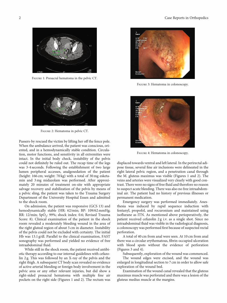
Emergency surgery was performed immediately. Anes-thesia was induced by rapid sequence induction withfentanyl, propofol, and rocuronium and maintained usingisoflurane as ITN. As mentioned above perioperatively, thepatient received cefazolin 2 g i.v. as a single shot. Since nointraabdominal fluid was visible in the radiological diagnosis,a colonoscopy was performed first because of suspected rectalperforation.
A total of 40 cm from anal were seen. At 10 cm from analthere was a circular erythematous, fibrin-occupied ulcerationwith blood spots without the evidence of perforation(Figures 3 and 4).
Subsequently, exploration of the wound was commenced.The wound edges were excised, and the wound was
enlarged in longitudinal section to 7 cm in order to allow safeexploration of the wound bed.
Examination of the wound canal revealed that the gluteusmaximus muscle was perforated and there was a lesion of thegluteus medius muscle at the margins.
Figure 1: Presacral hematoma in the pelvic CT.
Figure 2: Hematoma in pelvic CT.
Figure 3: Hematoma in colonoscopy.
Figure 4: Hematoma in colonoscopy.
2 Case Reports in Orthopedics

A coagulated hematoma was removed by careful andrepeated irrigation of the perforation channel with constantpalpatory control.
The perforation canal could be palpated through theforamen ischiadicum as far as the area of the rectum. Afurther coagulated hematoma was found in the area ofthe foramen ischiadicum. Here, there was no indication ofactive bleeding.
Apart from the muscular lesions, there was no evi-dence of injury to any of the pathways of the vascular andnerval tracts.
Under palpatory control, a size-16 Charrière Redon drainwas inserted and repeated careful and extensive irrigation ofthe penetration canal was carried out. No splinters of the for-eign body were detected, either by palpation or by irrigation.
A second Redon drain, Charrière 12, was introducedsubcutaneously after closure of the gluteal fascia.
The wound was closed by dermal suture with inserteddrains (Prolene 2.0).
Postoperatively, the patient was extubated promptly.The blood flow and the motor and sensory system of the
right leg was always intact during inpatient treatment.Postoperatively, the patient received piperacillin/-
tazobactam 4.5 g three times daily for six days i.v.Further treatment in our IMCU was carried out for five
days. The drains did not collect any relevant amount of secre-tion or blood and were removed four days postoperatively.
Initially, the patient was mobilized with partial weight-bearing and from the eighth postoperative day pain-adaptedfull weight-bearing was permitted.
Thrombosis prophylaxis was performed with enoxaparin-natrium 40mg once daily until safe full weight-bearingwas possible.
The patient remained in the hospital for 12 dayspostoperatively.
Approximately two and a half weeks after the trauma, anout-patient follow-up colonoscopy was performed. Here, theulcerations described above had left a scar which presentedno complications.
The patient returned for out-patient follow-up examina-tion seven months after discharge.
Using VAS, the patient reported the following pain inten-sities for the respective time points: immediately aftertrauma—1; shock room—2; immediately post op—6; twodays post op—3; and 8 months after trauma—1.
In an evaluation according to the SF36 score, thepatient reached 95% for physical function seven monthsafter trauma.
The range of motion of the hips at that time is asfollows: Ext/Flex left—10-0-110; Ext/Flex right—10-0-110;ER/IR left—40-0-30; ER/IR right—40-0-30; Abd/Addleft—45-0-30; and Abd/Add right—30-0-15. The scar hadhealed without irritation.
Overall, follow-up examination revealed a favorablelong-term outcome without any residual effects which wouldlimit the patient’s quality of life. Only the range of abductionand adduction in the right hip appeared to be reduced incomparison with the contralateral hip. The patient’s gaitwas normal.
3. Discussion
We consider the following points to be crucial for the treat-ment of an impaling injury in the pelvic area.
In the preclinical phase, the procedure described in thePTLS recommendations for blunt pelvic trauma should befollowed, since in this setting the exact nature and extent ofthe injury cannot be sufficiently differentiated [6].
In the first clinical phase, we consider the procedureaccording to the ATLS to be the best strategy.
Hornez et al. in 2016 [7] designed a flowchart for thetreatment of penetrating injuries.
While we largely agree with this chart, we would like toadd a few points. First, when a hemodynamically unstablepatient is delivered to the emergency department, the proce-dure according to the principles of damage control is withoutalternative [7]. The hemodynamically unstable patient is at agreat risk of dying and all attention must be focused on pre-venting death by stopping the loss of blood. With hemody-namically stable patients, in contrast, there is more time tofocus on the long-term outcome and the possibility of restitu-tio ad integrum. Second, in addition to standard diagnostics(X-ray, CT angiography), the foreign body itself should alsobe at the center of attention. The already high risk of infec-tion due to penetrating injuries should not be unnecessarilyincreased by the fact that the properties of the foreign bodyare not taken into account. Metal has a high radiopacity,but a wooden foreign object is insufficiently visualized byX-ray diagnostics. Due to the nature of wood, there is a highrisk of splintering or incomplete recovery of the foreign body.For wooden foreign bodies, a maximum sensitivity of 77.2%can be assumed in radiological CT diagnosis [8]. In our opin-ion, this weakness in imaging diagnostics of wooden foreignbodies must be compensated by thorough and comprehen-sive surgical exploration. Even with a negative result fromimaging diagnostics, the surgical procedure should alwaysbe chosen so as not to overlook fragments of a foreign bodyand thereby risk an unnecessary foreign body-associatedinfection [9].
We therefore see the necessity for larger studies withmore patients to develop more advanced and differentiatedconcepts for the treatment of penetrating pelvic trauma bywooden foreign objects.
4. Conclusions
(i) Preclinical: treatment according to the recommenda-tions for polytrauma or blunt pelvic trauma.
(ii) Emergency department: treatment according to ATLSprinciples and rapid differentiation between vitallyvulnerable patients and vitally less endangeredpatients.
(a) Vitally endangered patients: immediate surgicaltreatment according to damage control princi-ples and hemodynamic stabilization [7].
(b) Vitally less endangered patients: rapid butextensive preoperative diagnosis (X-ray, CT
3Case Reports in Orthopedics

angiography, and if necessary, sonography) andcalculated preoperative antibiotic therapy (amox-icillin/clavulan acid 1000/2000mg+metronida-zole 500mg i.v.). Surgical removal of the foreignbody with extensive surgical exploration, micro-biological samples, debridement, local antiseptictherapy, drainage, and continuation of thepreoperatively commenced antibiotic therapyuntil results of the microbiological samples areavailable.
Conflicts of Interest
The authors declare that there is no conflict of interestregarding the publication of this paper.
References
[1] M. Moncure, J. A. Konie, A. B. Kretzer, P. J. DiPasco, and C. C.Braxton, “Survival following rectal impalement through thepelvic, abdominal, and thoracic cavities: a case report,” CaseReports in Medicine, vol. 2009, Article ID 361829, 4 pages, 2009.
[2] Jahresbericht, Trauma Register DGU, 2018.
[3] Z. Arthurs, R. Kjorstad, P. Mullenix, Rush RM Jr., J. Sebesta, andA. Beekley, “The use of damage-control principles for penetrat-ing pelvic battlefield trauma,” American Journal of Surgery,vol. 191, no. 5, pp. 604–609, 2006.
[4] W. Song, D. Zhou, W. Xu et al., “Factors of pelvic infection anddeath in patients with open pelvic fractures and rectal injuries,”Surgical Infections, vol. 18, no. 6, pp. 711–715, 2017.
[5] R. Niola, A. Pinto, A. Sparano, R. Ignarra, L. Romano, andF. Maglione, “Arterial bleeding in pelvic trauma: priorities inangiographic embolization,” Current Problems in DiagnosticRadiology, vol. 41, no. 3, pp. 93–101, 2012.
[6] N. Davarinos, P. Ellanti, S. Morris, and J. P. Mc Elwain, “Epide-miology of pelvic and acetabular trauma in a Dublin tertiaryhospital: a 10-year experience,” Irish Journal of Medical Science,vol. 181, no. 2, pp. 243–246, 2012.
[7] E. Hornez, T. Monchal, G. Boddaert et al., “Penetrating pelvictrauma: initial assessment and surgical management inemergency,” Journal of Visceral Surgery, vol. 153, 4 Suppl,pp. 79–90, 2016.
[8] N. G. Venter, N. Jamel, R. G. Marques, F. Djahjah, and L. D. S.Mendonça, “Avaliação de métodos radiológicos na detecção decorpo estranho de madeira em modelo animal,” Acta CirurgicaBrasileira, vol. 20, suppl 1, pp. 19–26, 2005.
[9] U. F. Coales, P. Tandon, and A. F. Hinton, “Limitations ofimaging for foreign bodies in parapharyngeal abscess and theimportance of surgical exploration,” The Journal of Laryngologyand Otology, vol. 113, no. 7, pp. 683–685, 1999.
4 Case Reports in Orthopedics

Stem Cells International
Hindawiwww.hindawi.com Volume 2018
Hindawiwww.hindawi.com Volume 2018
MEDIATORSINFLAMMATION
of
EndocrinologyInternational Journal of
Hindawiwww.hindawi.com Volume 2018
Hindawiwww.hindawi.com Volume 2018
Disease Markers
Hindawiwww.hindawi.com Volume 2018
BioMed Research International
OncologyJournal of
Hindawiwww.hindawi.com Volume 2013
Hindawiwww.hindawi.com Volume 2018
Oxidative Medicine and Cellular Longevity
Hindawiwww.hindawi.com Volume 2018
PPAR Research
Hindawi Publishing Corporation http://www.hindawi.com Volume 2013Hindawiwww.hindawi.com
The Scientific World Journal
Volume 2018
Immunology ResearchHindawiwww.hindawi.com Volume 2018
Journal of
ObesityJournal of
Hindawiwww.hindawi.com Volume 2018
Hindawiwww.hindawi.com Volume 2018
Computational and Mathematical Methods in Medicine
Hindawiwww.hindawi.com Volume 2018
Behavioural Neurology
OphthalmologyJournal of
Hindawiwww.hindawi.com Volume 2018
Diabetes ResearchJournal of
Hindawiwww.hindawi.com Volume 2018
Hindawiwww.hindawi.com Volume 2018
Research and TreatmentAIDS
Hindawiwww.hindawi.com Volume 2018
Gastroenterology Research and Practice
Hindawiwww.hindawi.com Volume 2018
Parkinson’s Disease
Evidence-Based Complementary andAlternative Medicine
Volume 2018Hindawiwww.hindawi.com
Submit your manuscripts atwww.hindawi.com