Embed Size (px)
Citation preview
Pelvic muscles and pelvic fascia
Prepared by:
Dr. Talib Jawad
Dr. Bafrkhan Abdulla
Pelvic fascia:
Pelvic fascia is distributed in the extra peritoneal space of the pelvis ,it covers the lateral pelvic wall and the pelvic floor (parietal pelvic fascia) &also
surrounds the pelvic viscera (visceral pelvic fascia).
Principles of this distribution: The fascia is dense and membranous over the lateral
pelvic wall and is lossely arranged over viscera and over the pelvic floor.
As a rule the fascia does not extend over bones & at the margins of the muscles it fuses with the periosteum.

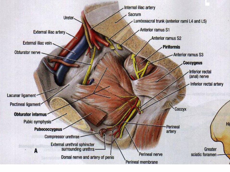
Parietal fascia of the lateral pelvic wall
The fascia covering the muscles of the lateral pelvic wall is condensed to form thick &strong membranes .
The fascia covering the obturator internus is called obturator
fascia , it shows a linear thickening for the origin of the levator ani.
The fascia covering the piriformis is thin.the nvs over the
piriformis (i,e the sacral plexus) lie external to the pelvic fascia.

Parietal fascia of the pelvic floor:
This fascia covers both surfaces of the pelvic diaphragm ,forming the superior and inferior layers, the inferior fascia also known as the anal fascia.
in general the fascia of the pelvic floor is loosely arranged between the peritoneum &the pelvic floor.
However the fascia is condensed at places to form fibromuscular ligaments which support the pelvic fascia.
Visceral pelvic fascia
• This fascia surrounds the extra peritoneal parts
of the pelvic viscera ,its loose and cellular
arround distensible pelvic organs like
(bladder, rectum and vagina), while its dense
around non distensible organs ,like prostate.
Pelvic muscles:
The pelvic muscles include two groups:
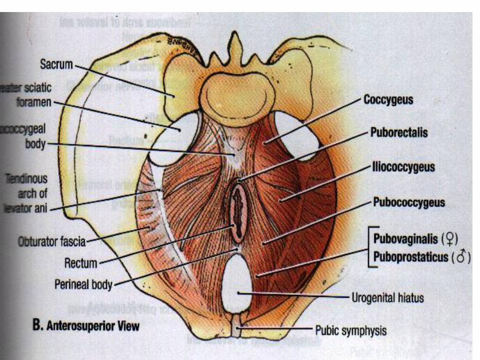
1. The piriformis and obturator internus,
2. The levator ani and coccygeus ,which with the corresponding muscles of the opposite side ,form the pelvic diaphragm.( The diaphragm separates the pelvis from the perineum).
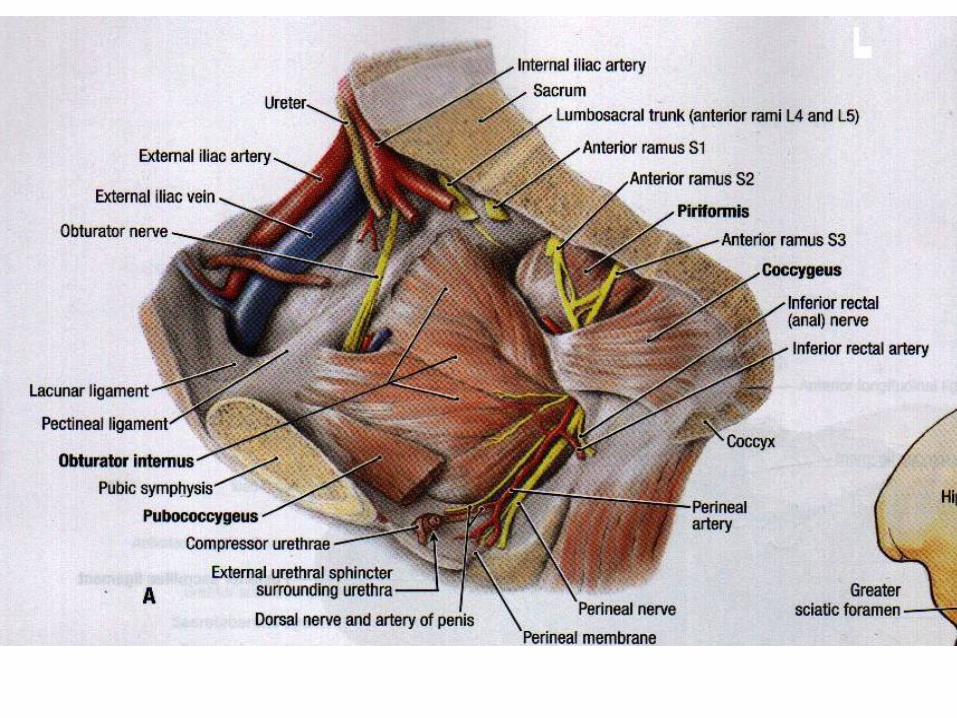
the Piriformis lies on the posterolateral pelvic wall and leaves the lesser pelvis through the greater sciatic foramen.

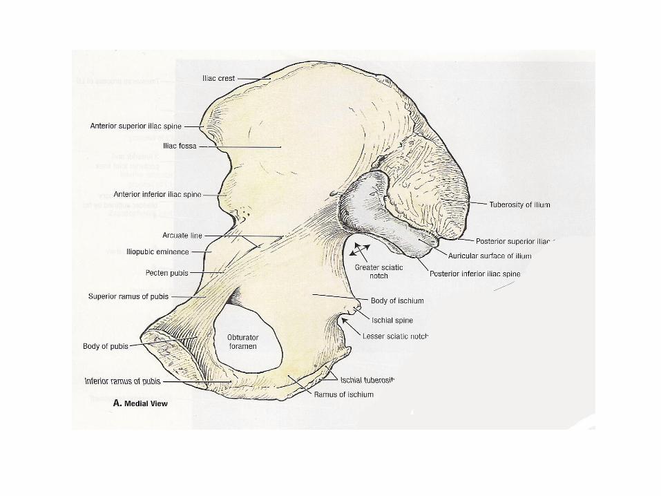
4. On the lateral pelvic wall the obturator foramen is closed by the obturator membrane and the obturator internus m. which attaches mainly to the obturator memb. And exits the lesser pelvis through the lesser sciatic foramen; & the obturator fascia lies on the medial surface of the muscle.
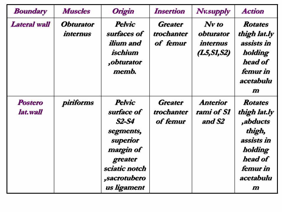
Action Nv.supply Insertion Origin Muscles Boundary
Rotates thigh lat.ly assists in holding head of femur in
acetabulum
Nv to obturator internus
(L5,S1,S2)
Greater trochanter of femur
Pelvic surfaces of ilium and ischium
,obturator memb.
Obturator internus
Lateral wall
Rotates thigh lat.ly ,abducts
thigh, assists in holding head of femur in
acetabulum
Anterior rami of S1
and S2
Greater trochanter of femur
Pelvic surface of
S2-S4 segments, superior
margin of greater
sciatic notch ,sacrotuberous ligament
piriforms Postero lat.wall

Forms most of pelvic diaphragm that helps support pelvic viscera& resists increase in intra abd. pressure
Nv.to lev.ani( br. of S4) ,inferior rectal nv,&coccygeal plexus
Perineal body ,coccyx,anococcygeal lig.,walls of prostate or vagina, rectum,and anal canal
Body of pubis,tendinous arch of obturator fascia, ischial spine
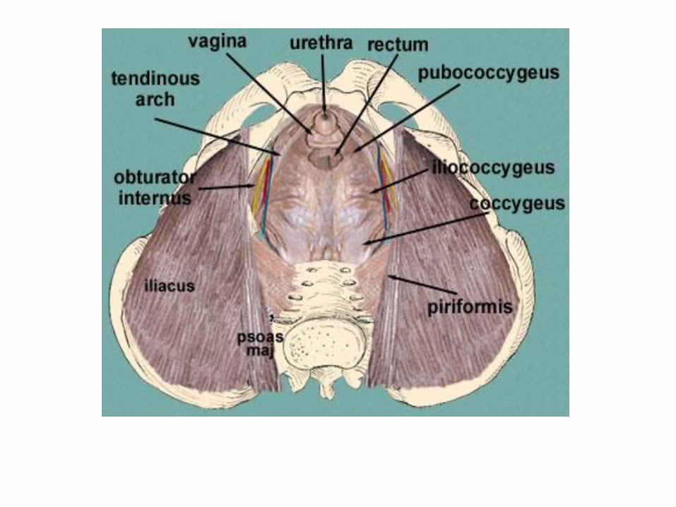
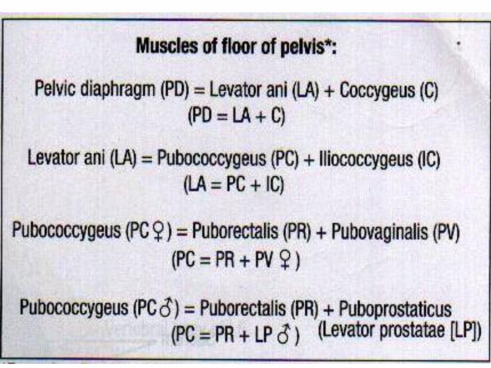
Levator ani (pubococcygeus,puborectalis&
illiococcygeus
floor
Forms small part of pelvic diaphragm that supports pelvic viscera
Branches of S4and S5 spinal nerves
Inferior end of sacrum and coccyx
Ischial spine
Coccygeus (ischiococcygeus)
floor

The levator ani and coccygeus may be regarded
as one morphological entity ,divisible from
before backwards into pubococcygeus,
the iliococcygeus and the (coccygeus) . they
have a continuous linear origin from the pelvic
surface of the body of the pubis.
The muscle fibers slope downwards and
backwards to the midline making pelvic floor.
• The Levator Ani :
• The muscle is divisible into a pubococcygeus
part ,and an iliococcygeus part.
A- pubo coccygeus part:
1. The anterior fibers of this part arise from the medial part of the body of the pubis.in females these fibers surround the vagina and form the sphincter vagnae .the anterior fibers are inserted into the perineal body .
2. The posterior fibers of the pubococcygeus arise from the lateral part of the pelvic surface of the pubis. they partly form aloop (or sling) around the anorectal junction;

• And are partly continuous with the longitudinal
muscle coat of the rectum .
3.The middle fibers constitute the puborectalis.
B-Iliococcygeus part:
The fibers of this part arise from:(a) the posterior
half of the white line on the obturator fascia ;and
(b) the pelvic surface of the ischial spine .they
are inserted into the anococcygeal ligament and
into the tip of the coccyx.
The coccygeus muscles:
Also called (ischiococcygeus),this muscle represents the posterior part of the pelvic diaphragm .it is triangular in shape .it is partly muscular and partly tendinous.
Its fibers arise from: a-the pelvic surface of the ischial spine,and b-the sacrospinous ligament.it is inserted into the side of the coccyx ,and into the fifth sacral vertebra.

Action of levator ani and coccygei :
1. The levator ani &coccygei close the posterior
part of pelvic outlet.
2. The levator ani fix the perineal body and
support the pelvic viscera.
3. During coughing ,sneezing ,lifting and other
muscular efforts, the levator ani and coccygei
counteract (resist) increased intra –abdominal
pressure, and help to maintain continence of
the bladder and the rectum.
4. in micturition ,defeacation and parturition,
particularly the pelvic outlet is open ,but
contraction of fibers around other openings
resists increased intra-abdominal pressure and
prevents any prolapse through the pelvic floor.
5. The coccygei pull forwards and support the
coccyx ,after it has been pressed backwards
during defaecation or parturition.
Relations of the levator ani :
1. The superior or pelvic surface is covered with
pelvic fascia which separates it from the
bladder ,the prostate in male ,the rectum and
peritoneum.
2. The inferior or perineal surface is covered
with anal fascia and forms the medial
boundary of the ischiorectal fossa.
3.The anterior borders of the two muscles are
seperated by atriangular space for the passage
of the urethra and vagina.
4. The posterior border is free and lies against
the anterior margin of the coccygeus.
Clinical notes:
• the muscles of the pelvic floor may be injured
during parturition .when the perineal body is
torn ,and has not been repaired satisfactorily,
• the contraction of anterior fibers of the levator
ani increases the normal gap in the pelvic floor
,instead of decreasing it. this results in
abnormalities like cystocele , or prolapse of
uterus.

Thank you for listening