Embed Size (px)
Citation preview

Aliso: A Journal of Systematic and Evolutionary Botany
Volume 12 | Issue 4 Article 3
1990
Wood Anatomy of Ascarina (Chloranthaceae) andthe Tracheid-vessel Element TransitionSherwin CarlquistPomona College; Rancho Santa Ana Botanic Garden
Follow this and additional works at: http://scholarship.claremont.edu/aliso
Part of the Botany Commons
Recommended CitationCarlquist, Sherwin (1990) "Wood Anatomy of Ascarina (Chloranthaceae) and the Tracheid-vessel Element Transition," Aliso: AJournal of Systematic and Evolutionary Botany: Vol. 12: Iss. 4, Article 3.Available at: http://scholarship.claremont.edu/aliso/vol12/iss4/3
ALISO ALISO 12(4), 1990, pp. 667-684
WOOD ANATOMY OF ASCARINA (CHLORANTHACEAE) AND THE TRACHEID-VESSEL ELEMENT TRANSITION
SHERWIN CARLQUIST
Rancho Santa Ana Botanic Garden and
Department of Biology, Pomona College Claremont, California 91711
ABSTRACT
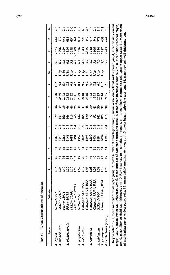
Quantitative and qualitative features are presented for 13 collections of 8 species of Ascarina. Wood anatomy is maximally primitive in most respects; moderate exception occurs in the imperforate tracheary elements, which range from tracheidlike (A. solmsiana) to fiber-tracheids (septate in two species). Perforation plates are scalariform, average more than 100 bars per plate, and have bordered bars. Even more significantly, portions of the primary walls in perforations characteristically fail to dissolve; these pit membrane portions range from nearly intact (much like the pit membranes in pits on end walls of tracheids of vesselless dicotyledons) to remnant strands or flakes. Dissolution of pit membranes in perforations is apparently inhibited by deposition of resinlike substances in some species; the rugose surfaces formed by these deposits may account for a report of vesturing on vessel walls of Ascarina. Axial parenchyma is diffuse, with only very small expressions of diversification; apotracheal banded parenchyma is, however, present in A. swamyana. Wood of Ascarina is highly mesomorphic. With age of plant, vessels increase in diameter, vessel elements and fiber-tracheids increase in length, and rays become wider and have a higher proportion of procumbent cells; uniseriate rays decrease in abundance. The implications of wood anatomy data on generic distinctions within the family and on the systematic position ofChloranthaceae will be examined when monographs on woods of the other genera have been completed.
Key words: Ascarina, Chloranthaceae, tracheids, vessel elements, wood anatomy.
INTRODUCTION
The genus Ascarina is composed of 13 species at present. The Madagascan species, A. coursii (Humb. & Capuron) Leroy & Jeremie, was formerly recognized under the genus Ascarinopsis, but is now considered the sole species of section Mascarina (Jeremie 1980). The remaining species occur in the Pacific, and have an insular distribution that extends from the Philippines (A. philippinensis C. B. Rob.) in the north to New Zealand (A. Iucida Hook f.) in the south; the range terminates eastwards in the Marquesas (A. marquesensis A. C. Smith) and Tahiti (A. polystachya J. R. & G. Forst.). The monograph by Swamy (1953b) has been supplanted by that of Smith (1976); since Smith's monograph, a New Guinean species, A. subsessilis Verdcourt, has been added (V erdcourt 1985). The nomenclatural changes offered by Smith (1976) have been accepted here.
Wood of Ascarina was studied by Thierry (1912) and by Swamy (1953a). Although Swamy (1953a) figures wood of"A. lanceolata Hook. f." (probably A. diffusa A. C. Smith in current nomenclature), he does not reveal if any other species of the genus were studied with respect to wood anatomy; his data are expressed only in terms of the genus. Detailed data on wood anatomy are available for A. Iucida thanks to the work of Patel ( 197 5) and Meylan and Butterfield (1978), and therefore I have not included that species in the present study. The data of

668 ALISO
these authors for A. Iucida reveal no features not observed in the material of the species studied here. In fact, no strongly marked differences of a taxonomic nature-only a feature or two at best-differentiate the species of Ascarina from each other with respect to wood anatomy. Differences can be observed among the wood samples studied, but these mostly prove to be related to ontogenetic change in wood features, for the samples differ in size and therefore maturity of wood features. Few if any features by which the wood samples differ from each other reflect distinctions in ecology. One must characterize the genus as a whole as typical of wet forest, particularly cloud forest. I have observed A. rubricaulis Solms in open areas at margins of the cloud forest in New Caledonia, but water availability in those places probably differs but little from that in such sites as the wet understory localities near the summit of Mt. Panie where A. solmsiana Schlechter occurs.
At the ultrastructural level, there is a surprising source of diversity in wood anatomy within Ascarina: the degree of presence of primary wall portions (pit membranes) in the perforations of vessel elements. Expressions of pit membrane presence in Ascarina perforations range from virtual absence (as would be expected) to presence of pit membranes much like those in end wall pitting of tracheids of vesselless dicotyledons. Meylan and Butterfield (1978) figured "microfibrillar webs" (nearly intact pit membranes) in pits transitional to perforations at the end wall-lateral wall juncture of vessel elements of Ascarina Iucida. Pit membranes in pits transitional between the two walls are not unexpected, but Ascarina proves characteristically to have primary wall fragments throughout perforations of a perforation plate. Such fragments were figured earlier for A. rubricaulis (Carlquist 1988). Many more variations in primary wall presence in perforations are reported in the present paper. Where primary walls are present most extensively in perforations, the vessel elements are notably tracheidlike. Because such perforation plates demonstrate the nature of the transition between tracheids and vessel elements, Ascarina wood is of exceptional interest. The primitive nature of wood ofChloranthaceae has been noted by such authors as Swamy and Bailey ( 1950) and Metcalfe ( 1987) prior to discovery of this feature. Sarcandra of the Chloranthaceae was once regarded as extraordinarily primitive because its stems are vesselless (Swamy and Bailey, 1950) but the stems are relatively short lived, and vessels do, in fact, occur in roots of Sarcandra (Carlquist 1987).
Although the woods of Chloranthaceae do contain many aspects deemed primitive for dicotyledons, that does not certify other features of the family as primitive for dicotyledons. Leroy (1983a, 1983b) has attempt~d to interpret elongate stamen-bearing structures of Hedyosmum (Chloranthaceae) as strobili like those of gymnosperms rather than catkins characteristic of angiosperms, but Endress (1987) has adequately ntfuted Leroy's contentions. Endress has shown that the flowers of Chloranthaceae represent various types of reduction related to shift into particular modes of pollination; Ascarina is anemophilous according to Endress (1987) and this agrees with my field observations. As Endress shows, Leroy's (1983a, 1983b) interpretations create more complexities than they resolve. Chloranthaceae can be regarded as interesting precisely because there is a curious admixture of primitive features in wood and nodal anatomy (Swamy 1953a) together with reductions in floral structures (Swamy 1953a, Endress 1987).
VOL(
Tt lectic tions tesy Risboffer• lized di./ftc. slope mahe porti piner ippin stach_ (Car! RSA; Mt. I Panie tea, S. RSA)
We Sectic staine mane: slides. croscc are be
Quo A sin_ select: fiber-1 Fewer ativel: longer shorte ments:
Ten Termi source
Growt.
Groamour: afews_

ALISO VOLUME 12, NUMBER 4 669
MATERIALS AND METHODS
The samples studied were all preserved in dried form. In addition to my collections, wood samples were available from the MAD-SJRw and MADw collections of the Forest Products Laboratory, Madison, Wisconsin, through the courtesy of Regis B. Miller and Donna Christensen. Samples from the Princes Risborough Laboratory (PRFw) were provided by J.D. Brazier, and W. E. Hillis offered specimens from the CSIRO, Highett, Victoria (FPAw). The samples utilized are listed in Table I. Provenances of these specimens are as follows: A. diffusa (SJRw-27993), Viti Levu, Fiji; A. maheshwarii Swamy (MADw-29478), slopes of Lake Loloru crater, Bougainville, Solomon Islands (Schodde 3699); A. maheshwarii (PRFw-28911), same as preceding collection, but probably a different portion of the wood; A. phi/ippinensis (FPAw-30135), Sabah, Malaysia; A. philippinensis (MADw-25301), New Guinea (Kalkman 4908); A. philippinensis (Philippine Bureau of Science 37 32 3, Arnold Arboretum), Luzon, Philippines; A. polystachya (SJRw-25501), Lake Vaheria, 500 m, Tahiti, Society Islands; A. rubricau/is (Carlquist 743, RSA), Mt. Mou, New Caledonia; A. rubricaulis(Carlquist 15315, RSA), Mt. Do, New Caledonia; A. solmsiana (Carlquist 15577, RSA); summit of Mt. Panie, New Caledonia; A. solmsiana (Carlquist 15579), near summit ofMt. Panie, New Caledonia; A. subfalcata J. W. Moore (SJRw-24864), Opoa Mt., Raiatea, Society Islands, J. W. Moore 657; A. swamyana A. C. Smith (Car/quist 15620, RSA), summit ofMt. Tomanivi, Viti Levu, Fiji.
Wood samples were boiled in water and stored in aqueous 50% ethyl alcohol. Sections were prepared on a sliding microtome. Some of these sections were stained in safranin, lightly counterstained with fast green, and made into permanent slides. Other sections of each collection were dried between clean glass slides, coated with gold, and observed with an lSI WB-6 scanning electron microscope. Radial sections were used for this purpose, because perforation plates are best observed on radial sections.
Quantitative features reported in Table 1 are means based on 25 measurements. A single measurement was made for features not easy to quantifY, for which selection of a typical condition was considered adequate: vessel wall thickness, fiber-tracheid diameter, fiber-tracheid wall thickness, and ray cell wall thickness. Fewer than 25 measurements were made where a cell type or structure was relatively infrequent. In some cases, a selective sampling was employed. For example, longer vessel elements and fiber-tracheids tend to be fractured more often than shorter ones in macerations. Therefore, in order not to present biased measurements, cells from assorted length classes were selected for measurement.
Terminology is according to the IA WA Committee on Nomenclature (1964). Terminology for primary wall fragments in perforations is not offered by that source, so simple descriptive terms are used.
ANATOMICAL FEATURES
Growth Rings
Growth rings are absent in most species of Ascarina (Fig. 1, 11, 21). A small amount of fluctuation in vessel diameter as seen in transactions may be seen in a few specimens (A. swamyana, Fig. 31 ). Such minor fluctuation in vessel diameter

670 ALISO
can be seen in Patel's ( 197 5) figure of A. Iucida. He calls the wood diffuse porous, which is an acceptable term for a wood with a small degree of vessel diameter change with respect to seasonal events.
Vessel Elements
Vessels in Ascarina are mostly solitary (Fig. 1, 11, 21, 31). The mean number of vessels per group (Table 1, column 1) ranges from 1.15 (A. diffusa, Fig. 1) to 1.52 (A. polystachya). The mean number of vessels per group for the genus as a whole is 1.28. One must note that vessels appear to be paired in instances where one is, in fact, seeing the overlapping ends of superposed vessel elements of a single vessel; where a circular vessel is bisected by a perforation plate as seen in a transection, a single vessel is present.
Vessel density for Ascarina is shown in Table 1, column 2. Number of vessels per mm2 ranges from 18 to 102. Vessel density ought to be roughly inversely proportional to vessel diameter, and it is (with deviations from a straight-line relationship in certain collections: compare columns 2 and 3 of Table 1). Mean vessel diameter in Ascarina is moderate, and ranges from 46 to 81 Jtm; the mean for the genus as a whole is 64, whereas the mean in dicotyledons as a whole is 94 Jtm (Metcalfe and Chalk 1950).
Vessels may be restricted to particular portions of the secondary xylem, a phenomenon termed vessel restriction patterns (Carlquist 1988). Although the term can apply to various conditions, in Ascarina it can connote the tendency in A. so/msiana (Car/quist 15577) for patches of early secondary xylem to be free of vessels (Fig. 23).
Mean vessel element length (Table 1, column 4) ranges from 801 to 2340 Jtm in Ascarina. Reliable figures on stem diameters of the xylarium samples used in the present study cannot be offered, so species-by-species comparison between vessel element lengths and sample sizes is not possible. Nevertheless, greater vessel element length can be seen in some specimens for which greater stem diameter is known. The shortest vessel element length reported for the genus here is in A. philippinensis (Philippine Bureau of Science 37 323), a twig specimen taken from an herbarium specimen. The longest vessel elements, by contrast, come from a specimen for which a photocopied herbarium label indicates trunk diameter of 15 em-relatively large for the genus (A. maheshwarii, Schodde 3699). The mean vessel element length for all samples studied is 1790 Jtm, which is close to the figure reported by Patel (1975) for A. Iucida (1840 Jtm). The vessel element length reported by Swamy (1953a) for the genus, 823 Jtm, may represent either juvenile material or an error in calcuation.
Vessel wall thickness in Ascarina is much less than that of fiber-tracheids (Table 1, column 5), as can be seen in transections (Fig. 1, 21, 23, 31). The mean vessel wall thickness for the genus is 2.6 Jtm, which is only a third of the wall thickness of fiber-tracheids (7. 7 Jtm) in the genus (Table 1, column 8).
The perforation plates of Ascarina are long and scalariform (Fig. 5). Bars are bordered to various degrees, but all are definitely bordered. There is some fluctuation in bar thickness within a sample; one frequently sees fluctuation of this sort in a species with scalariform perforation plates. The scanning electron micrographs do not reveal the bordered nature of the bars well because the photographs are perpendicular to the plates. More oblique angles in photographing, as
VOLl
.. I II II .. ~
I &
Fig. are me A. rna. intact: Porous plate (s magnil bracke·

ALISO
; the mean a whole is
xylem, a the
VOLUME 12, NUMBER 4 671
Fig. 1-5. Wood sections of Ascarina. 1, 2, 5. A. diffusa (SJRw-27993).-1. Transection; vessels are mostly solitary.-2. Tangential section; both multiseriate and uniseriate rays are present.-3, 4. A. maheshwarii (MADw-2478), SEM photographs of perforation plate portions.-3. Primary walls intact in perforations, probably due to a varnishlike deposition, droplets of which are evident.-4. Porous primary wall portions present at ends of perforations, which are otherwise open.- 5. Perforation plate (a few perforations at top not shown) from radial section; axial parenchyma at right. (Fig. I , 2, magnification scale above Fig. I [divisions = ~tm]; Fig. 3, 4, bracket at top of Fig. 3 = 10 ~tm; Fig. 5, bracket at top of Fig. 5 = 10 ~tm.)
Tab
le 1
. W
ood
Cha
ract
eris
tics
of A
scar
ina.
Spe
cies
C
olle
ctio
n I
2 3
4 5
6 7
8 9
10
II
12
13
14
A.
diff
usa
SJR
w-2
7993
1.
15
46
63
1560
5.
1 79
34
17
90
11.5
U
SP
5.
9 23
23
644
2.3
A.
mah
eshw
arii
M
AD
w-2
9478
1.
26
35
60
2340
2.
3 15
1 39
25
69
5.1
US
p 6.
2 47
96
577
1.8
PR
Fw
-289
11
1.40
36
65
22
86
1.8
165
39
2743
6.
4 U
Sp
8.1
2729
94
1 2.
4 A
. ph
ilip
pine
nsis
F
PA
w-3
0135
1.
30
18
81
2146
2.
8 14
6 46
26
52
8.1
US
p 6.
1 45
24
688
2.4
MA
Dw
-253
01
1.17
29
77
16
81
2.8
116
46
2432
6.
9 U
sp
5.8
2658
40
4 3.
0 P
hil.
B.
Sci.
3732
3 1.
28
102
46
801
1.4
65
23
1021
4.
6 U
s 6.
8 >
50
00
34
1 2.
2 A
. po
lyst
achy
a SJ
Rw
-255
01
1.52
49
72
19
52
2.5
144
39
2323
8.
1 U
sp
6.1
3576
81
4 2.
8 A
. ru
bric
aulis
C
arlq
uist
743
, R
SA
1.
17
70
64
2025
3.
0 10
5 37
24
48
9.2
Usp
4.
0 16
43
728
4.6
Car
lqui
st 1
5315
, R
SA
1.
48
42
61
1898
2.
5 12
0 48
23
16
9.2
US
P
3.7
2355
44
7 2.
3 A
. so
lmsi
ana
Car
lqui
st 1
5577
, R
SA
1.
08
66
48
1250
2.
1 73
30
13
73
4.6
US
P
3.2
1240
61
5 1.
8 C
arlq
uist
155
79,
RS
A
1.31
40
61
14
09
2.6
92
41
1928
8.
1 U
sp
5.6
1606
54
6 2.
4 A
. su
bfal
cata
SJ
Rw
-248
64
1.39
65
64
18
94
2.8
142
30
2219
6.
2 U
sp
7.0
3534
97
8 3.
4 A
. sw
amya
na
Car
lqui
st 1
5620
, R
SA
1.
18
42
64
2034
2.
5 10
5 39
23
44
11.5
U
sp
5.5
2287
65
1 2.
1 A
ll co
llec
tion
s (m
ean)
1.
28
49
64
1790
2.
4 11
5 38
21
65
7.7
5.7
2783
64
4 2.
6
Key
to
colu
mns
: 1,
mea
n nu
mbe
r o
f ves
sels
per
gro
up;
2, m
ean
num
ber
of v
esse
ls p
er m
m2
; 3,
mea
n ve
ssel
dia
met
er a
t w
ides
t poi
nt, ~
tm;
4, m
ean
vess
el e
lem
ent
leng
th, ~t
m; 5
, m
ean
vess
el w
all
thic
knes
s, ~tm;
6, m
ean
num
ber
of b
ars
per
perf
orat
ion
plat
e; 7
, m
ean
fibe
r-tr
ache
id d
iam
eter
, ~t
m; 8
, m
ean
fibe
r-tr
ache
id l
engt
h,
~tm;
9,
mea
n fi
ber-
trac
heid
wal
l th
ickn
ess,
~tm
; 10
, Ray
his
tolo
gy (
u =
up
rig
ht,
s =
squ
are,
p =
pro
cum
ben
t, c
omm
ones
t cel
l ty
pes
in u
pper
cas
e);
11, m
ean
wid
th
ofm
ulti
seri
ate
ray
at w
ides
t po
int,
cel
ls;
12,
mea
n he
ight
mul
tise
riat
e ra
ys; ~t
m; 1
3, m
ean
heig
ht u
nise
riat
e ra
ys, ~tm; 1
4, m
ean
ray
cell
wal
l th
ickn
ess,
~tm
.
-0\~
fr?"~
:E'E
! ~
........__
--r·-
----·
;~ ..-.;:..~.
.... .,.
_
0\
-..I IV ~ ~

ALISO VOLUME 12, NUMBER 4 673
,
' '
10 Fig. 6-10. SEM photographs of perforation plates from radial sections of A. philippinensis (Fig. 6,
FPAw-30315, Fig. 7-10, MADw-25301).-6. Perforation plate portion with primary walls intact, probably because dissolution of pit membranes has been inhibited by presence of varnishlike material which is evident as droplets and agglutinations. - 7. Perforations that contain primary walls with nontraumatic micropores of various sizes; rift at left, middle, probably due to mechanical damage.-8. Perforations that illustrate the range of micropore sizes in primary walls; rift above due to mechanical damage.- 9. Perforations with primary walls with a moderate number ofmicropores.- I 0. Perforations with a small number ofmicropores, perhaps due to interference of a secondary product deposit. (Fig., 6, bracket at top = 10 !Lm ; Fig. 7-10, bracket at top of Fig. 3.)

674 ALISO
employed by Meylan and Butterfield (1978, Fig. 168) in A. Iucida, show the borders well.
Mean number of bars per perforation plate (Table 1, column 6) is from 65 to 165. The greatest number observed was 222 (A. maheshwarii, PRFw-28911). The range in bar number reported by Patel (1975) for A. Iucida was 80-210. One notes with interest that the mean number of bars per perforation plate for all specimens studied here is 115. Number of bars seems clearly related to length of vessel elements, which in tum is related to ontogeny. Had only wood samples from the periphery of older stems been studied, the mean number of bars per perforation plate might have approached 200. Very few, if any, other dicotyledon genera with scalariform perforation plates have so many bars per perforation plate; only a few genera (mostly with one or two species), such as Aextoxicon or Euptelea, have comparable plates.
Lateral wall pitting of vessels in Ascarina is predominantly scalariform on vessel-to-vessel interfaces, scalariform to opposite on vessel-to-ray contacts (Fig. 12), and sparse (but often composed of horizontally elongate pits) on vessel-toaxial parenchyma and vessel-to-fiber-tracheid contacts.
Primary Wall Presence in Perforations
Special attention is given to this phenomenon because of the inherent interest with regard to the phylogenetic transition between tracheids and vessel elements, as mentioned in the Introduction. Perforations containing various degrees of primary wall (pit membrane) presence are illustrated here in Figures 3, 4, 6-10, 19, 20, and 26-30. These primary wall portions have sometimes been mentioned as occurring where perforations grade into lateral wall pitting, as described by Meylan and Butterfield (1978) for A.lucida. However, in surveyingAscarina wood, I discovered that remnants of primary wall material in perforations are more widespread than one would expect on the basis of such descriptions; they can be found throughout a perforation plate, and to varying degrees depending on the species of Ascarina.
In describing these instances, one must first deal with apparent inhibition of perforation plate removal by enzymatic action on account of apparent chemical overlay. In Figures 3 and 6, one sees not only presence of primary walls in perforations, one also sees overlay of walls with a vamishlike substance. If one examines such sections with a light microscope, one sees a yellowish or brownish color in these deposits where they are more massive. A logical explanation for these appearances is that prior to death of the protoplast in the vessel element, deposition ofthe vamishlike substances had occurred, so that enzymatic removal of the pit membranes in the perforations was prevented. Depositions of this material can mimic vesturing when seen under the scanning electron microscope. Bailey (1933), Wheeler (1981) and Gale (1982) warn against interpretation of relief due to deposition of secondary plant products as vesturing. On this account, I believe that the report by Ohtani et al. (1983) of vesturing in vessel walls of Ascarina should be discounted as a probable instance of secondary plant product deposition.
If one removes instances in which deposits may have hindered dissolution of primary walls in perforations, one still sees a number of instances in which pit membranes remain in perforations of mature vessel elements. In A. maheshwarii
vo
Fie II. T ofda relati_ multi [divi!
ALISO
the borders
VOLUME 12, NUMBER 4 675
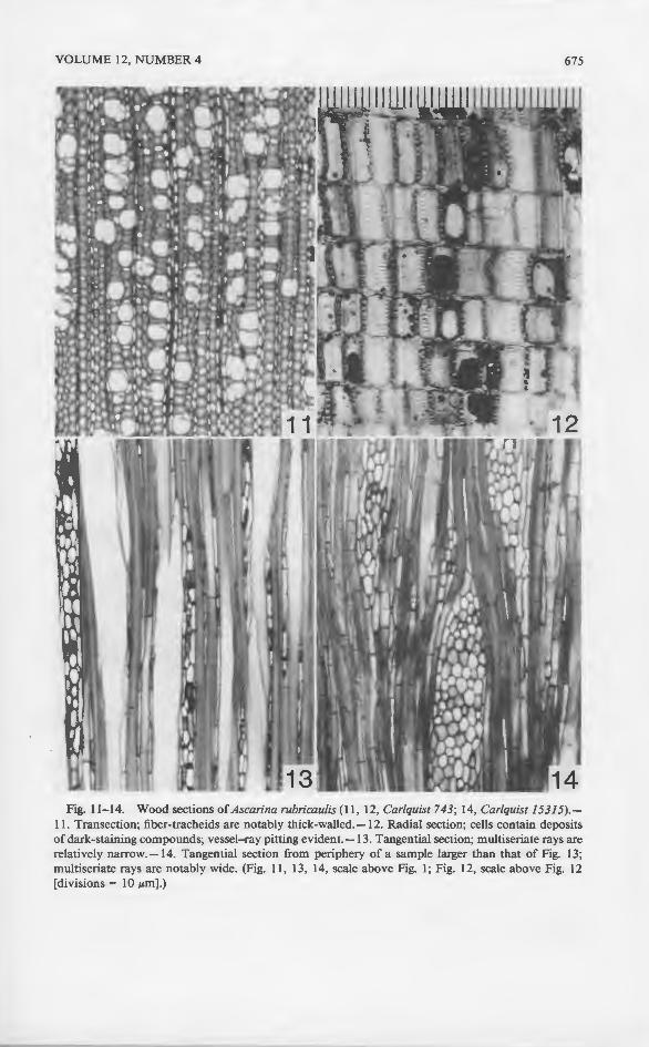
Fig. 11-14. Wood sections of Ascarina rubricaulis (11 , 12, Carlquist 743; 14, Carlquist 15315).-11. Transection; fiber-tracheids are notably thick-walled.-12. Radial section; cells contain deposits of dark-staining compounds; vessel-ray pitting evident.-13. Tangential section; multiseriate rays are relatively narrow.-14. Tangential section from periphery of a sample larger than that of Fig. 13; multiseriate rays are notably wide. (Fig. 11 , 13, 14, scale above Fig. 1; Fig. 12, scale above Fig. 12 [divisions= 10 JLm] .)
676 ALISO
' -. . \ -'
-~
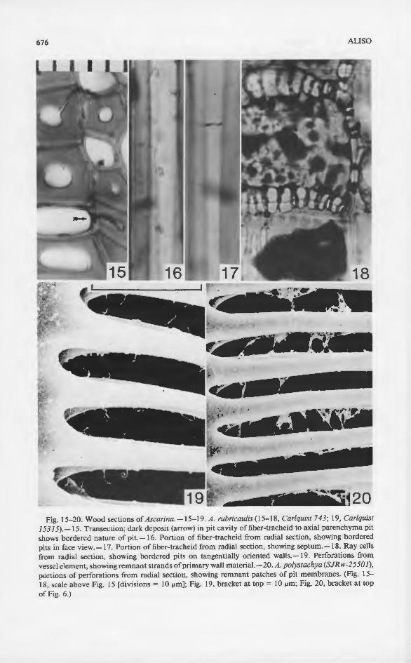
Fig. 15-20. Wood sections of Ascarina. -15-19. A. rubricau/is (15-18, Carlquist 743; 19, Carlquist 15315).-15. Transection; dark deposit (arrow) in pit cavity of fiber-tracheid to axial parenchyma pit shows bordered nature of pit.-16. Portion of fiber-tracheid from radial section, showing bordered pits in face view.-17. Portion of fiber-tracheid from radial section, showing septum. -18. Ray cells from radial section, showing bordered pits on tangentially oriented walls.-19. Perforations from vessel element, showing remnant strands of primary wall materiaL -20. A. polystachya (SJRw-25501), portions of perforations from radial section, showing remnant patches of pit membranes. (Fig. 15-18, scale above Fig. 15 [divisions= 10 JLm]; Fig. 19, bracket at top= 10 JLm; Fig. 20, bracket at top of Fig. 6.)
VOl
(Fi~ thn whi rati· inst In t like 3.7. perJ freq
R shw fora Rift duri pon
Imp
T tracl (19~
5 to tracl here spar spar Chl< tracl tracl iana one
Fi 7. 0 tracl stem 373~
Fi CO ill!
tend ouu
n wall~
and . 1, co
Axia
Ax and I
ALISO VOLUME 12, NUMBER 4 677
(Fig. 4), primary wall portions containing micropores occur at ends of perforations throughout perforation plates. In A. philippinensis (Fig. 7-10), primary walls in which micropores of various sizes occur are characteristically retained in perforations. The micropores may be large (Fig. 7, 9) or small (Fig. 10). In the latter instance, micropores might have failed to form because of some type of inhibition. In the remaining species of Ascarina studied here, one tends to find appearances like that illustrated for A. rubricaulis in Figure 19 (see also Carlquist 1988, Fig. 3.7.2), A. polystachya (Fig. 20), and A. solmsiana (Fig. 26-28). In these examples, perforations are open, but strands or fragments of primary wall material are frequent.
Relatively uncommonly, one can see in species of Ascarina other than A. maheshwarii occasional perforation plates with relatively intact primary walls in perforations and no associated deposition of secondary plant products (Fig. 29, 30). Rifts in these plates may be caused secondarily as the result of drying and wetting during preparation or as the result of mechanical stress during sectioning. Micropores can be observed in primary walls in these perforations.
Imperforate Tracheary Elements
The imperforate tracheary elements of Ascarina can mostly be termed fibertracheids according to the definitions ofthe IAWA Committee on Nomenclature (1964). Pit cavity diameter in the fiber-tracheids of Ascarina is large, ranging from 5 to 7 ~m, mostly the latter. Pit cavity diameter tends to be greater on fibertracheid-to-ray contacts than on fiber-tracheid interfaces. These pits are illustrated here in sectional view in Figure 15, and in face view in Figures 16 and 25. The sparseness of pits in Figure 16 characterizes fiber-tracheids in most species. This sparseness of pits connotes a nonconductive status for the fiber-tracheids of Chloranthaceae (for a discussion, see Carlquist 1984). Septa were observed in fibertracheids of two species, A. diffusa and A. rubricaulis (Fig. 17); septa in fibertracheids is yet another evidence of their nonconductive nature. Only in A. solmsiana are pits densely placed enough on the imperforate tracheary elements so that one might designate these as tracheids (Fig. 25).
Fiber-tracheid diameter is given for the collections studied in Table 1, column 7. One sees that mean diameter ranges from 23 to 48 ~m. The narrowest fibertracheids characterize the collections for which twig material rather than mature stem material was available, as for A. philippinensis (Philippine Bureau of Science 37323).
Fiber-tracheid length (Table 1, column 8) parallels vessel element length. The comments above on vessel element length apply here. The longest fiber-tracheids tend to characterize the largest wood samples, although correlations are not without exception.
Thickness of walls in fiber-tracheids is not related to length. The thickness of walls in fiber-tracheids is greater in A. diffusa (Fig. 1), A. rubricaulis (Fig. 11, 15), and A. swamyana, and is somewhat to markedly less in the other species (Table 1, column 9).
Axial Parenchyma
Axial parenchyma of Ascarina is characterized by both Patel ( 197 5) and Meylan and Butterfield (1978) as including diffuse, diffuse-in-aggregates, and vasicentric
678 ALISO
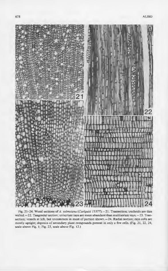
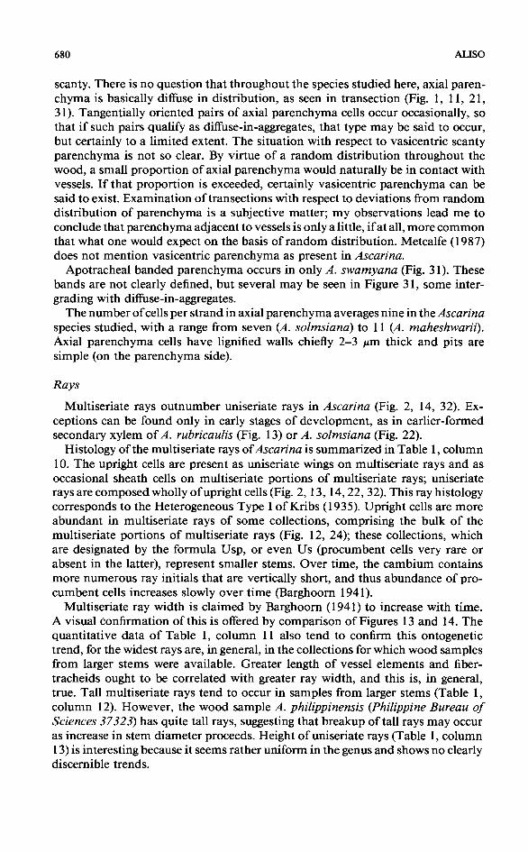
Fig. 21-24. Wood sections of A. so/msiana (Car/quist 15577).-21. Transection; tracheids are thin walled.-22. Tangential section; uniseriate rays are more abundant than multiseriate rays.-23. Transection; vessels at left, but uncommon in most of portion shown.-24. Radial section; rays cells are mostly upright; deposits of secondary plant compounds present in only a few cells. (Fig. 21 , 22, 24, scale above Fig. I; Fig. 23, scale above Fig. 12.)
vou.:
Fig. 25-sections, sections.Vertical s tions. (Fig 6.)

ALISO
22
VOLUME 12, NUMBER 4 679
Fig. 25-28. Wood sections of Ascarina solmsiana (Carlquist 15577).-25. Tracheids from tangential sections, showing pits in face view.-26-28. SEM photographs of perforation plates from radial sections.-26. Flakelike pit membrane portions occur on edges and at ends of perforations.-27. Vertical strands relatively abundant in perforations.-28. Slender strands occur sparsely in perforations. (Fig. 25 , scale above Fig. 15; Fig. 26, 28, bracket at top of Fig. 3; Fig. 27, bracket at top of Fig. 6.)
/
680 ALISO
scanty. There is no question that throughout the species studied here, axial parenchyma is basically diffuse in distribution, as seen in transection (Fig. 1, 11, 21, 31). Tangentially oriented pairs of axial parenchyma cells occur occasionally, so that if such pairs qualify as diffuse-in-aggregates, that type may be said to occur, but certainly to a limited extent. The situation with respect to vasicentric scanty parenchyma is not so clear. By virtue of a random distribution throughout the wood, a small proportion of axial parenchyma would naturally be in contact with vessels. If that proportion is exceeded, certainly vasicentric parenchyma can be said to exist. Examination oftransections with respect to deviations from random distribution of parenchyma is a subjective matter; my observations lead me to conclude that parenchyma adjacent to vessels is only a little, if at all, more common that what one would expect on the basis of random distribution. Metcalfe (1987) does not mention vasicentric parenchyma as present in Ascarina.
Apotracheal banded parenchyma occurs in only A. swamyana (Fig. 31 ). These bands are not clearly defined, but several may be seen in Figure 31, some intergrading with diffuse-in-aggregates.
The number of cells per strand in axial parenchyma averages nine in the Ascarina species studied, with a range from seven (A. solmsiana) to 11 (A. maheshwarii). Axial parenchyma cells have lignified walls chiefly 2-3 Jtm thick and pits are simple (on the parenchyma side).
Rays
Multiseriate rays outnumber uniseriate rays in Ascarina (Fig. 2, 14, 32). Exceptions can be found only in early stages of development, as in earlier-formed secondary xylem of A. rubricaulis (Fig. 13) or A. solmsiana (Fig. 22).
Histology of the multiseriate rays of Ascarina is summarized in Table 1, column 10. The upright cells are present as uniseriate wings on multiseriate rays and as occasional sheath cells on multiseriate portions of multiseriate rays; uniseriate rays are composed wholly of upright cells (Fig. 2, 13, 14, 22, 32). This ray histology corresponds to the Heterogeneous Type I ofKribs (1935). Upright cells are more abundant in multiseriate rays of some collections, comprising the bulk of the multiseriate portions of multiseriate rays (Fig. 12, 24); these collections, which are designated by the formula Usp, or even Us (procumbent cells very rare or absent in the latter), represent smaller stems. Over time, the cambium contains more numerous ray initials that are vertically short, and thus abundance of procumbent cells increases slowly over time (Barghoom 1941).
Multiseriate ray width is claimed by Barghoom (1941) to increase with time. A visual confirmation ofthis is offered by comparison of Figures 13 and 14. The quantitative data of Table 1, column 11 also tend to confirm this ontogenetic trend, for the widest rays are, in general, in the collections for which wood samples from larger stems were available. Greater length of vessel elements and fibertracheids ought to be correlated with greater ray width, and this is, in general, true. Tall multiseriate rays tend to occur in samples from larger stems (Table 1, column 12). However, the wood sample A. philippinensis (Philippine Bureau of Sciences 37 323) has quite tall rays, suggesting that breakup of tall rays may occur as increase in stem diameter proceeds. Height ofuniseriate rays (Table 1, column 13) is interesting because it seems rather uniform in the genus and shows no clearly discernible trends.
VOl
Fig. 2S perforati· rations.a few po1 31-32. A! bands (ar: (Fig. 29,

ALISO
14, 32). Ex-
l , column rays and as
uniseriate histology
VOLUME 12, NUMBER 4 681
Fig. 29-32. Wood sections of Ascarina. -29. A. so/msiana (Car/quist 15577), SEM photograph of perforation plate from radial section, showing flakelike remnants of pit membranes occluding perforations.-30. A. subfa/cata (SJRw-24864), SEM photograph of perforation plate; pit membranes have a few pores, occlude most perforations (rifts may represent mechanical damage during processing).-31-32. A. swamyana (Car/quist 15620).-31. Transection; inconspicuous apotracheal parenchyma bands (arrows) are present in top half.-32. Tangential section; multiseriate rays shown lack wings. (Fig. 29, 30, bracket at top of Fig. 6; Fig. 31, 32, scale above Fig. 1.)

682 ALISO
Ray cell wall thickness is not uniform in the genus (Table 1, column 14). Notably thick ray cell walls are reported for one collection of A. rubricaulis. Bordered pits are common on tangentially oriented ray cell walls of Ascarina (Fig. 18). Bordered pits were observed to be uncommon on tangentially oriented walls of ray cells only in A. diffusa, A. philippinensis (both collections), A. rubricaulis (Carlquist 15 315), and A. swamyana. Bordered pits on horizontally oriented ray cell walls were commonly observed only for A. polystachya and A. solmsiana, but even in these species, simple pits predominate on horizontal ray cell walls. Note should be taken that borders on ray cells should be observed in sectional view in radial sections. Borders on ray cell walls are common in dicotyledons, but are not often reported, probably because they have not been studied in sectional view. Sectional views of pits in ray cells are best seen in radial sections.
Other Wood Features
Cell contents of ray cells of Ascarina consist of droplets or massive accumulations of dark-staining compounds (Fig. 12, 18). These deposits are common throughout the genus except in A. solmsiana (Fig. 24), where only an occasional ray cell contains these materials. Deposits of this nature are less common in axial parenchyma than in ray cells of Ascarina. No crystals or silica bodies were observed in ray cells or axial parenchyma. Wood of Ascarina is nonstoried.
CONCLUSIONS
Ecology
The wood of Ascarina is clearly mesomorphic, notably devoid of any adaptation that would confer conductive safety. If one computes the Mesomorphy ratio (vessel diameter times vessel element length divided by vessel density) for Ascarina as a whole, one obtains the figure 2338. Higher Mesomorphy figures are found only in trees ofwet forests that are warm at least part of the year. Species with those higher Mesomorphy figures (which are due in large part to larger vessel diameter) would be expected to have greater transpiration rates than Ascarina, which characteristically occurs in cool cloud forest, often in understory locations. If computed for collections individually, the lowest Mesomorphy figure for the genus (361) would occur in the collection Philippine Bureau of Science 37323 of A. philippinensis. This reflects the tendency, common throughout dicotyledons but not often noted, for vessels to increase in diameter as stems increase in diameter. Production of progressively larger vessels corresponds to the ability of plants to tap deeper soil levels as they grow larger and to transpire greater volumes of water as they approach and reach canopy levels. The absence of vessels in patches early in ontogeny in the secondary xylem of A. solmsiana may be merely a chance occurrence, but it could relate to the presence in this species of imperforate tracheary elements that are tracheids capable of conduction. Figures by Thierry (1912) for stems of A. polystachya show secondary xylem apparently devoid of vessels for three growth rings. Thierry does figure vessels in A. lanceolata. Thierry's figures for A. polystachya may be in error, but if they are valid, the occurrence of a secondary xylem consisting of imperforate tracheary elements without vessel elements for several years may be like the patchy occurrence of vessels in earlier secondary xylem of A. solmsiana. It also recalls Sarcandra, in
VOL
whi' 1951
T1 ofb• with prirr pote ably
Ont.
Ti QUal
and norr pres in tt sterr: rays obse
Phy!
Tl: (198 occu latio betw ("Fr the c woul extre ratio trach pose• ence cond hist() ofFr
Tb Sept<: seem pauci what restri
As the g• anth~:
grapt

VOLUME 12, NUMBER 4 683
which the stems-which are of finite duration-are vesselless (Swamy and Bailey 1950; Carlquist 1987), although older roots do have vessels (Carlquist 1987).
The occurrence in Ascarina of scalariform perforation plates with large numbers of bars has been regarded not merely as a primitive feature, but one that correlates with mesic ecology in dicotyledons as a whole (Carlquist 1975). The remnants of primary walls in perforations, to the extent they are present in Ascarina, represent potential obstructions to conduction tolerable because conductive rates are probably low.
Ontogeny
The wood samples of Ascarina are of various ages and sizes. Comparisons of quantitative features with respect to these factors demonstrate that vessel elements and fiber-tracheids increase in length as stems increase in diameter. This is the normal progression for woody plants, as shown by curves such as those first presented by Bailey and Tupper ( 1918). Vessels increase in diameter, as mentioned in the preceding section, and fiber-tracheids increase in diameter with increase in stem diameter. Multiseriate rays increase in width during stem growth; uniseriate rays decrease in frequency. In all of these respects, Ascarina corresponds with observations made in a wide range of woody dicotyledons.
Phylogeny and Systematic Relationships
The wood of Chloranthaceae is proclaimed to be unspecialized by Metcalfe (1987). I agree with this assessment. The discovery that pit membrane remnants occur in perforations of Ascarina vessels adds yet another feature to the constellation of primitive characteristics of Ascarina wood. If one computes a ratio between length of imperforate tracheary elements and length of vessel elements ("F/V ratio") for Ascarina as a whole, one obtains the figure 1.21; the range in the collections studied here is from 1.10 to 1.44. A F/V ratio of less than 1.10 would indicate a higher degree of primitiveness, but such ratios below 1.10 are extremely few, even in the most primitive dicotyledon woods. The fact that the ratio rises above 1.30 in Ascarina may be related to the fact that imperforate tracheary elements are a nonconductive system (or nearly so), with sparsely disposed bordered pits, and are thus fiber-tracheids rather than tracheids. The presence of septa in fiber-tracheids of A. diffusa and A. rubricaulis indicates a nonconductive nature. The vessel features, axial parenchyma configurations, and ray histology of Ascarina all qualify as maximally primitive according to the criteria ofFrost (1930) and Kribs (1935, 1937).
There is little difference among the species with respect to wood anatomy. Septate fibers were found only in A. diffusa and A. rubricaulis, but this does not seem an important feature. The tracheidlike imperforate tracheary elements and paucity of deposits ofvamishlike substances inA. solmsiana may represent somewhat more distinctive features. Banded apotracheal parenchyma is apparently restricted to A. swamyana.
As for implications of data from wood anatomy concerning distinctions among the genera of the family and with respect to phylogenetic relationships of Chloranthaceae, comments are best deferred until further studies are completed. Monographs on wood anatomy of Hedyosmum and Chloranthus are planned.

684 ALISO
LITERATURE CITED
Bailey, I. W. 1933. The cambium and its derivative tissues. VII. Structure, distribution, and diagnostic significance of vestured pits in dicotyledons. J. Arnold Arb. 14:259-273.
---,and W. W. Tupper. 1918. Size variation in tracheary cells. I. A comparison between the secondary xylems of cryptogams, gymnosperms, and angiosperms. Proc. Amer. Acad. Arts Sci. 54:149-204.
Barghoorn, E. S. 1941. The ontogenetic development and phylogenetic specialization of rays in the xylem of dicotyledons. Amer. J. Bot. 28:273-282.
Carlquist, S. 1975. Ecological strategies of xylem evolution. Univ. Calif. Press, Berkeley. 259 p. ---. 1984. Vessel grouping in dicotyledon woods: significance and relationship to imperforate
tracheary elements. Aliso 10:505-525. ---. 1987. Presence of vessels in Sarcandra (Chloranthaceae); comments on vessel origins in
angiosperms. Amer. J. Bot. 74:1765-1771. ---. 1988. Comparative wood anatomy. Springer Verlag, Berlin, Heidelberg, and New York.
436 p. Endress, P. K. 1987. The Chloranthaceae: reproductive structures and phylogenetic position. Bot.
Jahrb. Syst. 109:153-226. Frost, F. H. 1930. Specialization in secondary xylem of dicotyledons. I. Origin of vessel. Bot. Gaz.
(Crawfordsville) 89:67-94. Gale, R. 1982. Some pitfalls in wood indentifications, with reference to Nothofagus. lAW A Bull.,
n.s., 3:179-184. IAWA Committee on Nomenclature. 1964. Multilingual glossary of terms used in wood anatomy.
Verlagsanstalt Buchdruckerei Konkordia, Winterthur, Switzerland. 185 p. Jeremie, J. 1980. Notes sur le genre Ascarina (Chloranthaceae) en Nouvelle Caledonie eta Mada
gascar. Adansonia, ser. 2, 20:273-285. Kribs, D. A. 1935. Salient lines of structural specialization in the wood rays of dicotyledons. Bot.
Gaz. (Crawfordsville) 96:547-557. ---. 19 3 7. Salient lines of structural specialization in the wood parenchyma of dicotyledons. Bull.
Torrey Bot. Club 64:177-186. Leroy, J.-F. 1983a. Interpretation nouvelle des appareils sexuels chez les Chloranthacees (Chlor
anthales, Magnoliidees). Compt. Rend. Hebd. Seances Acad. Sci., Ser. III 296:747-752. ---. 1983b. The origin of angiosperms: an unrecognized ancestral dicotyledon, Hedyosmum
(Chloranthales), with a strobiloid flower is living today. Taxon 32:169-175. Metcalfe, C. R. 1987. Anatomy of the dicotyledons. Ed. 2. Vol. 3. Magnoliales, Illiciales, and Laurales.
Clarendon Press, Oxford. 224 p. ---,and L. Chalk. 1950. Anatomy of the dicotyledons. Clarendon Press, Oxford. 1500 p. Meylan, B. A., and B. G. Butterfield. 1978. The structure of New Zealand woods. DSIR Bull. 222.
New Zealand DSIR, Wellington. 250 p. Ohtani, J., B. A. Meylan, and B. G. Butterfield. 1983. Occurrence of warts in the vessel elements
and fibers of New Zealand woods. New Zealand J. Bot. 21:359-372. Patel, R.N. 1975. Wood anatomy ofthe dicotyledons indigenous to New Zealand. 10. Chlorantha
ceae. New Zealand J. Bot. 13:141-148. Smith, A. C. 1976. Studies of Pacific island plants. XXXIII. The genus Ascarina (Chloranthaceae)
in the South Pacific. J. Arnold Arb. 57:405-425. Swamy, B. G. L. 1953a. The morphology and relationships of the Chloranthaceae. J. Arnold Arb.
34:375-408. ---. 1953b. A taxonomic revision of the genus Ascarina Forst. Proc. Nat. Inst. Sci. India 19:
371-388. ---, and I. W. Bailey. 1950. Sarcandra, a vesselless genus ofthe Chloranthaceae. J. Arnold Arb.
31:117-129. Thierry, R. 1912. Contribution a !'etude anatomique des Chloranthacees. Thesis, Paris. 158 p. Verdcourt, B. 1985. Notes on Malesian Chloranthaceae. Kew Bull. 40:213-224. Wheeler, E. A. 1981. Intervascular pitting in Fraxinus americana L. lAW A Bull., n.s., 2:169-174.
The_ study,analyze regions which • Reliant center c
Key wOo
The: estim~::
per hat en den: most c flora o 1987). on con: unexpl known geogra richne~
Any demic. biogeo1 in this or regie one or ered er: Rzedm. marily familie: conflue: occurs floristic