Embed Size (px)
Citation preview
INDIAN JOURNAL OF CARDIOVASCULAR DISEASES JOURNAL in women (IJCD) 2017 VOL2 ISSUE 1 INTERVENTIONAL ROUNDS
1
WINCARS
DEFINITION:
The ‚No-reflow phenomenon‛ is classically defined as
lack of myocardial perfusion despite opening up the
Epicardial vessel in the setting of primary percutaneous
coronary intervention (PCI). Broadly it refers to sudden
loss of Epicardial flow i.e. abrupt onset of TIMI- zero
flow after balloon dilatation or placement of a stent.
Angiographic no-reflow was defined as TIMI flow grade
0, 1, and 2 after PCI. Angiographic No-Reflow is defined
as the presence of TIMI 0-1 in absence of dissection,
spasm, stenosis or thrombus of the epicardial vessel.
Lesser degree of reduction of coronary flow (i.e.TIMI 2
flow) is defined as Slow-flow.
HISTORICAL ASPECT:
The term no-reflow was first coined by Ames et al in
their experimental work on cerebral ischemia. Kloner et
al. described coronary no-reflow for the first time in a
canine model after prolonged (90 min) coronary
occlusion followed by reperfusion. However, the same
couldn’t be directly extrapolated to the human situation
where myocardial infarction results from occlusive
superimposed coronary thrombosis. The no-reflow after
AMI in humans was first described by Ito et al, who had
assessed it by myocardial contrast echocardiography
(MCE).
INCIDENCE:
No-reflow has a prevalence ranging from 5% up to 50%,
depending on the method of assessment and the
population studied. It is seen in about 10% cases of
Primary PCI. It occurs with a lesser frequency in the
setting of non-ST-elevation myocardial infarction
(NSTEMI) or during elective PCI (1.5%); though
incidence may be higher in elective saphenous venous
graft interventions (4%).
Article received on 01 JAN 2017, published on 31JAN 2017
Maddury Jyotsna1,Nemani Lalita2
1Professor & HOU-IV, Department of Cardiology, NIMS, India, NIMS. 2Associate Professor, Department of Cardiology, NIMS.
Corresponding Author: Maddury Jyotsna
Email: [email protected]
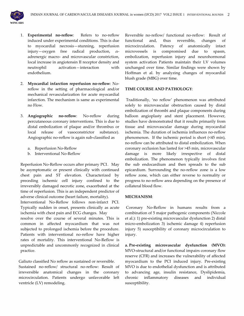
IMPORTANCE OF NO OR SLOW FLOW (Including
prognosis):
No-reflow or slow-flow phenomenon has been found to
be significantly associated with poor clinical and
functional outcomes and prolonged hospitalization.
Prognosis: No-reflow phenomenon is associated with
the poor short-term and long-term prognosis. Its
negative impact on outcomes dampens the potential
benefits of PPCI. The in- hospital course may be
complicated with malignant arrhythmias, re- infarction,
cardiac rupture and pump failure. There is an increased
risk of death at 30 days. LV remodeling is absent in no-
reflow resulting in LV dilation and heart failure. Five-
year rates of repeated hospitalization and mortality are
also high (Fig 7).
Fig 7: Survival curves showing less survival with no
ECG resolution cases.
CLASSIFICATION OF NO FLOW:
Eeckhout and Kern have suggested classifying no-
reflow into 1) experimental no-reflow and 2) MI
reperfusion no-reflow and 3) angiographic no-reflow.
NO-REFLOW Maddury Jyotsna, Nemani Lalita
INDIAN JOURNAL OF CARDIOVASCULAR DISEASES JOURNAL in women (IJCD) 2017 VOL2 ISSUE 1 INTERVENTIONAL ROUNDS
2
WINCARS
1. Experimental no-reflow: Refers to no-reflow
induced under experimental conditions. This is due
to myocardial necrosis—stunning, reperfusion
injury—oxygen free radical production, α-
adrenergic macro- and microvascular constriction,
local increase in angiotensin II receptor density and
neutrophil activation—interaction with
endothelium.
2. Myocardial infarction reperfusion no-reflow: No-
reflow in the setting of pharmacological and/or
mechanical revascularization for acute myocardial
infarction. The mechanism is same as experimental
no Flow.
3. Angiographic no-reflow: No-reflow during
percutaneous coronary interventions. This is due to
distal embolization of plaque and/or thrombus or
local release of vasoconstrictor substance).
Angiographic no-reflow is again sub-classified as
a. Reperfusion No-Reflow
b. Interventional No-Reflow
Reperfusion No-Reflow occurs after primary PCI . May
be asymptomatic or present clinically with continued
chest pain and ST elevation. Characterized by
preceding ischemic cell injury confined to the
irreversibly damaged necrotic zone, exacerbated at the
time of reperfusion. This is an independent predictor of
adverse clinical outcome (heart failure, mortality).
Interventional No-Reflow follows non-infarct PCI.
Typically sudden in onset, presents clinically as acute
ischemia with chest pain and ECG changes. May
resolve over the course of several minutes. This is
common in affected myocardium that was not
subjected to prolonged ischemia before the procedure.
Patients with interventional no-reflow have higher
rates of mortality. This interventional No-Reflow is
unpredictable and uncommonly recognized in clinical
practice.
Galiuto classified No reflow as sustained or reversible.
Sustained no-reflow/ structural no-reflow: Result of
irreversible anatomical changes in the coronary
microcirculation. Patients undergo unfavorable left
ventricle (LV) remodeling.
Reversible no-reflow/ functional no-reflow: Result of
functional and, thus reversible, changes of
microcirculation. Patency of anatomically intact
microvessels is compromised due to spasm,
embolization, reperfusion injury and neurohormonal
system activation Patients maintain their LV volumes
unchanged over time. Similar findings were shown by
Hoffman et al. by analyzing changes of myocardial
blush grade (MBG) over time.
TIME COURSE AND PATHOLOGY:
Traditionally, ‘no reflow’ phenomenon was attributed
solely to microvascular obstruction caused by distal
embolization of thrombi and plaque components during
balloon angioplasty and stent placement. However,
studies have demonstrated that it results primarily from
tissue and microvascular damage during myocardial
ischemia. The duration of ischemia influences no-reflow
phenomenon. If the ischemic period is short (<45 min),
no-reflow can be attributed to distal embolization. When
coronary occlusion has lasted for >45 min, microvascular
damage is more likely irrespective of distal
embolization. The phenomenon typically involves first
the sub endocardium and then spreads to the sub
epicardium. Surrounding the no-reflow zone is a low
reflow zone, which can either reverse to normality or
progress to no-reflow area depending on the presence of
collateral blood flow.
MECHANISM:
Coronary No-Reflow in humans results from a
combination of 5 major pathogenic components (Niccole
et al.): 1) pre-existing microvascular dysfunction 2) distal
micro-embolization 3) ischemic damage 4) reperfusion
injury 5) susceptibility of coronary microcirculation to
injury.
a. Pre-existing microvascular dysfunction (MVO):
MVO-structural and/or functional impairs coronary flow
reserve (CFR) and increases the vulnerability of affected
myocardium to the PCI induced injury. Pre-existing
MVO is due to endothelial dysfunction and is attributed
to advancing age, insulin resistance, Dyslipidemia,
chronic inflammatory diseases and individual
susceptibility.
INDIAN JOURNAL OF CARDIOVASCULAR DISEASES JOURNAL in women (IJCD) 2017 VOL2 ISSUE 1 INTERVENTIONAL ROUNDS
3
WINCARS
b. Distal Micro-embolization: Refer to the downstream
migration of thrombus debris or micro-material from
fissured and ruptured atheromatous plaques during
balloon dilatation or stenting. When more than 50% of
the capillaries are blocked, myocardial perfusion starts
falling. When the number of emboli is 25–200 or the size
of micro-emboli is >200 mm, it can cause severe MVO.
c. Ischemic injury: Prolonged ischemia causes
endothelial and myocardial degenerative changes.
Endothelial protrusions and membrane-bound bodies
contribute to capillaries luminal obliteration, the
endothelial gaps allow extravascular erythrocytes
migration and compression. In addition, endothelial
activation promotes expression of new adhesion
molecules which contributes to leucocytes accumulation.
Decreased production of adenosine triphosphate
resulting from ischemia impairs the sodium-potassium
pump (Na+/K+-ATPase) and results in cardiac myocyte
swelling. This irreversible damage leads to edema and
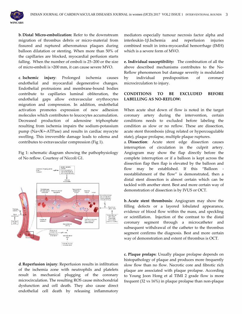
contributes to extravascular compression (Fig 1).
Fig 1: schematic diagram showing the pathophysiology
of No reflow. Courtesy of Niccoli G1.
d. Reperfusion injury: Reperfusion results in infiltration
of the ischemia zone with neutrophils and platelets
result in mechanical plugging of the coronary
microcirculation. The resulting ROS cause mitochondrial
dysfunction and cell death. They also cause direct
endothelial cell death by releasing inflammatory
mediators especially tumour necrosis factor alpha and
interleukin-1β.Ischemia and reperfusion injuries
combined result in intra-myocardial hemorrhage (IMH)
which is a severe form of MVO.
e. Individual susceptibility: The combination of all the
above described mechanisms contributes to the No-
Reflow phenomenon but damage severity is modulated
by individual predisposition of coronary
microcirculation to injury.
CONDITIONS TO BE EXCLUDED BEFORE
LABELLING AS NO-REFLOW:
When acute shut down of flow is noted in the target
coronary artery during the intervention, certain
conditions needs to excluded before labeling the
condition as slow or no reflow. These are dissection,
acute stent thrombosis (drug related or hypercoagulable
state), plaque prolapse, multiple plaque ruptures.
a. Dissection: Acute stent edge dissection causes
interruption of circulation in the culprit artery.
Angiogram may show the flap directly before the
complete interruption or if a balloon is kept across the
dissection flap then flap is elevated by the balloon and
flow may be established. If this ‚Balloon -
reestablishment of the flow‛ is demonstrated, then a
distal stent dissection is almost certain which can be
tackled with another stent. Best and more certain way of
demonstration of dissection is by IVUS or OCT.
b. Acute stent thrombosis: Angiogram may show the
filling defects or a layered lobulated appearance,
evidence of blood flow within the mass, and speckling
or scintillation. Injection of the contrast to the distal
coronary segment through a microcatheter and
subsequent withdrawal of the catheter to the thrombus
segment confirms the diagnosis. Best and more certain
way of demonstration and extent of thrombus is OCT.
c. Plaque prolaps: Usually plaque prolapse depends on
histopathology of plaque and produces more frequently
slow flow than no flow. Necrotic core and fibrotic rich
plaque are associated with plaque prolapse. According
to Young Joon Hong et al TIMI 2 grade flow is more
frequent (32 vs 16%) in plaque prolapse than non-plaque
INDIAN JOURNAL OF CARDIOVASCULAR DISEASES JOURNAL in women (IJCD) 2017 VOL2 ISSUE 1 INTERVENTIONAL ROUNDS
4
WINCARS
prolapse lesions [9a]. On IVUS tissue extrusion through
the stent struts will be demonstrated.
d. Multiple ruptured plaques: Angiographically it
simulates thrombus but IVUS shows plaque ruptures
separated by a >5-mm length of artery containing
smooth lumen contours. Hong Y et al showed that IVUS-
detected multiple PRs and plaque prolapse are
associated with no-reflow after PCI for PR-containing
culprit lesion in infarct-related arteries in AMI patients.
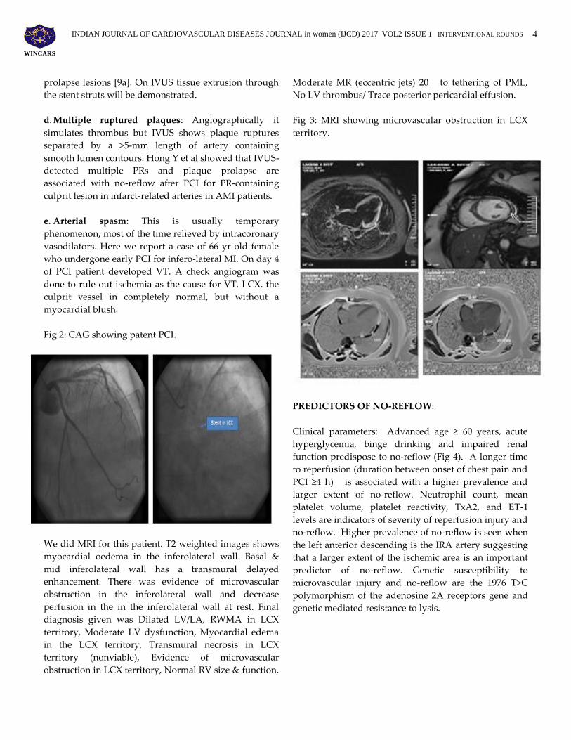
e. Arterial spasm: This is usually temporary
phenomenon, most of the time relieved by intracoronary
vasodilators. Here we report a case of 66 yr old female
who undergone early PCI for infero-lateral MI. On day 4
of PCI patient developed VT. A check angiogram was
done to rule out ischemia as the cause for VT. LCX, the
culprit vessel in completely normal, but without a
myocardial blush.
Fig 2: CAG showing patent PCI.
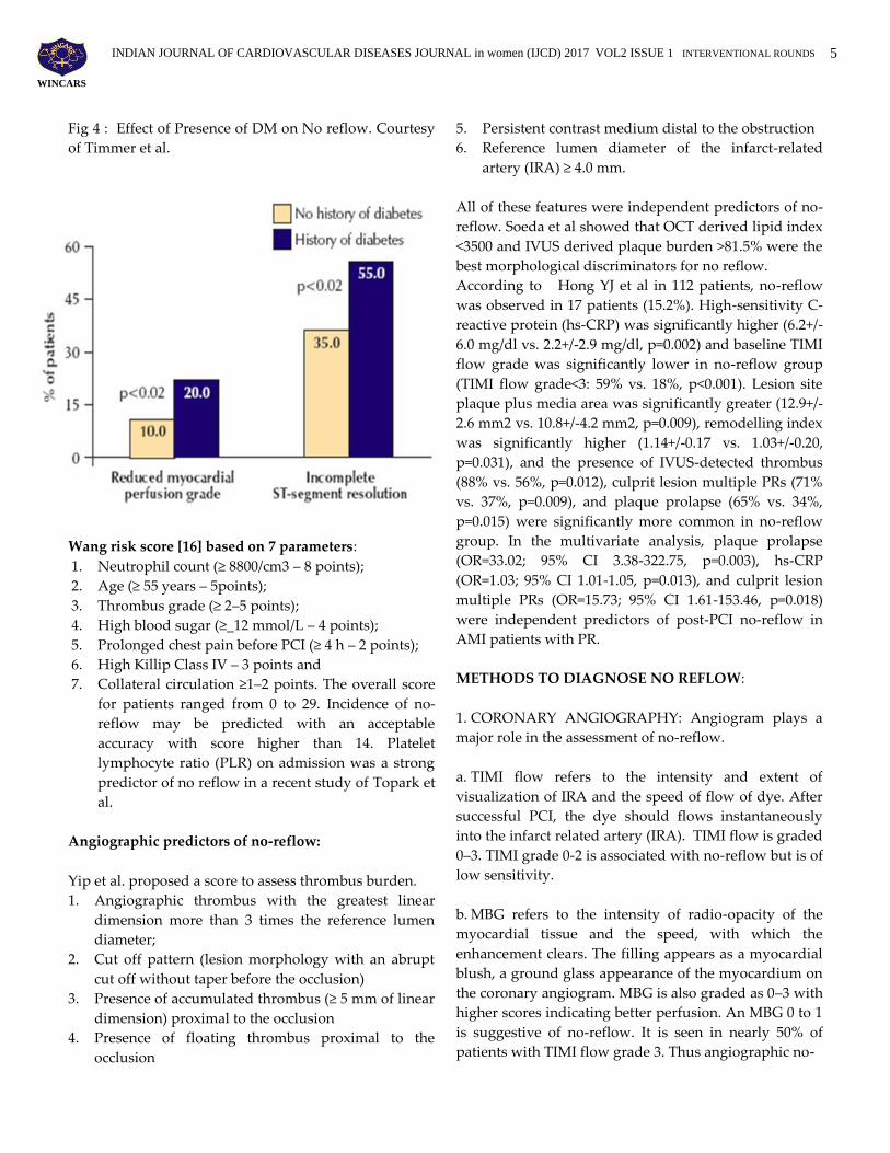
We did MRI for this patient. T2 weighted images shows
myocardial oedema in the inferolateral wall. Basal &
mid inferolateral wall has a transmural delayed
enhancement. There was evidence of microvascular
obstruction in the inferolateral wall and decrease
perfusion in the in the inferolateral wall at rest. Final
diagnosis given was Dilated LV/LA, RWMA in LCX
territory, Moderate LV dysfunction, Myocardial edema
in the LCX territory, Transmural necrosis in LCX
territory (nonviable), Evidence of microvascular
obstruction in LCX territory, Normal RV size & function,
Moderate MR (eccentric jets) 20 to tethering of PML,
No LV thrombus/ Trace posterior pericardial effusion.
Fig 3: MRI showing microvascular obstruction in LCX
territory.
PREDICTORS OF NO-REFLOW:
Clinical parameters: Advanced age ≥ 60 years, acute
hyperglycemia, binge drinking and impaired renal
function predispose to no-reflow (Fig 4). A longer time
to reperfusion (duration between onset of chest pain and
PCI ≥4 h) is associated with a higher prevalence and
larger extent of no-reflow. Neutrophil count, mean
platelet volume, platelet reactivity, TxA2, and ET-1
levels are indicators of severity of reperfusion injury and
no-reflow. Higher prevalence of no-reflow is seen when
the left anterior descending is the IRA artery suggesting
that a larger extent of the ischemic area is an important
predictor of no-reflow. Genetic susceptibility to
microvascular injury and no-reflow are the 1976 T>C
polymorphism of the adenosine 2A receptors gene and
genetic mediated resistance to lysis.
INDIAN JOURNAL OF CARDIOVASCULAR DISEASES JOURNAL in women (IJCD) 2017 VOL2 ISSUE 1 INTERVENTIONAL ROUNDS
5
WINCARS
Fig 4 : Effect of Presence of DM on No reflow. Courtesy
of Timmer et al.
Wang risk score [16] based on 7 parameters:
1. Neutrophil count (≥ 8800/cm3 – 8 points);
2. Age (≥ 55 years – 5points);
3. Thrombus grade (≥ 2–5 points);
4. High blood sugar (≥_12 mmol/L – 4 points);
5. Prolonged chest pain before PCI (≥ 4 h – 2 points);
6. High Killip Class IV – 3 points and
7. Collateral circulation ≥1–2 points. The overall score
for patients ranged from 0 to 29. Incidence of no-
reflow may be predicted with an acceptable
accuracy with score higher than 14. Platelet
lymphocyte ratio (PLR) on admission was a strong
predictor of no reflow in a recent study of Topark et
al.
Angiographic predictors of no-reflow:
Yip et al. proposed a score to assess thrombus burden.
1. Angiographic thrombus with the greatest linear
dimension more than 3 times the reference lumen
diameter;
2. Cut off pattern (lesion morphology with an abrupt
cut off without taper before the occlusion)
3. Presence of accumulated thrombus (≥ 5 mm of linear
dimension) proximal to the occlusion
4. Presence of floating thrombus proximal to the
occlusion
5. Persistent contrast medium distal to the obstruction
6. Reference lumen diameter of the infarct-related
artery (IRA) ≥ 4.0 mm.
All of these features were independent predictors of no-
reflow. Soeda et al showed that OCT derived lipid index
<3500 and IVUS derived plaque burden >81.5% were the
best morphological discriminators for no reflow.
According to Hong YJ et al in 112 patients, no-reflow
was observed in 17 patients (15.2%). High-sensitivity C-
reactive protein (hs-CRP) was significantly higher (6.2+/-
6.0 mg/dl vs. 2.2+/-2.9 mg/dl, p=0.002) and baseline TIMI
flow grade was significantly lower in no-reflow group
(TIMI flow grade<3: 59% vs. 18%, p<0.001). Lesion site
plaque plus media area was significantly greater (12.9+/-
2.6 mm2 vs. 10.8+/-4.2 mm2, p=0.009), remodelling index
was significantly higher (1.14+/-0.17 vs. 1.03+/-0.20,
p=0.031), and the presence of IVUS-detected thrombus
(88% vs. 56%, p=0.012), culprit lesion multiple PRs (71%
vs. 37%, p=0.009), and plaque prolapse (65% vs. 34%,
p=0.015) were significantly more common in no-reflow
group. In the multivariate analysis, plaque prolapse
(OR=33.02; 95% CI 3.38-322.75, p=0.003), hs-CRP
(OR=1.03; 95% CI 1.01-1.05, p=0.013), and culprit lesion
multiple PRs (OR=15.73; 95% CI 1.61-153.46, p=0.018)
were independent predictors of post-PCI no-reflow in
AMI patients with PR.
METHODS TO DIAGNOSE NO REFLOW:
1. CORONARY ANGIOGRAPHY: Angiogram plays a
major role in the assessment of no-reflow.
a. TIMI flow refers to the intensity and extent of
visualization of IRA and the speed of flow of dye. After
successful PCI, the dye should flows instantaneously
into the infarct related artery (IRA). TIMI flow is graded
0–3. TIMI grade 0-2 is associated with no-reflow but is of
low sensitivity.
b. MBG refers to the intensity of radio-opacity of the
myocardial tissue and the speed, with which the
enhancement clears. The filling appears as a myocardial
blush, a ground glass appearance of the myocardium on
the coronary angiogram. MBG is also graded as 0–3 with
higher scores indicating better perfusion. An MBG 0 to 1
is suggestive of no-reflow. It is seen in nearly 50% of
patients with TIMI flow grade 3. Thus angiographic no-

INDIAN JOURNAL OF CARDIOVASCULAR DISEASES JOURNAL in women (IJCD) 2017 VOL2 ISSUE 1 INTERVENTIONAL ROUNDS
6
WINCARS
reflow can be defined as a TIMI flow grade _3 or 3 with
an MBG 0 to 1. In practice, TIMI flow grade and MBG
are most commonly used.
c. TMPG is used to characterize the filling and clearance
of the myocardial perfusion. The filling appears as
myocardial blush (or ground glass appearance of the
myocardium). TMPG defines the intensity of the blush
and then focuses on the clearance of the contrast opacity
from the myocardium. TMPG is graded 0–3.
d. Corrected TIMI frame count (cTFC) is defined as the
number of cine frames required for dye to reach
standardized distal markers of the coronary tree. Lower
cTFC after PPCI have been associated with more
favorable prognosis. Thus TIMI flow grading and CTFC
evaluate the Epicardial flow, while MBG and TMPG
evaluate the microvascular flow.
4. ELECTROCARDIOGRAPHY: Study of ST resolution
(STR) in serial ECGs is a bedside method of assessing
myocardial perfusion following PCI. A rapid decrease of
ST elevation is highly suggestive of reperfusion. STR at
60 min after PCI should exceed 70%. STR <70% at 60 min
is a marker of no reflow. A rapid decrease of ST segment
is highly specific (91%) and fairly sensitive (77%)
parameter of myocardial reperfusion.
3. MYOCARDIAL CONTRAST
ECHOCARDIOGRAPHY (MCE):
MCE is one of the best methods to predict no reflow.
Intra-myocardial contrast opacification is visualized and
recorded after injecting intravenously an ultrasound
contrast agent containing small micro bubbles. Absence
of opacification detects no reflow/dysfunctional
microvascular circulation. MCE is best recorded after
24–48 h after PPCI since MCE performed immediately
after PPCI may underestimate the size and extent of no
reflow (Fig 5).
Fig 5: Myocardial contrast echo demonstrating
opacification on defects due to No reflow.
5. CORONARY MAGNETIC RESONANCE
IMAGING (CMRI): CMRI is the most sensitive
and specific method to assess the extent of no
reflows. The ideal time is 1 week after myocardial
infarction although it can be done at 48–72 h after
PCI. Necrotic or fibrotic myocardium enhances
gadolinium distribution into the interstitium,
which appears as a bright signal:
hyperenhancement. The severe microvascular
damage in the context of MVO prevents
gadolinium from entering the injured myocardium
which appears dark and hypo enhanced,
surrounded by the hyper-enhanced infarcted
myocardium This a non-invasive Gadolinium
based technique performed in two steps (i) Early
MVO is performed soon after injecting gadolinium
and recorded during the first pass of the contrast
agent and (ii) late or delayed contrast enhanced
MVO is performed 10–20 min after the injection of
contrast. The early phase represents no reflow,
while the delayed phase depicts the extent of
myocardial necrosis.
6. CORONARY COMPUTED TOMOGRAPHY
ANGIOGRAPHY (CCTA): Kinohira et al. showed
in a small study on 26 patients that low-attenuation
plaque in CCTA can predict PCI complications,
mainly slow-flow. In the small study of Nakazawa
et al. MDCT images of the culprit lesion were
assessed in 51 patients who underwent PCI. In
patients in whom no-reflow occurred after PCI
INDIAN JOURNAL OF CARDIOVASCULAR DISEASES JOURNAL in women (IJCD) 2017 VOL2 ISSUE 1 INTERVENTIONAL ROUNDS
7
WINCARS
there was significantly lower culprit plaque density
and signet-ring sign was found more frequently.
Rafał Wolny et al in a case report also showed the
similar complication in CCTA proved vulnerable
plaque.
6. INTRAVASCULAR IMAGING: They have a role in
investigating the potential predictors of No-Reflow
phenomenon.
a. Intravascular ultrasound (IVUS) - Echo-attenuated
plaque, containing more fibrofatty tissue and necrotic
core strongly correlated with No-reflow (HORIZONS-
AMI trial). other IVUS findings associated with higher
risk of slow-flow are intraluminal thrombus, thin-
capped fibro-atheroma morphology (TCFA) and large
plaque burden.
b. Optical Coherence Tomography (OCT) – The thin-cap
fibro atheroma (TCFA), identified by OCT, have been
more frequently associated with no-reflow.
7. INTRACORONARY DOPPLER : Intracoronary
Doppler guidewire is used to assess micro vascular
function by measuring coronary flow velocity (CFV) and
coronary flow reserve (CFR) from which
microcirculatory index can be calculated. The three
characteristic components seen in no reflow are (a)
systolic flow reversal, (b) reduced systolic antegrade
flow and (c) forward diastolic flow with rapid
deceleration slope.
Intracoronary pressure measurement: A double lumen
catheter with a side hole is employed to measure
intracoronary pressure gradient in IRA. Absence of
pressure gradient indicates absence of obstruction in
IRA. By using pressure/thermostat guidewire placed
distally in IRA, index of micro-circulatory resistance
(IMR) can be calculated which is related to acute
microvascular damage in no-reflow.
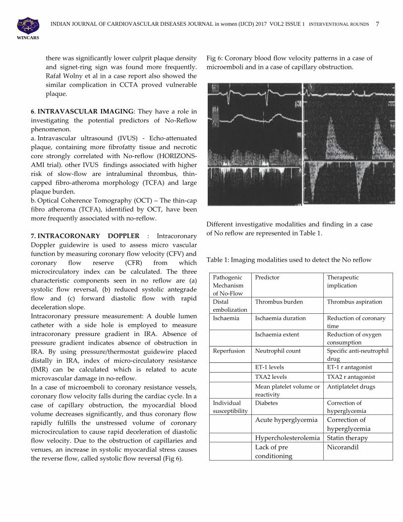
In a case of microemboli to coronary resistance vessels,
coronary flow velocity falls during the cardiac cycle. In a
case of capillary obstruction, the myocardial blood
volume decreases significantly, and thus coronary flow
rapidly fulfills the unstressed volume of coronary
microcirculation to cause rapid deceleration of diastolic
flow velocity. Due to the obstruction of capillaries and
venues, an increase in systolic myocardial stress causes
the reverse flow, called systolic flow reversal (Fig 6).
Fig 6: Coronary blood flow velocity patterns in a case of
microemboli and in a case of capillary obstruction.
Different investigative modalities and finding in a case
of No reflow are represented in Table 1.
Table 1: Imaging modalities used to detect the No reflow
Pathogenic
Mechanism
of No-Flow
Predictor Therapeutic
implication
Distal
embolization
Thrombus burden Thrombus aspiration
Ischaemia Ischaemia duration Reduction of coronary
time
Ischaemia extent Reduction of oxygen
consumption
Reperfusion Neutrophil count Specific anti-neutrophil
drug
ET-1 levels ET-1 r antagonist
TXA2 levels TXA2 r antagonist
Mean platelet volume or
reactivity
Antiplatelet drugs
Individual
susceptibility
Diabetes Correction of
hyperglycemia
Acute hyperglycemia Correction of
hyperglycemia
Hypercholesterolemia Statin therapy
Lack of pre
conditioning
Nicorandil
INDIAN JOURNAL OF CARDIOVASCULAR DISEASES JOURNAL in women (IJCD) 2017 VOL2 ISSUE 1 INTERVENTIONAL ROUNDS
8
WINCARS
PHARMACOLOGICALAPPROACH:
Local vasodilator therapy and local antiplatelet therapy
form the sheet anchor of treatment in the cath lab for no-
reflow. The 2011 ACC PCI guidelines give a class IIa
recommendation for administration of an intracoronary
vasodilator (adenosine, calcium channel blocker, or
nitroprusside) to treat PCI-related no-reflow that occurs
during primary or elective PCI. Current guidelines
recommend use of GpIIb/IIIa only as a bailout procedure
in no-reflow patients.
1. Adenosine: It is a short acting endogenous
nucleoside that increases microvascular flow owing
to its vasodilator properties. It inhibits neutrophils
adhesion and migration, exerts antiplatelet effects,
and inhibits oxygen free radical formation.
Adenosine has shown to be beneficial for both
intravenous and intracoronary administration.
a. The AMISTAD trial showed that I.V adenosine
infused before initiation of thrombolytic therapy
resulted in 33% relative reduction in infarct size
( 67% RR in patients with anterior MI).
b. AMISTAD-II trial aimed to assess the benefit of
adenosine routine use in 2118 patients
presenting with STEMI - No significant
difference in clinical outcome but a significant
reduction in infarct size with I.V high dose of
70mcg/kg/min).
c. REOPEN TRIAL proved that intracoronary
adenosine 120 mg bolus followed by 2 mg in 33
ml saline slow infusion over 2 min, was more
beneficial than nitroprusside in treating no-
reflow. STR >70% at 90 min was found in 71%
adenosine vs. 54% in nitroprusside vs. 51% in
saline placebo group. 30 days MACE (heart
failure and recurrent MI) was 10% in adenosine
group vs. 14% in nitroprusside group and 20%
in saline group.
d. In Su et al. Analysis of 11 RCTs involving 1027
STEMI patients, there was no evidence that
adenosine reduces short term or long term all
cause mortality or recurrent myocardial
infarction. However adenosine as treatment did
reduce angiographic no-reflow.
e. e.Gao et al. - PRISMA COMPLAINT meta-
analysis of 15 RCTs with 1736 patients revealed
better STR and improved TIMI flow grade after
adenosine but no definite improvement in LVEF
or mortality.
f. f.REFLOW STEMI is an ongoing trial to compare
the effects of adenosine, nitroprusside and
placebo. Primary end point is the measurement
of infarct size at the end of 48–72 h by CMRI.
The results are awaited.
Yetgin et al howed that no-reflow and infarct size
depended on dose of adenosine. The optimal IC bolus
dose of adenosine was 100 mg in right coronary artery
(RCA) and 200 mg in left coronary artery (LCA) to
induce maximum hyperemia and with minimal side
effects (Adjedj et al .
2. Calcium channel blockers: These drugs act by
blocking L-type channels in the cell membrane of
myocardium and cause endothelial dependent
relaxation of micro-vessels. These also reduce
oxygen demand by the myocardium and minimize
the damage caused by oxygen free radicals.
a. Verapamil- 100-250 mg intracoronary bolus
followed by 100 mg/min (max 1000mg) in
STEMI patients with no-reflow improved TIMI
flow with reduction of corrected TIMI frame
count (CTFC)
b. In a randomized study of 150 STEMI patients,
verapamil and adenosine were equally effective
for prevention of no-reflow and improving
TIMI frame count.
c. In the RECOVER-AMI trial, both verapamil
and diltiazem were equally effective in no-
reflow. Diltiazem was administered 400 mg
(diluted in NS) intracoronary followed by 90
mg BD orally.
d. Wang et.al’s *11+ meta-analysis of 8 RCTs
showed that both verapamil and diltiazem
equally reduced no-reflow but no improvement
in LVEF at 6months.
e. Nicardepine 360-460 mg intracoronary
improved TIMI flow grade and reduced CTFC
in 72 patients with no-reflow. Prophylactic
intra-graft Nicardepine followed immediately
by direct stenting of SVGs without mechanical
distal protection in a series of 68 elective
INDIAN JOURNAL OF CARDIOVASCULAR DISEASES JOURNAL in women (IJCD) 2017 VOL2 ISSUE 1 INTERVENTIONAL ROUNDS
9
WINCARS
patients dramatically reduced the incidence of
no-reflow.
f. All the three calcium channel blockers
(Verapamil, Diltiazem and Nicardepine) are
well tolerated and have produced good results
in the treatment of no reflow.
3. Nitroprusside: It is a nitric oxide donor with vaso-
dilatators, antiplatelet, and anti-inflammatory
properties; the potential benefits of this agent should
be weighed against the possible harm of systemic
hypotension. Doses of 50-100 mcg have been studied
in various trials but results proved inferior to CCBs
and Adenosine.
4. Nicorandil: It is an ATP-sensitive potassium channel
opener and a nitric oxide (NO) donor and is a potent
vasodilator. It also modulates neutrophil activation
and suppresses formation of oxygen free radicals.
Chen et.al showed that it reduces reperfusion injury
and improved LV function in patients with no
reflow. Nicorandil at a dose of 2 mg intracoronary
bolus and/or 8 mg/h infusion is beneficial in the
prevention and treatment of no reflow and reducing
major cardiac adverse events (MACE) at 5 years.
Intracoronary nicorandil was more effective than IC
verapamil in preventing no-reflow in 61 patients (63
lesions) undergoing rotational atherectomy.
5. Glycoprotein IIB/IIIA inhibitors: Abciximab,
tirofiban and eptifibatide have been used for the
prevention and treatment of no reflow. Though
theoretically these drugs appear as attractive options
for no-reflow, their efficacy has not been proven in
randomized trials.
a. In a study of 90 STEMI [18] patients, no reflow
occurred less frequently in abciximab group
(7%) compared with control group (17%). A
recent randomized trial of intracoronary
abciximab demonstrated a significantly smaller
infarct size when assessed by CMRI. In the
AIDA-STEMI , intracoronary or intravenous
abciximab did not result in any difference in the
combined end point of death, re-infarction or
congestive heart failure.
b. Tirofiban given as upstream intravenous
infusion achieved better STR and MBG
following PCI in ON TIME TRIAL.
c. Eptifibatide improved reperfusion by TIMI flow
grading in PROTECT TIMI trial.
The COCTAIL TRIAL to assess the safety and efficacy of
IC cocktail injection combined with thrombus aspiration
in STEMI patients treated with primary PCI are under
study. The cocktail includes bivalirudin, tirofiban and
tenectaplase. The trial is sponsored and conducted at
Xijing Hospital, China. The trial is currently recruiting
participants and the results are still awaited. COCTAIL
II study has compared standard vs CLEARWAY infused
abciximab in myocardial infarction.
6. Nitroglycerine: Intracoronary nitroglycerin failed to
show any benefit in treatment of no-reflow. This is
due to the fact that nitroglycerin is predominantly a
venodilator and has little impact on arteriolar tone.
Nitroglycerine needs to be metabolized by the
vascular wall to derive its nitric oxide, which the
micro vascular resistance arterioles are unable to.
Mechanical therapies: There is no data to treat no
reflow with any of the mechanical therapies.
PREVENTION OF NO-REFLOW:
The aim should be to counteract the various
mechanisms of no-reflow. Prevention of No-reflow.
a. BEFORE THE ONSET OF INFARCTION PAIN
b. BEFORE REPERFUSION: by decreasing the
ischemic time with minimum door to balloon time
no flow can be decreased. By reducing the severity
of ischemia and improving myocardial perfusion
with drugs that reduce myocardial oxygen
consumption can also be tried. The beneficial effects
of Carvedilol, Fosinopril, and Valsartan on coronary
no-reflow have indeed been recently demonstrated.
c. IN THE CATH LAB: Patients at high risk of No-
Reflow on the basis of the presence of reperfusion-
related injury can be treated with drugs like
Glycoprotein IIb/IIIa antagonists, Adenosine,
Nicorandil aimed at counteracting endothelial
platelet and neutrophil activation. Selective ET-1 or
TxA2 antagonism might represent novel therapeutic
approaches (25).
INDIAN JOURNAL OF CARDIOVASCULAR DISEASES JOURNAL in women (IJCD) 2017 VOL2 ISSUE 1 INTERVENTIONAL ROUNDS
10
WINCARS
Abciximab: Among glycoprotein IIb/IIIa antagonists,
abciximab has been found to improve myocardial
perfusion when started during PPCI and infused for 12
h thereafter, as assessed by a higher rate of STR 50% at
60 min after PCI (73% vs.57%, p < 0.05). Intracoronary
abciximab has been proven to be superior to intravenous
abciximab in patients treated by primary PPCI
approaches (26, 27). Ellis and colleagues analysed 102
vein graft stenosis from the EPIC and EPILOG trials and
failed to demonstrate any clinical benefit with the active
drug treatment with an 18•6% incidence of death,
myocardial infarction and urgent revascularization at 30
days compared to 16•3% for placebo. They
hypothesized that distal embolization of athermomatous
plaque from the vein graft wall is less sensitive to the
antiplatelet effect of abciximab. But this is not true in the
case saphenous vein graft PCI.
A. PREVENTION OF DISTAL EMBOLISM:
1. Direct stenting- Placement of a stent without prior
balloon predilation has been advocated as a means
to reduce no-reflow. Direct stent implantation, by
entrapping athero-thrombotic debris, reduces the
risk of distal embolization, but unfortunately only a
few subsets of patients are suitable for direct
stenting. In elective cases direct stenting has shown
no benefit, where as in primary PCI, a small
randomized trial showed decrease rates of slow flow
or no-reflow as compared to placebo.
2. Aspiration Thrombectomy: Thrombectomy devices
have initially shown promising results, especially in
the subset of patients with large thrombus burden.
More than 20 randomized trials have investigated
the benefits of manual thrombectomy over
conventional PCI. Three landmarks that have
influenced the present role of this strategy are the
TAPAS, TASTE and TOTAL studies.
a. TAPAS trial - Manual thrombus aspiration
group fare better in angiographic outcomes and
reduction in cardiac death.
b. TASTE trial - Failed to demonstrate any
difference in mortality, recurrent myocardial
infarction and stent thrombosis at 30 days in the
two groups.
c. TOTAL trial - No statistical difference in the
primary outcome of recurrent MI, heart failure
stent thrombosis or mortality between the two
groups. There was an increase in the incidence
of stroke at 48 h and 30 days in the thrombus
aspiration.
Base on the recent evidence, ACC/AHA/SCAI have
modified their earlier recommendation. Manual
aspiration thrombectomy is reasonable for patients
undergoing primary PCI is now class IIb
recommendation (level of evidence C-LD). There is no
role for Routine aspiration thrombectomy before
primary PCI –Class III (level of evidence- A).
3. Rheolytic thrombectomy: Rheolytic thrombectomy
(ANGIOJET/X-SIZER) has shown no benefit in
primary PCI.
4. Distal Filters: Distal protection with filters has not
shown to have any benefit in preventing no reflow in
the setting of ST elevation MI whereas it has been
shown to be beneficial in preventing no reflow in
elective saphenous vein PCI.
a. SAFER trial tested the Percusurge in patients
undergoing stent placement in saphenous vein
grafts. In comparison to patients who were
randomized to no device, Percusurge reduced
MACE by 42% at 1 month (9.6% vs. 16.5%), with
most of the reduction being in post-procedure MI
(8.6% vs. 14.7%) and no-reflow phenomenon
(3%vs.9%).
b. Filter wire Ex device was assessed in 230
saphenous vein graft patients and its use was
associated with reduced MACE (11.3%) and TIMI
O/1 flow rate and reduced distal micro-
embolization. Filter wire EX reduced distal
embolization and was associated with improved
TIMI 3 myocardial blush and ST segment
resolution post PCI in 53 patients treated for
acute MI.
5. Deferred stenting: Deferred stenting (6-12 hrs) in
selected high risk patients may reduce the incidence of
no reflow during which the patient receives supportive
therapy. In a recent randomized study of STEMI
patients, deferred stenting reduced the incidence of no
INDIAN JOURNAL OF CARDIOVASCULAR DISEASES JOURNAL in women (IJCD) 2017 VOL2 ISSUE 1 INTERVENTIONAL ROUNDS
11
WINCARS
reflow as judged by TIMI flow grade and cardiac MRI in
high risk patients.
6. M-guard stent: M guard was a technology, designed
to prevent distal embolization and no reflow following
PCI. It consists of a bare metal stent with cobalt
chromium strut and polyethylene theraphthalate mesh
(micronet) covering anchored to the external surface of
the strut. Three non-randomized and two randomized
trials (Piscione Trial, MAGICAL trial, REWARD and
MASTER-I and II) have shown it to be beneficial in
STEMI patients with large thrombus burden, restoring
myocardial reperfusion and reducing no-reflow.
However, M-guard stent has exhibited three drawbacks:
(i) high rates of instant restenosis(ii) difficulty in
negotiation (4.1%) and (iii) stent dislodgement (0.9%).
Hence, the stent has been recalled and the MASTER II
Trial cancelled. The manufacturers are now trying to
develop a new drug eluting version of M-guard stent.
B. REDUCTION OF ISCHEMIA: Measures to reduce
time interval from onset of chest pain to PPCI are
probably the best measure to improve myocardial
salvage and reduce risk of no reflow. The use of the
intra-aortic balloon pump is probably the last measure.
It may be beneficial when hemodynamic instability is
present by improving flow but only after the epi-cardial
occlusion is successfully relieved.
C. PREVENTION OF REPERFUSION INJURY:
a. Hyperoxemic reperfusion: Intracoronary
hyperoxemic reperfusion has been advocated for
prevention of reperfusion injury. Hyperoxemic
reperfusion improved microvascular blood flow and
decreased infarct size in a canine model of ischemia
reperfusion. In the AMIHOT-II (Acute Myocardial
Infarction With Hyperoxemic Therapy II) trial this
approach reduced infarct size but was not associated
with improved tissue perfusion as assessed by ST-
segment resolution. Because the clinical benefit of
hyperoxemic reperfusion has yet to be shown, the
routine use of this invasive strategy in the current era
of routine thrombectomy cannot be recommended at
present.
b. Myocardial post-conditioning: by use of intermittent
low-pressure balloon inflations in the infarct-related
artery reduced infarct size and improved
microvascular perfusion as assessed by myocardial
blush, and long-term functional recovery.
Pharmacological post-conditioning by intravenous
administration of cyclosporine, a direct MPTP blocker,
versus placebo , at the time of primary PCI decreased
infarct size. Remote post-conditioning by intermittent
inflations of a blood pressure cuff on the upper limb
before reperfusion improved ST-segment resolution
following primary PCI, an effect that was enhanced by
administration of morphine. These preliminary studies
suggest a beneficial role for conditioning strategies in
the setting of primary infarct PCI. However, their
efficacy in patients undergoing thrombectomy and
receiving glycoprotein IIb/IIIa inhibitors remains to be
defined.
c. It might be reasonable for patients with multiple
vulnerable plaques (detected by any imaging
modality) to be scheduled for coronary artery bypass
grafting (CABG) rather than PCI. If PCI is chosen,
avoiding balloon pre-dilation and performing direct
stenting should be encouraged. The use of distal
protection devices should be considered. Its significant
impact on reduction of major adverse cardiac events
after PCI in saphenous vein grafts was shown in the
SAFER trial ; however, there was no such relationship
in the setting of primary PCI of native coronary
arteries in the EMERALD trial . On the other hand,
when only patients with angio-scopically-detected
ruptured plaque were analysed, distal protection
reduced microcirculation damage and left ventricular
dysfunction. Another concern is the potential use of
additional antiplatelet drugs. Pre-procedural
glycoprotein IIb/IIIa receptor inhibitor administration
may be beneficial in some high-risk patients
undergoing saphenous graft angioplasty.
In conclusion, prevention of No reflow is better than the
treatment. So, we have to indentify the conditions which
are likely to develop No reflow before PCI itself and
target them to have good long term results.
REFERENCES:
1. Ames A III, Wright RL, Kowada M, Thurston JM,
Majno G. Cerebral ischemia II: the No-reflow
phenomenon. Am J Pathol 1968; 52:437-53.
INDIAN JOURNAL OF CARDIOVASCULAR DISEASES JOURNAL in women (IJCD) 2017 VOL2 ISSUE 1 INTERVENTIONAL ROUNDS
12
WINCARS
2. Kloner RA, Ganote CE, Jenning RB. The ‚no-reflow‛
phenomenon after temporary coronary occlusion in
dogs. J Clin Invest 1974;54:
3. 1496–508 Ito H, Tomooka T, Sakai N, Yu H, Higashino
Y, Fujii K, Masuyama T, Kitabatake A, Minamino T.
Lack of myocardial perfusion immediately after
successful thrombolysis: a predictor of poor recovery of
left ventricular function in anterior myocardial
infarction. Circulation 1992; 85:1699–705.
4. Lee CH, Wong HB, Tan HC, et al. Impact of
reversibility of no reflow phenomenonon 30-day
mortality following percutaneous re- vascularizationfor
acute myocardial infarction-insights from a 1,328patient
registry. J Interv Cardiol 2005; 18: 261-266.
5. Piana RN, Paik GY, Moscucci M, Cohen DJ, Gibson
CM, Kugelmass AD, Carroza JP Jr, Kuntz RE, Baim DS.
Incidence and treatment of 'no-reflow' after
percutaneous coronary intervention. Circulation.
1994;89:2514-8.
6.Niccoli G, Scalone G, Lerman A, Crea F. Coronary
microvascular obstruction in acute myocardial
infarction. Eur Heart J. 2015.
http://dx.doi.org/10.1093/eur- heartj/ehv484.
7. Galiuto L. Optimal therapeutic strategies in the setting
of post-infarct no reflow: the need for a pathological
classification. Heart. 2004;90:123–125.
8. Eeckhout E, MJ.Kern. The coronary no-reflow
phenomenon. a review of mechanisms and therapies.
Eur Heart J 2001; 22: 729–39.
9. Tranum-Jensen J, Janse MJ, Fiolet WT, et al. (1981)
Tissue osmolality, cell swelling, and reperfusion in acute
regional myocardial ischemia in the isolated porcine.
Circ Res 49: 364–381.
10. Danesh Sani HD, Eshraghi A, Sahri B, Vejdanparast
M. No-reflow phenomenon in patients with ST-elevation
acute myocardial infarction, treated with primary
percutaneous coronary intervention: a study of
predictive factors. J Cardiothorac Med. 2014;2(4):221–
226. 25.
11. Abdi S, Rafizadeh O, Peighambari M, Basiri H,
Bakhshandeh H. Evaluation of clinical and procedural
predictive factors of no-reflow phenomenon following
primary percutaneous coronary intervention. Res
Cardiovasc Med. 2005;4(2): e25414. 26.
12. Mazhar J, Maschicharan M, Farshid A. Predictors
and outcome of no-reflow post primary percutaneous
coronary intervention for ST elevation myocardial
infarc-tion. IJC Heart Vasc. 2016;10:8–12.
13. Iwakura K, Ito H, Kawano S, et al. Predictive factors
for development of the no-reflow phenomenon in
patients with reperfused anterior wall acute myocardial
infarction. J Am Coll Cardiol 2001;38:472–7.
14. Vignali L, Talanas G, Saia F, et al. Genetic
association between the 1976T>C polymorphism in the
adenosine A2 receptor and angiographic no-reflow
phenomenon (abstr). Il giornale italiano di Cardiologia
Invasiva 2007;3 Suppl 1:109.
15. Zalewski J, Undas A, Godlewski J, Stepien E,
Zmudka K. No-reflow phenomenon after acute
myocardial infarction is associated with reduced clot
permeability and susceptibility to lysis. Arterioscler
Thromb Vasc Biol 2007;27:2258–65.
16. Wang JW, Zhoud ZQ, Chen YD, Want CH, Zhu XL.
A risk score for no reflow in patients with ST-segment
elevation myocardial infarction after percutaneous
coronary intervention. Clin Cardiol. 2015;38(4):208–215.
17. Topark C, Tabakci MM, Zimsek Z, et al.
Platelet/lymphocyte ratio was associated with impaired
myocardial perfusion and both in-hospital and long-
term ad- verse outcome in patients with ST-segment
elevation acute myocardial infarction undergoing
primary coronary intervention. Postep Kardiol
Interwencyjnej. 2015; 11(4):288–297.
18. Yip HK, Chen MC, Chang HW, et al. Angiographic
morphologic features of infarct-related arteries and
timely reperfusion in acute myocardial infarction:
INDIAN JOURNAL OF CARDIOVASCULAR DISEASES JOURNAL in women (IJCD) 2017 VOL2 ISSUE 1 INTERVENTIONAL ROUNDS
13
WINCARS
predictors of slow-flow and no-reflow phenomenon.
Chest 2002;122:1322–32.
19. Soeda T, Higuma T, Abe N, et al. Intravascular
ultrasound, but not optical coherence tomography,
predicts no reflow phenomenon after primary percuta-
neous coronary intervention in patients with ST-segment
elevation myocardial infarction cause by plaque rupture.
J Am Coll Cardiol. 2015;67(10S). 1244-071.
20. Soeda T, Higuma T, Abe N, et al. Morphological
predictors for no-reflow phenom-enon after primary
percutaneous coronary intervention in patients with ST-
segment elevation myocardial infarction caused by
plaque rupture. Eur Heart J Cardiovasc Imaging.
2016;(January). pii:jev341 [epub ahead of print].
21. Wu X, Mintz GS, Xu K, et al. (2011) The relationship
between attenuated plaque identified by intravascular
ultrasound and no-reflow after stenting in acute
myocardial infarction: the HORIZONS-AMI
(Harmonizing Outcomes With Revascularization and
Stents in Acute Myocardial Infarction) trial. JACC
Cardiovasc Interv.
22. Tanaka A, Imanishi T, Kitabata H, et al. (2009)
Lipid-rich plaque and myocardial perfusion after
successful stenting in patients with non-ST-segment
elevation acute coronary syndrome: an optical coherence
tomography study. Eur Heart J 30: 1348–1355.
23. Morishima I, Sone T, Okumura K, et al.
Angiographic no-reflow phenomenon as a predictor of
adverse long-term outcome in patients treated with
percutaneous transluminal coronary angioplasty for first
myocardial infarction. J Am Coll Cardiol. 2000;36:1202–
1209.
24. Bolognese L, Carrabba N, Parodi G, et al. Impact of
microvascular dysfunction on left ventricular
remodelling and long-term clinical outcome after
primary coro-nary angioplasty for acute myocardial
infarction. Circulation. 2004;109:1121– 1126.
25. Ndreppa G, Tiroch K, Fusaro M, et al. 5-Year
prognostic value of no-reflow phenomenon after
percutaneous coronary intervention in patients with
acute myocardial infarction. J Am Coll Cardiol.
2010;55:2383–2389.
26. Young Joon Hong, MD; Myung Ho Jeong, MD; Sang
Wook Kim, MD*; Yun Ha Choi; Eun Hae Ma; Jum Suk
Ko, MD; Min Goo Lee, MD; Keun Ho Park, MD; Doo
Sun Sim, MD; Nam Sik Yoon, MD; Hyun Ju Yoon, MD;
Kye Hun Kim, MD; Hyung Wook Park, MD; Ju Han
Kim, MD; Youngkeun Ahn, MD; Jeong Gwan Cho, MD;
Jong Chun Park, MD; Jung Chaee Kang, MD. Relation
Between Plaque Components and Plaque Prolapse.
27. After Drug-Eluting Stent Implantation – Virtual
Histology – Intravascular Ultrasound. Circ J 2010; 74:
1142 – 1151.
28. Higashikuni Y, Tanabe K, Tanimoto S, Aoki J,
Yamamoto H, Nakazawa G, et al. Impact of culprit
plaque composition on the noreflow phenomenon in
patients with acute coronary syndrome: An
intravascular ultrasound radiofrequency analysis. Circ J
2008; 72: 1235 – 1241.
29. Watanabe T, Nanto S, Uematsu M, Ohara T,
Morozumi T, Kotani J, et al. Prediction of no-reflow
phenomenon after successful percutaneous coronary
intervention in patients with acute myocardial
infarction: Intravascular ultrasound findings. Circ J 2003;
67: 667 – 30. 671.
30. Okura H, Taguchi H, Kubo T, Toda I, Yoshida K,
Yoshiyama M, et al. Atherosclerotic plaque with
ultrasonic attenuation affects coronary reflow and
infarct size in patients with acute coronary syndrome:
An intravascular ultrasound study. Circ J 2007; 71: 648 –
653.
31. Niccoli G1, Burzotta F, Galiuto L, Crea F. Myocardial
no-reflow in humans. J Am Coll Cardiol. 2009 Jul
21;54(4):281-92.
32. Hong YJ1, Jeong MH, Choi YH, Ko JS, Lee MG,
Kang WY, Lee SE, Kim SH, Park KH, Sim DS, Yoon NS,
Youn HJ, Kim KH, Park HW, Kim JH, Ahn Y, Cho JG,
Park JC, Kang JC. Predictors of no-reflow after
percutaneous coronary intervention for culprit lesion
with plaque rupture in infarct-related artery in patients

INDIAN JOURNAL OF CARDIOVASCULAR DISEASES JOURNAL in women (IJCD) 2017 VOL2 ISSUE 1 INTERVENTIONAL ROUNDS
14
WINCARS
with acute myocardial infarction. J Cardiol. 2009
Aug;54(1):36-44.
33. Rafał Wolny, Artur Dębski, Mariusz Kruk, and
Cezary Kępka. Slow-flow phenomenon after elective
percutaneous coronary intervention of computed
tomography-detected vulnerable coronary lesion.
Postepy Kardiol Interwencyjnej. 2014; 10(3): 181–184.
34. Kinohira Y, Akutsu Y, Li HL, et al. Coronary arterial
plaque characterized by multislice computed
tomography predicts complications following coronary
intervention. Int Heart J. 2007;48:25–3.
35. Maurovich-Horvat P, Hoffmann U, Vorpahl M, et al.
The napkin-ring sign: CT signature of high-risk coronary
plaques? JACCCardiovasc Imaging. 2010;3:440–4. 12.
36. Baim DS, Wahr D, George B, et al. Randomized trial
of a distal embolic protection device during
percutaneous intervention of saphenous vein aorto-
coronary bypass grafts. Circulation. 2002;105:1285–90.
37. Stone GW, Webb J, Cox DA, et al. Distal
microcirculatory protection during percutaneous
coronary intervention in acute ST-segment elevation
myocardial infarction: a randomized controlled trial.
JAMA. 2005;293:1063–72.
38. Mizote I, Ueda Y, Ohtani T, et al. Distal protection
improved reperfusion and reduced left ventricular
dysfunction in patients with acute myocardial infarction
who had angioscopically defined ruptured plaque.
Circulation. 2005;112:1001–7.
39. Jonas M, Stone GW, Mehran R, et al. Platelet
glycoprotein IIb/IIIa receptor inhibition as adjunctive
treatment during saphenous vein graft stenting:
differential effects after randomization to occlusion or
filter-based embolic protection. Eur Heart J. 2006;27:920–
8.
40. Rezkalla SH1, Kloner RA. Coronary No-reflow
Phenomenon. Curr Treat Options Cardiovasc Med. 2005
May;7(1):75-80.
41. Michael Gibson, James A. de Lemos, Sabina A.
Murphy, Susan J. Marble, Carolyn H. McCabe,
Christopher P. Cannon, Elliott M. Antman, Eugene
Braunwald. Combination Therapy With Abciximab
Reduces Angiographically Evident Thrombus in Acute
Myocardial Infarction. A TIMI 14 Substudy. Circulation.
2001;103:2550-2554.
42. C. Michael Gibson, Cafer Zorkun, Vijayalakshmi
Kunadian . Intracoronary Administration of Abciximab
in ST-Elevation Myocardial Infarction. Circulation.
2008;118:6-8.
43. Allan M. Ross, MD⁎ (FACC), Raymond J. Gibbons,
MD† (FACC), Gregg W. Stone, MD‡ (FACC), Robert A.
Kloner, MD, PhD§ (FACC), R. Wayne Alexander, MD,
PhD. A Randomized, Double-Blinded, Placebo-
Controlled Multicenter Trial of Adenosine as an Adjunct
to Reperfusion in the Treatment of Acute Myocardial
Infarction (AMISTAD-II). Journal of the American
College of Cardiology, Volume 45, Issue 11, 7 June 2005,
Pages 1775–1780.
44.Quintana M1, Kahan T, Hjemdahl P. Pharmacological
prevention of reperfusion injury in acute myocardial
infarction. A potential role for adenosine as a therapeutic
agent. Am J Cardiovasc Drugs. 2004;4(3):159-67.
45. G Amit et al. Intracoronary Nitroprusside for the
Prevention of the No-Reflow Phenomenon After
Primary Percutaneous Coronary Intervention in Acute
Myocardial Infarction. A Randomized, Double-Blind,
Placebo-Controlled Clinical Trial . Am Heart J 152 (5),
887.e9-887.14. 11 2006.
46. Mahaffey KW, Puma JA, Barbagelata NA, et al.
Adenosine as an adjunct to thrombolytic therapy for
acute myocardial infarction: results of a multicenter,
randomized, placebo-controlled trial: the Acute
Myocardial Infarction STudy of ADenosine (AMISTAD)
trial. J Am Coll Cardiol 1999; 34: 1711-20.
47. Ross AM, Gibbons RJ, Stone GW, et al. (2005) A
randomized, double-blinded, placebo-controlled
multicenter trial of adenosine as an adjunct to
reperfusion in the treatment of acute myocardial
infarction (AMISTAD-II). J Am Coll Cardiol 45: 1775–
1780.
INDIAN JOURNAL OF CARDIOVASCULAR DISEASES JOURNAL in women (IJCD) 2017 VOL2 ISSUE 1 INTERVENTIONAL ROUNDS
15
WINCARS
48. Niccoli G, Rigattieri S, De Vita MR, et al. Open-
label, randomized, placebo-controlled evaluation of
intracoronary adenosine or nitroprusside after thrombus
aspiration during primary percutaneous coronary
intervention for the prevention of microvascular
obstruction in acute myocardial infarction: the REOPEN-
AMI study (Intracoronary Nitroprusside Versus
Adenosine in Acute Myocardial In-farction). J Am Coll
Cardiol Cardiovasc Interv. 2013;6:580–589.
49. Su Q, Nyi S, Li L. Adenosine and verapamil for no
reflow during primary percuta-neous coronary
intervention in people with acute myocardial infarction.
Cochrane Database Syst Rev. 2015;18(May
(5)):CD009503.
50. Gao Q, yang B, Guo Y, Zheng F. Efficacy of
adenosine in patients with acute myocardial infarction
undergoing primary percutaneous coronary
intervention. Medicine (Baltimore). 2015;94(August
(32)):e1279.
51. Yetgin T, Uitterdijk A, Hekkert ML, et al. Limitation
of infarct size and no-reflow by intracoronary adenosine
depends critically on dose and duration. J Am Coll
Cardiol Interv. 2015;8:1990–1999.
52. Adjedj J, Toh GG, Johnson NP, et al. Intracoronary
adenosine: dose–response relationship with hyperaemia.
J Am Coll Cardiol Interv. 2015;8:1422–1430.
53. Werner GS, Lang K, Kuehnert H, et al.
Intracoronary verapamil for reversal of no-reflow during
coronary angioplasty for acute myocardial infarction.
Catheter Cardiovasc Interv. 2002; 57:444–451.
54. Vijaylakshmi K, Whittaker VJ, Kunadian B, et al.
Prospective, randomized con-trolled trial to study the
effect of intracoronary injection of verapamil and
adenosine on coronary blood flow during percutaneous
coronary intervention in patients with acute coronary
syndromes. Heart. 2006; 92(9):1278–1284.
55. Huang D, Qian I, Gee L, et al. Restoration of
coronary flow in patients with no reflow after primary
coronary intervention of acute myocardial infarction
(RE-COVER AMI). Am Heart J. 2012; 164:394–401.
56. Wang L, Cheng Z, Gu Y, Peng D. Review article:
short-term effects of verapamil and diltiazem in the
treatment of no reflow phenomenon: a meta-analysis of
randomized controlled trials. Hindawi. 2015382086.
http://dx.doi.org/10.1155/ 2015/382086.
57. Huang RI, Patel P, Walinsky P, et al. Efficacy of
intracoronary nicardipine in the treatment of no-reflow
during percutaneous coronary intervention. Catheter
Cardiovasc Interv. 2006; 68:671–676.
58. Fischell T, Haller S, Ashraf K. Intragraft nicardipine
prophylaxis to prevent no-reflow in triple-vessel
saphenous vein graft intervention. J Invasive Cardiol
2005;17:334–337.
59. Chen Z, Chen X, Li S, Huo X, Fu X, Dong X.
Nicorandil improves myocardial function by regulating
plasma nitric oxide and enothelin-1 in coronary slow
flow. Coron Artery Dis. 2015;26(March (2)):114–120.
60. Ezhilan J, Juneja MS, George T, Ramkumar S,
Viswanathan V, Badrinath A. Prevention of no
flow/slow flow phenomenon in primary PCI by
nicorandil. Indian Heart J. 2007;59(3):246–249.
61. Ishii H, Ichimiya S, Kanashior M, et al. Impact of
single intravenous administration of nicorandil before
reperfusion in patients with ST-segment-elevation
myocar-dial infarction. Circulation. 2005;112:1284–1288.
62. Tsubokawa A, Ueda K, Sakamoto H, et al. Effect of
intracoronary nicorandil administration on preventing
no-reflow/slow flow phenomenon during rotational
atherectomy. Circ J 2002;66:1119–1123.
63. Petronio AS, de Carlo M, Ciabatti N, et al. Left
ventricular remodelling after primary coronary
angioplasty in patients treated with abciximab or
intracor-onary adenosine. Am Heart J. 2005;150:1015.
64. Thiele H, Wohrle J, Hambrecht R, et al.
Intracoronary versus intravenous bolus abciximab
during primary percutaneous coronary intervention in
patients with acute ST-elevation myocardial infarction: a
randomized trial. Lancet. 2012;379(9819):923–931.
INDIAN JOURNAL OF CARDIOVASCULAR DISEASES JOURNAL in women (IJCD) 2017 VOL2 ISSUE 1 INTERVENTIONAL ROUNDS
16
WINCARS
65. Van’t Hoff AW, ten Berg J, Heestermans T, et al.
Prehospital initiation of tirofiban in patients with ST-
elevation myocardial infarction undergoing primary
angio-plasty (On-TIMI 2): a multicentre, double-blind,
randomized controlled trial. Lancet. 2008;373:537–546.
66. Gibson CM, Morrow DA, Murphy SA, et al. A
randomized trial to evaluate the relative protection
against post-percutaneous coronary intervention micro-
vascular dysfunction, ischemia, and inflammation
among antiplatelet and antithrombotic agents: the
PROTECT-TIMI-30 trial. J Am Coll Cardiol.
2006;47:2364–2373.
67. ClinicalTrials.gov. Intracoronary cocktail injection
combined with thrombus aspiration in STEMI patients
treated with primary angioplasty. http://
clinicaltrials.gov/ct2/show/NCT02592694.
68. Prati F, Di Vito L, Ramazzotti V, et al. Randomized
trial of standard versus clearway-infused abciximab and
thrombectomy in myocardial infarction: ratio-nale and
design of the COCKTAIL II study. J Cardiovasc Med
(Hagerstown). 2013;14(May (5)):364–371.
69. Loubeyre C, Morice M, Lefevre T et al. A
Randomized Comparison of Direct Stenting With
Conventional Stent Implantation in Selected Patients
With Acute Myocardial Infarction. J Am Coll Cardiol
2002; 39: 15-21.
70. Vlaar PJ, Svilaas T, van der Horst IC, et al. Cardiac
death and reinfarction after 1 year in the Thrombus
Aspiration during Percutaneous coronary intervention
in Acute myocardial infarction Study (TAPAS): a 1-year
follow-up study. Lancet 2008; 371: 1915–20.
71. Frobert O, Lagerqvist B, Olivecrona GK, et al. (2013)
TASTE Trial. Thrombus aspiration during ST-segment
elevation myocardial infarction. N Engl J Med 369: 1587–
1597.
72. Jolly SS, Cairns JA, Yusuf S, et al. (2015) TOTAL
Investigators. Randomized trial of primary PCI with or
without routine manual thrombectomy. N Engl J
Med372: 1389–1398.
73. Baim DS, Wahr D, George B, Leon MB, Greenberg J,
Cutlip DE, Kaya U, Popma JJ, Ho KK, Kuntz RE.
Randomized trial of a distal embolic protection device
during percutaneous intervention of saphenous vein
aorto-coronary bypass grafts. Circulation 2002; 105:
1285– 90.
74. Stone GW, Rogers C, Ramee S, White C, Kuntz RE,
Popma JJ, George J, Almany S, Bailey SR. Distal filter
protection during saphenous vein graft stenting.
technical and clinical correlates of efficacy. J Am Coll
Cardiol 2002; 40: 1882–8.
75. Carrick D, Oldroyd KG, McEntegart M, et al. A
randomized trial of deferred stenting versus immediate
stenting to prevent no or slow-flow in acute ST-segment
elevation myocardial infarction (DEFER-STEMI). J Am
Coll Cardiol. 2014;63:2088–2098.
76. Piscione F, Danzi GB, Cassese S, et al. Multicentre
experience with MGuard net protective stent in ST-
elevation myocardial infarction: safety, feasibility and
impact on myocardial infarction. Catheter Cardiovasc
Interv. 2010;75(5):715–721.
77. Dudek D, Dziewierz A, Rzeszutko L, et al. Mesh
covered stent in ST-segment elevation myocardial
infarction. EuroIntervention. 2010;6(5):582–589.
78. Fernandez-Cisnal A, Cid-Alvarez B, Alvarez-
Alvarez B, et al. Real world compari-son of the MGuard
stent versus the bare metal stent for ST elevation
myocardial infarction (the REWARD-MI study).
Catheter Cardiovasc Interv. 2015;85(1):E1–E9.
79. Stone GW, Abizaid A, Silber S, et al. Prospective,
randomized, multicentre evalua-tion of a polyethylene
terephthalat micronet mesh-covered stent (MGuard) in
ST-segment myocardial infarction (The MASTER trial). J
Am Coll Cardiol. 2012;60(19): 1975–1984.
80. Costa RA, Abizaid A, Lotan C, et al. Impact of
thrombus burden on outcomes after standard versus
mesh-covered stents in acute myocardial infarction
(from the MGuard for acute ST elevation reperfusion
trial). Am J Cardiol. 2015;115(2):161–166.
INDIAN JOURNAL OF CARDIOVASCULAR DISEASES JOURNAL in women (IJCD) 2017 VOL2 ISSUE 1 INTERVENTIONAL ROUNDS
17
WINCARS
81. Stone GW, Martin JL, de Boer MJ, et al. Effect of
supersaturated oxygen delivery on infarct size after
percutaneous coronary intervention in acute myocardial
infarction. Circ Cardiovasc Interv 2009;2:366 –75.
82. Staat P, Rioufol G, Piot C, et al. Postconditioning the
human heart. Circulation 2005; 112:2143– 8.
83. Piot C, Croisille P, Staat P, et al. Effect of
cyclosporine on reperfusion injury in acute myocardial
infarction. N Engl J Med 2008;359:473– 81.
84. Rentoukas I, Giannopoulos G, Kaoukis A, et al.
Cardioprotective role of remote ischemic
periconditioning in primary percutaneous coronary
intervention. Enhancement by opioid action. J Am Coll
Cardiol Intv 2010;3:49 –55.
85. No reflow phenomenon by dr. deepchandh slide
share.