Embed Size (px)
Citation preview

Whole Joint Embedding in Polymethylmethacrylate: a Method for
Preparing Intact Musculoskeletal Organ Systems for Histomorphology and 3-D Reconstruction
John R. Matyas, Michael C. Anton and Cyril B. Frank McCaig Centre for joint Injury and Arthritis Research, University of Calgary, 3330 Hospital Drive,
Calgary, Alberta/ Canada T2N 4N7
ABSTRACT. Large and medium size unde- calcified joints were embedded in methylmethac- rylate resin. Sections of 600 p m prepared from polymethylmethacrylate blocks show minimal distortion and are suitable for surface staining and three-dimensional reconstruction. The 5 Fm sections prepared from these slabs retain good cy- tological detail. This method permits the exami- nation of musculoskeletal organ systems at both macroscopic and microscopic levels.
Key words: bone, cartilage, embedding, histology, joint, ligament, methylmethacrylate, muscle, three dimensional reconstruction
omputed tomography, ultrasonography, C and magnetic resonance imaging are useful techniques for studying the organization of in- tact organs and organ systems, but they are generally inadequate for resolving the details of tissue microstructure. The microscopic struc- ture of whole organs such as brain and liver has been investigated by encasing them in a supporting medium, such as gelatin or collo- dion, and slicing them (Gundersen and Jensen 1987, Suzuki et al. 1988). These embeddingme- dia, however, do not provide sufficient support for slicing organs of the musculoskeletal sys- tem, which are rich in the fibrous protein colla- gen and are often mineralized.
Organ architecture and tissue microstruc- ture often become distorted during sectioning,
although the choice of an appropriate embed- ding medium can minimize these cutting arti- facts. Typically, distortion is minimized when the material properties of the embedding me- dium match those of the t issues being sec- tioned. For musculoskeletal organs, the choice of an embedding medium generally depends on whether or not any mineralized t issues a re present. “Undecalcified” bone demands a rela- tively rigid embedding medium while decalcified bone and soft tissues may be embedded in a softer medium. A method has been described for decalcifylng and embedding whole joints in paraffin based wax (Ohrt et al. 19861. Routine methods of sectioning and mounting paraffin embedded tissues, however, can result in con- siderable distortion (compression and stretch- ing) of sections that renders them less suitable for histomorphometry and three dimensional reconstruction. In contrast, plastic embedding media have considerably improved dimensional stability during sectioning (Moller et al. 1990).
The aim of the study reported here was to devise a method to minimize distortions of whole musculoskeletal organ systems caused by embedding and sectioning. Polymethylmeth- acrylate, a plastic resin widely used for embed- ding undecalcified bone specimens (Emmanual et al. 19871, is rigid enough to provide adequate support to undecalcified bony tissues and has excellent dimensional stability. Hence, we
Address correspondenceto:John R. Matyas, Ph.D., Departmentsof Pathology and Anatomy, 3330 Hospital Drive, N.W., Calgary, Alberta T2N 4N1, CANADA
chose polymethylmethacrylate as an embed- ding medium to develop protocol for embedding whole hind limbs and ioints of medium size lab-
1052-0295/97/15Z-157/$3.00 BlOTECHNlC & HISTOCHEMISTRY Copyright 0 1997 by Williams & Wilkins
Volume Number 72 3 oratory animals for cdmbined studies of organ- ology. histology. and morphometry.
152
Bio
tech
His
toch
em D
ownl
oade
d fr
om in
form
ahea
lthca
re.c
om b
y T
he U
nive
rsity
of
Man
ches
ter
on 1
1/01
/14
For
pers
onal
use
onl
y.

Whole Joint Embedding 153
MATERIALS AND METHODS
Nineteen adult New Zealand White rabbits and four cats were cared for under the supervision of a veterinarian and were euthanized by an in- travenous injection of barbiturate. Hind limbs were disarticulated at the hip joints. Based on kinematic studies, stifle (femorotibial) joints were splinted a t either 70" (rabbits) or 90" (cats) of joint flexion (values chosen are within the normal range of movement) and b e d in 10% neutral buffered formalin for 72 hr at room tem- perature. Hind limbs were dehydrated in one change of 70% ethanol for 1 day, two changes of 70% acetone for 1 day each, one change of 95% acetone for 1 day and three changes of 100% acetone for 1 day each. Specimens were infiltrated with one change of 50% absolute ace- tone:50% methylmethacrylate monomer for 2 days and two changes of 100% methylmethac- rylate monomer (uncleaned monomer, hydro- quinone inhibited, Fisher Scientific, Fair Lawn, N J , USA, catalog #0-3629) for a t least 1 week each.
Partially polymerized methylmethacrylate was made in 500 ml batches similar to a method described by Villenueva (1980). Briefly, 6 g of benzoyl peroxide (Fisher B-274) was dissolved in 300 ml of uncleaned methyl methacrylate monomer and 120 g of polymethylmethacrylate beads (low molecular weight, BDH Chemicals Ltd., BDH Inc., Toronto, ONT, Canada) were added slowly over the course of 1 hr. This mix- ture was stirred rapidly for 48 hr to suspend the beads until they dissolved. This was done in a fume hood in 500 ml QorpaklR) bottles (Fisher) using a 3-inch-long Teflon coated mag- netic stir bar on a powerful stirring plate. Cov- ered tightly, this medium can be refrigerated and stored in the dark for several months.
Partially polymerized methylmethacrylate was poured into suitably shaped polypropylene containers (Rubbermaid) and the joints were covered with embedding medium to a depth of 3-5 cm. The containers with the rabbit joints (approximately 5 X 8 X 10 cm) were tightly cov- ered and stored a t 4 C for 5 days. When removed from the cold, the containers were placed in a chemical fume hood where they remained tightly covered for 2-3 days at room tempera- ture. The lid was cracked opened for several days and opened further progressively as poly- merization proceeded. The containers with the cat hind limbs (approximately 20 X 30 X 15 cm) were placed in a chemical fume hood either a t
room temperature or cooled in a bath of running tap water for 3 weeks with care to keep the water out of thl-container. Once the surface had hard- ened, the blocks were allowed to cure in a fume hood a t room temperature for 5 days, then placed in a 37 C oven overnight before removing them from their molds.
Small drill holes were made in the blocks be- fore cutting them into slabs. These drill holes were made perpendicular to the plane of section and served as fiducial points in three dimen- sional reconstructions. Parallel 600 pm thick slabs were cut through embedded rabbit stifle joints with a diamond wafer saw (Buehler, Ltd., Lake Bluff, IL, USA, Isomet). Cat hind limbs were cut into parallel slabs with a fine tooth band saw a t a thickness of 1.5 mm. The saw blade was rotated at slow speed and lubricated with water during cutting. The surfaces of the slabs were stained with Gomori's one-step tri- chrome (Gomori 1950) or toluidine blue 0 and rinsed with water. The slabs were photographed and the profiles of relevant objects (bones, mus- cles, cartilages, and ligaments) were traced from photographic enlargements or with a camera lucida on a digitizing board, and reconstructed using software (HVEM-3D version 1.2, Labora- tory for High Voltage Electron Microscopy a t the University of Colorado) on a microcomputer.
Slabs or parts of slabs were cemented using cyanoacrylate cement (Krazy Glue 11) onto wooden or plastic chucks and were c u t at a thickness of 5 pm for histomorphology using a sledge microtome (Reichert J u n g Model K) equipped with a tungsten carbide blade. Sec- tions were floated on gelatinized glass slides flooded with 95% ethanol, covered with a small piece of Glad@ plastic wrap, and flattened with firm t h u m b pressure. Slides were s tacked, clamped firmly in a small vice, and placed in a 60 C oven overnight. The polymethylmethacry- late in the sections was dissolved in methyl ace- tate (Fisher) for 30 min before staining with to- luidine blue 0, Harris' hematoxylin and eosin, or Goldner's trichrome.
The right hind limbs of seven rabbits were used to assess deformation from the fixation and embedding procedure. The ankle joints of these animals were fixed a t 90" to control the position of the gastrocnemius muscle. Stifle joint angle was measured with a goniometer ac- curate to 0.5 degrees and linear measurements were made with a vernier caliper accurate to 0.05 mm. Femoral length, the length and width of the medial collateral ligament, and the length
Bio
tech
His
toch
em D
ownl
oade
d fr
om in
form
ahea
lthca
re.c
om b
y T
he U
nive
rsity
of
Man
ches
ter
on 1
1/01
/14
For
pers
onal
use
onl
y.
154 Biotechnic & Histochemistry
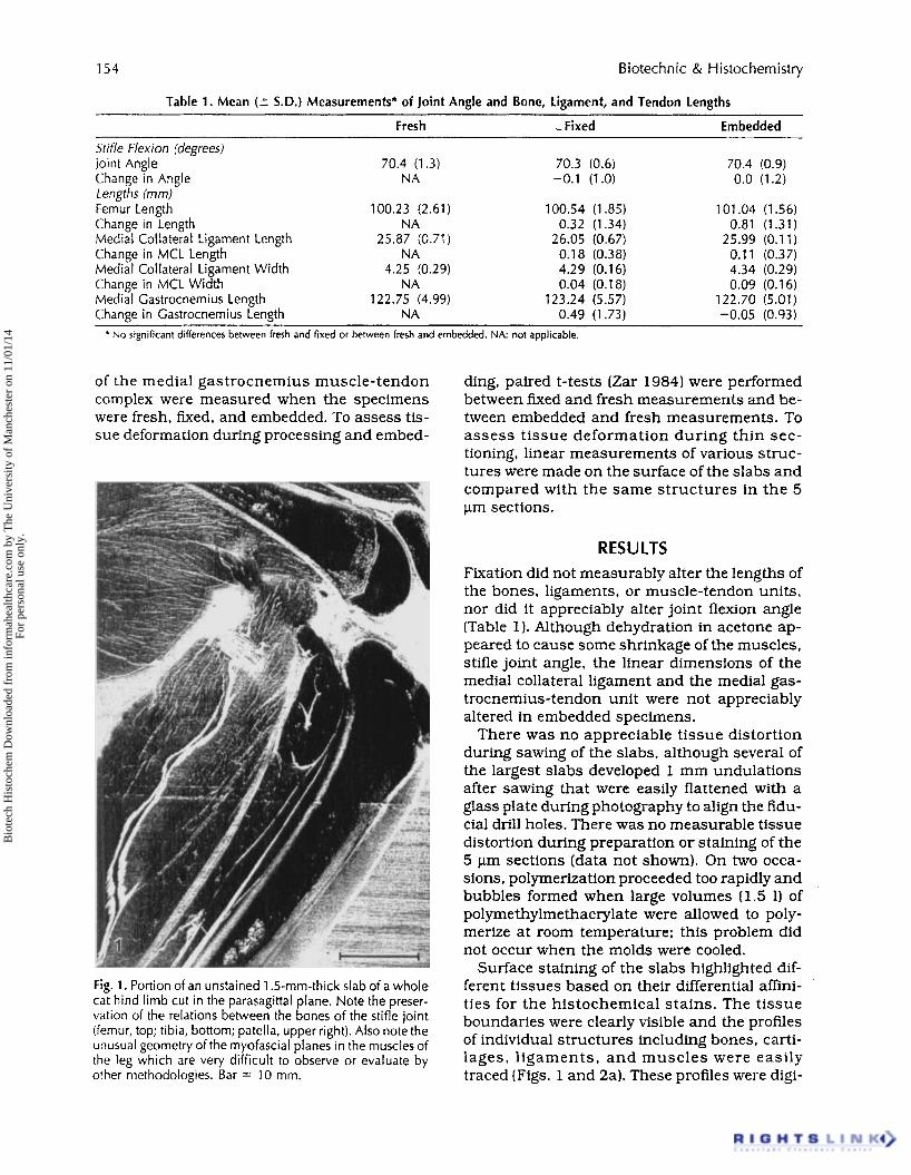
Table 1 . Mean (2 S.D.) Measurements* of Joint Angle and Bone, Ligament, and Tendon Lengths
Fresh .-Fixed Embedded ~~ ~
Stifle Flexion (degrees) Joint Angle 70.4 (1.3) 70.3 (0.6)
Lengths (rnm) Femur Length 100.23 (2.61) 100.54 (1 35 ) Change in Length NA 0.32 (1.34) Medial Collateral Ligament Length 25.87 (0.7’1) 26.05 (0.67) Change in MCL Length NA 0.18 (0.38) Medial Collateral Ligament Width 4.25 (0.29) 4.29 (0.16) Change in MCL Width NA 0.04 (0.18) Medial Gastrocnemius Length 122.75 (4.99) 123.24 (5.57) Change in Gastrocnemius Length NA 0.49 (1.73)
Change in Angle NA -0.1 (1.0)
* No significant differences between fresh and fixed or between fresh and embedded. NA not applicable.
of the medial gastrocnemius muscle-tendon complex were measured when the specimens were fresh, fixed, and embedded. To assess tis- sue deformation during processing and embed-
Fig. 1 . Portion of an unstained 1.5-mm-thick slab of a whole cat hind limb cut in the parasagittal plane. Note the preser- vation of the relations between the bones of the stifle joint (femur, top; tibia, bottom; patella, upper right). Also note the unusual geometry of the myofascial planes in the muscles of the leg which are very difficult to observe or evaluate by other methodologies. Bar = 10 mm.
70.4 (0.9) 0.0 (1.2)
101.04 (1.56) 0.81 (1.31)
25.99 (0.11) 0.11 (0.37) 4.34 (0.29) 0.09 (0.16)
122.70 (5.01) -0.05 (0.93)
ding, paired t-tests (Zar 1984) were performed between fixed and fresh measurements and be- tween embedded and fresh measurements. To assess tissue deformation during thin sec- tioning, linear measurements of various struc- tures were made on the surface of the slabs and compared with the same structures in the 5 pm sections.
RESULTS Fixation did not measurably alter the lengths of the bones, ligaments, or muscle-tendon units, nor did it appreciably alter joint flexion angle (Table 1). Although dehydration in acetone ap- peared to cause some shrinkage of the muscles, stifle joint angle, the linear dimensions of the medial collateral ligament and the medial gas- trocnemius-tendon unit were not appreciably altered in embedded specimens.
There was no appreciable tissue distortion during sawing of the slabs, although several of the largest slabs developed 1 mm undulations after sawing that were easily flattened with a glass plate during photography to align the fidu- cia1 drill holes. There was no measurable tissue distortion during preparation or staining of the 5 pm sections (data not shown). On two occa- sions, polymerization proceeded too rapidly and bubbles formed when large volumes (1.5 11 of polymethylmethacrylate were allowed to poly- merize at room temperature; this problem did not occur when the molds were cooled.
Surface staining of the slabs highlighted dif- ferent tissues based on their differential affini- ties for the histochemical stains. The tissue boundaries were clearly visible and the profiles of individual structures including bones, carti- lages, l igaments, and muscles were easily traced (Figs. 1 and 2al. These profiles were digi-
’
Bio
tech
His
toch
em D
ownl
oade
d fr
om in
form
ahea
lthca
re.c
om b
y T
he U
nive
rsity
of
Man
ches
ter
on 1
1/01
/14
For
pers
onal
use
onl
y.

Whole Joint Embedding 155
Fig. 2. A) A 600-pm-thick slab of a whole rabbit stifle joint cut in the coronal plane and surface stained with Gomori’s one- step trichrome. The mineralized tissues stain light green (femur, top; tibia, bottom; fibular head also visible on left), the soft connective tissues stain pink to red (menisci separating femur and tibia), and the muscles remain unstained. Bar = 10 mm. B) Three dimensional reconstruction of the same joint as in panel (A) taken from a computer monitor. This reconstruction includes the slab shown in panel (A). The bones are outlined (femur at the top, tibia and fibula at the bottom) and the four major ligaments are solid (medial collateral ligament on right, lateral collateral ligament on left, anterior and posterior cruciate ligaments cross each other in the middle of the joint). Approximately the same magnification as panel (A).
tized and used to reconstruct the internal archi- tecture of the stifle joint (Fig. 2b) and the three dimensional myotendinous network of the gas- trocnemius. I t was possible to trace tissue pro- files from both sides of each slab and to digitize them separately. The thickness of the saw blade was used as the dis tance between adjacent slabs and the thickness of the slabs were used as the distance between digitized surfaces for the three dimensional reconstructions.
The 5 pm sections cut from the slabs revealed that cytological detail was preserved by this em- bedding procedure (Fig. 3) and was comparable to embedding protocols for small samples.
Fig. 3. Fibrochondrocytes in a 5 p m section taken from a 600-pm-thick slab. The area is the ligament-bone interface of the anterior cruciate ligament. H & E stain; partially polar- ized, Nomarski optics. Bar = 100 pm.
DISCUSSION The function of musculoskeletal organs is clearly dependent on their architectural and
Bio
tech
His
toch
em D
ownl
oade
d fr
om in
form
ahea
lthca
re.c
om b
y T
he U
nive
rsity
of
Man
ches
ter
on 1
1/01
/14
For
pers
onal
use
onl
y.

156 Biotechnic & Histochemistry
cellular organization, yet few methods are avail- able that faithfully preserve musculoskeletal or- gan architecture and tissue microstructure for histological analysis. We describe here a method that minimizes distortion during pro- cessing and allows the assessment of both or- gan architecture and cytomorphology.
It should be noted that the precise quantifica- tion of three dimensional geometry is problem- atic and that evaluating tissue deformation us- ing l inear measu remen t s is l imited. For example, we noted radial shrinkage of the gas- trocnemius muscle after tissue dehydration, but the overall muscle-tendon length was not measurably altered. Furthermore, despite the accuracy of the measuring device, the variation in the sequential measurements was relatively high and the statistical power for resolving dif- ferences was correspondingly low. Thus, while tissue deformation was controlled satisfactorily for the present study, more exacting experi- ments using this method might require fur- ther validation.
Methylmethacrylate embedding is a routine procedure for laboratories processing bone bi- opsies. Typically, however, biopsy specimens and the volumes of methacrylate that are used to embed them are relatively small. For embed- ding the large specimens described here, as in other reports (Emmanual et al. 19871, the criti- cal modification of routine methodologies is the prolongation of dehydration and infiltration steps, and control of the rate of polymerization. Although there are a number of ways to control the rate of polymerization, we chose to control temperature in this study. Our initial problem with premature polymerization and bubbling has been experienced by others (Hahn et al. 1991) and is likely related to the volume of the embedding medium and the geometry of the embedding molds. Polymerization is exothermic (Liu 1987) and small blocks seem to dissipate the heat of polymerization faster than large blocks, presumably because of their large sur- face area to volume ratio (Baliga et al. 1992). Cooling large blocks during the initial phases of polymerization prevented this problem.
Polymerized methylmethacrylate provided ex- cellent support for undecalcified and collagen- dense tissues, and the slabs preserved the over- all architectural organization of the hind limbs allowing them to be studied by low power mi- croscopy and radiography. As reported pre- viously (Bab et al. 19831, surface staining of plastic blocks can reveal important histochemi-
cal features of the cut surfaces, and because polymethylmethacrylate is transparent, many tissue features within the slab are also visible. This technique has already proven useful in several studies of ligament and muscle struc- ture and function (Matyas 1990, Anton 1991, Matyas et al. 1995).
For studies at higher magnification, the poly- methylmethacrylate embedded tissues were re- cut into thin tissue sections and examined at the microscopic level revealing additional func- tional information such as sarcomere length and cellular organization. This re-cutting tech- nique is also very useful for stereological studies (Gundersen and Jensen 1987) that previously were difficult for musculoskeletal tissues.
Emerging technologies such as ultrafine CT (Feldkamp et al. 1989) hold great promise for examining organ architecture, particularly in unfixed organs and living subjects. Such tech- nology is likely to improve and become more widely available and affordable in the future; however, at present it seems unlikely that CT, MFU, or ultrasound will be able to provide the important information that is readily available by polarized light (e.g., collagen birefringence), histochemistry (e.g., proteoglycan distribution), and fluorescent microscopy (e.g., bone fluor- ochrome patterns) in embedded musculoskele- tal tissues. We believe that techniques, as de- scribed here for embedding whole organs will continue to be useful in the future.
ACKNOWLEDGMENT The authors gratefully acknowledge the finan- cial support of the Medical Research Council (Canada). the Alberta Heritage Foundation for Medical Research, The Arthritis Society, and the University of Calgary (Thesis Research Grant, M. G. A.) . Thanks also to Stephanie Grassman for access to some of the animal material.
REFERENCES Anton, M. G. 1991. Mechanical Models of Skeletal
Muscle. Doctoral Thesis, University of Calgary. Bab, I., Ashton, B. A.. Owen, M. E. and Boyde, A.
1983. Incident light microscopy of surfaces of plastic embedded hard tissues. J. Microsc.
Baliga, B. R., Rose, P. L. and Ahmed, A. M. 1992. Thermal modeling of polymerizing polymethyl- methacrylate. considering temperature-depen- dent heat generation. J. Biomech. Eng. 114:
134: 49-53.
25 1-259.
Bio
tech
His
toch
em D
ownl
oade
d fr
om in
form
ahea
lthca
re.c
om b
y T
he U
nive
rsity
of
Man
ches
ter
on 1
1/01
/14
For
pers
onal
use
onl
y.
Whole Joint Embedding 157
Emmanual, J.. Hornbeck, C. and Bloebaum, R. D. 1987. A polymethyl methacrylate method for large specimens of mineralized bone with im- plants. Stain Technol. 62: 401-410.
Feldkamp, L. A., Goldstein, S. A., Parfitt, M., Jesion. G. and Kleerekoper, M. 1989. The direct exami- nation of three-dimensional bone architecture in vitro by computed tomography. J. Bone Miner. Res. 4: 3-1 1.
Gomori, G. 1950. A rapid one-step trichrome stain. Am. J. Clin. Pathol. 20: 661-664.
Gunderson, H. J. G. and Jensen. E. B. 1987. The ef- ficiency of systematic sampling in stereology and its prediction. J. Microsc. 147: 229-263.
Hahn, M., Vogel. M. and Delling, G. 1991. Unde- calcified preparation of bone tissue: report of technical experience and development of new methods. Virchows Arch. A. Pathol. Anat. Histol.
Liu. C-C. 1987. A simplified technique for low tem- perature methyl methacrylate embedding. Stain Technol 62: 155-159.
Matyas, J. R. 1990. Ligament Insertion Structure and Function. Doctoral Thesis. University of Calgary.
418: 1-7.
Matyas, J. R., Anton, M. G., Shrive, N. G. andFrank. C. B. 1995. Stress governs tissue phenotype at the femoral insertion of the rabbit MCL. J. Bio- mech. 28: 147-157.
Mdler, A.. Stange, P. and Cundersen, H. J. G. 1990. Efficient estimation of cell volume and number using the nucleator and disector. J. Microsc.
Ohrt, C., Mahowald, M. L. and Valls, A. A. 1986. Preparation of whole rabbit knee joints for micro- scopic examination. Stain Technol. 61: 353-360.
Suzuki. M., Takahashi, T. and Ohuchi, K. 1988. Three-dimensional reconstruction of semi-gross biostructures using “macroserial”- 1 -mm-thick serial organ slices. J. Microsc. 149: 175-183.
Villanueva, A. R. 1980. Basic preparation and stain- ing of undecalcified bone. In: Theory and Practice ofHistotechnology. D. C. Sheehan and B. Hrap- chak, Eds. Mosby, St. Louis, MO. pp. 96-107.
Zar, J. H. 1984. Paired sample hypotheses. In: Bto- statlstkal Analysis. 2nd edition. Prentice-Hall, Inc. Englewood Cliffs, N J . pp. 150-161.
159: 61-71.
Bio
tech
His
toch
em D
ownl
oade
d fr
om in
form
ahea
lthca
re.c
om b
y T
he U
nive
rsity
of
Man
ches
ter
on 1
1/01
/14
For
pers
onal
use
onl
y.