Embed Size (px)
Citation preview

www.sysmex.com/us
Getting beyond the flags
WHITE PAPER
IMMATURE GRANULOCYTE (IG) POPULATIONS MAY IMPROVE THE ASSESSMENT OF INFECTION AND SEPSIS.
2 | IG
IntroductionFor many decades, physicians have relied on the Complete Blood Count (CBC) and differential (diff) to be a simple and effective screening tool for detecting sample abnormalities. When interpreting CBC and diff results, the presence of a ‘left shift’ has been considered indicative of a number of conditions (Table 1). However, the term ‘left shift’, though universally accepted as the presence of immature neutrophils, lacks specificity. Is a left shift present if non-segmented neutrophils (bands) are the only immature cells? If so, what percentage of bands is required? Or, do more immature forms such as metamyelocytes, myelocytes and promyelocytes also need to be present? When 100-cell manual differentials are performed, the results can be imprecise due to the low number of cells being counted, particularly in leukopenic samples. Furthermore, results may not be reproducible between operators because of differences in morphology interpretation.
In recent years, it has become possible to flag and enumerate immature granulocytes by the automated cell counters. The Sysmex® XN-Series™ analyzers utilize fluorescent flow cytometry and a proprietary software algorithm to generate an accurate and reproducible Immature Granulocyte (IG) count that includes metamyelocytes, myelocytes and promyelocytes. These three cell types are separated from the neutrophil population based on their increased fluorescence emission due to higher levels of DNA and RNA. The measurement is rapid and accurate and is reported as part of the automated differential results.
Correlation studies: gaining agreementEarly studies describing the use of flow cytometry to differentiate immature granulocytes from mature forms also determined that accuracy and reproducibility of white blood cell differentiation were improved by counting 10,000 cells compared to the 100-cell manual differential. These studies used flow cytometry and monoclonal antibodies CD45, CD16 and CD11b to identify immature granulocytes. CD45, the leukocyte common antigen, was used to isolate cells of hematopoietic origin. Promyelocytes and myelocytes resisted staining with both CD16 and CD11b (CD16-/CD11b-), metamyelocytes stained as CD16-/CD11b+, and mature granulocytes were CD16+/CD11b+. This antibody combination allowed for the measurement of light scatter and fluorescence by flow cytometry rather than morphological characteristics only.
Figure 1 shows the flow cytometry results of a patient with immature granulocytes versus a normal, healthy volunteer.
The ability to automate quantitation of immature myeloid cells was described by Briggs et al. in 2003. This article compared manual light microscopy, flow cytometry and the Sysmex IG Master™ software that was introduced on the XE-Series™ analyzers. The flow principles that made IG enumeration possible in the earlier technology carried through to the XN-Series analyzers, which offers IG as part of the standard 6-part automated differential. Analysis is accomplished with the assistance of two specialized reagents: an erythrocyte lysing reagent which also acts upon white cells to prepare them for staining, and a proprietary fluorescent dye which stains nucleic acid in the treated leukocytes.
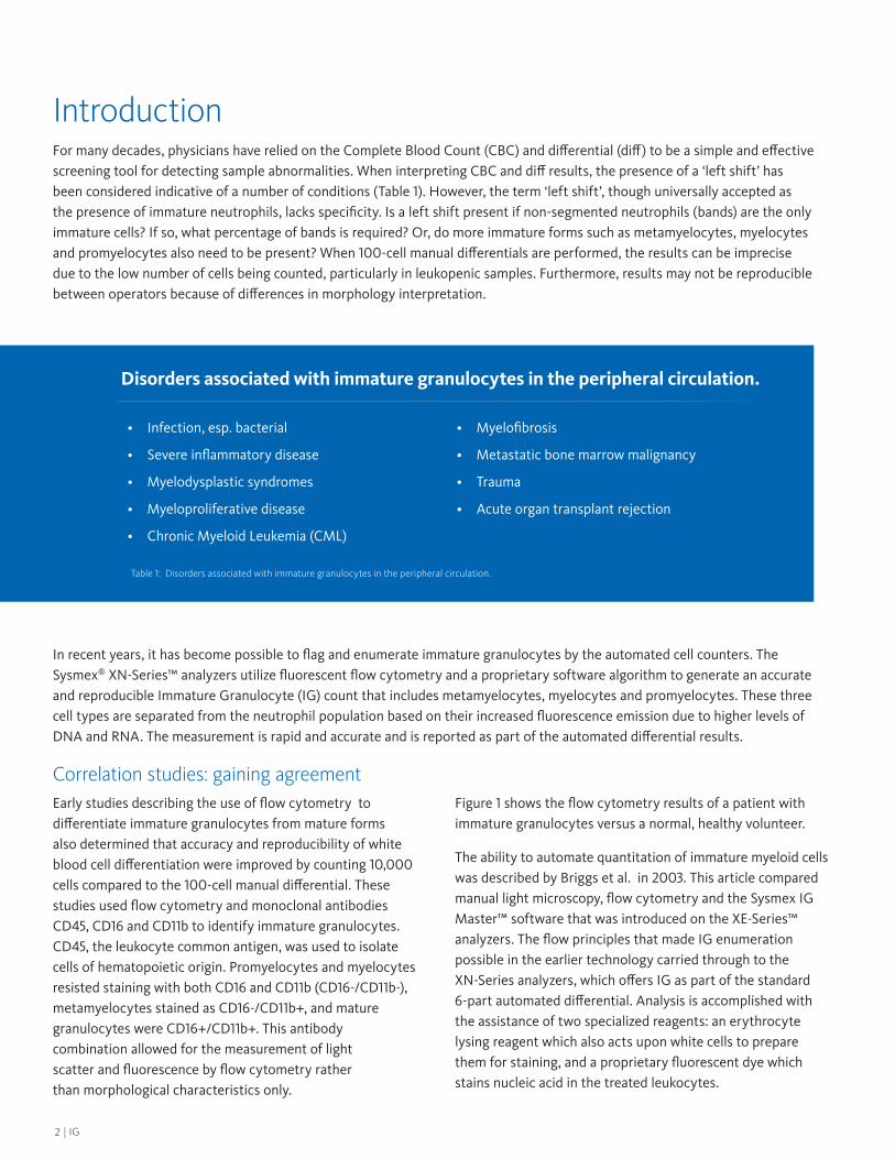
Disorders associated with immature granulocytes in the peripheral circulation.
• Infection, esp. bacterial
• Severe inflammatory disease
• Myelodysplastic syndromes
• Myeloproliferative disease
• Chronic Myeloid Leukemia (CML)
• Myelofibrosis
• Metastatic bone marrow malignancy
• Trauma
• Acute organ transplant rejection
Table 1: Disorders associated with immature granulocytes in the peripheral circulation.

3IG |
IG
Figure 1: Flow cytometric analysis of a patient sample containing immature granulocytes and a sample from a normal, healthy volunteer after staining with CD45-PerCP, CD16-FITC, and CD11b-PE. In step (A), granulocytes (IG + neutrophils + eosinophils) were isolated from other WBCs. Step (B) demonstrates the separation of neutrophils and IGs from eosinophils. In the third step, (C), IG stage 1 (CD16-/CD11b-) and stage 2 (CD16-/CD11b+) were identified as well as mature neutrophils (CD16+/CD11b+). No IGs were seen in the normal sample.1
Figure 2: The correlation of the percentage of Immature Granulocytes (IG%) determined by the flow cytometry reference method and XE-IG Master software. Flow cytometric IG count based on a 3-color method using FITC-CD16, FITC-CD11b, and PerCP-CD45 was used as the reference method.2
Figure 3: The correlation of the percentage of immature granulocytes (IG%) determined by the manual count reference method and XE-IG Master software. The manual IG count was determined on a 400-cell leukocyte differential.2
In the XN’s flow cell, a laser diode measures side light scatter and side-fluorescence to classify the white blood cells (WBC) in the sample.
Immature granulocytes are identified by their high side-scattered light readings which distinguish them as granulocytic cells, and their elevated fluorescence intensity which reflects the excess of nuclear material. The IG parameter is reported in relative (IG%) and absolute (IG#) numbers. Figure 2 compares IG percentages from traditional flow cytometry studies and the XE-IG Master software while Figure 3 shows the correlation between the manual 400-cell differential and the XE-IG Master software.
Briggs et al. determined that the automated IG count demonstrated excellent correlation with the immature granulocytes identified with the 3-color flow cytometry method, but that correlation with the manual method was slightly lower. This discrepancy was likely due to the smaller number of cells counted with the manual method and to the subjectivity involved in the manual morphologic classification of immature granulocytes. Briggs’s investigation also reported that the Sysmex automated IG parameter demonstrated excellent
reproducibility. The mean Coefficient of Variation (CV) was 7.02% and 6.93% for the IG percent (IG%) and absolute IG count (IG#), respectively. Similar studies performed on the 100-cell manual differential showed that the manual method displayed poorer precision with CV values ranging from 39.7% to 65.7%.1 Fernandes and Hamaguchi also reported excellent correlation between flow cytometry and the automated IG count from the Sysmex analyzer with correlation coefficients of 0.93 and 0.95 for IG# and IG%, respectively. Both studies reported that samples were stable when refrigerated or stored at room temperature.

4 | IG
Acute Phase ReactantsTable 2 lists positive acute phase reactants (APRs) that may be helpful in assessing inflammation and infection. In their 2003 study, Briggs et al.2 investigated possible correlation between an elevated IG count (> 2%) and other inflammatory markers. Forty-three patient samples with high IG and normal absolute neutrophil count (ANC) were studied; of these, 15 patients had an infectious disease and 14 had post- operative bleeding. The mean IG count for these 43 samples was 3.9% (range 2.0% – 11.0%) and the mean neutrophil count was 5.21 x 10⁹/L (range 2.01-7.49 x 10⁹/L). The IG result (considered “positive” by the authors) in each sample was compared to qualitative values for C-reactive protein (CRP), erythrocyte sedimentation rate (ESR), CD64 and interleukin 6 (IL-6). Samples showed good correlation with CRP, ESR, and CD64 (84%, 95%, and 80% respectively) despite neutrophil counts in the normal range. Correlation between an elevated IG% and IL-6 positivity was only 58%. (Figure 4) In light of these findings, the authors concluded that the automated IG count could be used as part of the CBC to highlight a potential inflammatory or infectious process even in the absence of leukocytosis or neutrophilia.
Positive Acute Phase Reactants
• WBC (high, low or normal)
• Immature granulocytes (peripheral blood)
• C-Reactive Protein
• Interleukin 1 (IL-1)
• Interleukin 6 (IL-6)
• Tumor Necrosis Factor (TNF)
• CD-64
• ESR (“sed rate”)
• Ceruloplasmin
• Components of the complement cascade
• Serum amyloid A
• Fibrinogen
• Alpha-1 antitrypsin
• Haptoglobin
• Ferritin
Table 2: Acute Phase Reactants3,4
Figure 4: Positive sample rates in samples with high immature granulocytes count (2.0% or higher) and normal neutrophil levels (2.0-7.5 x 10⁹/L). CRP indicates C-reactive protein; ESR, erythrocyte sedimentation rate; IL-6, interleukin 6.2

5IG |
IG
Immature Granulocytes in Adult Sepsis PatientsIn a separate study published in 2003, Ansari-Lari, Kickler and Borowitz investigated the use of IG% as a predictor of infection or sepsis. Blood samples from 102 infected individuals and 69 non-infected individuals were analyzed on the Sysmex XE-2100 instrument. The study showed that IG% was significantly higher in infected versus non- infected patients (Table 3) as well as patients with positive versus negative blood culture results (Table 4). The demonstrated specificity of IG was high, showing that 11 of 12 patients with IG% values greater than 3% had proven septicemia.
Table 3: Comparison of WBC Count and Percentage of Immature Granulocytes Measurements as Predictors of Infection4.
Similar to Brigg’s findings, IG% correlated better than the absolute white cell count and was comparable to the absolute neutrophil count, although neither parameter had the sensitivity to be used as a sole screening tool. Rather, it was suggested that the value of an increased IG% of greater than 3% could be useful as part of an algorithm-driven panel of tests.
Table 4: Comparison of WBC Count and Percentage of Immature Granulocytes Measurements as Predictors of Sepsis4.
A 2013 study by Nierhaus et al.6 measured the IG count in surgical intensive care patients (SICU) to determine whether the parameter could discriminate between Systemic Inflammatory Response Syndrome (SIRS) and sepsis. In an assessment of 70 patients, the IG parameter had a sensitivity of 89.2% and a specificity of 76.4% in distinguishing infected from non-infected patients (P < 0.0001). These SICU patients also had CRP, lipopolysaccharide binding protein (LBP) and interleukin-6 (IL-6) concentrations measured and it was found that the IG count had a stronger ability to discriminate SIRS and sepsis than these biomarkers. The researchers concluded that the IG parameter was a good marker to discriminate SICU patients with sepsis, especially very early after SIRS symptoms presented. However, the IG count was not found to demonstrate any prognostic value for mortality in these patients. Other studies have observed similar results in ED (emergency department) patients: Ha et al. concluded that IG% was able to reflect sepsis severity without additional cost, however, was not a useful biomarker for predicting 28-day mortality.
More recently, a study by van der Geest et al. looked at 46 critically ill patients with a suspicion of infection based on temperature, WBC count and an elevated CRP value. These authors determined that IG percentage is a better predictor of infection compared to WBC count alone, and that it is as effective as CRP in predicting microbial infection, even in the critically ill. Additionally, adding IG% to a model using WBC and CRP to determine infection increased its predictive value with an AUC of 0.80-0.88, and this combination showed a sensitivity of 80% with 90% specificity.
An additional study of intensive care unit (ICU) patients with liver impairment and suspected sepsis concluded that leukocyte parameters may prove valuable in the diagnostic approach to these patients. This investigation compared CBCs from 115 adult patients admitted to the ICU and 200 healthy subjects. Compiled data showed a median IG# value of 0.02 x 10³/µL in healthy subjects versus 0.14, 0.29, and 0.34 x 10³/µL in non-septic, septic and septic shock ICU patients, respectively.

6 | IG
Immature Granulocytes in Diagnosing Neonatal SepsisA study of pediatric patients by Iddles et al.10 compared CRP, IG ratio ([total IG count/total white cell count] X 100) and IT ratio ([total IG count/total neutrophil count] X 100) to determine their effectiveness as early indicators of neonatal sepsis. A total of 58 patients were evaluated: 31 with positive blood culture results; 7 with negative blood cultures, but clinically- diagnosed sepsis; 20 negative for sepsis.
The patients diagnosed with sepsis and showing a positive blood culture result all had elevated IG and IT ratios, the latter particularly raised and in one case elevated to 37.5%. The additional group of patients diagnosed with sepsis but with negative blood cultures (perhaps as a result of antibiotic treatment) showed elevated IG and IT ratios compared to the negative controls. Negative control patients did not have ratios higher than 3%.
Using cut-off points of 0.35 for the IG ratio and 0.65 for the IT ratio, both calculations showed a sensitivity of greater than 70%, albeit with low specificity. The IT ratio appeared to be somewhat more sensitive and had better positive and negative predictive values than the IG ratio, perhaps because it is calculated using the neutrophil count rather than the leukocyte count, which allows for better diagnostic sensitivity in neutropenic samples. This study also evaluated CRP levels in some of the patients: 21 of 38 septic patients and 17 of 20 in the negative control group. Using an upper limit of greater than 11mg/L, 68% of the septic patients had an increased CRP, although because of the low amount of patients studied, this number should be interpreted conservatively. Nonetheless, it was determined that the sensitivity of CRP, in this study at least, was lower than for IT and IG ratios at 71%. The specificity of CRP was also low at 35% compared to the IT ratio of 50% in this pediatric population.
In this study, the IG and IT ratios were calculated using the automated IG from the Sysmex analyzer. The authors deemed that the automated count offered advantages over the manual differential because it is rapid, analyzes more WBCs than the traditional 100 cells, and also avoids statistical error caused by inconsistent operator interpretation. The authors concluded that using the automated IG count in the calculated IT ratio is a fast, inexpensive, reliable indicator of sepsis in a pediatric patient.
These findings were confirmed by MacQueen et al. (2016) in a study to determine reference intervals for extended leukocyte
differential parameters in non-infected neonates. The research also compared the statistical performance of the following indicators of “left shift” and their effectiveness in predicting infection: I/T ratio, absolute band count, IG% and IG#. The authors concluded that the statistical performance of the I/T ratio and the IG% was essentially identical in identifying neonates with infection and that, for most purposes, an automated differential should serve as well as a manual differential in neonatal medicine.
In the Surviving Sepsis Campaign: International Guidelines for the Management of Severe Sepsis and Septic Shock: 2012 12, bands have been removed as an inflammatory variable for both adult and pediatric patients. In adults, bands have been replaced by the presence of >10 percent immature (WBC) forms even with a normal WBC count. In pediatric patients, bands have been replaced by age-specific WBC cutoffs.
SummaryDiagnosis is complex in sepsis due to a broad range of non-specific symptoms and absence of a definitive diagnostic biomarker. Microbiologic testing in the form of blood cultures may border on being a “gold standard,” but it may take up to four days to receive final results. The test can also provide false positive and false negative results, frequently due to faulty blood collection techniques. Therefore, the search for quick and economical alternatives to this and the many tests cited here continues.
Now that the laboratory is able to report the immature granulocyte population with a high degree of accuracy and precision, the WBC count and differential have become increasingly valuable tests in the clinician’s sepsis investigation toolkit. Physicians will have additional diagnostic parameters to consider when assessing infection so that timely intervention with appropriate therapeutics can be made.
As previously noted, hematologic and biochemical tests can be used to contribute to a sepsis diagnosis, and the highly precise and accurate IG parameter from Sysmex is a well-studied biomarker of infection and inflammation. As investigators continue to evaluate IG count and IG% along with other acute phase reactants, it may be possible to develop a diagnostic algorithm for laboratory results to aid in the identification of acute inflammation, infection, and sepsis.

7IG |
IG
1 Fujimoto H, Sakata T, Hamaguchi Y, et al. Flow cytometric method for enumeration and classification of reactive immature granulocyte populations. Cytometry. 2000 Dec 15;42:371-378.
2 Briggs C, Kunka S, Fujimoto H, et al. Evaluation of Immature Granulocyte Counts by the XE-IG Master: Upgraded Software for the XE-2100 Automated Hematology Analyzer. Lab Hematol. 2003;9:117-124.
3 Fernandes B, Hamaguchi Y. Automated Enumeration of Immature Granulocytes. Am J Clin Pathol. 2007 Sep;128:454-463.
4 http://www.uptodate.com/patients/content/topic.do?topicKey=~22ip2ecYo1z07Yp
5 Ali Ansari-Lari M, Kickler TS, Borowitz MJ. Immature Granulocyte Measurement using the Sysmex XE-2100. Am J Clin Pathol. 2003 Nov;120(5):795-799.
6 Nierhaus A, Klatte S, Linssen J, et al. Revisiting the white blood
cell count: immature granulocytes count as a diagnostic marker to discriminate between SIRS and sepsis - a prospective, observational study. BMC Immunol. 2013;14:8.
7 Ha S, Park S, Park S, Park J, et al. Fraction of immature granulocytes reflects severity but not mortality in sepsis. Scan J Clin Lab Invest. 2015 Jan;75(1):36-43.
8 Van der Geest P, Mohseni M, Brouwer R, Van der Hoven B, Steyerberg E, Groeneveld, A. Immature granulocytes predict microbial infection and its adverse sequelae in the intensive care unit. J Crit Care. 2014;29:523-7.
9 Buoro S, Mecca T, Azzara G, et al. Extended leukocyte differential count and C-reactive protein in septic patients with live impairment: diagnostic approach to evaluate sepsis in intensive care unit. Ann Transl Med. 2015;3(17):244.
10 Iddles C, Taylor J, Cole R, Hill FGH. Evaluation of the Immature Granulocyte Count in the Diagnosis of Sepsis Using the Sysmex XE-2100. Sysmex Journal International. 2007;17:Suppl. 1/No. 1.
11 MacQueen B, Christensen R, Yoder B, et al. Comparing automated vs manual leukocyte differential counts for quantifying the ‘left shift’ in the blood of neonates. J Perinatol. 2016 Oct;36(10):843-8.
12 Dellinger RP, et al. Surviving Sepsis Campaign: international guidelines for management of severe sepsis and septic shock, 2012. Intensive Care Med. 2013;39:2,165-228.
Bibliography
Program availability varies by location. Programs and specification subject to change without notice.© 2018 Sysmex America, Inc.
MKT-10-1066, REV. 2
Sysmex America, Inc.577 Aptakisic Road, Lincolnshire, IL 60069, U.S.A. · Phone +1 800 379-7639 · www.sysmex.com/us
Sysmex Canada, Inc.5700 Explorer Drive Suite 200, Mississauga, ON L4W0C6 Canada · Phone +1 905 366-7900 · www.sysmex.ca
DisclaimerThe uses or clinical applications described in this paper may not have been approved or cleared by the FDA. It is the clinician’s responsibility to validate any off-label applications for use in routine clinical practice.