Embed Size (px)
Citation preview

Core Curriculum V5
When the fracture is more: Pathologic Fractures in the
Pediatric PopulationAdrienne R Socci, MD
Assistant ProfessorYale School of Medicine

Core Curriculum V5
Disclaimer
• All clinical and radiographic images provided are used with permission of Adrienne Socci, MD and Chris Souder, MD, unless otherwise specified

Core Curriculum V5
Objectives
• How to recognize a pathologic fracture• How to evaluate a pathologic fracture• Treatment principles in the management of pathologic fractures• Common pathologies encountered in the pediatric population

Core Curriculum V5
Recognizing a pathologic fracture
• Be suspicious when• The mechanism of injury seems disproportionately minor
• Example: a fall from standing results in a humerus fracture• The fracture pattern is atypical for location
• Example: transverse fracture in a femur after a fall• The bone doesn’t look normal at the fracture site
• Example: any lesion, or appearance that is cystic, lucent, sclerotic, or with surrounding reaction such as periosteal elevation

Core Curriculum V5
Evaluating a suspected pathologic fracture
• Obtain a detailed history• Local and constitutional symptoms: confirm if any pain, particularly night
pain, fever, weight loss, swelling, or lack of these• Current or past medical conditions• Family history
• Physical exam• A thorough exam includes evaluation for skin changes, swelling, soft tissue
mass, lymphadenopathy

Core Curriculum V5
Evaluating a suspected pathologic fracture
• Diagnostic imaging• Plain x-rays are typically done to identify the fracture• Ensure complete imaging of the whole bone
• Advanced imaging can be useful• MRI, CT, bone scan

Core Curriculum V5
Evaluating a suspected pathologic fracture
• Diagnostic imaging• Evaluate the lesion
• Location• Diagnosis can be specific to
epiphysis/metaphysis/diaphysis• Character
• Consider the presence of cyst, calcification, or matrix in the bone
• Reaction of the underlying bone• Is there periosteal elevation, is the shape of the
bone normal, is there a clear border to the lesion

Core Curriculum V5
Evaluating a suspected pathologic fracture
• Biopsy• May be needed for diagnosis or treatment planning• Sometimes performed at the time of operative fracture
management • Principles of oncology must be followed in performing a
biopsy• Consider possibility of malignancy and need for future resection• Send cultures as well as tissue specimen
• Consider consultation with an orthopaediconcologist prior to biopsy, particularly if there is concern for malignancy.

Core Curriculum V5
Treating a suspected pathologic fracture
• Treat the fracture• Not all pathologic fractures require operative management• Some pathologic fractures can be treated as though there
were no lesion
• Treat the pathology• Some pathologies can be observed, or treated in a delayed
fashion, i.e. after fracture healing.

Core Curriculum V5
Common benign pathologies by location
• Epiphysis:• Chondroblastoma• Giant cell tumor
• Metaphysis• Osteomyelitis• Nonossifying fibroma• Unicameral bone cyst• Aneurysmal bone cyst• Fibrous dysplasia
• Diaphysis:• Fibrous dysplasia• Osteofibrous dysplasia

Core Curriculum V5
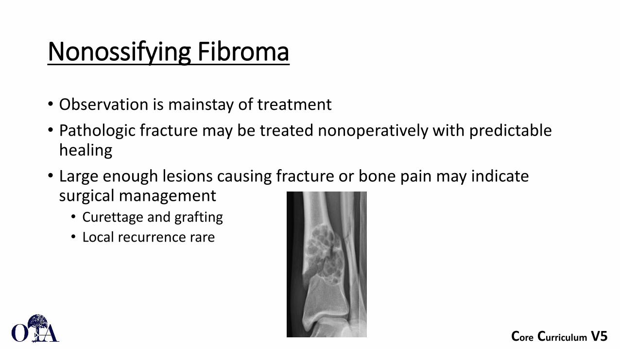
Nonossifying Fibroma
• Most common benign lesion • Common incidental finding on xrays
• Eccentric, metaphyseal, multilocular lucency with a sclerotic border• Often classic enough in appearance that no further imaging is
necessary• Differential diagnosis: UBC, ABC• Other names for this include:
• Fibrous cortical defect• Fibroxanthoma
Core Curriculum V5
Nonossifying Fibroma
• Observation is mainstay of treatment • Pathologic fracture may be treated nonoperatively with predictable
healing• Large enough lesions causing fracture or bone pain may indicate
surgical management• Curettage and grafting • Local recurrence rare

Core Curriculum V5
Unicameral Bone Cyst (UBC)
• Benign cystic lesion• 2:1 male:female ratio• Common between ages 4-10• Proximal humerus (59%) and proximal
femur (26%) most common• Uncommon to grow or recur after skeletal
maturity

Core Curriculum V5
Unicameral Bone Cyst (UBC)
• Well-defined, radiolucent lesion centrally located within the metaphysis
• Cortical thinning• No periosteal reaction
• Mild cortical expansion• NOT wider than the epiphysis
• Fallen leaf sign• Fragment of bone floating inside the fluid-
filled cavity• Typical after a fracture
Fallen leaf sign: cortical fragment within the lesion

Core Curriculum V5
Unicameral Bone Cyst (UBC)
• Most commonly identified at time of fracture (85% of cases)
• Thin cortical rim• Microfracture of this can lead to pain• Pathologic fracture possible after
minor trauma, e.g. fall from standing
• More potential for growth when closer to the physis
• Active lesions are near physis• Latent lesions are remote from physis

Core Curriculum V5
Unicameral Bone Cyst (UBC)
• X-rays usually sufficient for diagnosis
• CT or MR can be considered if diagnosis uncertain
• Axial skeleton
• Aspiration of straw-colored fluid is diagnostic

Core Curriculum V5
Unicameral Bone Cyst (UBC)
• Treatment of the fracture can be guided by typical fracture treatment principles
• Conservative treatment is most often indicated
• Low rates of spontaneous healing of cyst with fracture healing

Core Curriculum V5
Unicameral Bone Cyst (UBC)• Treatment of the cyst can proceed upon fracture healing• Treatment goals include preventing recurrent fracture
• Prevent complications of fracturedeformity or growth arrest

Core Curriculum V5
Unicameral Bone Cyst (UBC)
• Treatment options include injection, decompression, and curettage with grafting
• Comparable healing rates (~80%)• Bone marrow or steroid injection• Curettage and grafting with either autograft, allograft, or
bone substitutes • Decompression with IM nails or cannulated screw

Core Curriculum V5
Aneurysmal Bone Cyst (ABC)
• Benign, but locally aggressive tumor• Most commonly in teenagers (80%)• Much less common than UBC• Most common in long bones and spine
• 50% long bones• 30% spine
• Typically metaphyseal• Occasionally found in the epiphysis or
diaphysis

Core Curriculum V5
Aneurysmal Bone Cyst (ABC)
• Radiolucent, eccentric, expansile lesion, most commonly in the metaphysis
• May see periosteal reaction due to aggressive nature of this benign tumor
• Fluid/fluid levels on MRI characteristic• Differential diagnoses: telangiectatic
osteosarcoma, giant cell tumor, UBC, secondary ABC
• Biopsy recommended to confirm diagnosis

Core Curriculum V5
Aneurysmal Bone Cyst (ABC)
• Treatment is most commonly curettage and grafting, with or without adjuvant therapy
• Embolization used pre-operatively to reduce bleeding
• High rate of local recurrence• Radiation limited to inoperable lesions

Core Curriculum V5
Fibrous Dysplasia
• Benign lesion• Most common in long bones, pelvis
• Long lesion in a long bone
• Mutation in Gsα gene• Failure of maturation of bone
• Immature matrix leads to decreased mechanical strength of bone
• This can lead to pain, pathologic fracture, deformity
• Clinical presentation with bone pain or fracture

Core Curriculum V5
Fibrous Dysplasia
• Monostotic more common• Femur most common site
• Polyostotic form can be more severe• Larger lesions and secondary deformity
• Deformity can be caused by microfracture and progressive structural deformation
• Shepherd’s crook deformity of the proximal femur• Can be associated with endocrine abnormalities
• McCune-Albright syndromeprecocious puberty

Core Curriculum V5
Fibrous Dysplasia –Imaging
• Xrays will demonstrate a “ground glass” • Irregular, metaplastic woven bone replacing trabecular bone
• Lesion may appear expansile, with endosteal scalloping• Periosteal reaction not typically seen• Differential diagnoses: UBC, NOF
”ground glass” appearance Endosteal scalloping

Core Curriculum V5
Fibrous Dysplasia - Treatment
• Observation of asymptomatic or incidental lesions is recommended• Follow XR needed to ensure no progression or development of deformity
• Progressive deformity is common in polyostotic disease• Rare in monostotic
• Bisphosphonates can be used in polyostotic form to decrease bone pain
Proximal femoral varusdeveloping

Core Curriculum V5
Fibrous Dysplasia – Conservative Treatment
• Treatment of fracture• Conservative treatment most commonly indicated• Fractures heal rapidly
• Good periosteal bone formation• Poor endosteal bone
• Underlying bone will remain dysplastic• Curettage and grafting is NOT indicated
• Graft is converted to FD bone

Core Curriculum V5
Fibrous Dysplasia – Surgical Treatment
• Most often indicated for• Lower extremity/weightbearing bones• Polyostotic cases with deformity
• Intramedullary fixation is ideal• Load-sharing implant• Protect the entire bone
• Especially in polyostotic cases• Additional osteotomies often needed for associated
deformities
• Fixed angle constructs required in periarticular fractures when IM fixation is not sufficient

Core Curriculum V5
Fibrous Dysplasia – Prophylactic Treatment
• Prevention of fracture• Prophylactic fixation in weightbearing bones with large lesions and/or
progressing deformity• May also present with bone pain in the absence of visible fracture• Minor trauma can lead to pathologic fracture

Core Curriculum V5
Fibrous Dysplasia - Augmentation
• Augmentation with cortical strut allograft may also improve mechanical strength
• Bone graft will be resorbed and replaced by fibrous dysplasia over time• Nonstructural allograft does not have a role in treating this condition
Resorption of cortical strut graft

Core Curriculum V5
Malignant Tumors & Metastases
• Malignant bone tumors may cause pathologic fractures
• Osteosarcoma and Ewing’s sarcoma most common• Metastases
• Signs of malignancy include • Aggressive appearance• Periosteal reaction• Bone forming and/or destructive lesions with poorly
delineated borders• Not well circumscribed• Permeative appearance
• Associated soft tissue mass

Core Curriculum V5
Malignant Tumors & Metastases
• Identification and diagnosis is crucial for appropriate treatment
• Work-up should be done by the treating orthopedic oncologist, especially biopsy
• Do not perform fixation before diagnosis is made• Treatment of malignancy can include chemotherapy,
radiation, and/or surgery• Limb salvage is often possible
Core Curriculum V5
Pediatric pathologic fractures
• Pathologic fractures can happen from many different etiologies, both benign and malignant
• Treatment of the fracture and treatment of the pathology are dependent on the etiology
• Many benign etiologies do not require treatment distinct from the fracture treatment
• An orthopaedic oncologist should be consulted when dealing with a suspected malignancy

Core Curriculum V5
References
• Kadhim, M., Thacker, M., Kadhim, A. et al. Treatment of unicameral bone cyst: systematic review and meta analysis. J Child Orthop 8, 171–191 (2014). https://doi.org/10.1007/s11832-014-0566-3
• Rapp TB, Ward JP, Alaia MJ.J Am Acad Orthop Surg. 2012 Apr;20(4):233-41. doi: 10.5435/JAAOS-20-04-233.
• DiCaprio MR, Enneking WF. Fibrous dysplasia. Pathophysiology, evaluation, and treatment. J Bone Joint Surg Am. 2005 Aug;87(8):1848-64. doi: 10.2106/JBJS.D.02942. PMID: 16085630.
• John A Herring. Tachdjian's Pediatric Orthopaedics: from the Texas Scottish Rite Hospital for Children. 5th edition. 2014. Elsevier Saunders: Philadelphia ISBN: 978-1-4377-1549-1.