Embed Size (px)
Citation preview
FRACTURE PROXIMAL HUMERUS
5% of all fractures of all the fractures of the proximal humerus.
Court-Brown found that 70% of all 3- and 4-part fractures are seen in patients aged
over 60 years and 50% in patients aged over 70 years. These results indicate that poor
bone quality or even advanced osteoporosis will be found in the majority of patients
with humeral head fractures.
In case of a humeral head fracture, the following issues are of interest:
1. Anatomy and Vascularity
2. X rays
3. Classification
3. Reduction
4. Implant characteristics
5. Bone quality
1. Vascularity
Gerber stated that in the case of an existing avascular necrosis, it is the deformity
rather than necrosis that causes disability. Therefore, the risk of limited blood supply
of the articular fragment does not influence our decision making in terms of
treatment.
It is believed that the alignment of the tuberosities is very important in cases in which
prosthetic replacement might be necessary as a secondary procedure because of head
necrosis.
1 Anterior circumflex artery; 2 Axillary artery; 3 Ascending branch of PCHA 4 Branch of thoraco-‐acromial artery 5 Anterolateral branch of the ACHA
Gerber 72A:1486-‐1494
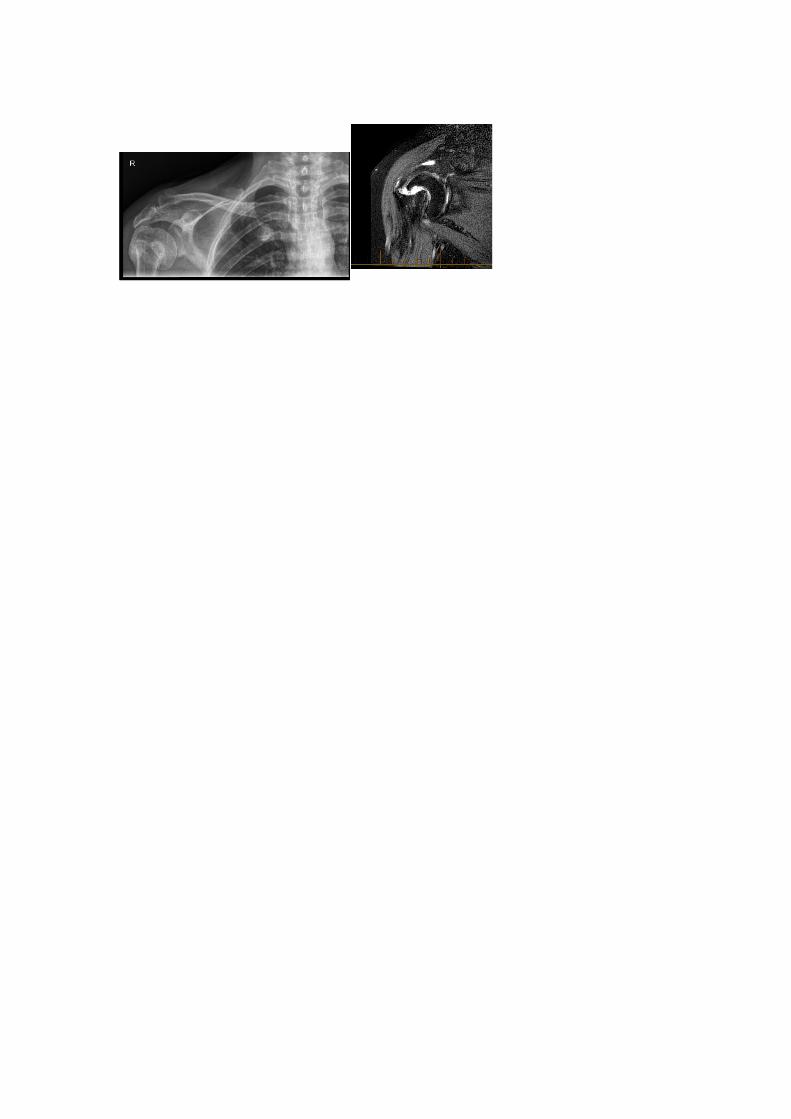
2. Trauma Series X rays:
3. Classification
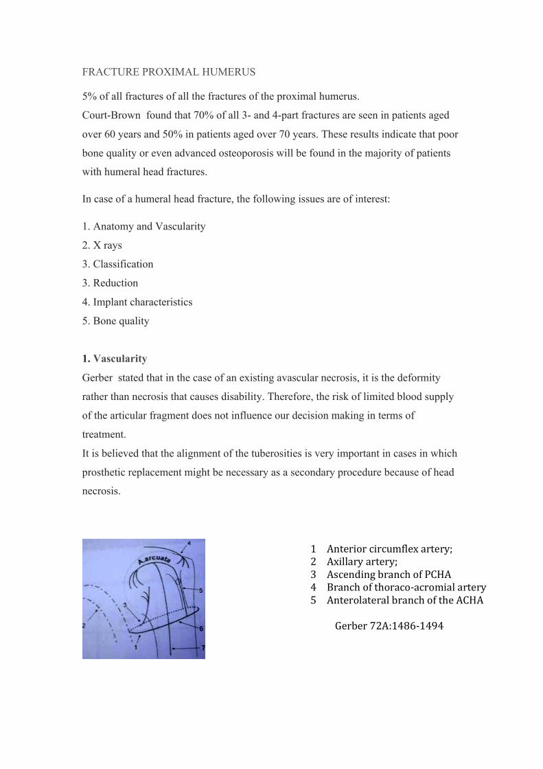
a. Neer’s Classification
In 1993, Siebenrock and Gerber22 and Sidor et al21 found very low interobserver
reliability for the existing and commonly used classification systems. Therefore, a
new classification system characterized by 3 features:
1. It should be easy to understand.
2. It should include the second plane.
3. It should include accepted findings of recent years, such as Varus/valgus deformity
and length and displacement of the medial hinge.
b. HCTS Classification system [Resch]
H stands for head, C for the medial calcar, T for the tuberosities, and S for the shaft.
Each region is described separately, and all regions are finally assembled. The system
n Anatomical or Surgical Neck n Greater or lesser Tuberosity n Fracture or dislocation, Head
splitting Accordingly:
n 2 part, 3 part, 4 part, Articular surface
n Displacement is 10 mm separation/45° angulation
n Intra and interobserver difference is significant.
CT definition is better
provides information on the expected vascularity and the expected difficulties during
reduction and fixation.
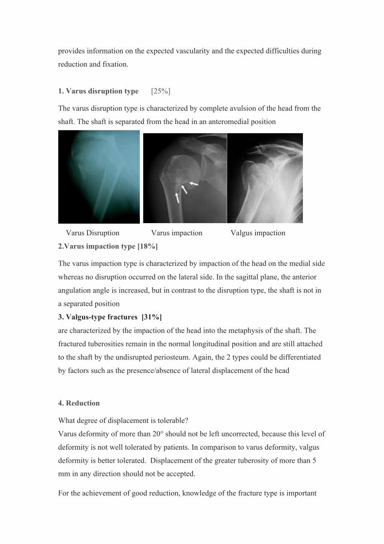
1. Varus disruption type [25%]
The varus disruption type is characterized by complete avulsion of the head from the
shaft. The shaft is separated from the head in an anteromedial position
Varus Disruption Varus impaction Valgus impaction
2.Varus impaction type [18%]
The varus impaction type is characterized by impaction of the head on the medial side
whereas no disruption occurred on the lateral side. In the sagittal plane, the anterior
angulation angle is increased, but in contrast to the disruption type, the shaft is not in
a separated position
3. Valgus-type fractures [31%]
are characterized by the impaction of the head into the metaphysis of the shaft. The
fractured tuberosities remain in the normal longitudinal position and are still attached
to the shaft by the undisrupted periosteum. Again, the 2 types could be differentiated
by factors such as the presence/absence of lateral displacement of the head
4. Reduction
What degree of displacement is tolerable?
Varus deformity of more than 20° should not be left uncorrected, because this level of
deformity is not well tolerated by patients. In comparison to varus deformity, valgus
deformity is better tolerated. Displacement of the greater tuberosity of more than 5
mm in any direction should not be accepted.
For the achievement of good reduction, knowledge of the fracture type is important
because this provides information on the preserved periosteum.
1.Varus-impacted fractures: are characterized by residual primary stability, as a result
of the periosteum still being preserved on the lateral side. The calcar on the medial
side has to be reduced, which can usually be achieved just by traction and
manipulation of the arm.
2. Varus disruption type: with additional fracture of the greater tuberosity presents
quite often with the head in an internally rotated position (3-part fracture according to
Neer). Reduction of this fracture type can only be achieved by a step-by-step
procedure. At first, the shaft has to be brought into alignment with the head, and then
the head has to be derotated by pulling on the lesser tuberosity with a hooked
instrument. At the moment when alignment and derotation are achieved, either
temporarily or permanently, Humerusblock K-wires (Synthes, Bettlach, Switzerland)
are introduced through the shaft into the head. As the last step, the greater tuberosity
is pulled downward by means of a hooked instrument and fixed with cannulated
screws. All of the maneuvers are performed percutaneously (but even with an open
procedure, the various steps remain the same).
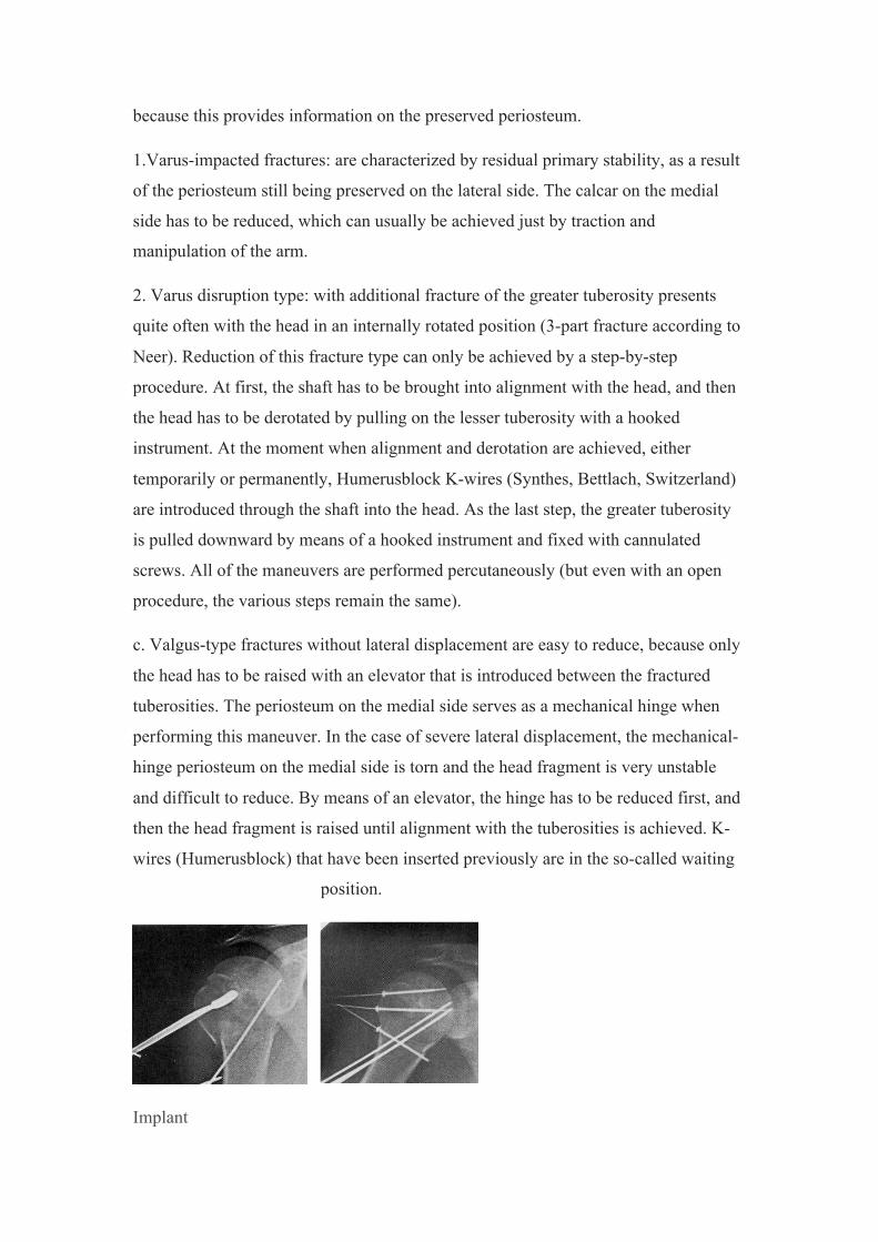
c. Valgus-type fractures without lateral displacement are easy to reduce, because only
the head has to be raised with an elevator that is introduced between the fractured
tuberosities. The periosteum on the medial side serves as a mechanical hinge when
performing this maneuver. In the case of severe lateral displacement, the mechanical-
hinge periosteum on the medial side is torn and the head fragment is very unstable
and difficult to reduce. By means of an elevator, the hinge has to be reduced first, and
then the head fragment is raised until alignment with the tuberosities is achieved. K-
wires (Humerusblock) that have been inserted previously are in the so-called waiting
position.
Implant
1. Closed K Wires
2. Traditional plate
3. Locking plate
4. Nailing
5. Humerusblock implant. Semi rigidity
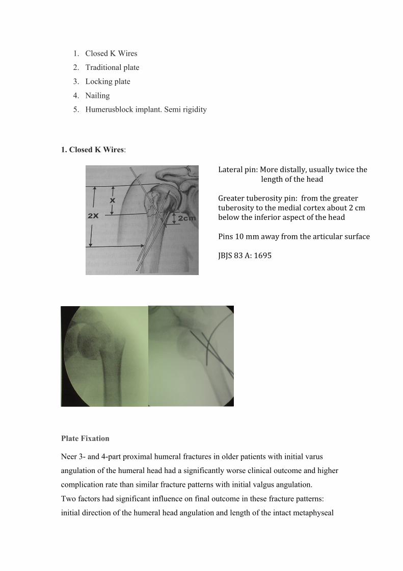
1. Closed K Wires:
Plate Fixation
Neer 3- and 4-part proximal humeral fractures in older patients with initial varus
angulation of the humeral head had a significantly worse clinical outcome and higher
complication rate than similar fracture patterns with initial valgus angulation.
Two factors had significant influence on final outcome in these fracture patterns:
initial direction of the humeral head angulation and length of the intact metaphyseal
Lateral pin: More distally, usually twice the length of the head Greater tuberosity pin: from the greater tuberosity to the medial cortex about 2 cm below the inferior aspect of the head Pins 10 mm away from the articular surface JBJS 83 A: 1695
segment attached to the articular fragment.
The best clinical outcomes were obtained in valgus impacted fractures with a
metaphyseal segment length of greater than 2 mm, and this was independent of Neer
fracture type. Humeral head angulation had the greatest effect on final outcomes (P <
0.001), whereas metaphyseal segment length of less than 2 mm was predictive of
developing avascular necrosis (J Orthop Trauma. 2009 Feb;23(2):113-9.)
2. Traditional Plate fixation
3. Locking Plates
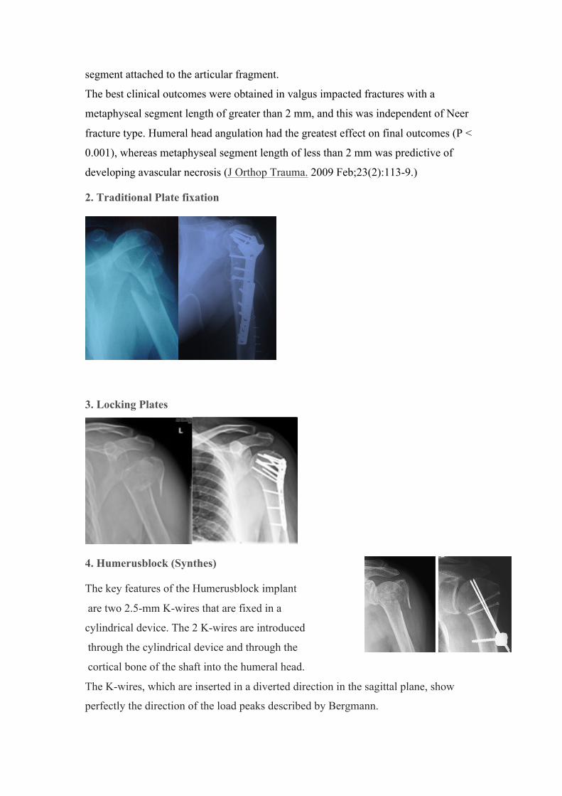
4. Humerusblock (Synthes)
The key features of the Humerusblock implant
are two 2.5-mm K-wires that are fixed in a
cylindrical device. The 2 K-wires are introduced
through the cylindrical device and through the
cortical bone of the shaft into the humeral head.
The K-wires, which are inserted in a diverted direction in the sagittal plane, show
perfectly the direction of the load peaks described by Bergmann.
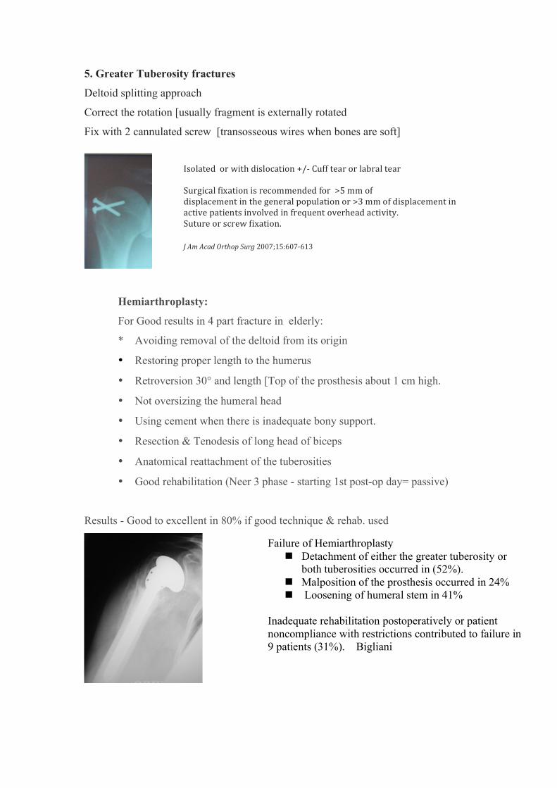
5. Greater Tuberosity fractures
Deltoid splitting approach
Correct the rotation [usually fragment is externally rotated
Fix with 2 cannulated screw [transosseous wires when bones are soft]
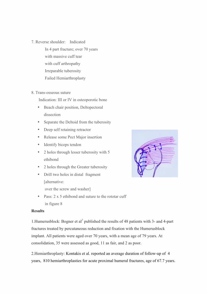
Hemiarthroplasty:
For Good results in 4 part fracture in elderly:
* Avoiding removal of the deltoid from its origin
• Restoring proper length to the humerus
• Retroversion 30° and length [Top of the prosthesis about 1 cm high.
• Not oversizing the humeral head
• Using cement when there is inadequate bony support.
• Resection & Tenodesis of long head of biceps
• Anatomical reattachment of the tuberosities
• Good rehabilitation (Neer 3 phase - starting 1st post-op day= passive)
Results - Good to excellent in 80% if good technique & rehab. used
Failure of Hemiarthroplasty n Detachment of either the greater tuberosity or
both tuberosities occurred in (52%). n Malposition of the prosthesis occurred in 24% n Loosening of humeral stem in 41%
Inadequate rehabilitation postoperatively or patient noncompliance with restrictions contributed to failure in 9 patients (31%). Bigliani
Isolated or with dislocation +/-‐ Cuff tear or labral tear Surgical fixation is recommended for >5 mm of displacement in the general population or >3 mm of displacement in active patients involved in frequent overhead activity. Suture or screw fixation. J Am Acad Orthop Surg 2007;15:607-‐613
7. Reverse shoulder: Indicated
In 4 part fracture; over 70 years
with massive cuff tear
with cuff arthropathy
Irreparable tuberosity
Failed Hemiarthroplasty
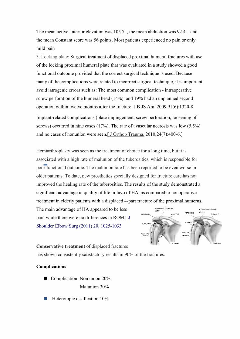
8. Trans-osseous suture
Indication: III or IV in osteoporotic bone
• Beach chair position, Deltopectoral
dissection
• Separate the Deltoid from the tuberosity
• Deep self retaining retractor
• Release some Pect Major insertion
• Identify biceps tendon
• 2 holes through lesser tuberosity with 5
ethibond
• 2 holes through the Greater tuberosity
• Drill two holes in distal fragment
[alternative:
over the screw and washer]
• Pass: 2 x 5 ethibond and suture to the rototar cuff
in figure 8
Results
1.Humerusblock: Bogner et al3 published the results of 48 patients with 3- and 4-part
fractures treated by percutaneous reduction and fixation with the Humerusblock
implant. All patients were aged over 70 years, with a mean age of 79 years. At
consolidation, 35 were assessed as good, 11 as fair, and 2 as poor.
2.Hemiarthroplasty: Kontakis et al. reported an average duration of follow-up of 4
years, 810 hemiarthroplasties for acute proximal humeral fractures, age of 67.7 years.
The mean active anterior elevation was 105.7_, the mean abduction was 92.4_, and
the mean Constant score was 56 points. Most patients experienced no pain or only
mild pain
3. Locking plate: Surgical treatment of displaced proximal humeral fractures with use
of the locking proximal humeral plate that was evaluated in a study showed a good
functional outcome provided that the correct surgical technique is used. Because
many of the complications were related to incorrect surgical technique, it is important
avoid iatrogenic errors such as: The most common complication - intraoperative
screw perforation of the humeral head (14%) and 19% had an unplanned second
operation within twelve months after the fracture. J B JS Am. 2009 91(6):1320-8.
Implant-related complications (plate impingement, screw perforation, loosening of
screws) occurred in nine cases (17%). The rate of avascular necrosis was low (5.5%)
and no cases of nonunion were seen.[ J Orthop Trauma. 2010;24(7):400-6.]
Hemiarthroplasty was seen as the treatment of choice for a long time, but it is
associated with a high rate of malunion of the tuberosities, which is responsible for
poor functional outcome. The malunion rate has been reported to be even worse in
older patients. To date, new prosthetics specially designed for fracture care has not
improved the healing rate of the tuberosities. The results of the study demonstrated a
significant advantage in quality of life in favo of HA, as compared to nonoperative
treatment in elderly patients with a displaced 4-part fracture of the proximal humerus.
The main advantage of HA appeared to be less
pain while there were no differences in ROM.[ J
Shoulder Elbow Surg (2011) 20, 1025-1033
Conservative treatment of displaced fractures
has shown consistently satisfactory results in 90% of the fractures.
Complications
n Complication: Non union 20%
Malunion 30%
n Heterotopic ossification 10%
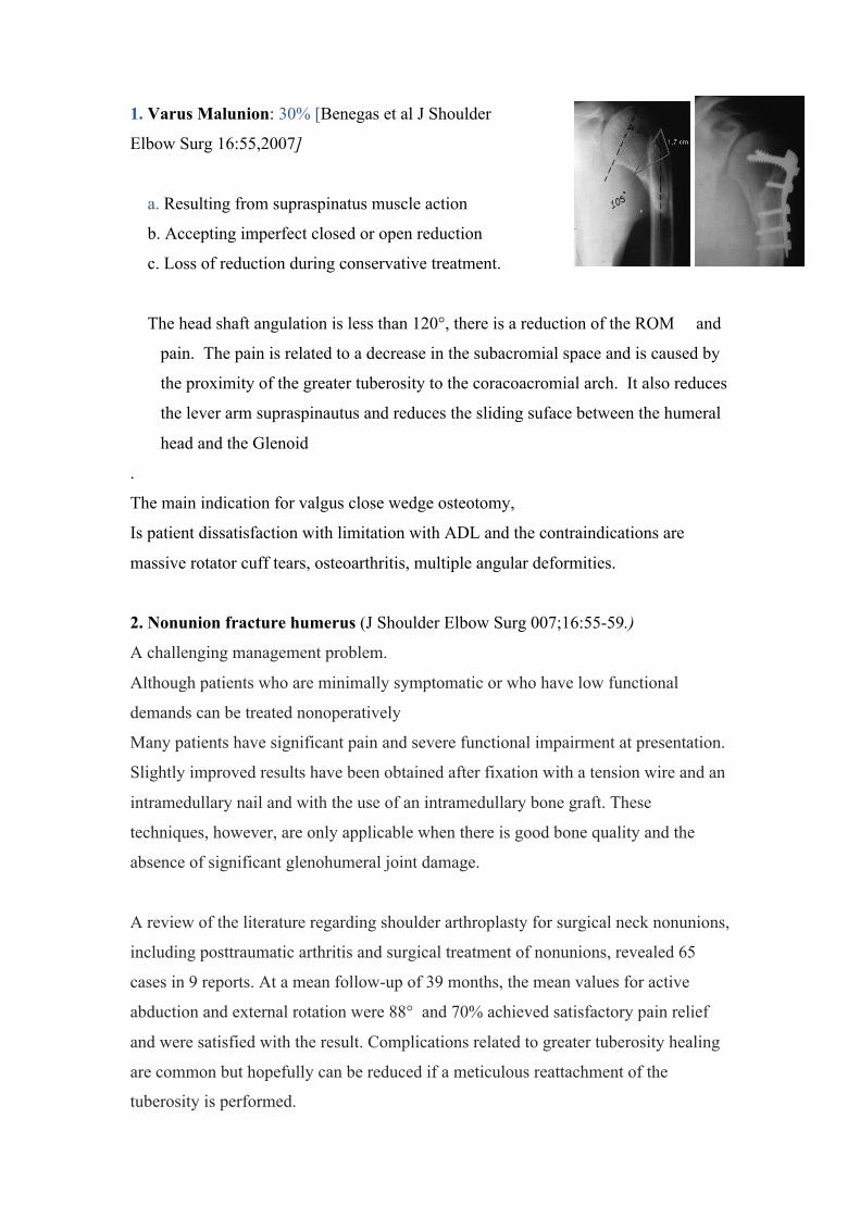
1. Varus Malunion: 30% [Benegas et al J Shoulder
Elbow Surg 16:55,2007]
a. Resulting from supraspinatus muscle action
b. Accepting imperfect closed or open reduction
c. Loss of reduction during conservative treatment.
The head shaft angulation is less than 120°, there is a reduction of the ROM and
pain. The pain is related to a decrease in the subacromial space and is caused by
the proximity of the greater tuberosity to the coracoacromial arch. It also reduces
the lever arm supraspinautus and reduces the sliding suface between the humeral
head and the Glenoid
.
The main indication for valgus close wedge osteotomy,
Is patient dissatisfaction with limitation with ADL and the contraindications are
massive rotator cuff tears, osteoarthritis, multiple angular deformities.
2. Nonunion fracture humerus (J Shoulder Elbow Surg 007;16:55-59.)
A challenging management problem.
Although patients who are minimally symptomatic or who have low functional
demands can be treated nonoperatively
Many patients have significant pain and severe functional impairment at presentation.
Slightly improved results have been obtained after fixation with a tension wire and an
intramedullary nail and with the use of an intramedullary bone graft. These
techniques, however, are only applicable when there is good bone quality and the
absence of significant glenohumeral joint damage.
A review of the literature regarding shoulder arthroplasty for surgical neck nonunions,
including posttraumatic arthritis and surgical treatment of nonunions, revealed 65
cases in 9 reports. At a mean follow-up of 39 months, the mean values for active
abduction and external rotation were 88° and 70% achieved satisfactory pain relief
and were satisfied with the result. Complications related to greater tuberosity healing
are common but hopefully can be reduced if a meticulous reattachment of the
tuberosity is performed.


![Management of proximal humerus fractures in adults · 2017-05-05 · traobserver reproducibility of proximal humerus fracture classification systems have been shown to be poor[15],](https://img.dokumen.tips/doc/110x75/5f03e0727e708231d40b3493/management-of-proximal-humerus-fractures-in-adults-2017-05-05-traobserver-reproducibility.jpg)