Embed Size (px)
Citation preview

3/21/2016
1
Pinakin G Davey PhD, OD, FAAO
Professor and Director of Research
Disclosures Principal investigator for iVue OCT trial
Principal investigator Topcon FDA trials for Maestro and OCT 2000
Consultant for Topcon
Speakers bureau Sanofi- Genzyme and Allergan
Decision making
DiagnosisPrognosisFollow-upTreatment
Risk assessment
Disease evaluation
Differential diagnosis
Ocular examination
Clinical evaluation
Intraocular pressureNerve head analysisVisual fieldsImagingGonioscopyPachymetry
Modifiable Non modifiable
TreatmentMedical Surgical
What is glaucoma? Definition:
“Ocular tissue damage at least partially related to intraocular pressure”
Where glaucoma is concerned agreement is limited among clinicians and scientists.
Types of glaucoma
Glaucoma
Open angle Closed angle
Primary or secondary
Primary Open angle glaucoma
Normal tension glaucoma
Ocular hypertension
Glaucoma suspects
Primary angle closure
Visual fields
Secondary angle closure
Optic disc
Secondary glaucoma
Congenital glaucoma
Prevalence studies Prevalence in different studies varies
Different populations
Different methods used to obtain a sample
Definition of glaucoma

3/21/2016
2
Prevalence of POAG in Caucasians
Study Age range Prevalence %
Roscommon Over 50 1.9
Beaver Dam 43-84 2.1
Rotterdam Over 55 1.1
Dalby 55-69 0.9
Blue Mountain Over 49 2.4
Barbados Caucasians 40-84 0.8
Baltimore Caucasians Over 40 1.3
Prevalence of POAG in African American & African Caribbean
Study Age range Prevalence %
Barbados 40-84 7.1
Baltimore Over 40 4.2
St Lucia Over 30 8.8
London Over 35 3.9
African-Caribbean
Prevalence of OAG in LALES
Risk factors for glaucoma examined in population based studies
Demographic Age
Gender
Race
Ocular IOP
Optic nerve head
Myopia
Hypermetropia
Systemic Diabetes
Systemic hypertension
Genetic Family history
Other Cigarette smoking
Alcohol intake
Socio economic factors
Intraocular pressure Major risk factor
Not as fundamental as once thought.
Prevalence increases with increase in IOP
Visual field loss slows down with decrease in IOP
Even if both eyes have IOP lower than 21. The eye with greater IOP will lose field quicker.

3/21/2016
3
Systemic hypertension and glaucoma Blood pressure and pathogenesis of glaucoma
Hospital based study
Baltimore Eye Survey examined perfusion pressure
Ocular Perfusion pressure= Blood pressure-IOP
(Systolic or Diastolic or mean pressure)
Tielsch et al Hypertension perfusion pressure and primary open angle glaucoma Arch
ophthalmol 1995
Genetic factors Positive family history
Bias:
+ ve Family history makes a person have frequent check ups
Recall bias
Sibling with glaucoma odds ratio 3.69
Parents with glaucoma odds ratio 2.67
Children with glaucoma odds ratio 1.12
Summary Prevalence of POAG is Caucasians over 40 years of age
2% and in African American and African Caribbean is “four times” that.
Hispanics greater risk than African American as they grow older
Overall quite underdiagnosed- 50% unknown
Glaucoma suspects- increases need for care dramatically
Intraocular pressure Diagnosis- not helpful
Treatment- only proven method
Progression- very closely associated with IOP
Risk factor- without a doubt most important risk factor
In fact only alterable risk factor!
Intraocular pressurePascal dynamic contour tonometer
Ocular Response Analyzer
3/21/2016
4
1. Observe the scleral ring to identify the limits of the optic disc and evaluate its size.
Measure Disc Size Observe the scleral ring to identify the limits of the
optic disc and evaluate its size. 66D 1 X magnification
Cup size is associated with disc size
Effects any casual observer for cup to disc ratio measurement
Rim thickness varies with disc size
Disc size Small < 1.5 mm 2
Medium > 1.5 but <2.5 mm 2
Large > 2.5 mm 2
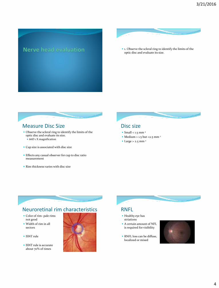
Neuroretinal rim characteristics Color of rim- pale rims
not good
Width of rim in all sectors
ISNT rule
ISNT rule is accurate about 70% of times
RNFL Healthy eye has
striations
A certain amount of NFL is required for visibility
RNFL loss can be diffuse, localized or mixed

3/21/2016
5
RNFL cont… Diffuse – reduction in
RNFL brightness
Localized – wedge shaped defect
Localized RNFL defects should traced back the disc
Peripapillary atrophy Where
How large
1/8, ¼, ½ , ¾, 1, > 1 DD
α
β
Optic disc hemorrhages Transient
Inferior temporal or superior temporal regions mainly
Record present or absent
If present where
Retinal vessels
Look for this in patients that you suspect NTG
CD ratio Vertical
Horizontal
Largest
CD ratio of imaging devices will not match your findings!

3/21/2016
6
Focal atrophy of neural rim Focal atrophy of neural rim-2
Optic disc hemorrhages
Optic disc hemorrhages-33 years later
Barring of circumlinear vessels Vessels that runs along
margin between cup and neural rim.
Found supero and infero temporally

3/21/2016
7
Barring of circumlinear vessels As rim becomes thinner
it leaves an area of pallor between the rim and the circumlinear blood vessel.
Barring of circumlinear vessels
Nasal cupping Usually seen in
advanced glaucoma.
Space between Nasal rim and blood vessels.
Laminar dot sign
Bayonetting Double angulation of
blood vessel.
Nerve fiber bundle defect Rim changes also
produces RNFL defects.
Dark stripes or wedge shapes defect paralleling the normal striations.
Common after disc hemorrhages

3/21/2016
8
1Watch out forPupil sizeReliability indexType of test24-2 SITA STDage refractive error
10
9
2
1
8
7
6
5
4
3
2 Grey ScaleLook for patternsGlobal view Not for diagnosisTypes of visual field defects
3 Raw dataNormals centrally low 30’sPeripherally high 20’s
10
9
2
1
8
7
6
5
4
3
4 Total deviationDevidation from average
5 Total deviation probability plot
6 Pattern deviationRemoves any generalized defectsCataractPupil miosis
7 Pattern deviation probability plot
8 glaucoma hemifield testOutside normal limits Borderline Generalized reduction in sensitivity
9 global indices
MD mean deviationPSD pattern Standard Devaition
10 gaze tracking
10
9
2
1
8
7
6
5
4
3

3/21/2016
9
Criteria for glaucomatous damage GHT outside normal limits in at least two occasions
PSD < 5% of normal individuals
A cluster of three or more non-edge points (pattern deviation plot) all of which are depressed at a p<5% and one of which is depressed at a p<1% on two occasions (respecting horizontal meridian)
This criterion was written for 30-2, if 24-2 field is analyzed edge points are included.
49
Staging based on MD Better than-6 db- Mild
Worse than -6.o dB but better than -12 dB – Moderate
Worse than -12.0 dB severe
50
Gonioscopy
Iris insertion
Curvature of periheral iris
Angle approachA = Above Schwalbe line, totally occluded angle.B = Behind the Schwalbe line, peripheral iris is in contact with TM.C = Scleral spur Iris root at the level of scleral spur
D = Deep anterior ciliary body
seen.
E = extremely deep

3/21/2016
10
Van Herrick angle estimation 1:1 – Open angle, VH grade
4
1:1/2 – Open angle, VH grade 3
1:1/4 – Narrow angle, VH grade 2 (Angle Closure Possible)
1: <1/4 – Angle closure likely, VH grade 1
Ultrasound pachymetry is standard As central data as
possible
Greater number of measurements increase your reproducibility of data
Always use lowest data
Why lowest data? Perpendicular
measurements are lowest or smallest in value
Why not average the data? Average 484 microns
Lowest 473 microns
Averaging helps decrease error but does not eliminate it.
A Haun, P Gunvant, M Baskaran, L Vijaya: Central corneal thickness measurement using a pachometer: Mean or lowest values? Invest Ophthalmol Vis Sci, 2004, 45: E-Abstract 137
3/21/2016
11
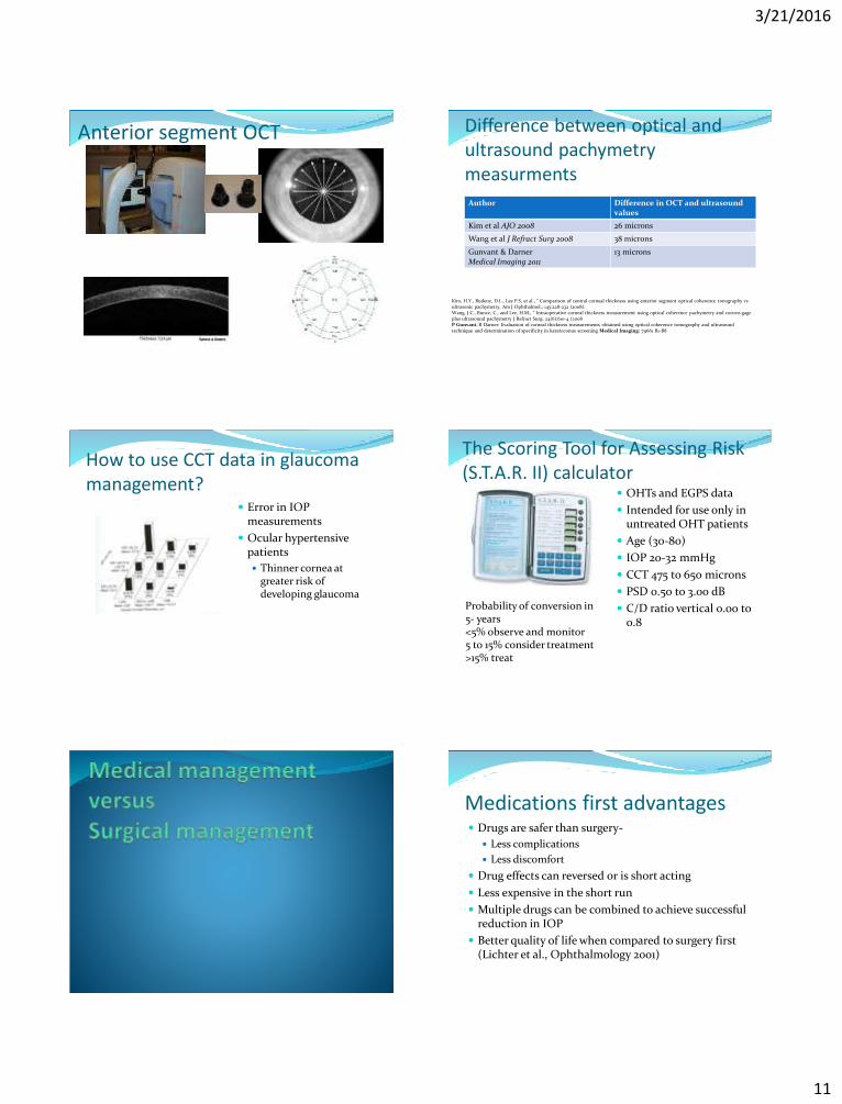
Anterior segment OCT
Author Difference in OCT and ultrasound values
Kim et al AJO 2008 26 microns
Wang et al J Refract Surg 2008 38 microns
Gunvant & Darner Medical Imaging 2011
13 microns
Difference between optical and ultrasound pachymetrymeasurments
Kim, H.Y., Budenz, D.L., Lee P.S, et al., “ Comparison of central corneal thickness using anterior segment optical coherence tonography vsultrasonic pachymetry, Am J Ophthalmol,; 145:228-232 (2008).Wang, J.C., Bunce, C., and Lee, H.M., “ Intraoperative corneal thickness measurement using optical coherence pachymetry and corneo-gage plus ultrasound pachymetry J Refract Surg. 24(6):610-4 (2008P Gunvant, R Darner: Evaluation of corneal thickness measurements obtained using optical coherence tomography and ultrasound technique and determination of specificity in keratoconus screening Medical Imaging: 79661 B1-B8
How to use CCT data in glaucoma management?
Error in IOP measurements
Ocular hypertensive patients
Thinner cornea at greater risk of developing glaucoma
The Scoring Tool for Assessing Risk (S.T.A.R. II) calculator
OHTs and EGPS data
Intended for use only in untreated OHT patients
Age (30-80)
IOP 20-32 mmHg
CCT 475 to 650 microns
PSD 0.50 to 3.00 dB
C/D ratio vertical 0.00 to 0.8
Probability of conversion in 5- years<5% observe and monitor 5 to 15% consider treatment>15% treat
Medications first advantages Drugs are safer than surgery-
Less complications
Less discomfort
Drug effects can reversed or is short acting
Less expensive in the short run
Multiple drugs can be combined to achieve successful reduction in IOP
Better quality of life when compared to surgery first (Lichter et al., Ophthalmology 2001)

3/21/2016
12
Medications first disadvantages May be more expensive in the long run
Multiple drugs Compliance, adherence and persistence issues
Chronic drug uses and its effect on future surgical outcomes? Preservatives effect?
Inflammation leading to failure of future procedures*
Increased chances of cataract formation
*Broadway DC et al., Adverse effects of topical antiglaucoma medications: II Arch Ophthalmol 1994
Surgery first - advantages If successful and large drop in IOP may be obtained
No issues related to patient compliance, adherence and persistence
Good in situations where obtaining continuous supply of medications is a problem
May be cheaper long term
Surgery first - disadvantages Outcomes may be variable
Long term may loose efficacy
May still require additional topical medications
Complications may be dire
Comfort and quality of life may be lower
Chances of cataract formation is greater than topical medications
Age- young vs. older individuals
Race and management options Race – white versus individuals with greater pigment
Individuals with greater pigment- greater risk of
post-operative scarring* Medications –first choice
*Broadway DC et al., Racial differences in the results of glaucoma filtration surgery: are racial differences in conjunctival cell profile important? BJO 1994
Age and management options Younger individuals
Accelerated wound healing systems
Thick fleshy periocular tissues heals rapidly
Thus older individuals better suited for surgical options
Current practice patterns Unacceptable high pressures will inevitably destroy
optic nerve tissue
Safe levels of IOP by any means warranted
If these don’t work or not sufficient
drugs like – prostaglandins
reduction in inflow – beta blockers
Maximal medical therapy
Consider surgery
3/21/2016
13
Maximal tolerated medical therapy β-Blockers
Timolol
Betaxolol
Levobunolol
Carteolol
Metipranolol
Carbonic Anyhydrase Inhibitors (CAIs)
Systemic:
Acetazolamide
Methazolamide
Topical:
Dorzolamide
Brinzolamide
Adrenergic Agonists
Nonspecific:
Dipivefrin (epinephrine) – also increases conventional outflow
α2-Agonists:
Brimonidine – also increases uveoscleral outflow
Apraclonidine – also increases uveoscleral outflow
Conventional/Trabecular
Cholinergic agonists (parasymphathomimetics): Pilocarpine
Echothiophate iodide
Carbachol
Prostaglandin derivatives:
Bimatoprost
Latanoprost
Nonspecific adrenergic agonists:
Dipivefrin (epinephrine)
Nonconventional/Uveoscleral
Prostoglandin derivatives:
Latanoprost
Bimatoprost
Travoprost
α2-Agonists:
Brimonidine
Tsai and Forbes Medical management of
glaucoma Third edition 2009
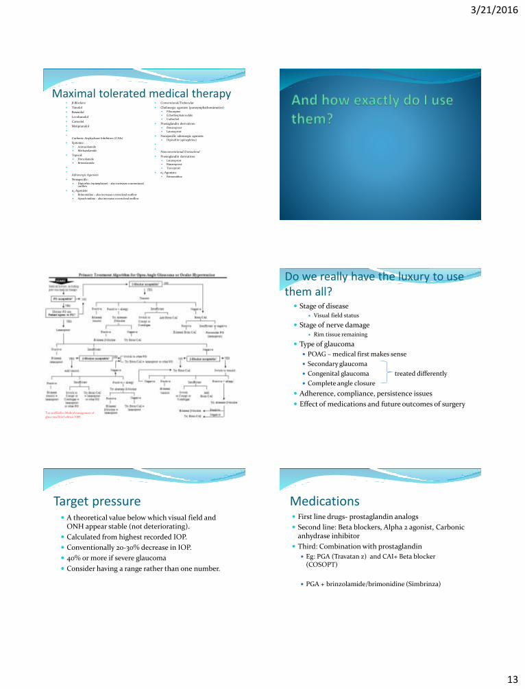
Do we really have the luxury to use them all?
Stage of disease Visual field status
Stage of nerve damage Rim tissue remaining
Type of glaucoma
POAG – medical first makes sense
Secondary glaucoma
Congenital glaucoma treated differently
Complete angle closure
Adherence, compliance, persistence issues
Effect of medications and future outcomes of surgery
Target pressure A theoretical value below which visual field and
ONH appear stable (not deteriorating).
Calculated from highest recorded IOP.
Conventionally 20-30% decrease in IOP.
40% or more if severe glaucoma
Consider having a range rather than one number.
Medications First line drugs- prostaglandin analogs
Second line: Beta blockers, Alpha 2 agonist, Carbonic anhydrase inhibitor
Third: Combination with prostaglandin
Eg: PGA (Travatan z) and CAI+ Beta blocker (COSOPT)
PGA + brinzolamide/brimonidine (Simbrinza)

3/21/2016
14
Where should the IOP be? No real number
Start with 30% drop
Monitor for progression
Advanced glaucoma you want IOP to be less than 12
Pressure should not fluctuate much
ALT versus SLT SLT preferred
Unlike ALT, SLT does not scar
Autopsy specimens – confirm no coagulative damage after SLT
SLT can be repeated
Mechanisms of action SLT 5-8 fold increase in monocytes and macrophages in
TM
after treatment with SLT
Hypothesis
Injury via laser causes releasing of chemoattractant
This in turn recruits monocytes that are transformed into macrophages
Macrophages clear pigment granules and exit via Schlemm’s canal
Alvardo and Murphy Outflow obstruction in pigmentary and primary open angle glaucoma Arch
Ophthalmol 1992
Selective Laser trabeculoplasty Selectively targets melanin pigment of TM
More safe compared to ALT (because lower power)
Equally effective as ALT
Can be repeated if first attempt is not effective

3/21/2016
15
Triggerfish
Cost 500 Euro. Not available for sale in USA86
Main adverse events Blurred vision 82 %Hyperemia 80%SPK 15%
Correlation for repeatability Overall r=59That is r-square = 35%
87 88
Overall sensimed is “sensitive” to IOP fluctuations
89
Summary of contact lens IOP devices Long way to go
The ability of device to capture peak accurately is not excellent.
Cautiously optimistic.
FDA cleared on March 2016 not yet available for sale
Prediction …
90

3/21/2016
16
Latanoprostene Bunod- Bausch and Lomb Latanoprostene bunod (LBN, BOL-303259-X) is a
nitric oxide (NO)-donating prostanoid FP receptor agonist
Mechanism of actionLatanoprostene Bunod
Cellular esterase
latanoprost acid 4-hydroxybutyl nitrate (Butanediol mononitrate )
1,4 butanediol Nitric oxideActs on changing ECM
Changes trabecular meshwork cells that are highly contractile in nature
Timolol Maleate 0.5% or Latanoprostene Bunod 0.024%IOP measured at 8 AM , 12 noon and 4 PM at week 2, 6, and 3 months

3/21/2016
17
Treatment protocol-Acute angle closure- ABC procedure Alpha -2 agonist- Brimonidine
Beta blocker- Timolol (caution in asthmatics ) or Betaxolol
Carbonic anhydrase inhibitor – Dorzolamide (Caution sulpha allergy contraindication)
Each medication given every 15 minutes
Perform 3 times
Oral medications Oral Carbonic anhydrase inhibitor
Two tablets of 250 mg acetozolamide (Caution sulphaallergies contraindication)
Works good when patient can retain medication -Vomiting common with angle closure glaucoma
Check intraocular pressure after 1 hour if lower than 40
Add Pilocarpine every 15 minutes for 45 minutes and repeat procedure ABC procedure
Seek ophthalmologist opinion-refer patient

3/21/2016
18
Take home medication Prednisolone acetate 1% q1-6 hours (approx every 3
hours)
Acetazolamide 500 mg sequel BID
Alpha agonist or beta blocker BID
Pilocarpine 2% QID
Laser therapy


![eCare Clinical Protocols - Overview Dorset V1.2[1] · Twinfield SPARK R full (MD -1.2, PSD 0.4, GHT WNL) L possible early nasal step (MD -2.8, PSD 1.3, GHT BORDERLINE) 20 Photos:](https://img.dokumen.tips/doc/110x75/5c5d64c709d3f2a1498cd327/ecare-clinical-protocols-overview-dorset-v121-twinfield-spark-r-full-md.jpg)