Embed Size (px)
Citation preview
cst
sirtmncMec
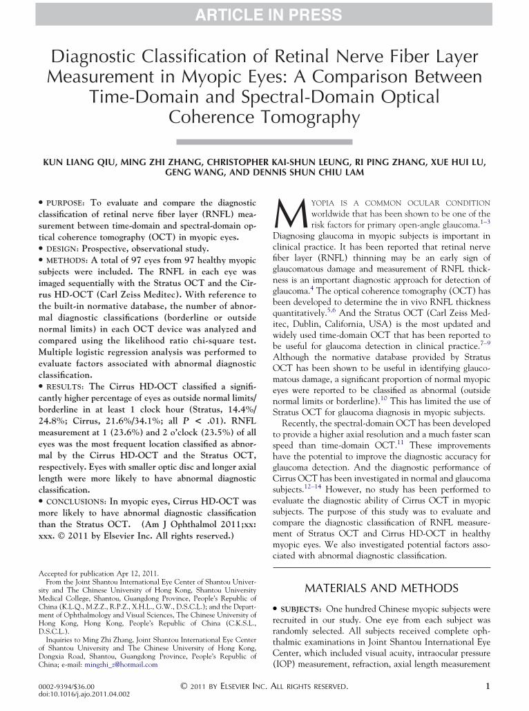
Diagnostic Classification of Retinal Nerve Fiber LayerMeasurement in Myopic Eyes: A Comparison Between
Time-Domain and Spectral-Domain OpticalCoherence Tomography
KUN LIANG QIU, MING ZHI ZHANG, CHRISTOPHER KAI-SHUN LEUNG, RI PING ZHANG, XUE HUI LU,
GENG WANG, AND DENNIS SHUN CHIU LAMbq
● PURPOSE: To evaluate and compare the diagnosticlassification of retinal nerve fiber layer (RNFL) mea-urement between time-domain and spectral-domain op-ical coherence tomography (OCT) in myopic eyes.
● DESIGN: Prospective, observational study.● METHODS: A total of 97 eyes from 97 healthy myopicubjects were included. The RNFL in each eye wasmaged sequentially with the Stratus OCT and the Cir-us HD-OCT (Carl Zeiss Meditec). With reference tohe built-in normative database, the number of abnor-al diagnostic classifications (borderline or outsideormal limits) in each OCT device was analyzed andompared using the likelihood ratio chi-square test.ultiple logistic regression analysis was performed to
valuate factors associated with abnormal diagnosticlassification.
● RESULTS: The Cirrus HD-OCT classified a signifi-cantly higher percentage of eyes as outside normal limits/borderline in at least 1 clock hour (Stratus, 14.4%/24.8%; Cirrus, 21.6%/34.1%; all P < .01). RNFLmeasurement at 1 (23.6%) and 2 o’clock (23.5%) of alleyes was the most frequent location classified as abnor-mal by the Cirrus HD-OCT and the Stratus OCT,respectively. Eyes with smaller optic disc and longer axiallength were more likely to have abnormal diagnosticclassification.● CONCLUSIONS: In myopic eyes, Cirrus HD-OCT wasmore likely to have abnormal diagnostic classificationthan the Stratus OCT. (Am J Ophthalmol 2011;xx:xxx. © 2011 by Elsevier Inc. All rights reserved.)
Accepted for publication Apr 12, 2011.From the Joint Shantou International Eye Center of Shantou Univer-
sity and The Chinese University of Hong Kong, Shantou UniversityMedical College, Shantou, Guangdong Province, People’s Republic ofChina (K.L.Q., M.Z.Z., R.P.Z., X.H.L., G.W., D.S.C.L.); and the Depart-ment of Ophthalmology and Visual Sciences, The Chinese University ofHong Kong, Hong Kong, People’s Republic of China (C.K.S.L.,D.S.C.L.).
Inquiries to Ming Zhi Zhang, Joint Shantou International Eye Centerof Shantou University and The Chinese University of Hong Kong,Dongxia Road, Shantou, Guangdong Province, People’s Republic of
China; e-mail: [email protected]© 2011 BY ELSEVIER INC. A0002-9394/$36.00doi:10.1016/j.ajo.2011.04.002
M YOPIA IS A COMMON OCULAR CONDITION
worldwide that has been shown to be one of therisk factors for primary open-angle glaucoma.1–3
Diagnosing glaucoma in myopic subjects is important inclinical practice. It has been reported that retinal nervefiber layer (RNFL) thinning may be an early sign ofglaucomatous damage and measurement of RNFL thick-ness is an important diagnostic approach for detection ofglaucoma.4 The optical coherence tomography (OCT) haseen developed to determine the in vivo RNFL thicknessuantitatively.5,6 And the Stratus OCT (Carl Zeiss Med-
itec, Dublin, California, USA) is the most updated andwidely used time-domain OCT that has been reported tobe useful for glaucoma detection in clinical practice.7–9
Although the normative database provided by StratusOCT has been shown to be useful in identifying glauco-matous damage, a significant proportion of normal myopiceyes were reported to be classified as abnormal (outsidenormal limits or borderline).10 This has limited the use ofStratus OCT for glaucoma diagnosis in myopic subjects.
Recently, the spectral-domain OCT has been developedto provide a higher axial resolution and a much faster scanspeed than time-domain OCT.11 These improvementshave the potential to improve the diagnostic accuracy forglaucoma detection. And the diagnostic performance ofCirrus OCT has been investigated in normal and glaucomasubjects.12–14 However, no study has been performed toevaluate the diagnostic ability of Cirrus OCT in myopicsubjects. The purpose of this study was to evaluate andcompare the diagnostic classification of RNFL measure-ment of Stratus OCT and Cirrus HD-OCT in healthymyopic eyes. We also investigated potential factors asso-ciated with abnormal diagnostic classification.
MATERIALS AND METHODS
● SUBJECTS: One hundred Chinese myopic subjects wererecruited in our study. One eye from each subject wasrandomly selected. All subjects received complete oph-thalmic examinations in Joint Shantou International EyeCenter, which included visual acuity, intraocular pressure
(IOP) measurement, refraction, axial length measurementLL RIGHTS RESERVED. 1

pastradwcot
go
by IOL master (Carl Zeiss Meditec), and dilated fundusstereoscopic examination. All the included eyes havespherical equivalent of less than �0.5 diopters (D) and noother concurrent diseases. Subjects with best-correctedvisual acuity of less than 20/40, IOP over 21 mm Hg,family history of glaucoma, intraocular surgery, myopicmacular degeneration, glaucoma, parapapillary atrophy(PPA) extending outside the measurement circle of theOCT, refractive surgery, neurological diseases, or diabeteswere excluded. The study was designed following theethical standards of the Declaration of Helsinki andapproved by the local ethical committee with informedconsent obtained before the study.
● VISUAL FIELD TESTING: All visual field tests wereperformed with the static automated white-on-whitethreshold 24-2 SITA standard strategy (Humphrey FieldAnalyzer II; Carl Zeiss Meditec). A visual field test wasconsidered to be reliable when fixation loss, false positive,and false negative were all less than 20%. All the visualfield tests of included eyes were those with pattern standarddeviation (PSD) with P � 5% and within normal limits inglaucoma hemifield test (GHT).
● CONFOCAL SCANNING LASER OPHTHALMOSCOPY
IMAGING: The optic disc was imaged with confocalscanning laser ophthalmoscopy (Heidelberg Retina To-mograph II [HRT 2]; Heidelberg Engineering, GmbH,Dossenheim, Germany). A 3-dimensional topographic im-age consisting of 384 � 384 � 16 up to 384 � 384 � 64ixels is constructed from multiple focal planes axiallylong the optic nerve head. An average of 3 consecutivecans is obtained and aligned to compose a single meanopography for analysis. Images obtained in this study wereeviewed carefully for image quality and selected fornalyses only when an average pixel height standardeviation was 30 �m or less. The margin of the optic discas defined as the inner edge of the Elschnig ring. Theontour line was manually drawn by a trained ophthalmol-gist (K.L.Q.). Optic disc area was recorded and used inhe present analysis.
● OPTICAL COHERENCE TOMOGRAPHY IMAGING: Time-domain OCT imaging was performed with Stratus OCT(software version 4.0.4, Carl Zeiss Meditec). The acquisi-tion rate of the Stratus OCT is 400 A-scans per second andthe axial resolution is 8 to 10 �m. RNFL thickness wasmeasured with the fast RNFL (3.4) scanning protocolconsisting of 256 A-scans. Average measurements of 3sequential circular scans of diameter 3.46 mm centered onthe optic disc were recorded. The RNFL thickness isdetermined by the difference in distance between thevitreoretinal interface and a posterior border, based on apredefined reflectivity signal level. All the included scans
had signal strength of at least 7.AMERICAN JOURNAL OF2
Spectral-domain OCT imaging was obtained using Cir-rus HD-OCT (software version 4.5.1.11; Carl Zeiss Med-itec). The scan speed for Cirrus is 27 000 A-scans persecond and the axial resolution is 5 �m.15 The parapapil-lary RNFL thickness was measured by Cirrus HD-OCTusing the Optic Disk Cube 200 � 200 protocol. Thisprotocol obtains data through an area of 6 � 6 mm at theoptic disc region by acquiring a series of 200 horizontalscan lines each composed of 200 A-scans. Eye movementswere monitored by real-time fundus images. Images withmisaligned vessels within the scanning circle were ex-cluded and retaken. The signal for the included images hada minimum signal strength of 7.
For both OCT measurements, all images were acquiredby 2 well-trained technicians during the same visit. Param-eters including average and clock-hour RNFL thicknesseswere generated automatically in the analysis reports of theStratus OCT and Cirrus HD-OCT. Based on the internalnormative databases, diagnostic classification (within nor-mal limits, borderline, or outside normal limits) of eachdevice was provided in the analysis printout. For bothOCTs, there were 4 diagnostic classes. The upper 5thpercentile was indicated in white, the 5th to 95th percen-tiles were indicated in green (within normal limits), the1st to 5th percentiles were indicated in yellow (border-line), and the lower 1st percentile was indicated in red(outside normal limits).15,16 White and green were re-arded as normal while yellow and red were abnormal inur analysis.
● STATISTICAL ANALYSIS: The statistical analyses wereperformed with commercially available software (SPSSver.13.0; SPSS Inc, Chicago, Illinois, USA). The relation-ship between Cirrus HD-OCT and Stratus OCT measure-ments was evaluated using Pearson correlation analysis.Based on the internal normative databases, diagnostic
TABLE 1. Comparison of Average and Clock-Hour RetinalNerve Fiber Layer Thickness Between Cirrus HD-OCT and
Stratus OCT in 97 Normal Myopic Eyes (Paired t Test)
Clock Hours Stratus OCT (um) Cirrus HD-OCT (um) P
12 135.5 � 25.56 112.9 � 30.45 �.01
1 125.8 � 22.68 102.1 � 20.84 �.01
2 92.1 � 25.79 74.7 � 14.43 �.01
3 68.6 � 19.05 58.5 � 10.28 �.01
4 75.3 � 20.70 58.9 � 9.95 �.01
5 106.4 � 23.10 91.5 � 18.60 �.01
6 134.4 � 26.40 127.6 � 26.90 �.01
7 164.1 � 22.36 157.7 � 30.70 �.01
8 100.2 � 24.98 89.6 � 21.85 �.01
9 75.1 � 16.5 65.5 � 11.72 �.01
10 110.7 � 23.47 100.8 � 20.80 �.01
11 155.9 � 24.37 144.6 � 20.96 �.01
Average thickness 111.7 � 11.34 98.7 � 9.02 �.01
classification (within normal limits, borderline, or outside
OPHTHALMOLOGY MONTH 2011

(�d1O(
otsm(
sC(PcAnb
normal limits) provided by each device were comparedusing the likelihood ratio chi-square test. Logistic regres-sion analysis was performed to investigate the factorsassociated with the diagnostic classification. A P � .05 wasconsidered statistically significant.
RESULTS
AFTER EXCLUDING 2 SUBJECTS WITH UNRELIABLE VISUAL
field tests and 1 for extensive PPA, 97 eyes from 97subjects (43 female and 61 right eyes) were analyzed. Themean age, axial length, and spherical equivalent were22.84 � 3.92 years (range, 18-40), 25.55 � 1.11 mmrange, 22.52–28.77 mm), and �4.93 � 2.17 D (range,1.00 to �12.75 D), respectively. The visual field mean
eviation and pattern standard deviation were �2.13 �.12 dB and 1.49 � 0.98 dB. The signal strength of StratusCT was significantly higher than that of Cirrus HD-OCT
9.1 � 0.8 vs 8.0 � 0.7, P � .01).The average and clock-hour RNFL thickness for both
OCTs were demonstrated in Table 1. RNFL thicknessbtained by Stratus OCT was significantly thicker thanhat of Cirrus HD-OCT in clock-hour and average mea-urements (all with P � .01). Average RNFL thicknesseasured by the 2 OCT devices was highly correlated
Figure 1, r � 0.794; P � .01).Figure 2 presents the proportion of eyes identified as
abnormal based on the normative databases provided inStratus OCT and Cirrus HD-OCT. For average RNFLthickness, 93 of 97 eyes (96%) were classified as normal (4
FIGURE 1. Scatterplot showing the relationship of average reeyes.
eyes as borderline) in Cirrus HD-OCT while all eyes were
DIAGNOSTIC CLASSIFICATION OF RETINAL NERVE FIBVOL. XX, NO. X
classified as normal in Stratus. For clock-hour RNFLthickness, the Cirrus HD-OCT had a significantly higherpercentage of eyes detected as outside normal limits/borderline in at least 1 clock hour (Stratus, 14.4%/24.8%;Cirrus, 21.6%/34.1%; all with P � .01). For Cirrus HD-OCT, RNFL measurement at 1 o’clock was most fre-quently classified as borderline/outside normal limits(4.1%/19.5%). For Stratus OCT, 2 o’clock was mostfrequently classified as borderline/outside normal limits(6.0%/17.5%). An example of abnormal diagnostic classi-fication with Stratus OCT and Cirrus OCT in a normalmyopic eye is demonstrated in Figure 3.
Logistic regression analysis showed that eyes withmaller optic disc size (Stratus, odds ratio: 0.23, P � .01;irrus, odds ratio: 0.17, P � .01) and longer axial length
Stratus, odds ratio: 2.75, P � .01; Cirrus, odds ratio: 1.80,� .017) were significantly associated with higher per-
entage of abnormal diagnostic classification (Table 2).ge, visual field mean deviation, and signal strength wereot associated with abnormal diagnostic classification inoth OCTs (Table 2).
DISCUSSION
IN THIS STUDY, WE FOUND THAT NASAL RNFL MEASURE-
ment was frequently classified as borderline or outsidenormal limits in both OCTs and that the frequency ofabnormal classification of RNFL measurement was sig-nificantly higher in Cirrus HD-OCT than Stratus OCT.
nerve fiber layer thickness between both OCTs in 97 myopic
tinalEyes with smaller optic disc size and longer axial length
ER LAYER MEASUREMENT IN MYOPIC SUBJECTS 3

ojcs(
fb
w
were found to be associated with significantly higherpercentages of abnormal diagnostic classification in bothdevices.
The Stratus OCT is a time-domain OCT that has beenshown reliable for detecting glaucoma in clinical prac-tice.7–9 Jeoung and associates17 reported that the StratusOCT diagnostic classification can detect localized RNFLdefects with moderate sensitivity and high specificity.Although the normative database provided by StratusOCT has been shown useful to detect glaucomatousdamage, Leung and associates10 reported that a significantproportion (44.2% at 2 o’clock) of normal myopic eyeswere classified as abnormal (outside normal limits orborderline). In agreement with Leung’s study, we foundthat 23.5% of eyes were classified as abnormal in myopiceyes, with 2 o’clock being the most frequent abnormalclock hour. With a higher scan resolution and a much
FIGURE 2. Clock-hour abnormal classification of retinal nervStratus OCT in 97 myopic eyes.
faster scan speed, the Cirrus HD-OCT has the potential to p
AMERICAN JOURNAL OF4
provide more accurate RNFL measurements and improvethe diagnostic accuracy for glaucoma detection. The diag-nostic performance of Cirrus HD-OCT has been investi-gated in normal and glaucoma subjects.12–14,18 However,nly a few studies have been performed on myopic sub-ects. We found that the frequency of having an abnormallassification (borderline or outside normal limits) wasignificantly higher in Cirrus HD-OCT than Stratus OCT55.7% vs 39.2 % in at least 1 clock hour).
According to the normative database from the manu-acturer, 1 out of 20 normal eyes (the thinnest 5%) woulde classified as abnormal.15 Kim and associates reported
that the false-positive rates (percentage of abnormal clas-sification in normal eyes) at the 5% level ranged from 0%to 5.1% with the Stratus OCT normative database.19 Inanother study, Sung and associates20 reported that no eyes
ere classified as abnormal in 60 healthy eyes. In the
er layer measurement with (Top) Cirrus OCT and (Bottom)
e fibresent study, however, sectorial RNFL measurement was
OPHTHALMOLOGY MONTH 2011

frequently classified as abnormal for both OCTs in normalmyopic eyes. These contradictory results may be explained
FIGURE 3. Diagnostic classification of retinal nerve fiber laynormal myopic eye. RNFL measurement report with (A) Cirrquadrants and clock hours. A2: RNFL deviation map. B2: VidB4: RNFL TSNIT data. Five clock hours (2 to 6 o’clock) wero’clock) were abnormal in Stratus (see A1, B1). Nasal RNFL
in part by the differences in refractive errors in the study
DIAGNOSTIC CLASSIFICATION OF RETINAL NERVE FIBVOL. XX, NO. X
population. Of note, only subjects with myopia (meanspherical equivalent was �4.93 � 2.17 D) were included
NFL) measurement using Cirrus and Stratus OCT in a leftd (B) Stratus. A1 and B1: RNFL diagnostic classification forage of optic disc. A3 and B3: Exacted RNFL profile. A4 and
ssified as abnormal in Cirrus while only 3 clock hours (2 to 4was detected by Cirrus OCT deviation map (A2).
er (Rus aneo ime claloss
in our study and the mean spherical equivalent in the
ER LAYER MEASUREMENT IN MYOPIC SUBJECTS 5

ilaacsdmo
a
snssripac
gnirsintacguSed3o
qmctlaCS
present study was 3.90 D less than that reported in Kim’sstudy (mean refraction, �1.03 � 2.08 D).19 In Leung’sstudy, the authors also reported that more eyes wereclassified as abnormal in the high myopia group than in thelow-to-moderate myopia group.10 These results indicatedthat refractive errors may play a significant role on diag-nostic classification in both OCTs.
The relationship between myopia and OCT RNFLmeasurements has been investigated. While some studiesfound a significant negative relationship between axiallength and RNFL thickness, others have reported differentresults.10,21,22 Hoh and associates22 did not find any signif-cant association between RNFL thickness and axialength or spherical equivalent. By using logistic regressionnalysis, we found that eyes with longer axial length weressociated with a higher percentage of abnormal diagnosticlassification in both OCTs. Our findings demonstrated aignificant association between axial length and abnormaliagnostic classification for both OCTs. According to theanufacturer, the RNFL normative database is adjusted
nly by age but not by axial length or refractive error.15
Due to high abnormal classification rates observed innormal myopic eyes, further studies on RNFL measure-ments in myopia are warranted to refine the confidencelimits in the current OCT normative databases. Refractivestatus should be taken into account when interpretingOCT reports.
Controversies exist regarding the association betweenoptic disc size and RNFL measurements. Some histologicstudies have reported a positive correlation between disksize and the number of nerve fibers, while other studieshave reported different results.23–25 Most studies demon-strated a positive correlation between disk area and RNFLmeasurement. Budenz and associates26 and Savini andssociates27 demonstrated a positive correlation between
average RNFL thickness and the disc area using StratusOCT., Medeiros and associates28 found that the diagnosticperformance of Stratus OCT was significantly influencedby the optic disc size. As both Stratus OCT and Cirrus
TABLE 2. Factors Associated With the DiagnosticClassification of Stratus OCT and Cirrus HD-OCT in 97
Normal Myopic Eyes (Multiple LogisticRegression Analysis)
Stratus OCT Cirrus HD-OCT
Odds Ratios
(95% CI) P
Odds Ratios
(95% CI) P
Age 1.07 (0.95–1.20) .25 1.04 (0.92–1.17) .48
Signal strength 2.04 (0.97–4.27) .06 1.09 (0.56–2.10) .79
Axial length 2.75 (1.61–4.69) �.01 1.80 (1.10–2.92) .017
Disc area 0.23 (0.07–0.70) .01 0.17 (0.05–0.50) �.01
Visual field MD 1.01 (0.66–1.54) .95 1.04 (0.69–1.55) .83
CI � confidence interval; MD � mean deviation.
HD-OCT measure RNFL at a fixed-diameter circle of w
AMERICAN JOURNAL OF6
1.73-mm radius around the optic disc, the optic disc size isnegatively correlated to distance between disc margin andthe measurement circle. On the other hand, it has beenshown that RNFL thickness decreases when the measure-ment distance increases from the disc.29 Therefore, it is notsurprising that eyes with smaller optic disc size wereassociated with a higher percentage of abnormal diagnosticclassification in both OCTs. Thus, disc size should betaken into consideration when interpreting the results ofStratus OCT and Cirrus HD-OCT in clinical practice.
Previous investigations reported that Cirrus HD-OCTmeasured a significantly thinner RNFL than the StratusOCT. We also found that Stratus OCT measured signifi-cantly thicker RNFL than the Cirrus in myopic eyes.Different technologies and segmentation algorithms ofthe 2 OCT systems may be accountable for the thinnerRNFL measurement observed in Cirrus HD-OCT. Signalstrength has been shown to affect RNFL measurement inStratus OCT. Cheung and associates30 reported that theignal strength was positively correlated with RNFL thick-ess in Stratus OCT. At the time of this writing, notudy has been performed to evaluate the effect of signaltrength on RNFL thickness in Cirrus HD-OCT. Toeduce the potential effect of signal strength, onlymages with signal strength �7 were included in theresent analysis and we did not find a significantssociation between signal strength and the diagnosticlassification in both OCTs.
One limitation in this study is that we did not have anyold standard for preperimetric glaucoma. Abnormal diag-ostic classification observed in the present study may
ndicate early glaucomatous damage not detected by pe-imetry or clinical optic disc examination. It has beenhown that the RNFL thickness of a normal eye is thickestn the superior and inferior sectors, and thinnest in theasal and temporal sectors (ISNT rule). Early glaucoma-ous defects are frequently observed in the inferotemporalnd superotemporal sectors. Abnormal diagnostic classifi-ation at the nasal sector may not be suggestive oflaucomatous damage. Longitudinal follow-up would beseful to address this issue. Another limitation is that bothtratus OCT and Cirrus HD-OCT did not consider theffect of ocular magnification on RNFL measurement. Theiameter of the measurement circle could be greater than.4 mm in myopic eyes and resulted in higher percentagesf eyes having an abnormal classification.In conclusion, the nasal RNFL measurement was fre-
uently classified as borderline or outside normal limits inyopic eyes and the frequency of such abnormal classifi-
ation was significantly higher in Cirrus HD-OCT thanhat in Stratus OCT. Eyes with smaller optic disc size andonger axial length were associated with higher percent-ges of abnormal diagnostic classification in both devices.aution should be exercised in interpreting the results oftratus OCT and Cirrus HD-OCT in highly myopic eyes
ith small optic disc.OPHTHALMOLOGY MONTH 2011

PUBLICATION OF THIS ARTICLE WAS SUPPORTED IN PART BY THE INTERNAL FUNDING (09-013) OF JOINT SHANTOUInternational Eye Center of Shantou University and The Chinese University of Hong Kong (JSIEC), Shantou, Guangdong Province, People’s Republicof China. The authors have no proprietary interest in the materials used in this study. The authors indicate no financial support or financial conflictof interest. Involved in study design and conduct (M.Z.Z., K.L.Q.); data collection, management, analysis (K.L.Q., G.W., X.H.L., R.P.Z.), andinterpretation (M.Z.Z., C.K.L., K.L.Q.); and manuscript preparation, review, or approval (D.S.L., M.Z.Z., C.K.L., K.L.Q.). The study was designedfollowing the ethical standards of the Declaration of Helsinki and approved by the ethical committee of Joint Shantou International Eye Center ofShantou University and The Chinese University of Hong Kong.
1
1
1
1
1
2
2
2
2
2
2
2
2
REFERENCES
1. Katz J, Tielsch JM, Sommer A. Prevalence and risk factorsfor refractive errors in an adult inner city population. InvestOphthalmol Vis Sci 1997;38(2):334–340.
2. Mitchell P, Hourihan F, Sandbach J, Wang JJ. The relation-ship between glaucoma and myopia: the Blue Mountains EyeStudy. Ophthalmology 1999;106(10):2010–2015.
3. Liang YB, Wong TY, Sun LP, et al. Refractive errors in arural chinese adult population: the Handan Eye Study.Ophthalmology 2009;116(11): 2119–2127.
4. Sommer A, Katz J, Quigley HA, et al. Clinically detectablenerve fiber atrophy precedes the onset of glaucomatous fieldloss. Arch Ophthalmol 1991;109(1):77–83.
5. Huang D, Swanson EA, Lin CP, et al. Optical coherencetomography. Science 1991;254(5035):1178–1181.
6. Schuman JS, Hee MR, Puliafito CA, et al. Quantification ofnerve fiber layer thickness in normal and glaucomatous eyesusing optical coherence tomography. Arch Ophthalmol1995;113(5):586–596.
7. Medeiros FA, Zangwill LM, Bowd C, Vessani RM, SusannaR Jr, Weinreb RN. Evaluation of retinal nerve fiber layer,optic nerve head, and macular thickness measurements forglaucoma detection using optical coherence tomography.Am J Ophthalmol 2005;139(1):44–55.
8. Medeiros FA, Zangwill LM, Bowd C, Weinreb RN. Com-parison of the GDx VCC scanning laser polarimeter,HRT II confocal scanning laser ophthalmoscope, andStratus OCT optical coherence tomograph for the detec-tion of glaucoma. Arch Ophthalmol 2004;122(2):827–837.
9. Budenz DL, Michael A, Chang RT, McSoley J, Katz J.Sensitivity and specificity of the Stratus OCT for perimetricglaucoma. Ophthalmology 2005;112(1):3–9.
10. Leung CK, Mohamed S, Leung KS, et al. Retinal nerve fiberlayer measurements in myopia: An optical coherence tomog-raphy study. Invest Ophthalmol Vis Sci 2006;47(12):5171–5176.
11. Wojtkowski M, Leitgeb R, Kowalczyk A, Bajraszewski T,Fercher AF. In vivo human retinal imaging by Fourierdomain optical coherence tomography. J Biomed Opt 2002;7(3):457–463.
12. Leung CK, Cheung CY, Weinreb RN, et al. Retinal nervefiber layer imaging with spectral-domain optical coherencetomography: a variability and diagnostic performance study.Ophthalmology 2009;116(7):1257–1263.
13. Park SB, Sung KR, Kang SY, Kim KR, Kook MS.Comparisonof glaucoma diagnostic Capabilities of Cirrus HD and Stratusoptical coherence tomography. Arch Ophthalmol 2009;127(12):1603–1609.
14. Leung CK, Ye C, Weinreb RN, et al. Retinal nerve fiber
layer imaging with spectral-domain optical coherenceDIAGNOSTIC CLASSIFICATION OF RETINAL NERVE FIBVOL. XX, NO. X
tomography: a study on diagnostic agreement withHeidelberg Retinal Tomograph. Ophthalmology 2010;117(2):267–274.
5. Carl Zeiss Meditec, Inc. Cirrus HD-OCT User Manual.Dublin, CA: Carl Zeiss Meditec, Inc; 2008;4.18-9. Rev. A.
6. Patella VM. StratusOCT—Establishment of NormativeReference Values for Retinal Nerve Fiber Layer ThicknessMeasurements. Dublin, CA: Carl Zeiss Meditec, Inc;2003.
7. Jeoung JW, Park KH, Kim TW, Khwarg SI, Kim DM.Diagnostic ability of optical coherence tomography with anormative database to detect localized retinal nerve fiberlayer defects. Ophthalmology 2005;112(12):2157–2163.
8. Jeoung JW, Park KH. Comparison of Cirrus OCT and StratusOCT on the ability to detect localized retinal nerve fiberlayer defects in preperimetric glaucoma. Invest OphthalmolVis Sci 2010;51(2):938–945.
9. Kim TW, Kim TW, Park KH, Kim DM. An unexpectedlylow Stratus optical coherence tomography false-positive ratein the non-nasal quadrants of Asian eyes: indirect evidenceof differing retinal nerve fibre layer thickness profiles accord-ing to ethnicity. Br J Ophthalmol 2008;92(6):735–739.
0. Sung KR, Kim DY, Park SB, Kook MS. Comparison ofretinal nerve fiber layer thickness measured by Cirrus HDand Stratus optical coherence tomography. Ophthalmology2009;116(7):1264–1270.
1. Vernon SA, Rotchford AP, Negi A, Ryatt S, Tattersal C.Peripapillary retinal nerve fibre layer thickness in highlymyopic Caucasians as measured by Stratus optical coherencetomography. Br J Ophthalmol 2008;92(8):1076–1080.
2. Hoh ST, Lim MC, Seah SK, et al. Peripapillary retinal nervefiber layer thickness variations with myopia. Ophthalmology2006;113(5):773–777.
3. Jonas JB, Schmidt AM, Müller-Bergh JA, Schlötzer-Schre-hardt UM, Naumann GO. Human optic nerve fiber countand optic disc size. Invest Ophthalmol Vis Sci 1992;33(6):2012–2018.
4. Mikelberg FS, Yidegiligne HM, White VA, Schulzer M.Relation between optic nerve axon number and axon diam-eter to scleral canal area. Ophthalmology 1991;98(1):60–63.
5. Quigley HA, Coleman AL, Dorman-Pease ME. Larger opticnerve heads have more nerve fibers in normal monkey eyes.Arch Ophthalmol 1991;109(10):1441–1443.
6. Budenz DL, Anderson DR, Varma R, et al. Determinants ofnormal retinal nerve fiber layer thickness measured byStratus OCT. Ophthalmology 2007;114(6):1046–1052.
7. Savini G, Zanini M, Carelli V, Sadun AA, Ross-CisnerosFN, Barboni P. Correlation between retinal nerve fibre layerthickness and optic nerve head size: an optical coherence
tomography study. Br J Ophthalmol 2005;89(4):489–492.ER LAYER MEASUREMENT IN MYOPIC SUBJECTS 7

3
28. Medeiros FA, Zangwill LM, Bowd C, Sample PA, WeinrebRN. Influence of disease severity and optic disc size on thediagnostic performance of imaging instruments in glaucoma.Invest Ophthalmol Vis Sci 2006;47(3):1008–1015.
29. Skaf M, Bernardes AB, Cardillo JA, et al. Retinal nerve fibre
layer thickness profile in normal eyes using third-generationAMERICAN JOURNAL OF8
optical coherence tomography. Eye (Lond) 2006;20(4):431–439.
0. Cheung CY, Leung CK, Lin D, Pang CP, Lam DS. Relation-ship between retinal nerve fiber layer measurement andsignal strength in optical coherence tomography. Ophthal-
mology 2008;115(8):1347–1351.OPHTHALMOLOGY MONTH 2011

Biosketch
Mingzhi Zhang, is a Professor of Glaucoma at Joint Shantou International Eye Center of Shantou University and TheChinese University of Hong Kong. She is experienced in glaucoma surgery and cataract and refractive surgery. Dr. Zhang’sprimary research focus is ocular imaging and neuroprotection in glaucoma.
DIAGNOSTIC CLASSIFICATION OF RETINAL NERVE FIBER LAYER MEASUREMENT IN MYOPIC SUBJECTSVOL. XX, NO. X 8.e1

Biosketch
Kunliang Qiu, is currently a resident at Department of Glaucoma, Joint Shantou International Eye Center of ShantouUniversity and The Chinese University of Hong Kong. Dr. Qiu is interested in ocular imaging with hopes of becominga proliferative clinician-scientist.
AMERICAN JOURNAL OF OPHTHALMOLOGY8.e2 MONTH 2011