Embed Size (px)
Citation preview
GENETIC'CARDIOMYOPATHY:'HOW'DO'DIFFERENT'DISEASE4CAUSING'MUTATIONS'INFLUENCE'TYPE'OF'CELL'DEATH'AND'FIBROSIS'PATTERN'OF'THE'MYOCARDIUM'
'''Author'' ' ' F.S.A.M.&Schuiringa''First'supervisor' ' Drs.&W.&Koning4Mulder&'Second'supervisors' ' Dr.&A.&Vink,&drs.&Z.J.&van&der&Klooster''Department' ' ' Pathology,&Heart&Transplantation&research&group&& & & & University&Medical&Centre&Utrecht&(UMCU)&& & & & Utrecht,&the&Netherlands''
' Period'' ' ' April&1st&2015&–&August&31st&2015'

2
TABLE'OF'CONTENTS 'Abstract& & & & & && & & & & & & 3''Dutch'summary' ' ' ' ' ' ' ' ' ' ' 4&&1.'Introduction& & & & & & & & & & & 5&
1.1&Cardiomyopathy& & & & & & & & & 5&& 1.2&Genetic&cardiomyopathy& & & & & & & & & 5&& & 1.2.1&Dilated&cardiomyopathy&(DCM)& & & & & & & 6&& & 1.2.2&Hypertrophic&cardiomyopathy&(HCM)& & & & & & 7&& & 1.2.3&Restrictive&cardiomyopathy&(RCM)& & & & & & 7&& & 1.2.3&Arrhythmogenic&cardiomyopathy&(ACM)& & & & & & 8&& 1.3&Cardiomyopathy&causing&mutations& & & & & & & 8&& & 1.3.1&Mutations&of&the&sarcomere& & & & & & & 9&& & 1.3.2&Mutations&of&the&calcium&pump& & & & & & & 9&& & 1.3.3&Mutations&of&the&nuclear&envelope& & & & & & 9&& & 1.3.4&Mutations&of&the&desmin&filament&network&& & & & & 9&& & 1.3.5&Mutations&of&the&desmosome& & & & & & & 10&& 1.4&Cell&death&of&the&cardiomyocyte& & & & & & & & 10&& & 1.4.1&Apoptosis& & & & & & & & & 10&& & 1.4.2&Oncosis& & & & & & & & & 11&& & 1.4.3&Autophagy&and&the&ubiquitin4proteasome&system& & & & & 11&& & & 1.4.3.1&The&role&of&autophagy&in&disease& & & & & 12&& 1.5&Introduction&research& & & & & & & & & 13&& & 1.5.1&What&is&known?& & & & & & & & 13&& & 1.5.2&Research&question& & & & & & & & 13&& &2.'Materials'and'methods' ' ' ' ' ' ' ' ' ' 14&' 2.1&Ethical&approval& & & & & & & & & 14&& 2.2&Data&collection&& & & & & & & & & 14&& 2.3&Inclusion&and&exclusion&criteria& & & & & & & & 14&& 2.4&Cell&death&determination&& & & & & & & & 14&& 2.5&Fibrosis&quantification& & & & & & & & & 15&& 2.6&Statistical&analysis& & & & & & & & & 16&&3.'Results' ' ' ' ' ' ' ' ' ' ' 16&' 3.1&Cell&death& & & & & & & & & &&&&&&&&&&&&&&& 17&
& 3.1.1&Apoptosis& & & & & & & & & 17&& 3.1.2&Oncosis& & & & & & & & & 18&& 3.1.3&Autophagy& & & & & & & & & 18&3.2&Fibrosis&quantification& & & & & & & & & 19&& 3.2.1&Total&amount&of&fibrosis& & & & & & & 19&& 3.2.2&Distribution&of&fibrosis&per&layer& & & & & & & 19& & &
'4.'Discussion' ' ' ' ' ' ' ' ' ' ' 20'' 4.1&Cell&death& & & & & & & & & & 20&& & 4.1.1&Apoptosis& & & & & & & & & 20&& & 4.1.2&Oncosis& & & & & & & & & 21&& & 4.1.3&Autophagy& & & & & & & & & 21&& 4.2&Fibrosis& & & & & & & & & & 22&& & 4.2.1&Total&amount&of&fibrosis& & & & & & & 22&& & 4.2.1&Distribution&of&fibrosis' ' ' ' ' ' ' ' 23&& 4.3.&Clinical&implications&and&limitations& & & & & & & 23&' '6.'Conclusion' ' ' ' ' ' ' ' ' ' ' 24'&7.'Acknowledgements' ' ' ' ' ' ' ' ' ' 25'' '' ' ' ' ' ' ' '8.'References' ' ' ' ' ' ' ' ' ' ' 26'''

3
ABSTRACT' OBJECTIVES The aim of this study is to identify myocardial remodeling parameters (type of cell death and fibrosis pattern) in apex biopsies of patients with end stage genetic cardiomyopathy and compare these between causative mutation groups. BACKGROUND Cell death and fibrosis lead to cardiac dysfunction in genetic cardiomyopathy. Over a hundred disease-causing genes have been identified. Currently, classification and treatment of genetic cardiomyopathy are based on morphology and function of the heart instead of genetics. We hypothesize that different mutations have distinct pathomechanisms leading to myocardial remodeling. Analysis of complete heart slices showed a clear correlation between mutation groups (nine groups of genes that are responsible for a specific mechanism in the cell, e.g. the nuclear envelope) and the parameters fibrosis and cell death: autophagy was upregulated in the group calcium pump and desmin filament network. The groups sarcomeric and nuclear envelope revealed mainly endocardial fibrosis. Outer myocardial fibrosis was more distinct in calcium pump and desmin filament network. Before explantation, only biopsies can be studied. Unraveling histological characteristics of several mutation groups in apex biopsies can provide guidance in genetic testing, which will positively effect time and costs, and gives insight in the different pathomechanisms; essential knowledge for the development of targeted drugs. METHODS 38 apex biopsies of patients with end stage genetic cardiomyopathy were analyzed. Patients were classified according to their mutation in the functional groups: sarcomeric, desmin filament network, calcium pump, nuclear envelope, other and double mutation. Apoptosis, oncosis and autophagy were detected with immunostaining of active caspase 3, C4d and P62, respectively. Digital fibrosis quantification was performed on Masson’s trichrome stained slides with an in-house developed MATLAB script (http://sourceforge.net/projects/fibroqua nt/). The amount of fibrosis was quantified for the total slice and for the three myocardial layers separate (endocardial, inner myocardium and outer myocardium). RESULTS No significant results were found concerning apoptosis and oncosis rates. Calcium pump [8.48% ± 1.46] and desmin filament network [58.00, N=1] showed a trend towards upregulation of autophagy compared to controls [1.20% ± 0.59], although not significant. The total amount of fibrosis was significantly elevated compared to control [3.00% ± 0.37] in calcium pump [67.28 ± 12.09]. However, all groups showed elevation of total fibrosis compared to the controls. Endocardial fibrosis was significantly higher in sarcomeric [34.14% ± 5.89], calcium pump [67.28% ± 12.09] and nuclear envelope [38.28; 6.25], compared to control. Concerning outer myocardial fibrosis, the group calcium pump [44.38% ± 10.14] showed a significant elevation compared to control [2.32% ± 0.23]. Desmin filament network (N=1) revealed outer myocardial fibrosis of 62.00%. CONCLUSION We objectified myocardial remodeling characteristics that can be assigned to specific mutation groups. The data of the apex biopsies concerning cell death and fibrosis are in line with the findings in complete heart plaques. This suggests that a prediction can be made about a patients’ mutation based on histology: fibrosis pattern and P62 (autophagy) of the apex. Unraveling the effect of a mutation on the myocardium is important in order to understand the pathomechanism. Furthermore, a prediction of the possible mutation can facilitate genetic testing. ' '
4
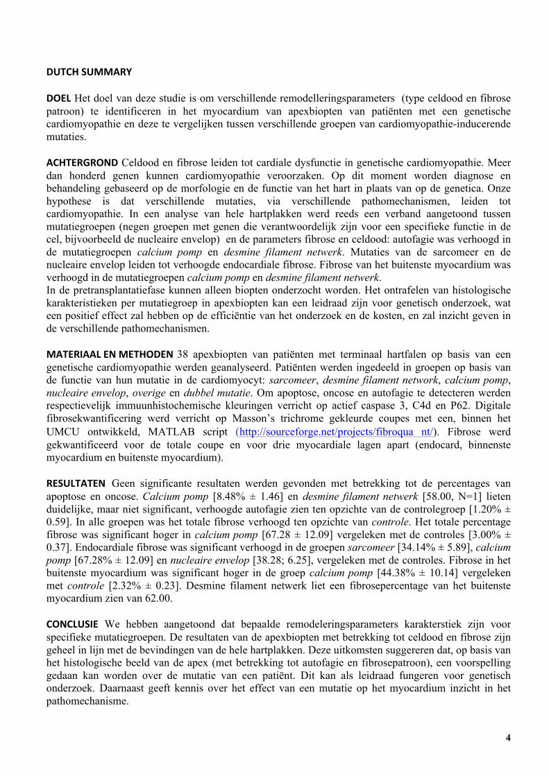
DUTCH'SUMMARY' DOEL Het doel van deze studie is om verschillende remodelleringsparameters (type celdood en fibrose patroon) te identificeren in het myocardium van apexbiopten van patiënten met een genetische cardiomyopathie en deze te vergelijken tussen verschillende groepen van cardiomyopathie-inducerende mutaties. ACHTERGROND Celdood en fibrose leiden tot cardiale dysfunctie in genetische cardiomyopathie. Meer dan honderd genen kunnen cardiomyopathie veroorzaken. Op dit moment worden diagnose en behandeling gebaseerd op de morfologie en de functie van het hart in plaats van op de genetica. Onze hypothese is dat verschillende mutaties, via verschillende pathomechanismen, leiden tot cardiomyopathie. In een analyse van hele hartplakken werd reeds een verband aangetoond tussen mutatiegroepen (negen groepen met genen die verantwoordelijk zijn voor een specifieke functie in de cel, bijvoorbeeld de nucleaire envelop) en de parameters fibrose en celdood: autofagie was verhoogd in de mutatiegroepen calcium pomp en desmine filament netwerk. Mutaties van de sarcomeer en de nucleaire envelop leiden tot verhoogde endocardiale fibrose. Fibrose van het buitenste myocardium was verhoogd in de mutatiegroepen calcium pomp en desmine filament netwerk. In de pretransplantatiefase kunnen alleen biopten onderzocht worden. Het ontrafelen van histologische karakteristieken per mutatiegroep in apexbiopten kan een leidraad zijn voor genetisch onderzoek, wat een positief effect zal hebben op de efficiëntie van het onderzoek en de kosten, en zal inzicht geven in de verschillende pathomechanismen. MATERIAAL'EN'METHODEN 38 apexbiopten van patiënten met terminaal hartfalen op basis van een genetische cardiomyopathie werden geanalyseerd. Patiënten werden ingedeeld in groepen op basis van de functie van hun mutatie in de cardiomyocyt: sarcomeer, desmine filament network, calcium pomp, nucleaire envelop, overige en dubbel mutatie. Om apoptose, oncose en autofagie te detecteren werden respectievelijk immuunhistochemische kleuringen verricht op actief caspase 3, C4d en P62. Digitale fibrosekwantificering werd verricht op Masson’s trichrome gekleurde coupes met een, binnen het UMCU ontwikkeld, MATLAB script (http://sourceforge.net/projects/fibroqua nt/). Fibrose werd gekwantificeerd voor de totale coupe en voor drie myocardiale lagen apart (endocard, binnenste myocardium en buitenste myocardium). RESULTATEN Geen significante resultaten werden gevonden met betrekking tot de percentages van apoptose en oncose. Calcium pomp [8.48% ± 1.46] en desmine filament netwerk [58.00, N=1] lieten duidelijke, maar niet significant, verhoogde autofagie zien ten opzichte van de controlegroep [1.20% ± 0.59]. In alle groepen was het totale fibrose verhoogd ten opzichte van controle. Het totale percentage fibrose was significant hoger in calcium pomp [67.28 ± 12.09] vergeleken met de controles [3.00% ± 0.37]. Endocardiale fibrose was significant verhoogd in de groepen sarcomeer [34.14% ± 5.89], calcium pomp [67.28% ± 12.09] en nucleaire envelop [38.28; 6.25], vergeleken met de controles. Fibrose in het buitenste myocardium was significant hoger in de groep calcium pomp [44.38% ± 10.14] vergeleken met controle [2.32% ± 0.23]. Desmine filament netwerk liet een fibrosepercentage van het buitenste myocardium zien van 62.00. CONCLUSIE We hebben aangetoond dat bepaalde remodeleringsparameters karakterstiek zijn voor specifieke mutatiegroepen. De resultaten van de apexbiopten met betrekking tot celdood en fibrose zijn geheel in lijn met de bevindingen van de hele hartplakken. Deze uitkomsten suggereren dat, op basis van het histologische beeld van de apex (met betrekking tot autofagie en fibrosepatroon), een voorspelling gedaan kan worden over de mutatie van een patiënt. Dit kan als leidraad fungeren voor genetisch onderzoek. Daarnaast geeft kennis over het effect van een mutatie op het myocardium inzicht in het pathomechanisme.
5
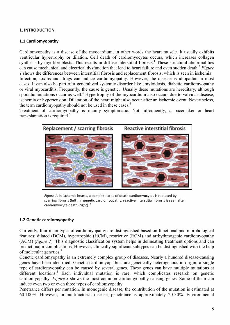
1.'INTRODUCTION' 1.1'Cardiomyopathy' Cardiomyopathy is a disease of the myocardium, in other words the heart muscle. It usually exhibits ventricular hypertrophy or dilation. Cell death of cardiomyocytes occurs, which increases collagen synthesis by myofibroblasts. This results in diffuse interstitial fibrosis.1 These structural abnormalities can cause mechanical and electrical dysfunction that lead to heart failure and even sudden death.2 Figure 1 shows the differences between interstitial fibrosis and replacement fibrosis, which is seen in ischemia. Infection, toxins and drugs can induce cardiomyopathy. However, the disease is idiopathic in most cases. It can also be part of a generalized systemic disorder like amyloidosis, diabetic cardiomyopathy or viral myocarditis. Frequently, the cause is genetic. Usually these mutations are hereditary, although sporadic mutations occur as well.3 Hypertrophy of the myocardium also occurs due to valvular disease, ischemia or hypertension. Dilatation of the heart might also occur after an ischemic event. Nevertheless, the term cardiomyopathy should not be used in these cases.4 Treatment of cardiomyopathy is mainly symptomatic. Not infrequently, a pacemaker or heart transplantation is required.5
1.2'Genetic'cardiomyopathy' Currently, four main types of cardiomyopathy are distinguished based on functional and morphological features: dilated (DCM), hypertrophic (HCM), restrictive (RCM) and arrhythmogenic cardiomyopathy (ACM) (figure 2). This diagnostic classification system helps in delineating treatment options and can predict major complications. However, clinically significant subtypes can be distinguished with the help of molecular genetics.7 Genetic cardiomyopathy is an extremely complex group of diseases. Nearly a hundred disease-causing genes have been identified. Genetic cardiomyopathies are genetically heterogenous in origin; a single type of cardiomyopathy can be caused by several genes. These genes can have multiple mutations at different locations.1 Each individual mutation is rare, which complicates research on genetic cardiomyopathy. Figure 3 shows the most common cardiomyopathy causing genes. Some of them can induce even two or even three types of cardiomyopathy. Penetrance differs per mutation. In monogenic disease, the contribution of the mutation is estimated at 60-100%. However, in multifactorial disease, penetrance is approximately 20-30%. Environmental
Figure'1.'In&ischemic&hearts,&a&complete&area&of&death&cardiomyocytes&is&replaced&by&scarring&fibrosis&(left).&In&genetic&cardiomyopathy,&reactive&interstitial&fibrosis&is&seen&after&cardiomyocyte&death&(right).&6&
6
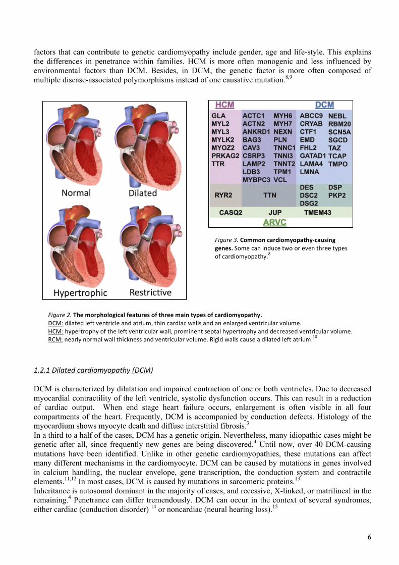
factors that can contribute to genetic cardiomyopathy include gender, age and life-style. This explains the differences in penetrance within families. HCM is more often monogenic and less influenced by environmental factors than DCM. Besides, in DCM, the genetic factor is more often composed of multiple disease-associated polymorphisms instead of one causative mutation.8,9
1.2.1'Dilated'cardiomyopathy'(DCM)'
DCM is characterized by dilatation and impaired contraction of one or both ventricles. Due to decreased myocardial contractility of the left ventricle, systolic dysfunction occurs. This can result in a reduction of cardiac output. When end stage heart failure occurs, enlargement is often visible in all four compartments of the heart. Frequently, DCM is accompanied by conduction defects. Histology of the myocardium shows myocyte death and diffuse interstitial fibrosis.3
In a third to a half of the cases, DCM has a genetic origin. Nevertheless, many idiopathic cases might be genetic after all, since frequently new genes are being discovered.4 Until now, over 40 DCM-causing mutations have been identified. Unlike in other genetic cardiomyopathies, these mutations can affect many different mechanisms in the cardiomyocyte. DCM can be caused by mutations in genes involved in calcium handling, the nuclear envelope, gene transcription, the conduction system and contractile elements.11,12 In most cases, DCM is caused by mutations in sarcomeric proteins.13
Inheritance is autosomal dominant in the majority of cases, and recessive, X-linked, or matrilineal in the remaining.4 Penetrance can differ tremendously. DCM can occur in the context of several syndromes, either cardiac (conduction disorder) 14 or noncardiac (neural hearing loss).15
Figure'2.'The'morphological'features'of'three'main'types'of'cardiomyopathy.&&DCM:&dilated&left&ventricle&and&atrium,&thin&cardiac&walls&and&an&enlarged&ventricular&volume.&HCM:&hypertrophy&of&the&left&ventricular&wall,&prominent&septal&hypertrophy&and&decreased&ventricular&volume.&RCM:&nearly&normal&wall&thickness&and&ventricular&volume.&Rigid&walls&cause&a&dilated&left&atrium.10&
Figure'3.'Common'cardiomyopathy4causing'genes.&Some&can&induce&two&or&even&three&types&of&cardiomyopathy.8&
7
1.2.2'Hypertrophic'Cardiomyopathy'(HCM)'
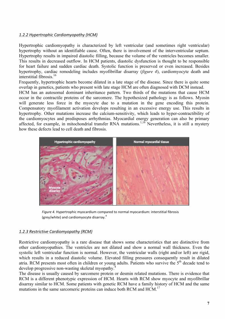
Hypertrophic cardiomyopathy is characterized by left ventricular (and sometimes right ventricular) hypertrophy without an identifiable cause. Often, there is involvement of the interventricular septum. Hypertrophy results in impaired diastolic filling, because the volume of the ventricles becomes smaller. This results in decreased outflow. In HCM patients, diastolic dysfunction is thought to be responsible for heart failure and sudden cardiac death. Systolic function is preserved or even increased. Besides hypertrophy, cardiac remodeling includes myofibrillar disarray (figure 4), cardiomyocyte death and interstitial fibrosis.16
Frequently, hypertrophic hearts become dilated in a late stage of the disease. Since there is quite some overlap in genetics, patients who present with late stage HCM are often diagnosed with DCM instead. HCM has an autosomal dominant inheritance pattern. Two thirds of the mutations that cause HCM occur in the contractile proteins of the sarcomere. The hypothesized pathology is as follows. Myosin will generate less force in the myocyte due to a mutation in the gene encoding this protein. Compensatory myofilament activation develops resulting in an excessive energy use. This results in hypertrophy. Other mutations increase the calcium-sensitivity, which leads to hyper-contractibility of the cardiomyocytes and predisposes arrhythmias. Myocardial energy generation can also be primary affected, for example, in mitochondrial transfer RNA mutations.1,16 Nevertheless, it is still a mystery how these defects lead to cell death and fibrosis.
1.2.3'Restrictive'Cardiomyopathy'(RCM)'
Restrictive cardiomyopathy is a rare disease that shows some characteristics that are distinctive from other cardiomyopathies. The ventricles are not dilated and show a normal wall thickness. Even the systolic left ventricular function is normal. However, the ventricular walls (right and/or left) are rigid, which results in a reduced diastolic volume. Elevated filling pressures consequently result in dilated atria. RCM presents most often in children or young adults. Patients who survive the 5th decade tend to develop progressive non-wasting skeletal myopathy.4
The disease is usually caused by sarcomere protein or desmin related mutations. There is evidence that RCM is a different phenotypic expression of HCM. Hearts with RCM show myocyte and myofibrillar disarray similar to HCM. Some patients with genetic RCM have a family history of HCM and the same mutations in the same sarcomeric proteins can induce both RCM and HCM.17
Figure'4.&Hypertrophic&myocardium&compared&to&normal&myocardium:&interstitial&fibrosis&
(grey/white)&and&cardiomyocyte&disarray.4'
8
1.2.4'Arrhythmogenic'Cardiomyopathy'(ACM)'
Arrhythmogenic cardiomyopathy is characterized by ventricular arrhythmias. There is myocyte loss followed by inflammation and fibrofatty replacement in the myocardium, often of the right ventricle, but also of the left ventricle. These signs of remodeling induce motion abnormalities that later develop into right ventricular dilatation.18 The prevalence is estimated between 1:1000 and 1:2000. However, because it is an under recognized clinical entity, the disease is rarely diagnosed. It often stays asymptomatic (reduced penetrance) and has a tendency to become apparent during heavy exercise.19 It is an important cause of sudden death (11% of all cases), especially in athletes and young adults.20 Most of the time, genes encoding desmosomal proteins are affected. There are two inheritance patterns of ACM: autosomal dominant and autosomal recessive. In the latter, ACM is part of a cardiocutaneous syndrome. Affected patients also exhibit hyperkeratosis of the palms and soles and woolly hair.21
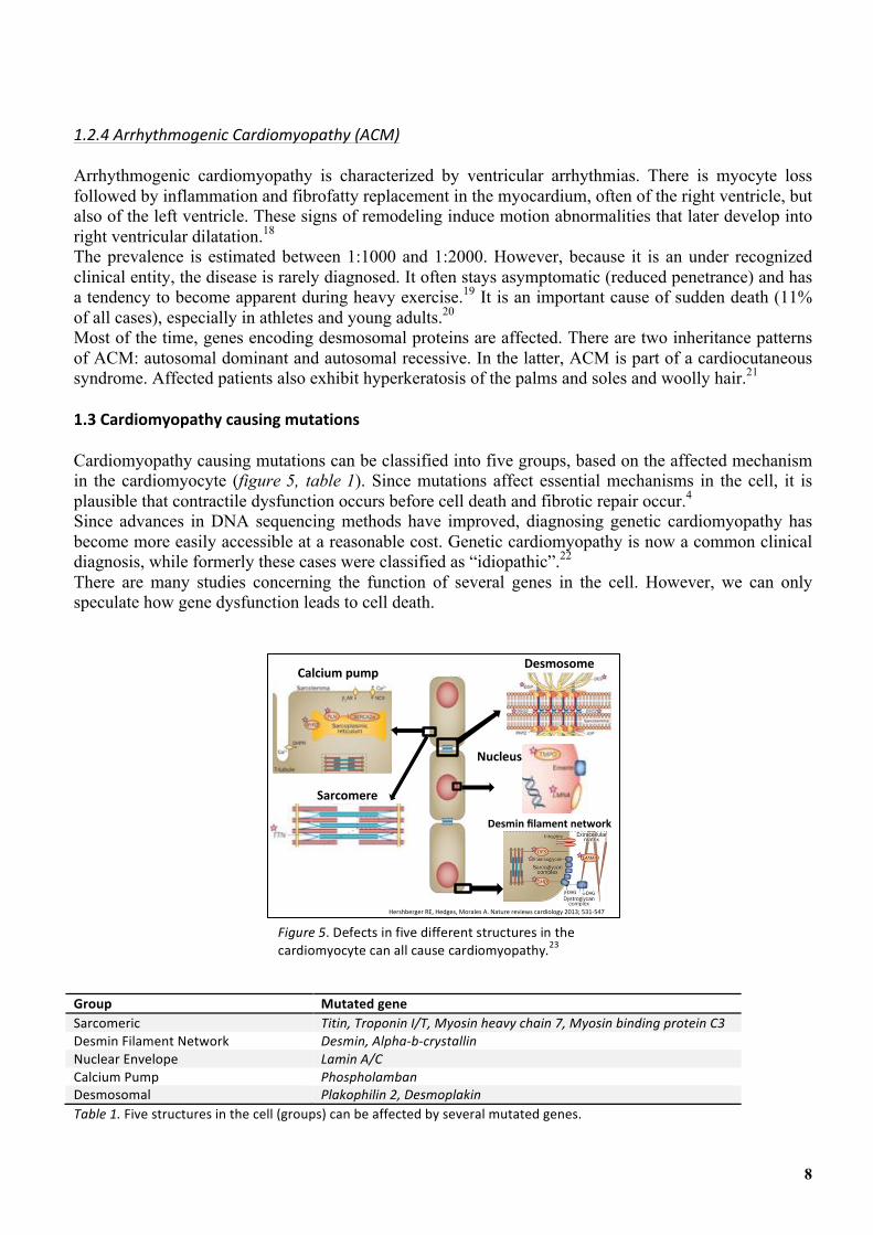
1.3'Cardiomyopathy'causing'mutations' Cardiomyopathy causing mutations can be classified into five groups, based on the affected mechanism in the cardiomyocyte (figure 5, table 1). Since mutations affect essential mechanisms in the cell, it is plausible that contractile dysfunction occurs before cell death and fibrotic repair occur.4
Since advances in DNA sequencing methods have improved, diagnosing genetic cardiomyopathy has become more easily accessible at a reasonable cost. Genetic cardiomyopathy is now a common clinical diagnosis, while formerly these cases were classified as “idiopathic”.22 There are many studies concerning the function of several genes in the cell. However, we can only speculate how gene dysfunction leads to cell death.
Group' Mutated'gene'Sarcomeric& Titin,'Troponin'I/T,'Myosin'heavy'chain'7,'Myosin'binding'protein'C3&Desmin&Filament&Network& Desmin,'AlphaHbHcrystallin&Nuclear&Envelope& Lamin'A/C&Calcium&Pump& Phospholamban&Desmosomal& Plakophilin'2,'Desmoplakin&Table'1.&Five&structures&in&the&cell&(groups)&can&be&affected&by&several&mutated&genes.&&
Hershberger(RE,(Hedges,(Morales(A.(Nature(reviews(cardiology(2013;(531A547(
Sarcomere(
Calcium(pump(Desmosome(
Nucleus(
Desmin(filament(network(
Figure'5.&Defects&in&five&different&structures&in&the&cardiomyocyte&can&all&cause&cardiomyopathy.23&

9
1.3.1'Mutations'of'the'sarcomere&
The heart muscle is composed of repeating sections of sarcomeres. Sarcomeres consist of long fibrous proteins that slide past each other during contraction and relaxation. The two main proteins are actin and myosin, forming the thin and the thick filament respectively. Contraction occurs when both filaments bind to each other after actin has bound to calcium ions and myosin has bound to ATP. The sarcomeric protein titin prevents overstretching.11
DCM, RCM and especially HCM can all be induced by mutations in sarcomeric proteins. A mutation in titin is responsible for 25% of the genetic DCM cases.11
Over 400 mutations have been found in genes encoding proteins that constitute the sarcomere of the cardiac muscle.1,8
1.3.2'Mutations'of'the'calcium'pump'
SERCA is a Ca2+ ATPase that controls the uptake of calcium from the cytoplasm into the sarcoplasmic reticulum (SR) during relaxation. Phospholamban (PLN) is responsible for the activity of SERCA; usually PLN inhibits SERCA. PLN mutation can give rise to a calcium imbalance; electrical instability arises, the contraction intervals become shorter and contractility goes up. SR stress can result in misfolding, accumulation of proteins and apoptosis.24 The p.Arg14del mutation in the gene encoding PLN is frequently seen in patients with genetic DCM.25 Recently, it has also been associated with ACM.23
1.3.3'Mutations'of'the'nuclear'envelope'
The nuclear envelope is a double membrane that surrounds the genetic material of the eukaryotic cells. It consists of an inner and outer membrane. Nuclear pores make exchange of cellular products possible. The nuclear lamina covers the inner membrane.24 The nuclear lamina determines size and shape of the nucleus, contributes to its stability and functions as a permeable barrier. Furthermore, it is involved in DNA replication, transcription and chromatin organization.26 Lamins, intermediate filament proteins, are major structural determinants of the lamina. The LMNA gene encodes lamin A and lamin C.27
Cardiomyopathy due to malfunction of the nuclear envelope is most often caused by mutations in lamin A/C (LMNA/C). Lamin A is expressed in all differentiated cells. LMNA/C mutations are associated with more than ten clinical entities, collectively called laminopathies. It can cause skeletal muscle dystrophy and accelerated aging (progeria). However, dilated cardiomyopathy with conduction defects is the most prevalent expression of laminopathy.28
LMNA mutations are the second most common cause of familial dilated cardiomyopathy (up to 11% of cases).29 LMNA cardiomyopathy has an aggressive clinical course compared to other cardiomyopathies and the rates of deadly arrhythmias and heart failure are high.30 Penetrance of disease causing LMNA mutations is high: evidence shows that 100% of mutation carriers are affected by the age of 60.31
1.3.4'Mutations'of'the'desmin'filament'network'
Desmin is a critical protein of the cytoskeleton. The cytoskeleton plays an essential role in the cellular structure, cytoplasmic organization, proliferation, and intra- and intercellular signaling. An intact and dynamic cytoskeleton is especially important in muscle cells. Signaling through the sarcolemma and the contractile apparatus rely on it. In desmin related cardiomyopathy, the cytoskeleton is disrupted. Symptoms are slowly progressive muscle weakness, conduction-system disease, cardiac arrhythmias and DCM.32
10
It is caused by a missense mutation in αB-crystallin (CryAB) or, less common, in genes encoding desmin itself. CryAB is a chaperone of desmin; it is responsible for folding and stabilizing the protein.33 Characteristic is protein misfolding and protein aggregates that lead to increased proteotoxic stress. Proteotoxicity will be discussed later. 1.3.5'Mutations'of'the'desmosome'
For proper functioning of the heart, electrical and mechanical activity needs to be synchronized. The desmosome protein is specialized in cell-to-cell adhesion. While gap junctions provide electrical continuity, desmosomes are responsible for the mechanical continuity.34 Mutated desmosomal proteins, mainly plakophilin 2 and desmoplakin, can lead to ACM and various cutaneous diseases. Desmosome impairment causes detachment of cardiomyocytes. Afterwards, inflammation and cell death occur followed by fibrofatty replacement.35
It is hypothesized that the right ventricle is more vulnerable to impaired cell adhesion, because of its thin walls. Desmosomes reside in the intercalated disks between the cardiomyocytes, where they interact with the gap junctions. Disruption of desmosomal integrity can therefore alter electric stability as seen in ACM.34
1.4'Cell'death'of'the'cardiomyocyte' As mentioned above, it is thought that the death of cardiomyocytes and subsequent reactive fibrosis induce dysfunction of the myocardium in genetic cardiomyopathy. Kostin et al. showed that myocytes die by multiple mechanisms in idiopathic cardiomyopathy: apoptosis, oncosis and autophagic cell death.36 Vigliano et al. concluded that oncosis and autophagy are independent predictors of death in patients with idiopathic cardiomyopathy.37 To what extent the types of cell death contribute to myocardial dysfunction in genetic cardiomyopathy is still unknown. 1.4.1'Apoptosis'
Apoptosis or programmed cell death is considered an essential mechanism in a vital human body. It plays an important role in processes like normal cell turnover, proper development and functioning of the immune system and embryonic development.38
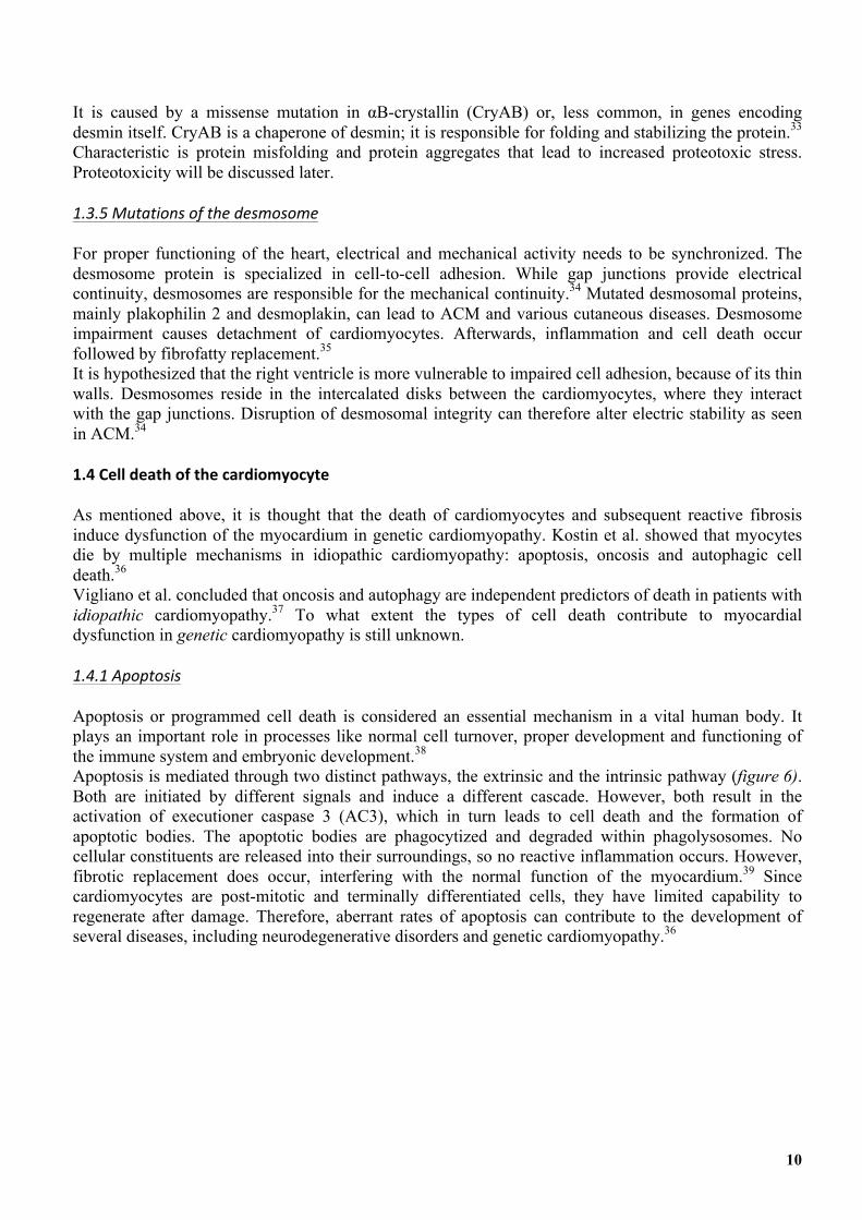
Apoptosis is mediated through two distinct pathways, the extrinsic and the intrinsic pathway (figure 6). Both are initiated by different signals and induce a different cascade. However, both result in the activation of executioner caspase 3 (AC3), which in turn leads to cell death and the formation of apoptotic bodies. The apoptotic bodies are phagocytized and degraded within phagolysosomes. No cellular constituents are released into their surroundings, so no reactive inflammation occurs. However, fibrotic replacement does occur, interfering with the normal function of the myocardium.39 Since cardiomyocytes are post-mitotic and terminally differentiated cells, they have limited capability to regenerate after damage. Therefore, aberrant rates of apoptosis can contribute to the development of several diseases, including neurodegenerative disorders and genetic cardiomyopathy.36
11
1.4.2'Oncosis'
Where apoptosis manifests itself by shrinkage and by breaking up in apoptotic bodies, oncosis seems to do the opposite. Oncosis is characterized by swelling, coagulation of the cytoplasm, blebbing (blistering of the membrane) and increased plasma membrane permeability. Typically, it is caused by ischemia. This results in failure of the ionic pumps in the plasma membrane and increased permeability. Environmental factors as mild heat or toxic agents can induce apoptosis, but in higher doses, cell death by oncosis will occur.41,42 Oncosis leads to inflammation and fibrotic repair. Elevated oncosis rates are found in genetic cardiomyopathy. Extensive fibrosis contributes to cardiac dysfunction and heart failure.36
Oncosis is often confused with the term necrosis. Necrosis is the degradation process following cell death. Signs of necrosis are usually visible after 12 to 24 hours.39
1.4.3'Autophagy'and'UbiquitinHProteasome'System'
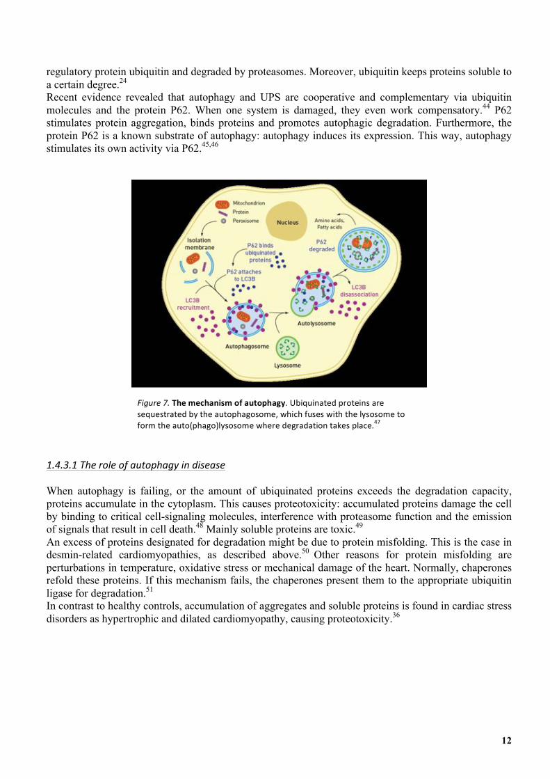
The third mechanism that promotes cell death in genetic cardiomyopathy is autophagy- or UPS-induced cell death. Two pathways are responsible for the degradation of most proteins in eukaryotic cells: autophagy and the ubiquitin-proteasome system (UPS). They form the protein quality control system of the cell. Disruption of these processes can contribute to a variety of diseases through accumulation of proteins or proteotoxicity.43 Autophagy, or macroautophagy, is the mechanism of the cell that is mainly responsible for de degradation of damaged organelles and protein aggregates. In all human cells a low rate of autophagy is present. In stressful situations, for example in starvation, autophagy is upregulated in order to maintain the cells energy level. Unnecessary or damaged cellular constituents are bound to the protein P62 and sequestrated into double membrane vacuoles: the autophagosomes. The autophagosome then fuses with a lysosome to form an autophagolysosome where degradation takes place (figure 7).36 UPS is more specific than autophagy. It is responsible for 80-90% of the turnover of cellular proteins, for example the misfolded and damaged ones. Proteins designated for degradation are labeled by the
Figure'6.&The'mechanism'of'apoptosis.'Both&intrinsic&and&extrinsic&pathways&lead&to&the&activation&of&effector&caspase&3.40&
12
regulatory protein ubiquitin and degraded by proteasomes. Moreover, ubiquitin keeps proteins soluble to a certain degree.24
Recent evidence revealed that autophagy and UPS are cooperative and complementary via ubiquitin molecules and the protein P62. When one system is damaged, they even work compensatory.44 P62 stimulates protein aggregation, binds proteins and promotes autophagic degradation. Furthermore, the protein P62 is a known substrate of autophagy: autophagy induces its expression. This way, autophagy stimulates its own activity via P62.45,46
1.4.3.1'The'role'of'autophagy'in'disease'
When autophagy is failing, or the amount of ubiquinated proteins exceeds the degradation capacity, proteins accumulate in the cytoplasm. This causes proteotoxicity: accumulated proteins damage the cell by binding to critical cell-signaling molecules, interference with proteasome function and the emission of signals that result in cell death.48 Mainly soluble proteins are toxic.49 An excess of proteins designated for degradation might be due to protein misfolding. This is the case in desmin-related cardiomyopathies, as described above.50 Other reasons for protein misfolding are perturbations in temperature, oxidative stress or mechanical damage of the heart. Normally, chaperones refold these proteins. If this mechanism fails, the chaperones present them to the appropriate ubiquitin ligase for degradation.51
In contrast to healthy controls, accumulation of aggregates and soluble proteins is found in cardiac stress disorders as hypertrophic and dilated cardiomyopathy, causing proteotoxicity.36 '''''
Figure'7.&The'mechanism'of'autophagy.&Ubiquinated&proteins&are&sequestrated&by&the&autophagosome,&which&fuses&with&the&lysosome&to&form&the&auto(phago)lysosome&where°radation&takes&place.47&

13
1.5'Introduction'research' 1.5.1'What'is'known?'
As previously described, in genetic cardiomyopathy, heart failure is mainly induced by cell death of cardiomyocytes followed by interstitial fibrosis.36 Genetic cardiomyopathy can be caused by over a hundred mutations. It is unclear what the differences are in the pathomechanisms of these mutations. From experience, we realized that certain mutations have specific histologic characteristics. Objectifying these characteristics provides insight in the disease and the pathomechanisms of the different mutations. Understanding of the pathomechanism will support new therapeutic modalities. The Heart Transplantation Research Group of the University Medical Center Utrecht performed several studies on this subject. Klooster et al. studied types of cell death in complete mid-ventricular heart slices. They concluded that P62 (related to failing autophagy) was upregulated in the functional mutation groups calcium pump, desmosomal and desmin filament network. Nuclear envelope and sarcomeric mutations showed relatively high rates of apoptosis. No relation between oncosis and mutations groups was found. Sepehrkhouy et al. studied the amount and the distribution of fibrosis in the myocardium in patients with genetic cardiomyopathy and compared these between mutation groups. Again, complete heart slices were studied. The myocardium was divided in three layers: endocardium, inner myocardium and outer myocardium. They showed that the distribution of fibrosis is related to the type of mutation. Mutations in the sarcomeric and nuclear envelope proteins especially induced inner myocardial fibrosis. The groups desmosome, calcium pump and desmin filament network, however, showed fibrosis in the outer myocardium, or epicardium. The two studies described above are just finished and not published yet. 1.5.2'Research'question'
As described above a relationship was found between disease causing mutation groups and the remodeling parameters type of cell death and fibrosis pattern. In these studies complete heart slices were analyzed. However, in the pretransplantation phase, only biopsies of apexes are available. If the same histopathological characteristics can be derived from biopsies this would allow for interesting new applications. For example, pathologists would be able to provide guidance to genetic research by predicting the patients mutation based on histopathological characteristics. This will allow for savings in both cost and time by narrowing the search for specific mutations. In idiopathic cardiomyopathy, a direct relationship between remodeling parameters and survival time was observed.37 This relationship is plausible in genetic cardiomyopathy as well, and requires further investigation. Currently, late gadolinium enhancement on cardiac magnetic resonance imaging is the noninvasive standard for detecting myocardial fibrosis.52 However, no studies have compared this technique with the histological assessment of myocardial apex biopsies. While the small amount of tissue acquired through these procedures provides only regional information, a distinction between the myocardial layers and the type of fibrosis can be made with adequate confidence. The aim of this study is to objectify remodeling parameters in apex biopsies of patients with genetic cardiomyopathy and compare the results between the causative mutation groups. As previously has been done for complete heart slices, we will study types of cell death (apoptosis, oncosis and autophagy) and fibrosis pattern (the amount of fibrosis and the location of fibrosis in the myocardium). We will compare our results with the results of the complete heart slices to see if the apex biopsy is representative for the whole heart. If this is the case, a prediction of a patients’ mutation can be made based on a biopsy. This will provide guidance in genetic testing, which will safe costs and time. Furthermore, objectifying the effect of a mutation on the myocardium will provide insight in the
14
pathomechanism. This knowledge is essential in order to develop targeted drugs in the future. To our best knowledge, this has never been investigated before. 2.'MATERIALS'AND'METHODS' 2.1'Ethical'approval'
The study met the criteria of the code of proper use of human tissue that is used in the Netherlands. The study was approved by the scientific advisory board of the biobank of the University Medical Center Utrecht, Utrecht, the Netherlands (protocol no. 12/387). 2.2'Data'collection'
Apex tissue of 38 patients with end stage genetic cardiomyopathy was collected between 1997 and 2015. In all cases, tissue was obtained during a Left Ventricular Assist Device (LVAD) procedure as a bridge to heart transplantation. In 3 patients, cardiomyopathy was classified as hypertrophic (HCM), in the rest of the cases as dilated (DCM). When the clinical diagnosis changed during the clinical course, the initial diagnosis was used. Five non-cardiomyopathy hearts served as controls; three of these were rejected for transplantation, one heart was rejected shortly after transplantation and one heart was obtained during autopsy of a patient with a non-cardiac cause of death. Unfortunately, no apexes of these hearts were available, only mid-ventricular slices. However, we assume that healthy mid-ventricular myocardium is equal to healthy apex myocardium. The average data of one slice of the anterior left ventricular wall, one of the posterior left ventricular wall and one of the ventricular septum was used. Unfortunately, stained slides were sometimes lost or the quality had decreased with time. The in paraffin embedded tissue was at times finished, or untraceable. Therefore, analyzes could not be performed in all cases. For this reason, the group sizes can differ per analysis and are mentioned in the graphs. 2.3'Inclusion'and'exclusion'criteria'
To be eligible for the study, patients had to meet the following criteria: cardiomyopathy had to be clinically diagnosed and the causative mutated gene had to be known. Patients were excluded in case of other causes of heart muscle disease than genetic, for example post-viral or after myocardial infarction. 2.4'Cell'death'determination'
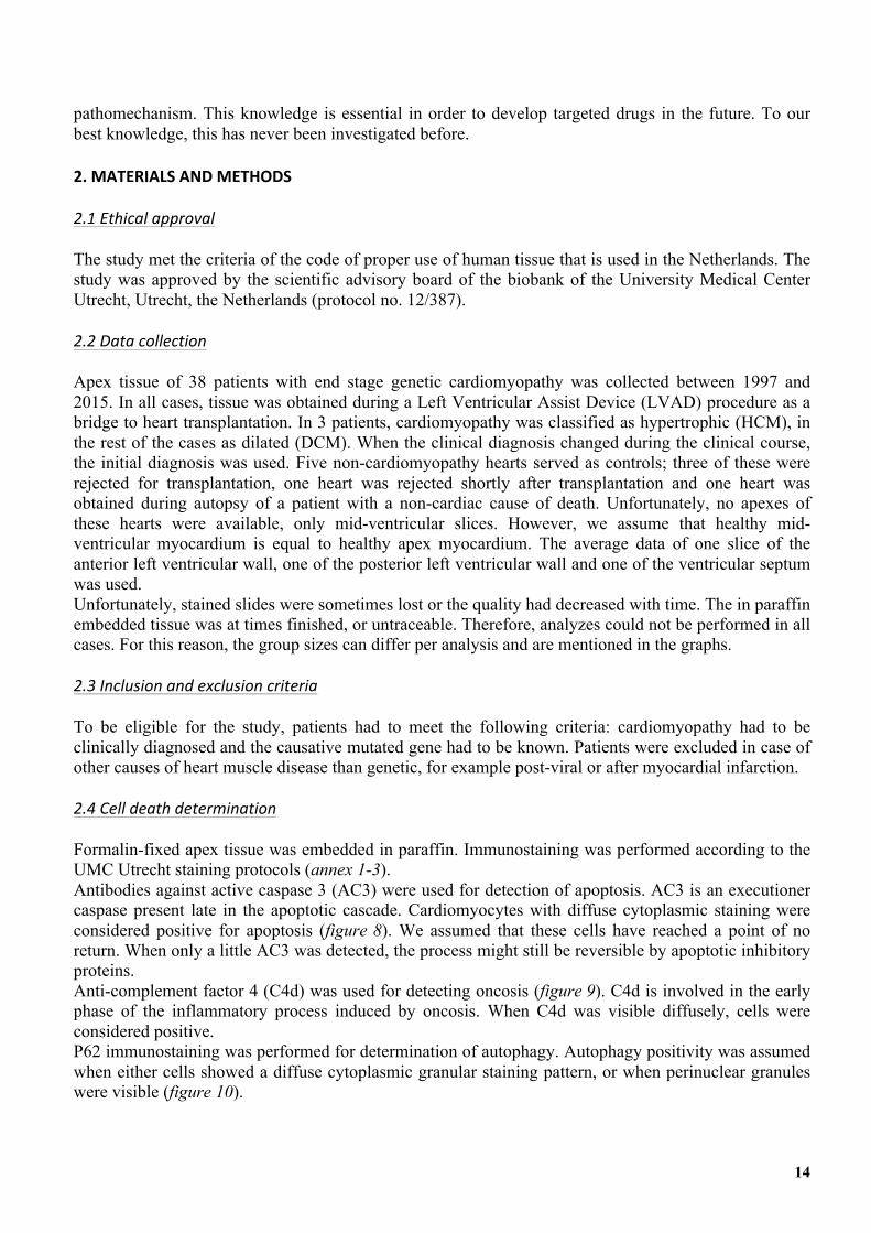
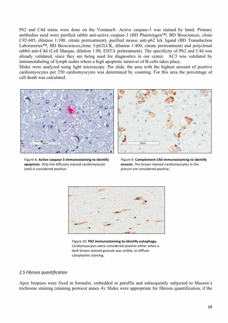
Formalin-fixed apex tissue was embedded in paraffin. Immunostaining was performed according to the UMC Utrecht staining protocols (annex 1-3). Antibodies against active caspase 3 (AC3) were used for detection of apoptosis. AC3 is an executioner caspase present late in the apoptotic cascade. Cardiomyocytes with diffuse cytoplasmic staining were considered positive for apoptosis (figure 8). We assumed that these cells have reached a point of no return. When only a little AC3 was detected, the process might still be reversible by apoptotic inhibitory proteins. Anti-complement factor 4 (C4d) was used for detecting oncosis (figure 9). C4d is involved in the early phase of the inflammatory process induced by oncosis. When C4d was visible diffusely, cells were considered positive. P62 immunostaining was performed for determination of autophagy. Autophagy positivity was assumed when either cells showed a diffuse cytoplasmic granular staining pattern, or when perinuclear granules were visible (figure 10).
15
P62 and C4d stains were done on the Ventana®. Active caspase-3 was stained by hand. Primary antibodies used were purified rabbit anti-active caspase-3 (BD Pharmingen™, BD Biosciences, clone C92-605, dilution 1:100, citrate pretreatment), purified mouse anti-p62 Ick ligand (BD Transduction Laboratories™, BD Biosciences,clone 3/p62LCK, dilution 1:400, citrate pretreatment) and polyclonal rabbit anti-C4d (Cell Marque, dilution 1:80, EDTA pretreatment). The specificity of P62 and C4d was already validated, since they are being used for diagnostics in our center. AC3 was validated by immunolabeling of lymph nodes where a high apoptotic turnover of B-cells takes place. Slides were analyzed using light microscopy. Per slide, the area with the highest amount of positive cardiomyocytes per 250 cardiomyocytes was determined by counting. For this area the percentage of cell death was calculated.
2.5'Fibrosis'quantification'
Apex biopsies were fixed in formalin, embedded in paraffin and subsequently subjected to Masson’s trichrome staining (staining protocol annex 4). Slides were appropriate for fibrosis quantification, if the
Figure'8.&Active'caspase'3'immunostaining'to'identify'apoptosis.&Only&the&diffusely&stained&cardiomyocyte&(red)&is&considered&positive.&
Figure'9.&Complement'C4d'immunostaining'to'identify'oncosis.'The&brown&stained&cardiomyocytes&in&the&picture&are&considered&positive.&
Figure'10.&P62'immunostaining'to'identify'autophagy.&Cardiomyocytes&were&considered&positive&either&when&a&dark&brown&stained&granule&was&visible,&or&diffuse&cytoplasmic&staining.&

16
epicardial, myocardial and endocardial region were all present (figure 11, left). The slides were scanned at 20x magnification following the procedure described by Huisman et al..53 Images were extracted using Aperio ImageScope v12.0.0.5039 (Aperio, Vista, California, United States) as a TIFF file with lossless compression. The images were resized to 10% of their original size. For digitally analyzing the slides, an in-house developed open source MATLAB script was used.1,54 Slides were divided in four areas: epicardial, inner and outer compact myocardial and endocardial (or non-compact myocardial) (figure 12, right). The epicardium consists of the fatty layer bordered by the first row of cardiomyocytes. The trabeculated region forms the endocardium. The compact myocardium is the central region and is divided in an inner and outer area. For all four areas, the percentage of fibrosis (blue), cardiomyocytes (red) and fatty tissue (green) was quantified using MATLAB (Release R2012a, The MathWorks, Inc., Natick, Massachusetts, United States). The epicardial region was excluded from analysis since it does not contain cardiomyocytes.
2.6'Statistical'analysis'
Statistical analysis was performed using GraphPad Prism version 6.00. Data was analyzed using one-way ANOVA. Holm-Sidak Post Hoc testing was performed on significant ANOVAs. Groups with N<2 were excluded from statistical analysis. However, they are included in the graphs. In the case of fibrosis data, groups were compared using a two-way ANOVA, with mutation group as one factor and cell layer as another. Data are presented as median [interquartile range, IQR] or as mean ± standard error of the mean. Alpha was set at 0.05. 3.'RESULTS' Patients were classified according to their mutation(s) in one of the following functional groups: sarcomeric, desmin filament network, nuclear envelope, calcium pump, desmosomal, other or double
Figure'11.&Overview'of' fibrosis'quantification'methodology.&Left'picture:& four&layers& of& the& heart& bordered& by& the& colored& lines:& epicardium& (on& the& right),&outer& myocardium,& inner& myocardium& and& endocardium& (at& the& left).& Right'picture:& the&same&heart&slide&after&digital&slide&processing.&Fatty&tissue&(green),&myocardium&(red)&and&fibrosis&(blue).&
17
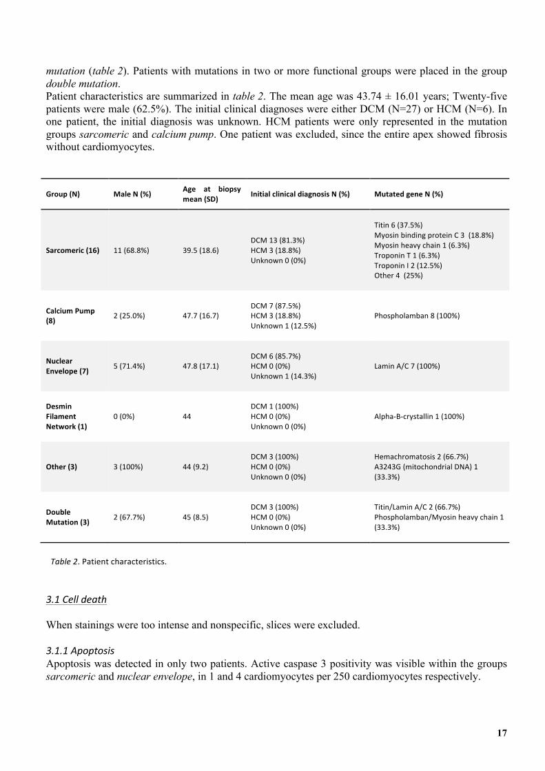
mutation (table 2). Patients with mutations in two or more functional groups were placed in the group double mutation. Patient characteristics are summarized in table 2. The mean age was 43.74 ± 16.01 years; Twenty-five patients were male (62.5%). The initial clinical diagnoses were either DCM (N=27) or HCM (N=6). In one patient, the initial diagnosis was unknown. HCM patients were only represented in the mutation groups sarcomeric and calcium pump. One patient was excluded, since the entire apex showed fibrosis without cardiomyocytes.
Group'(N)' Male'N'(%)' Age' at' biopsy'mean'(SD)' Initial'clinical'diagnosis'N'(%)' Mutated'gene'N'(%)'
Sarcomeric'(16)' 11&(68.8%)& 39.5&(18.6)&
&DCM&13&(81.3%)&HCM&3&(18.8%)&Unknown&0&(0%)&&
&Titin&6&(37.5%)&Myosin&binding&protein&C&3&&(18.8%)&Myosin&heavy&chain&1&(6.3%)&Troponin&T&1&(6.3%)&Troponin&I&2&(12.5%)&Other&4&&(25%)&&
Calcium'Pump'(8)' 2&(25.0%)& 47.7&(16.7)&
&DCM&7&(87.5%)&HCM&3&(18.8%)&Unknown&1&(12.5%)&&
Phospholamban&8&(100%)&
Nuclear'Envelope'(7)' 5&(71.4%)& 47.8&(17.1)&
&DCM&6&(85.7%)&HCM&0&(0%)&Unknown&1&(14.3%)&&
Lamin&A/C&7&(100%)&
Desmin'Filament'Network'(1)'
0&(0%)& 44&
&DCM&1&(100%)&HCM&0&(0%)&Unknown&0&(0%)&&
Alpha4B4crystallin&1&(100%)&
Other'(3)' 3&(100%)& 44&(9.2)&
&DCM&3&(100%)&HCM&0&(0%)&Unknown&0&(0%)&&
Hemachromatosis&2&(66.7%)&A3243G&(mitochondrial&DNA)&1&(33.3%)&
Double'Mutation'(3)' 2&(67.7%)& 45&(8.5)&
&DCM&3&(100%)&HCM&0&(0%)&Unknown&0&(0%)&&
Titin/Lamin&A/C&2&(66.7%)&Phospholamban/Myosin&heavy&chain&1&(33.3%)&
Table'2.&Patient&characteristics.' 3.1'Cell'death'
When stainings were too intense and nonspecific, slices were excluded. 3.1.1'Apoptosis'
Apoptosis was detected in only two patients. Active caspase 3 positivity was visible within the groups sarcomeric and nuclear envelope, in 1 and 4 cardiomyocytes per 250 cardiomyocytes respectively.
18
3.1.2'Oncosis'
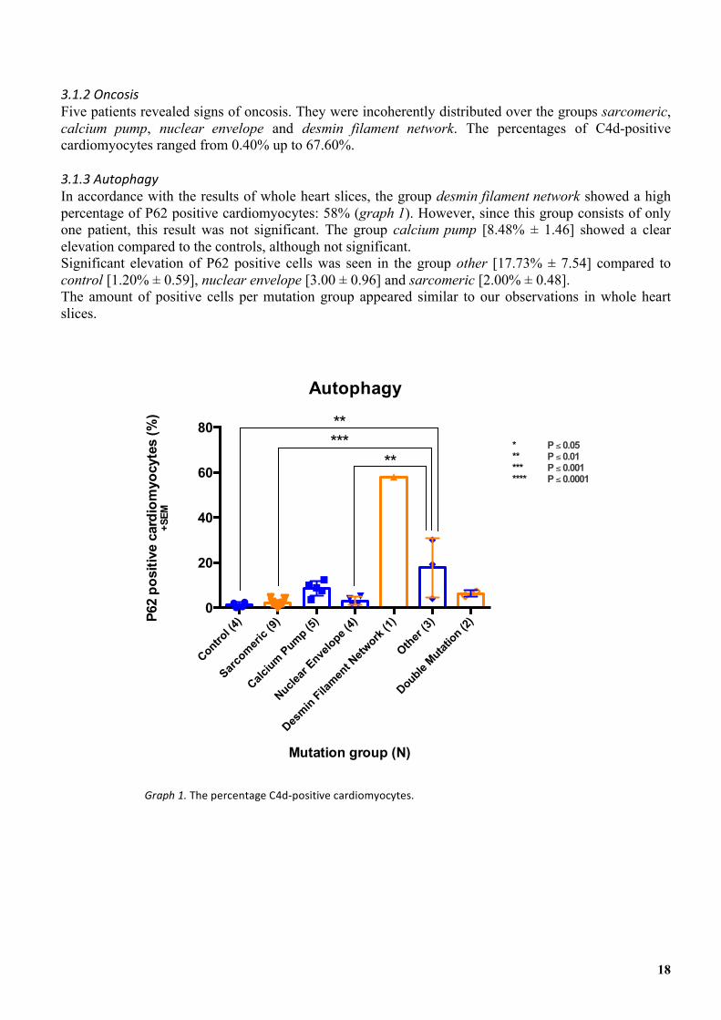
Five patients revealed signs of oncosis. They were incoherently distributed over the groups sarcomeric, calcium pump, nuclear envelope and desmin filament network. The percentages of C4d-positive cardiomyocytes ranged from 0.40% up to 67.60%. 3.1.3'Autophagy'
In accordance with the results of whole heart slices, the group desmin filament network showed a high percentage of P62 positive cardiomyocytes: 58% (graph 1). However, since this group consists of only one patient, this result was not significant. The group calcium pump [8.48% ± 1.46] showed a clear elevation compared to the controls, although not significant. Significant elevation of P62 positive cells was seen in the group other [17.73% ± 7.54] compared to control [1.20% ± 0.59], nuclear envelope [3.00 ± 0.96] and sarcomeric [2.00% ± 0.48]. The amount of positive cells per mutation group appeared similar to our observations in whole heart slices.
Control (4
)
Sarco
meric
(9)
Calcium Pump (5
)
Nuclear
Envelope (
4)
Desmin Fila
ment N
etwork
(1)
Other (3)
Double Mutat
ion (2)
0
20
40
60
80
P62
posi
tive
card
iom
yocy
tes
(%)
+SEM
Autophagy
Mutation group (N)
**
*****
* P ≤ 0.05 ** P ≤ 0.01 *** P ≤ 0.001**** P ≤ 0.0001
Graph'1.&The&percentage&C4d4positive&cardiomyocytes.&
19
3.2'Fibrosis'quantification'
3.2.1'Total'amount'of'fibrosis'
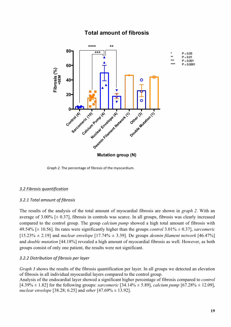
The results of the analysis of the total amount of myocardial fibrosis are shown in graph 2. With an average of 3.00% [± 0.37], fibrosis in controls was scarce. In all groups, fibrosis was clearly increased compared to the control group. The group calcium pump showed a high total amount of fibrosis with 49.54% [± 10.56]. Its rates were significantly higher than the groups control 3.01% ± 0.37], sarcomeric [15.23% ± 2.19] and nuclear envelope [17.74% ± 3.39]. De groups desmin filament network [46.47%] and double mutation [44.18%] revealed a high amount of myocardial fibrosis as well. However, as both groups consist of only one patient, the results were not significant.
3.2.2'Distribution'of'fibrosis'per'layer'
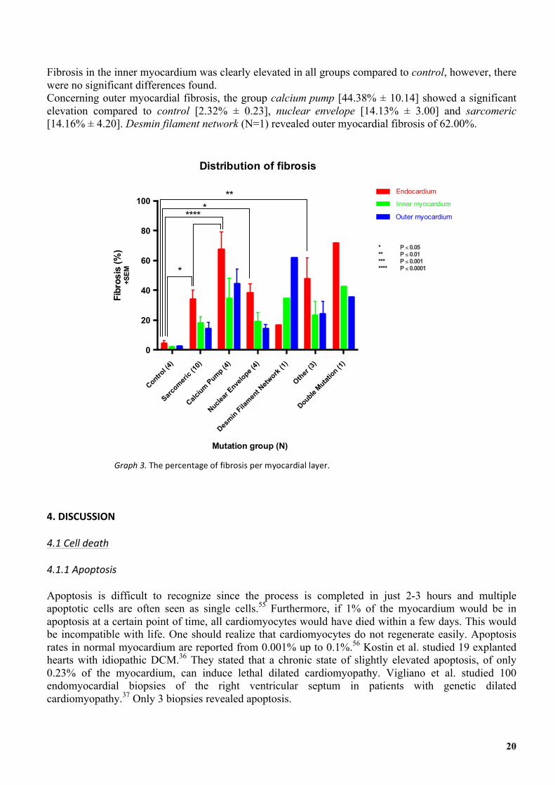
Graph 3 shows the results of the fibrosis quantification per layer. In all groups we detected an elevation of fibrosis in all individual myocardial layers compared to the control group. Analysis of the endocardial layer showed a significant higher percentage of fibrosis compared to control [4.39% ± 1.82] for the following groups: sarcomeric [34.14% ± 5.89], calcium pump [67.28% ± 12.09], nuclear envelope [38.28; 6.25] and other [47.69% ± 13.92].
Control (4
)
Sarco
meric
(10)
Calcium Pump (4
)
Nuclear
Envelope (
4)
Desmin Fila
ment N
etwork
(1)
Other (3)
Double Mutat
ion (1)
0
20
40
60
80
Mutation group (N)
Fibr
osis
(%)
+SEM
Total amount of fibrosis
******* **
* P ≤ 0.05 ** P ≤ 0.01 *** P ≤ 0.001**** P ≤ 0.0001
Graph'2.'The&percentage&of&fibrosis&of&the&myocardium.&
20
Fibrosis in the inner myocardium was clearly elevated in all groups compared to control, however, there were no significant differences found. Concerning outer myocardial fibrosis, the group calcium pump [44.38% ± 10.14] showed a significant elevation compared to control [2.32% ± 0.23], nuclear envelope [14.13% ± 3.00] and sarcomeric [14.16% ± 4.20]. Desmin filament network (N=1) revealed outer myocardial fibrosis of 62.00%.
''''4.'DISCUSSION&
4.1'Cell'death'
4.1.1'Apoptosis'
Apoptosis is difficult to recognize since the process is completed in just 2-3 hours and multiple apoptotic cells are often seen as single cells.55 Furthermore, if 1% of the myocardium would be in apoptosis at a certain point of time, all cardiomyocytes would have died within a few days. This would be incompatible with life. One should realize that cardiomyocytes do not regenerate easily. Apoptosis rates in normal myocardium are reported from 0.001% up to 0.1%.56 Kostin et al. studied 19 explanted hearts with idiopathic DCM.36 They stated that a chronic state of slightly elevated apoptosis, of only 0.23% of the myocardium, can induce lethal dilated cardiomyopathy. Vigliano et al. studied 100 endomyocardial biopsies of the right ventricular septum in patients with genetic dilated cardiomyopathy.37 Only 3 biopsies revealed apoptosis.
Control (4
)
Sarco
meric
(10)
Calcium Pump (4
)
Nuclear
Envelope (
4)
Desmin Fila
ment N
etwork
(1)
Other (3)
Double Mutat
ion (1)
0
20
40
60
80
100
Fibr
osis
(%)
+SEM
Distribution of fibrosis
Endocardium
Inner myocardium
Outer myocardium
Mutation group (N)
****
***
*
* P ≤ 0.05 ** P ≤ 0.01 *** P ≤ 0.001**** P ≤ 0.0001
Graph'3.'The&percentage&of&fibrosis&per&myocardial&layer.&
21
Besides this, active caspase 3 is only present late in the cascade, prior to the actual cell death or apoptosis. Several apoptosis inhibiting proteins are known, some of them targeting AC3.57 Therefore, apoptosis can be reversible in AC3-expressing cells. Nevertheless, we only considered myocytes AC3-postitive when the complete cell was stained. We assume that in these cells, apoptosis has reached a point of no return. Apoptosis does not seem to have a preferred site in the heart. Slices of complete hearts showed similar results as our apex biopsies and did not reveal a preference for a specific location in the heart. In complete heart slices, the nuclear envelope group showed the highest percentage of AC3-positive cells: 0.4%. Note that the complete slice was stained and that this percentage reflects the 250 cardiomyocytes with most apoptosis, not the mean percentage of apoptosis of the complete slice. Although AC3 is currently the most reliable and widely used marker for apoptosis58, the discovery of a manner to visualize the process of apoptosis over a longer time period, or even (days) after apoptosis, is desirable. At this moment, no assertions can be made about the genetic nature of a cardiomyopathy by immunostaining of the apex on apoptosis. However, this study shows that apoptosis appears to be elevated in apex biopsies of patients with genetic cardiomyopathy compared to healthy controls. This may have therapeutic implications, as there are currently several studies ongoing concerning the prevention and/or reversal of apoptosis in cardiomyopathy.59
4.1.2'Oncosis'
Only five apex biopsies revealed oncosis and there was no relation with a specific mutation group. Acute cell death by oncosis occurs in only one hour. The dead cell, however, is detectable for 48 hours.63 This short time span makes it hard to detect oncosis, especially in a small piece of tissue. In the study of complete heart slices almost all hearts were positive for oncosis, although the rates were low. Again, no location of preference was found. The two groups that scored highest were calcium pump and nuclear envelope with an average of 0.07%. There were no significant differences compared to the controls, although the controls showed no oncosis at all. Vigliano et al. detected oncosis in all forty-one biopsies with an overall average of 0.11%.37 Kostin et al. detected oncosis in all explanted hearts, but the average was only 0.06%.36
There are two mechanisms that can induce oncosis: defects in microvasculature causing ischemia and low-grade inflammation, both leading to complement activation. Which mechanism is responsible for oncosis in cardiomyopathy is still unclear, although is it thought they act in a complimentary fashion.61 Cell death due to vascular deficiencies was expected to be located endocardially, since the coronary arteries are situated at the epicardial side of the heart. However, the oncotic cardiomyocytes were completely randomly distributed over the myocardial layers. From our relatively small population, we can deduce that while oncosis is commonly seen in cardiomyopathy, we cannot find correlations between oncosis in apex biopsies and the patients’ mutation. One should realize that oncosis can also be induced by ischemia; it can be very hard to distinguish local scarring fibrosis from interstial fibrosis and often there is a combination of both types. 4.1.3'Autophagy'
Autophagy induced cell death arises when cytoplasmic proteins exceed degradation capacity, or when the mechanism of autophagy is defect. We used P62 to detect proteins marked for autophagy. We assume that an increase of this protein is related to failing autophagy, proteotoxicity and subsequent cell death. P62 binds proteins that need to be degraded by autophagy. In normal situations, P62 is quickly degraded in the autophagolysosome as well. When autophagy is defect, misfolded proteins and P62 will accumulate in the cytoplasm. However, we cannot be completely certain that an elevation of P62 is
22
caused by transcriptional factors due to an increase in misfolded proteins, which would lead to an increase of autophagy. It might also be elevated under different circumstances.61 Autophagy is an active process and histologically we only look at one point in time. The role of P62 and other autophagy markers needs to be further investigated in cardiomyocytes. In vitro experiments of vital cells could provide more information about the autophagy process. In the literature, widespread autophagic vacuolization or large deposits are used for autophagic cell death.62 However, one should realize that cell death is not proven with the detection of excessive (failing) autophagy. Nevertheless, cardiomyocytes are probably not able to function properly after extensive organelle loss. In complete heart slices, autophagy was significantly upregulated in the group calcium pump. In apex biopsies, we see this increase as well, although not significant. This might be due to the fact that we study such a small piece of tissue, compared to a complete heart plaque, where the chances of finding autophagy are higher. Furthermore, our study population was smaller. It can also be due to the location: the apex. Physiologically, the apex experiences less mechanical stress than the mid-ventricular part of the myocardium. This stress might play a role in autophagy upregulation. Only little is known about the pathomechanism of the p.Arg14del mutation. We assume that a high level of Ca2+ in the SR causes stress that results in protein misfolding. Further research is required to reveal the exact pathomechanism. Unfortunately, we could include only one patient in the group desmin filament network. Autophagy was extensively present in this biopsy, just as in the complete heart slices. This is an expected outcome since the CryAB mutation prevents proper folding of desmin, resulting in protein aggregates and proteotoxicity. A larger population is required to confirm our suspicion, but we state that extensive autophagy is indicative for a mutation in the group calcium pump or desmin filament network. The group other reveals high autophagy as well. These two rare mutations, with their associated pathomechanisms, are beyond the scope of this review. Furthermore, we cannot make statements about the results of the group double mutation, since we do not know which mutation is causative. 4.2'Fibrosis'
4.2.1'Total'amount'of'fibrosis'
Compared to controls, the total amount of fibrosis is elevated in all mutation groups. We found a non-significant but probable relationship between cell death and fibrosis. The groups sarcomeric and nuclear envelope show lower rates of fibrosis. In these two groups, all types of cell death were relatively low as well. It is hard to relate these results to the severity of the disease. In the sarcomeric group, the mean age at LVAD implantation was 39.5 years. In the nuclear envelope group, the mean age at LVAD implantation was 47.8 years; the youngest and the oldest group, respectively. Highest fibrosis rates were seen in the group calcium pump, in where the mean age at LVAD implantation was 47.7 years. Therefore, we can conclude that end stage heart failure is not strictly related to the amount of fibrosis at that time. However, all patients had fibrosis rates clearly elevated compared to the controls. These results are in line with the literature: the fractional area of collagen seems not related to death of the patient.63 If we compare the results of the apexes to the complete heart slices, the same trends are seen with respect to the amount and the location of fibrosis per mutation group. However, apex biopsies clearly show a higher overall amount of fibrosis. This fact can be explained in two ways. Physiologically, the apex is the least movable part of the heart. This often allows mural thrombi formation and subsequent decreased diffusion of blood at the endocardial site of the apex. Secondly, the apex is located at the utmost part of the cardiac blood supply. However, since the apex endures not as much stretch as de middle part of the heart, one would expect less fibrosis by cell death due to mechanical stress and
23
intercellular detachment. Although apical fibrosis is not expected to be seen in control hearts, it would have been interesting to compare our results with healthy apexes instead of mid-ventricular tissue. 4.2.2'Distribution'of'fibrosis'
We found patterns in the distribution of fibrosis for the different mutation groups that are similar to the results of the complete heart slices. This result is promising, because we showed that histological differences between mutation groups can be objectified in apex biopsies. An interesting question is why sarcomeric and nuclear envelope mutations lead to inner myocardial fibrosis and why calcium pump and desmin filament network mutations lead to outer (and also inner) myocardial fibrosis. Dilation of the cardiac muscle leads to stretch and possibly detachment of cardiomyocytes, especially in the outer layer. Since cardiomyocytes are dependent on cell-to-cell contact, cell death and fibrosis will occur. In hypertrophic hearts, however, the blood flow might not be able to supply the complete muscle wall, which might induce endocardial fibrosis. Unfortunately, we cannot support these statements with the data of our HCM patients, since only two were suitable for fibrosis quantification. Probably, more patients had hypertrophic cardiomyopathy in the beginning of their disease, and this eventually developed into dilated cardiomyopathy. Since most patients present in a late stage of disease, we are not able to distinguish between these different pathomechanisms. This makes it hard to make statements about the role of specific mutations as well. In several studies, the amount of fibrosis in genetic cardiomyopathy is analyzed for several individual mutations.64,65 Unfortunately, all of these studies focused on determining the area in the heart, e.g. left/right/posterior/anterior/ventricular/atrial/septal, and not on the specific layer of the myocardium that was effected. 4.3'Clinical'implications'and'limitations'
In recent years, the diagnostic possibilities using cardiac MRI with late gadolinium enhancement have improved tremendously. This is an non-invasive examination and can be performed in every stage of disease. Furthermore, it gives an overview of the complete heart. Apical biopsies, however, are only performed in the final stage, during LVAD implantation. It provides information of a very small part of the heart. However, by immunohistochemistry, specific types of cell death and the morphology of the cardiomyocytes can be made visible. Distinction can be made between interstitial and replacement fibrosis. Endocardial biopsies can be performed at any time, however, this is an invasive intervention and not all myocardial layers can be analysed. In this study, we related remodeling parameters to specific cardiomyopathy-causing mutations. The importance of the genetic origin of diseases will increase significantly in the coming decades as it will allow for the development of a new class of targeted drugs. Patients, on the other hand, often want to be informed about their prognosis and the risks concerned for their family and offspring. Currently, genetic testing is expensive and time consuming. If pathologists can predict the patients mutation based on histopathological features acquired through apex biopsy, genetic testing could be better targeted and thus allow for cheaper and more accessible diagnosis. However, this guidance might be misleading since a patient might have more than one mutation. We created groups of one or more genes with a specific function in the cell, e.g. involved in the sarcomere or the nuclear envelope. This might be misleading, since different genes involved in a single function, and even different mutations within one gene, can cause different types of cardiomyopathy. Only in a small number of patients the mutations underlying their cardiomyopathy is known. Therefore, patient databases currently contain only small groups. However, genetic testing is becoming more routine and new mutations are still frequently discovered.
24
5.'CONCLUSION' Several studies of the University Medical Centre Utrecht pathology research team revealed a relation between different cardiomyopathy causing mutations and types of cell death and fibrosis. This was found in complete heart slices. The aim of this study was to see if we are able to find these relations in apex biopsies as well. Objectifying the histological characteristics of different mutations can help pathologists to predict the patients’ mutation by examining the myocardium. Furthermore, it provides understanding of the pathomechanisms of different mutations, which might help in the development of targeted drugs. We can conclude from our small population that the location within the myocardial layers and the extent of apoptosis and oncosis are not correlated to a specific mutation group. This is probably due to the fact that these processes occur too fast to detect at a specific point in time. Nevertheless, the results of autophagy and fibrosis are more promising. Our data confirmed the findings in complete heart slices: autophagy is typically upregulated in the groups calcium pump and, to a lesser extent, in desmin filament network. Inner myocardial fibrosis is clearly elevated in the groups sarcomeric, calcium pump and nuclear envelope. Outer myocardial fibrosis is more outspoken compared to other groups in calcium pump and desmin filament network. Further research on a larger scale is necessary to confirm our data and to figure out why certain mutations lead to specific remodeling parameters. Furthermore, clarification is desirable concerning cardiac dysfunction: is it mainly caused by remodeling of the cardiomyocytes or do mutations induce dysfunction of the cardiomyocytes in an earlier stage? We recommend staining for different types of cell death in genetic cardiomyopathy. This way, more data becomes available upon which guidelines can be based. In the future, digital quantification of fibrosis in genetic cardiomyopathy should become a standard procedure. This will facilitate comparisons between patients, even between different medical centers. Right now, fibrosis quantification is dependent on the estimation of the pathologist. The program we used for quantifying fibrosis is easy to use and outcomes are hardly dependent on the executor.

25
ACKNOWLEDGEMENTS'
Hereby I would like to thank my supervisors, Joy van der Klooster, Aryan Vink and Wilma Koning, for their dedicated tutorship. This period has been extremely instructive to me, because I was able to perform a study from beginning to end and I learned and carried out numerous histological analyses.
In addition I would like to thank Petra van der Kraak (analyst at the department of pathology), Nicolas Stathonikos and René van Es (IT-specialists).
I would also like to thank all residents of the department of pathology for making it an encouraging atmosphere to work in.
Frederique Schuiringa
26
References'
1. Gho J, van Es R, Stathonikos N, Harakalova M, te Rijdt W, Suurmeijer A et al. High Resolution Systematic Digital Histological Quantification of Cardiac Fibrosis and Adipose Tissue in Phospholamban p.Arg14del Mutation Associated Cardiomyopathy. PLoS ONE. 2014;9(4):e94820.
2. Abelmann W. Classification and natural history of primary myocardial disease. Progress in Cardiovascular Diseases. 1984;27(2):73-94.
3. McNally E, Golbus J, Puckelwartz M. Genetic mutations and mechanisms in dilated cardiomyopathy. Journal of Clinical Investigation. 2013;123(1):19-26.
4. Schwartz R, Watkins H, Ashrafian H, Redwood C. Inherited Cardiomyopathies. New England Journal of Medicine. 2011;364(17):1643-1656.
5. Report of the WHO/ISFC task force on the definition and classification of cardiomyopathies. Heart. 1980;44(6):672-673.
6. Mewton N, Liu C, Croisille P, Bluemke D, Lima J. Assessment of Myocardial Fibrosis With Cardiovascular Magnetic Resonance. Journal of the
American College of Cardiology. 2011;57(8):891-903.
7. Morimoto S. Sarcomeric proteins and inherited cardiomyopathies. Cardiovascular Research. 2007;77(4):659-666.
8. McNally E, Barefield D, Puckelwartz M. The Genetic Landscape of Cardiomyopathy and Its Role in Heart Failure. Cell Metabolism. 2015;21(2):174-182.
9. Kimura A. Contribution of Genetic Factors to the Pathogenesis of Dilated Cardiomyopathy. Circulation Journal. 2011;75(7):1756-1765.
10. Torpy J. Cardiomyopathy. JAMA. 2004;292(23):2936.
11. McNally E, Golbus J, Puckelwartz M. Genetic mutations and mechanisms in dilated cardiomyopathy. Journal of Clinical Investigation.
2013;123(1):19-26.
12. Arimura T, Ishikawa T, Nunoda S, Kawai S, Kimura A. Dilated cardiomyopathy-associated BAG3 mutations impair Z-disc assembly and enhance sensitivity to apoptosis in cardiomyocytes. Human Mutation. 2011;32(12):1481-1491.
13. Kimura A. Contribution of Genetic Factors to the Pathogenesis of Dilated Cardiomyopathy. Circulation Journal. 2011;75(7):1756-1765.
14. Wolf C, Wang L, Alcalai R, Pizard A, Burgon P, Ahmad F et al. Lamin A/C haploinsufficiency causes dilated cardiomyopathy and apoptosis-
triggered cardiac conduction system disease. Journal of Molecular and Cellular Cardiology. 2008;44(2):293-303.
15. Schönberger J, Wang L, Shin J, Kim S, Depreux F, Zhu H et al. Mutation in the transcriptional coactivator EYA4 causes dilated cardiomyopathy and sensorineural hearing loss. Nature Genetics. 2005;37(4):418-422.
16. Kubo T, Gimeno J, Bahl A, Steffensen U, Steffensen M, Osman E et al. Prevalence, Clinical Significance, and Genetic Basis of Hypertrophic
Cardiomyopathy With Restrictive Phenotype. Journal of the American College of Cardiology. 2007;49(25):2419-2426.
17. Angelini A, Calzolari V, Thiene G, Boffa G, Valente M, Daliento L et al. Morphologic spectrum of primary restrictive cardiomyopathy. The American Journal of Cardiology. 1997;80(8):1046-1050.
18. Report of the 1995 World Health Organization/International Society and Federation of Cardiology Task Force on the Definition and
Classification of Cardiomyopathies. Circulation. 1996;93(5):841-842.
19. Gemayel C, Pelliccia A, Thompson P. Arrhythmogenic right ventricular cardiomyopathy. Journal of the American College of Cardiology. 2001;38(7):1773-1781.
20. Corrado D, Fontaine G, Marcus FI. Arrhythmogenic right ventricular dysplasia/cardiomyopathy: need for an international registry. Study Group
on Arrhythmogenic Right Ventricular Dysplasia/Cardiomyopathy of the Working Groups on Myocardial and Pericardial Disease and Arrhythmias of the European Society of Cardiology and of the Scientific Council on Cardiomyopathies of the World Heart Federation. Circulation 2000; 101:E101.
21. Protonotarios N, Tsatsopoulou A, Patsourakos P, Alexopoulos D, Gezerlis P, Simitsis S et al. Cardiac abnormalities in familial palmoplantar
keratosis. Heart. 1986;56(4):321-326.
22. Voelkerding K, Dames S, Durtschi J. Next Generation Sequencing for Clinical Diagnostics-Principles and Application to Targeted Resequencing for Hypertrophic Cardiomyopathy. The Journal of Molecular Diagnostics. 2010;12(5):539-551.
23. Hershberger R, Hedges D, Morales A. Dilated cardiomyopathy: the complexity of a diverse genetic architecture. Nat Rev Cardiol.
2013;10(9):531-547.
24. Isserlin R, Merico D, Alikhani-Koupaei R, Gramolini A, Bader G, Emili A. Pathway analysis of dilated cardiomyopathy using global proteomic profiling and enrichment maps. Proteomics. 2010;10(6):1316-1327.
25. Moe G, Naik G, Konig A, Lu X, Feng Q. Early and persistent activation of myocardial apoptosis, bax and caspases: insights into mechanisms of
progression of heart failure. Pathophysiology. 2002;8(3):183-192.
26. Dechat T, Adam S, Taimen P, Shimi T, Goldman R. Nuclear Lamins. Cold Spring Harbor Perspectives in Biology. 2010;2(11):a000547-a000547.
27
27. Lin F, Worman H. Structural Organization of the Human Gene (LMNB1) Encoding Nuclear Lamin B1. Genomics. 1995;27(2):230-236.
28. Lu J, Muchir A, Nagy P, Worman H. LMNA cardiomyopathy: cell biology and genetics meet clinical medicine. Disease Models & Mechanisms. 2011;4(5):562-568.
29. Parks S, Kushner J, Nauman D, Burgess D, Ludwigsen S, Peterson A et al. Lamin A/C mutation analysis in a cohort of 324 unrelated patients
with idiopathic or familial dilated cardiomyopathy. American Heart Journal. 2008;156(1):161-169.
30. Morales A, Hershberger R. Genetic Evaluation of Dilated Cardiomyopathy. Current Cardiology Reports. 2013;15(7).
31. Pasotti M, Klersy C, Pilotto A, Marziliano N, Rapezzi C, Serio A et al. Long-Term Outcome and Risk Stratification in Dilated Cardiolaminopathies. Journal of the American College of Cardiology. 2008;52(15):1250-1260.
32. Finsterer J, Stöllberger C. Primary myopathies and the heart. Scandinavian Cardiovascular Journal. 2008;42(1):9-24.
33. Vicart P, Caron A, Guicheney P. A missense mutation in the alphaB-crys- tallin chaperone gene causes a desmin- related myopathy. Nature
Genetics. 1998;20: 92-5.
34. Delmar M, McKenna W. The Cardiac Desmosome and Arrhythmogenic Cardiomyopathies: From Gene to Disease. Circulation Research. 2010;107(6):700-714.
35. Sen-Chowdhry S, Syrris P, McKenna WJ. Genetics of Right Ventricular Cardiomyopathy. Journal of Cardiovascular Electrophysiology.
2005;16(8):927-935.
36. Kostin S. Myocytes Die by Multiple Mechanisms in Failing Human Hearts. Circulation Research. 2003;92(7):715-724.
37. Vigliano C, Cabeza Meckert P, Diez M, Favaloro L, Cortés C, Fazzi L et al. Cardiomyocyte Hypertrophy, Oncosis, and Autophagic Vacuolization Predict Mortality in Idiopathic Dilated Cardiomyopathy With Advanced Heart Failure. Journal of the American College of Cardiology. 2011;57(14):1523-1531.
38. Elmore S. Apoptosis: A Review of Programmed Cell Death. Toxicologic Path. 2007;35(4):495-516.
39. Manjo G, Joris I. Apoptosis, Oncosis, and Necrosis, an Overview of Cell Death. The American Journal of Pathology. 1995 ; 146-1.
40. Lee Y, Gustafsson Å. Role of apoptosis in cardiovascular disease. Apoptosis. 2009;14(4):536-548.
41. Elmore S. Apoptosis: A Review of Programmed Cell Death. Toxicologic Path. 2007;35(4):495-516.
42. Hirsch T, Marchetti P, Susin S, Dallaporta B, Zamzami N, Marzo I et al. The apoptosis-necrosis paradox. Apoptogenic proteases activated after
mitochondrial permeability transition determine the mode of cell death. Oncogene. 1997;15(13):1573-1581.
43. Glick D, Barth S, Macleod K. Autophagy: cellular and molecular mechanisms. J Pathol. 2010;221(1):3-12.
44. Lilienbaum, A. Relationship between the proteasomal system and autophagy. International Journal of Biochemistry and Molecular Biology. 2013:4(1): 1–26.
45. Su H, Wang X. p62 Stages an Interplay Between the Ubiquitin-Proteasome System and Autophagy in the Heart of Defense Against Proteotoxic
Stress. Trends in Cardiovascular Medicine. 2011;21(8):224-228.
46. Zheng Y, Shahnazari S, Brech A, Lamark T, Johansen T, Brumell J. The Adaptor Protein p62/SQSTM1 Targets Invading Bacteria to the Autophagy Pathway. The Journal of Immunology. 2009;183(9):5909-5916.
47. http://www.lifetechnologies.com
48. Ross C, Poirier M. Protein aggregation and neurodegenerative disease. Nature Medicine. 2004;10(7):S10-S17.
49. Haass C, Selkoe D. Soluble protein oligomers in neurodegeneration: lessons from the Alzheimer's amyloid β-peptide. Nature Reviews Molecular
Cell Biology. 2007;8(2):101-112.
50. Willis M, Patterson C. Proteotoxicity and Cardiac Dysfunction — Alzheimer's Disease of the Heart?. New England Journal of Medicine. 2013;368(5):455-464.
51. Nixon R, Yang D. Autophagy failure in Alzheimer's disease—locating the primary defect. Neurobiology of Disease. 2011;43(1):38-45.
52. Mewton N, Liu C, Croisille P, Bluemke D, Lima J. Assessment of Myocardial Fibrosis With Cardiovascular Magnetic Resonance. Journal of the
American College of Cardiology. 2011;57(8):891-903.
53. Huisman A, Looijen A, van den Brink S, van Diest P. Creation of a fully digital pathology slide archive by high-volume tissue slide scanning. Human Pathology. 2010;41(5):751-757.
54. http://sourceforge.net/projects/fibroqua nt/
55. Whelan R, Kaplinskiy V, Kitsis R. Cell Death in the Pathogenesis of Heart Disease: Mechanisms and Significance. Annual Review of
Physiology. 2010;72(1):19-44.

28
56. Wencker D, Chandra M, Nguyen K, Miao W, Garantziotis S, Factor S et al. A mechanistic role for cardiac myocyte apoptosis in heart failure. Journal of Clinical Investigation. 2003;111(10):1497-1504.
57. Silke J, Vucic D. Iap family of cell death and signaling regulators. Methods in Enzymology. 2014; 545, 35-65.
58. Qi D1, Fu M. Cardiomyocyte apoptosis in heart development: methods and protocols. Methods in Molecular Biology. 2012;843:191-7.
59. Sala V, Bergerone S, Gatti S, Gallo S, Ponzetto A, Ponzetto C et al. MicroRNAs in myocardial ischemia: identifying new targets and tools for
treating heart disease. New frontiers for miR-medicine. Cell Mol Life Sci. 2013;71(8):1439-1452.
60. Gómez-Cabañas L, Delgado-Martín C, López-Cotarelo P, Escribano-Diaz C, Alonso-C L, Riol-Blanco L et al. Detecting apoptosis of leukocytes in mouse lymph nodes. Nat Protoc. 2014;9(5):1102-1112.
61. Mizushima N, Yoshimori T, Levine B. Methods in Mammalian Autophagy Research. Cell. 2010;140(3):313-326.
62. Knaapen M. Apoptotic versus autophagic cell death in heart failure. Cardiovascular Research. 2001;51(2):304-312.
63. Turner G. Detecting and Measuring Cotranslational Protein Degradation in Vivo. Science. 2000;289(5487):2117-2120.
64. Groeneweg J, van der Zwaag P, Olde Nordkamp L, Bikker H, Jongbloed J, Jongbloed R et al. Arrhythmogenic Right Ventricular
Dysplasia/Cardiomyopathy According to Revised 2010 Task Force Criteria With Inclusion of Non-Desmosomal Phospholamban Mutation Carriers. The American Journal of Cardiology. 2013;112(8):1197-1206.
65. Gramlich M, Michely B, Krohne C, Heuser A, Erdmann B, Klaassen S et al. Stress-induced dilated cardiomyopathy in a knock-in mouse model
mimicking human titin-based disease. Journal of Molecular and Cellular Cardiology. 2009;47(3):352-358.