Embed Size (px)
Citation preview

Massimo Gallucd Alessandro Bozzao
Alessandra Splendiani Carlo Masciocchi
Roberto Passariello
Received October 4, 1989; revision requested December 15, 1989; revision received February 2, 1990; accepted February 6, 1990.
' Cattedra di Radiologia, Universita dell' Aquila , Ospedale "S.M. di Collemaggio," 67100 L'Aquila, Italy. Address reprint requests to M. Gallucci.
0195-6108/90/ 1105-0887 © American Society of Neuroradiology
Wernicke Encephalopathy: MR Findings in Five Patients
887
Wernicke encephalopathy is a disease usually related to chronic alcoholism. The clinical diagnosis is often difficult to establish, and CT is unable to provide specific findings. MR follow-up studies in five patients affected by Wernicke encephalopathy were performed with the aim of establishing the sensitivity of MR in depicting the typical diencephalic/mesencephalic lesions. All subjects had MR imaging in the acute phase of the disease and were reexamined 6-12 months later, at which time they were in good health. Three of them also had CT scanning. On MR, hyperintense areas seen surrounding the third ventricle and aqueduct during the acute phase of the disease had disappeared or diminished on follow-up evaluations. The third ventricle and aqueduct were dilated. We suggest that these findings reflect the natural evolution of Wernicke encephalopathy.
The MR findings in Wernicke encephalopathy enable early diagnosis of the disease, which has a positive effect on both treatment and prognosis.
AJNR 11:887-892, September/October 1990; AJR 155: December 1990
Wernicke encephalopathy is the most severe neurologic complication of chronic alcoholism. It is caused by a nutritional deficiency of thiamine, which is why it is observed mainly, but not exclusively, in alcoholics.
Circumscribed mesencephalic/diencephalic lesions, which are typical of the disease [1-6], are responsible for a severe deterioration of consciousness as well as for focal neurologic symptoms. These symptoms, which may be single or multiple, usually occur suddenly. The clinical diagnosis is often difficult to establish, and CT findings are neither constant nor specific [7, 8]. M R is highly sensitive in detecting demyelinating, vascular, and degenerative diseases and therefore is a nearly ideal diagnostic tool for Wernicke encephalopathy. In this article, we report five cases of Wernicke encephalopathy in which typical mesencephalic and diencephalic lesions were documented by MR in the acute and chronic phases.
Subjects and Methods
Five patients, four men and one woman 22-47 years old (average age, 37), were admitted to the study. They had all been alcoholics at one time (with a daily consumption of ethanol of at least 80 g) and all presented suddenly with bi lateral sixth cranial nerve palsy (one case), bilateral third cranial nerve palsy (one case) , internuclear ophthalmoplegia (one case) , right sixth cranial nerve palsy (one case), and vertical nystagmus (one case).
The neurologic symptoms started 4-72 hr before our examination, increased gradually , and were always associated with headache and vomiting . Neurologic examination revealed a global confusional state in three and ataxia of gait in four . Four patients also exhibited vestibular signs (vestibular paresis). Possible differential diagnoses included brainstem neoplasms (possibly metastatic in one patient who had undergone a laryngectomy 2 years before) and demyelinating or vascular diseases. Plain and enhanced CT examinations were obtained in three patients.
MR was performed with an Ansaldo Esatomj MR 5000 unit (Esaote-Biomedica, Genova, Italy) operating at a 0.5-T magnetic field . Spin-echo (SE) sequences, 1800/50-150/2 (TR/TE/
888 GALLUCCI ET AL. AJNR :11 , September/October 1990
excitations), were obtained with a 5- or 7-mm thickness (axial) for the whole brain and a 5-mm thickness (coronal) through the periaqueductal regions and third and fourth ventricles. Sagittal SE 350{30{2 scans with a 4-mm thickness were obtained also.
The patients were reexamined after 6-12 months. Neurologic evaluation showed normal ocular motility. Nystagmus on extreme gaze persisted in two patients. All were in good general health .
Results
Acute Phase
CT scans in three subjects showed a nonenhancing hypodense area around the aqueduct in one case (Figs. 1 A and 1 B). In all subjects T2-weighted sequences revealed a signal hyperintensity surrounding the third ventricle and aqueduct (Figs. 1 C, 2A-2D, 3A, and 3B). In particular, the hyperintense area around the third ventricle showed a suggestive "doublewing" configuration (Figs. 2C, 2D, 3A, and 3B). Sagittal scans also showed no evidence of the mamillary bodies in four cases (Figs. 21-2K).
The presence of these lesions and their correlation with pathologic periaqueductal changes described in postmortem studies, together with the history of chronic alcohol abuse and accompanying clinical symptoms, led to the diagnosis of Wernicke encephalopathy.
Laboratory findings showed moderate macrocytic anemia and platelet reduction , with sideropenia in four patients, while a marked elevation of blood pyruvate was evident in all cases. Blood transketolase assay performed in four subjects showed a marked reduction of the enzyme levels with a thiamine pyrophosphate effect up to 50%. For these reasons, medical therapy was prescribed, with 50 mg thiamine and 30 mg folate intramuscularly every day.
Follow-up
MR , performed with the same equipment and techniques as in the original study, showed lower intensities or fewer regions of hyperintensity than previously, and revealed third ventricle enlargement with aqueductal dilatation. The tissue degeneration , suggested by the hyperintensities detected on the first examinations, appeared to have developed into tissue atrophy (Figs. 1 E, 1 F, 2E-2H, 3C, and 3D). The CT scan was normal in the only case in which the brainstem lesion was evident. Laboratory findings were normal.
Discussion
Clinical and Physiopathologic Aspects
The true prevalence of Wernicke encephalopathy is clearly higher than is diagnosed clinically , since pathognomonic lesions have been found in up to 2.2% of consecutive series of autopsies in adults [6, 9). It would seem that the prevalence of the disease is not influenced by the sex of the patients, though it is slightly higher in males [9] ; the age of onset ranges from 30 to 70 years [5] .
The symptomatology of Wernicke encephalopathy is characterized by nystagmus, abducens and conjugate gaze palsies, ataxia of the gait, and confusion [5, 9, 1 0). Consequently, the triad of clinical features described by Wernicke (ophthalmoplegia, ataxia, and mental confusion) is still diagnostically useful [11 ). These symptoms may follow a slowly evolving pattern or, conversely , be characterized by an abrupt onset. In either case, early thiamine therapy may successfully modify the evolution of the disease [5).
Capillary proliferation and myelin degeneration in Wernicke encephalopathy were described in 1928 [1). Marked intracellular edema with swelling of astrocytes, oligodendrocytes, myelin sheaths, and neuronal dendrites is considered to be the earliest finding [12). More advanced changes are represented by demyelination [1 0), petechial hemorrhage, and astrocytic, microglial proliferation with relative neuronal sparing. The lesions, always symmetric, are typically located around the third ventricle and involve the mamillary bodies, the pineal and periaqueductal regions, and the brainstem (Fig. 4)[1' 2, 1 0].
The exact mechanisms underlying the pathogenesis of the lesions observed in Wernicke encephalopathy are not understood completely. Thiamine's phosphoric esters are involved in the function of excitable membranes, glucose metabolism, and neurotransmitter production [2). Thiamine-deficient membranes are unable to maintain osmotic gradients, resulting in swelling of intra- and extracellular spaces [13). In the periventricular regions, the blood-brain barrier is defective [3, 14] and there is a high rate of thiamine-related glucose and oxidative metabolism [3) . For this reason thiamine deficiency may render this structure more susceptible to failure in transport mechanism [2] . Thiamine is administered to cause attenuation of the edema [15, 16) and promote reparative changes (glial and vascular proliferation) and remyelination [17, 18).
All these pathologic features , primarily resulting from vitamin B1 deprivation [4, 19], are generally, but not necessarily, related to chronic alcohol intoxication [20-22).
The diagnosis of Wernicke encephalopathy is generally based on the combination of history, clinical symptoms, and laboratory results; thus, many cases are misdiagnosed during the patient's lifetime, as has been reported by Harper [6).
Neuroradiologic Aspects
CT is usually of little help in the diagnosis of this disease [7] , even if in some cases it is possible to show brainstem lesions. In fact, in only one of our cases was CT able to detect a hypodense area around the aqueduct. However, we should mention that this lesion was the largest observed in our series, and that it was shown by MR even on T1-weighted sequences (Fig. 1 ). MR offered the possibility of demonstrating similar lesions in all the cases studied, giving more satisfactory criteria for the identification of Wernicke encephalopathy.
To our knowledge the only study concerning the MR evaluation of Wernicke encephalopathy was the one published by Drayer [23] in 1988. Drayer reported a single case of Wernicke encephalopathy in which hyperintense areas were seen

AJNR:11 , September/October 1990
Fig. 1.-A and 8, Axial (A) and sagittal (8) CT reconstructions in acute phase show hypodense area surrounding aqueduct (arrows).
C, T2-weighted MR image shows lesion to be hyperintense relative to normal parenchyma.
D, Lesion is also shown clearly on T1-weighted midsagittal scan (arrow).
E, Follow-up T2-weighted axial MR image 9 months later shows periaqueductal lesion to have almost disappeared. Aqueduct is now enlarged.
F, Midsagittal MR image confirms disappearance of periaqueductal lesion and aqueductal enlargement. Also note superior vermian atrophy.
MR IN WERNICKE ENCEPHALOPATHY
A
c
E
889
B
D
F

890 GALLUCCI ET AL. AJNR :11 , September/October 1990
A B c D
E F G H
J K Fig. 2.-A-0, Proton-density- and T2-weighted axial and coronal MR images in acute phase show hyperintense areas (arrows) surrounding aqueduct
(A and 8) and third ventricle (C and 0). E-H, On follow-up, both periaqueductal (E and F) and periventricular (G and H) hyperintensities have almost disappeared. Note enlargement of aqueduct
and third ventricle . Lack of signal and transverse artifact are attributed to flow effect (H). 1-K, Contiguous 4-mm sagittal T1-weighted MR images exhibit no evidence of mamillary bodies (arrows).
around the aqueduct and third ventricle; however, it was not specified whether the patient was observed in the acute or chronic phase or if the patient was an alcoholic.
In our study, MR was able to show many of the pathologic findings described in the medical literature, since typical changes were present both in the acute and chronic phases in the periventricular areas of all patients. Although the hy-
perintensities seen on the T2-weighted sequences were not specific to the pathology (edema, demyelination, gliosis), they can be considered typical of Wernicke encephalopathy, because their symmetric distribution permits differential diagnosis from other diseases (Fig . 5). Lack of MR evidence of hemorrhagic foci, as described in the pathology literature, is probably due to their presence in only a small percentage of
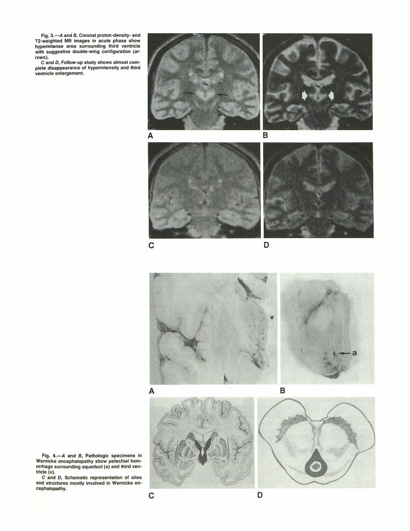
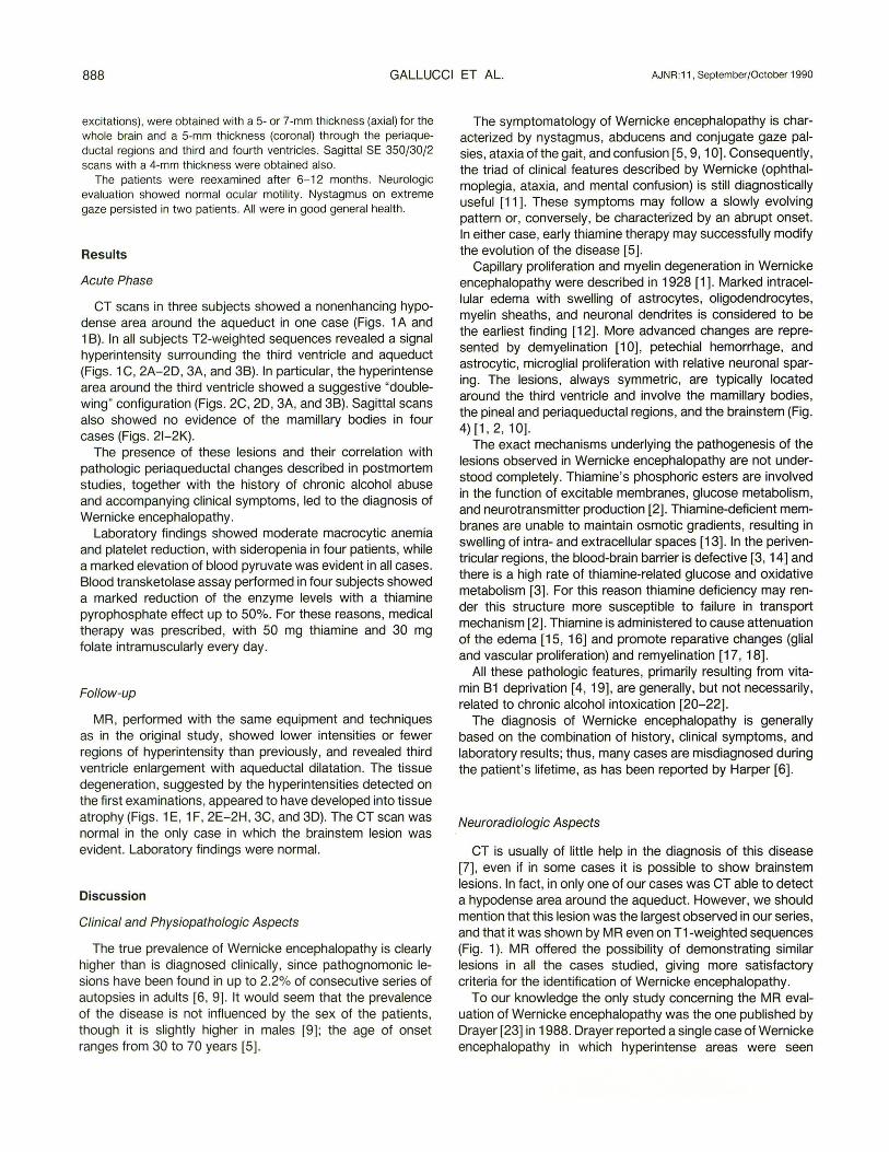
Fig. 3.-A and B, Coronal proton-density- and T2-weighted MR images in acute phase show hyperintense area surrounding third ventricle with suggestive double-wing configuration (ar· rows) .
C and D, Follow-up study shows almost complete disappearance of hyperintensity and third ventricle enlargement.
Fig. 4.-A and B, Pathologic specimens in Wernicke encephalopathy show petechial hemorrhage surrounding aqueduct (a) and third ventricle (v).
C and D, Schematic representation of sites and structures mostly involved in Wernicke encephalopathy.
A
c
A
c
D
v
8
D

892 GALLUCCI ET AL. AJNR:11, September/October 1990
A B
cases (20%), and when hemorrhage is present, many patients are agonal [5]. The reduction in hyperintense areas on the follow-up examination, also confirmed by ventricular and aqueductal dilatation, underscores the benign evolution of the pathology when appropriate therapy is applied and a correct diagnosis is made. Degeneration of the mamillary bodies and cerebellar atrophy were seen on CT in our patients and were in line with findings in other studies (Charness ME et al., presented at the annual meeting of the Radiological Society of North America, November 1986). However, these findings cannot be considered specific enough to enable more accurate diagnosis.
We did not find other focal lesions in the brain of patients affected by Wernicke encephalopathy, as was reported in one study of chronic alcohol abuse [24]. This may have been because of different pathophysiological mechanisms, different drinking habits, or to a younger age or longer duration of alcohol consumption.
In conclusion, the direct evidence of lesions provided by MR in Wernicke encephalopathy allows both early diagnosis and treatment, and therefore can successfully modify the prognosis of the disease. MR proved useful also when the history otherwise might have led to an incorrect diagnosis. Finally, we believe an early and definite identification of the disease may help establish the therapeutic approach as well.
REFERENCES 1. Gamper E. Zur Frage der Polio-encephalitis haemorragica des Chronischen
Alkoholier: Anatomische Befunde beim alkoholischen Korsakow und ihre Benziehungen zum Klinischen Bild. Dtsch Z Nervenheilk 1928;102: 122-129
2. Witt ED. Neuroanatomical consequences of thiamine deficiency: a comparative analysis. Alcohol Alcohol 1985;2:201-221
3. Gullotta F. Zur Localisation der Wernicke Encephalopatie im Kinder und Erwachsenelater. Eur Arch Psychiatry Neural Sci 1968;211 :88-108
4. Thomson AD, Ryle PR , Shaw GK. Ethanol, thiamine and brain damage. Alcohol Alcohol 1983;1 :27-43
5. Adams RD, Victor M. Principles of neurology. New York: McGraw-Hill, 1985
Fig. 5.-A and B, Thalamic infarction (A) and multiple sclerosis plaque (8). Both could simulate Wernicke encephalopathy at clinical onset; asymmetric involvement of periventricular and periaqueductal structures may help in the differentiation.
6. Harper C. Wernicke's encephalopathy: a more common disease than realised. A neuropathological study of 51 cases. J Neural Neurosurg Psychiatry 1978;42: 226-231
7. McDowell JR, Le Blanc HJ . Computed tomography finding in WernickeKorsakoff syndrome. Arch Neural 1984;41 :435-454
8. Gala LA, Mastaglia FL. Computerized axial tomography in the detection of brain damage. Med J Aust 1980;2:193-198
9. Victor M, Adams RD, Collins GH. The Wernicke-Korsakoff syndrome: a clinical and pathological study of 245 patients, 82 with post-mortem examinations. Philadelphia: Davis, 1971
10. Ghez C. Vestibular paresis: a clinical feature of Wernicke disease. J Neural Neurosurg Psychiatry 1969;178: 134-139
11 . Wernicke C. Lehrbuch der Gehirnkrankheiten fur Aertre und Studiezenk, vol. 2. Berlin: Theodor Fisher, 1881:229-242
12. Collins GH, Converse WK. Cerebellar degeneration in thiamine deficient rats . Am J Pathol 1970;29:57-69
13. Drey1us PM. Thiamine and nervous system: an overview. J Nutr Sci Vitaminol (Tokyo) 1976;22: 13-16
14. Hofer H. Zur Morphologie der Circumventrikulare Organen des Zwishenhirns der Saugetiere. Verh Dtsch Ges Zoot 1958;202
15. Watanabe I, Iwasaki Y, Aikawa H, Satayashi E, Davis JW. Hemorrhage of thiamine-deficient encephalopathy in the mouse. J Neuropathol Exp Neural 1981;40 :566-580
16. Watanabe I, Tomoto T, Hung KG , Iwasaki Y. Edematous necrosis in thiamine-deficient encephalopathy of the mouse. J Neuropathol Exp Neural 1981;40:454-471
17. Witt ED, Goldman-Rakic PS. Intermittent thiamine deficiency in the rhesus monkey. I. Progression of neurological signs and neuroanatomicallesions. Ann Neuro/1983;13:376-395
18. Witt ED, Goldman-Rakic PS. Intermittent thiamine deficiency in the rhesus monkey.ll. Progression of neurological signs and neuroanatomicallesions. Ann Neural 1983;13:396-401
19. Victor M, Adams RD. On the etiology of alcoholic neurologic disease, with special reference to the role of nutrition. Am J Clin Nutr 1961 ;9 : 379-397
20. Lopez Rl , Collins GH. Wernicke's encephalopathy. A complication of chronic hemodialysis. Arch Neural 1968;18:248-259
21 . Frantzen E. Wernicke's encephalopathy: three cases occurring in connection with severe malnutrition. Acta Neural Scand 1966;42: 426-441
22. Nixon PF. Is there a genetic component to the pathogenesis of the Wernicke-Korsakoff syndrome? Alcohol Alcohol 1984;3 :219-221
23. Drayer BP. Imaging of the aging brain. Part II. Pathologic conditions. Radiology 1988;166:797-806
24. Gallucci M, Amicarelli I, Rossi A, et al. MR imaging of white matter lessions in uncomplicated chronic alcoholism. J Comput Assist Tomogr 1989; 13(3) : 395-398
The reader 's attention is directed to the commentary on this article, which appears on pages 895-896.