Embed Size (px)
Citation preview

SKULLNEUROCRANIUM
26. 11. 2013
Kaan YücelM.D., Ph.D.
http://yeditepeanatomy1.org

Dr.Kaan Yücel http://yeditepeanatomy1.org Skull; Neurocranium
http://www.youtube.com/yeditepeanatomy
The skeleton of the head is the skull. We rather use the ancient Greek term “cranium”, e.g. the cranial nerves. The skull has 22 bones, excluding the ossicles of the ear. Except for the mandible, which forms the lower jaw, the bones of the skull are attached to each other by sutures, are immobile, and form the cranium.
The part that is covering the cranial cavity and the brain in it is called neurocranium. The skeleton of the face is called viscerocranium or facial skeleton. It is the lower part of the cranium.
Out of the 22 bones in the skull, 8 of them are in the neurocranium. They are:• 1 Frontal bone; the bone in the front of the head• 1 Occipital bone; the bone at the back of the head• 2 Parietal bones; “paries” means wall, and these though bones are on the lateral sides of the skull.• 2 Temporal bones; “temple” has two meanings “time” and “temple”. Time can make more sense for
the temporal bones, as where they are the hair becomes grey first.• 1 Sphenoid bone in the middle (Greek sphēnoeidēs wedge-shaped) • 1 Ethmoid bone again in the middle (In Moore’s textbook it is part of the facial skeleton,though)The skeleton of your face is made up by the remaining 14 bones of the cranium.The inferior and anterior parts of the frontal lobes of the brain occupy the anterior cranial fossa, the
shallowest of the three cranial fossae. The fossa is formed by the frontal bone anteriorly, the ethmoid bone in the middle, and the body and lesser wings of the sphenoid posteriorly. The butterfly-shaped middle cranial fossa has a central part composed of the sella turcicae on the body of the sphenoid and large, depressed lateral parts on each side. The bones forming the lateral parts of the fossa are the greater wings of the sphenoid and squamous parts of the temporal bones laterally and the petrous parts of the temporal bones posteriorly. The posterior cranial fossa, the largest and deepest of the three cranial fossae is formed mostly by the occipital bone, but the dorsum sellae of the sphenoid marks its anterior boundary centrally and the petrous and mastoid parts of the temporal bones contribute its anterolateral “walls.”
Sutura is that form of articulation where the contiguous margins of the bones are united by a thin layer of fibrous tissue; it is met with only in the skull. The major suturae in the skull are; coronal, lambdoid, and sagittal suturues.
The bones of the calvaria of a newborn infant are separated by membranous intervals. They include the anterior and posterior fontanelles and the paired sphenoidal and mastoid fontanelles.
2

Dr.Kaan Yücel http://yeditepeanatomy1.org Skull; Neurocranium
The skeleton of the head is the skull. We rather use the ancient Greek term “cranium”, e.g. the
cranial nerves. The skull has 22 bones, excluding the ossicles of the ear. Except for the mandible, which
forms the lower jaw, the bones of the skull are attached to each other by sutures, are immobile, and form
the cranium. Suture is also a term used in surgical practices as “surgical stitching of a wound”. Actually
suture in anatomy is a type of articulation where two articulation surfaces come together along a line, just
like you sew them with a needle.
We can divide the cranium into two or three parts. Generally into two!
Let’s see, there is one part enclosing the brain; protecting the brain, and there is another part which
makes the skeleton of your face. The part that is covering the cranial cavity and the brain in it is called
neurocranium. The skeleton of the face is called viscerocranium or facial skeleton. It is the lower part of
the cranium. Here the “viscera” means organ, and on your face there is a list of organs; your mouth, your
nose, your eyes. The prefix neuro in the term neurocranium just refers to the “nerve” telling you that this
part of the skeleton of the head covers the brain and meninges (the membrane covering the brain) within
the cranial cavity.
A third part of the skeleton of the head? The part that covers the upper part of the head; calvaria
(skullcap) “kafatası” in Turkish might be considered as a third part. If you add it into the neurocranium,
then the cranium has two parts. Ok? So the neurocranium has one roof (calvaria) and one floor; the base
(base of the skull) basicranium.
Question: which bones make up the neurocranium? Out of the 22 bones of the cranium, 8 of them
belong to the neurocranium. Some of them are paired which means you can find one on the right side, and
one on the left side, and some of them are single.
1 Frontal bone; the bone in the front of the head
1 Occipital bone; the bone at the back of the head
2 Parietal bones; “paries” means wall, and these though bones are on the lateral sides of the skull.
2 Temporal bones; “temple” has two meanings “time” and “temple”. Time can make more sense for the
temporal bones, as where they are the hair becomes grey first.
1 Sphenoid bone in the middle (Greek sphēnoeidēs wedge-shaped)
1 Ethmoid bone again in the middle
http://twitter.com/yeditepeanatomy3
1.SKULL

Dr.Kaan Yücel http://yeditepeanatomy1.org Skull; Neurocranium
As you see only the temporal bones and the parietal bones above them are paried (bilateral), and
the other four bones are single (unilateral) which make the eight bones of the neurocranium.
The neurocranium has a dome-like roof, the calvaria (skullcap), and a floor or cranial base
(basicranium).
The bones forming the calvaria are mainly the paired temporal and parietal bones, and parts of the
unpaired frontal, sphenoid, and occipital bones. The frontal bone, parietal bones, and occipital bone make
up the superior part of the calvaria or the calva (skullcap).
The bones forming the base of the cranium are mainly parts of the sphenoid, temporal, and
occipital bones.
The ethmoid bone is an irregular bone that makes a relatively minor midline contribution to the
neurocranium but is also part of the viscerocranium. Watch out! Although there is only a minor
contribution of the ethmoid bone to the neurocranium, it is counted under the bones of the neurocranium.
Clue: Smelling.
The skeleton of your face (viscerocranium; as we have three organs: from superior to inferior- eyes,
nose, tongue) is made up by the remaining 14 bones of the cranium.
Figure 1. Skull bones (lateral view)http://images.tutorvista.com/content/locomotion-animals/human-skull-structure.jpeg
http://www.youtube.com/yeditepeanatomy 4

Dr.Kaan Yücel http://yeditepeanatomy1.org Skull; Neurocranium
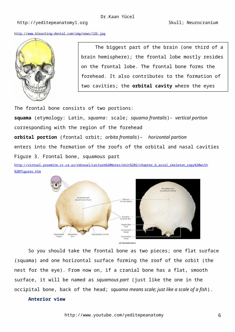
2.1. FRONTAL BONE (OS FRONTALE)Figure 2. Frontal bonehttp://www.bleaching-dental.com/img/news/126.jpg
The frontal bone consists of two portions:
squama (etymology: Latin, squama: scale; squama frontalis)- vertical portion
corresponding with the region of the foreheadorbital portion (frontal orbit; orbita frontalis)– horizontal partion
enters into the formation of the roofs of the orbital and nasal cavitiesFigure 3. Frontal bone, squamous parthttp://virtual.yosemite.cc.ca.us/rdroual/Lecture%20Notes/Unit%202/chapter_6_axial_skeleton_copy%20with%20figures.htm
So you should take the frontal bone as two pieces; one flat surface (squama) and one horizontal
surface forming the roof of the orbit (the nest for the eye). From now on, if a cranial bone has a flat,
smooth surface, it will be named as squamous part (just like the one in the occipital bone, back of the
head; squama means scale; just like a scale of a fish).
http://twitter.com/yeditepeanatomy5
2.BONES OF THE NEUROCRANIUM
The biggest part of the brain (one third of a brain hemisphere); the
frontal lobe mostly resides on the frontal lobe. The frontal bone forms the
forehead. It also contributes to the formation of two cavities; the orbital
cavity where the eyes are located and the nasal cavity (the cavity inside
your nose).
Dr.Kaan Yücel http://yeditepeanatomy1.org Skull; Neurocranium
Anterior viewThe supra-orbital margin of the frontal bone, the angular boundary between the squamous and
the orbital parts, has a supra-orbital foramen or notch in some crania for passage of the supra-orbital
nerve and vessels. Just superior to the supra-orbital margin and the rim of the orbit there is a ridge, the
superciliary arch. The prominence of these ridges, deep to the eyebrows, is generally greater in males.
Between the superciliary arches is a small depression (glabella). The superciliary arches extend laterally on
each side from the glabella. In some people, frontal eminences are also visible, giving the calvaria an
almost square appearance.
Medially, the frontal bone projects inferiorly forming a part of the medial rim of the orbit. Laterally,
the zygomatic process of the frontal bone projects inferiorly forming the upper lateral rim of the orbit.
This process articulates with the frontal process of the zygomatic bone.
In some adults a metopic suture, a persistent frontal suture or remnant of it, is visible in the midline
of the glabella, the smooth, slightly depressed area between the superciliary arches. The estimated
prevalence is 1 in 15,000 live births with a 3:1 male:female ratio.
Figure 4. Frontal bone (anterior view)http://www.csuchico.edu/anth/Module/frontal.html
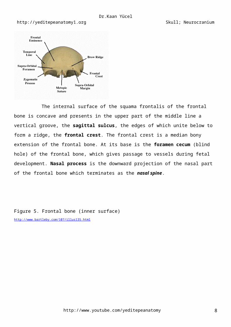
The internal surface of the squama frontalis of the frontal bone is concave and presents in
the upper part of the middle line a vertical groove, the sagittal sulcus, the edges of which unite below to
form a ridge, the frontal crest. The frontal crest is a median bony extension of the frontal bone. At its base
is the foramen cecum (blind hole) of the frontal bone, which gives passage to vessels during fetal
development. Nasal process is the downward projection of the nasal part of the frontal bone which
terminates as the nasal spine.
http://www.youtube.com/yeditepeanatomy 6

Dr.Kaan Yücel http://yeditepeanatomy1.org Skull; Neurocranium
Figure 5. Frontal bone (inner surface)http://www.bartleby.com/107/illus135.html
2.2. PARIETAL BONESThe two parietal bones unite and form the sides and roof of the cranium. Each bone is irregularly
quadrilateral in form. The external surface is convex, smooth, and marked near the center by an eminence,
the parietal eminence (tuber parietale). Crossing the middle of the bone in an arched direction are two
curved lines, the superior and inferior temporal lines. The parietal foramen is a small, inconstant aperture
located posteriorly in the parietal bone near the sagittal suture. Paired parietal foramina may be present.
Figure 6. Parietal bone (lateral view)http://medical-dictionary.thefreedictionary.com/parietal+bone
2.3. TEMPORAL BONES
http://twitter.com/yeditepeanatomy7
Figure 7. Parietal bones (anterior view)http://aftabphysio.blogspot.com/2010/09/bones-of-skull.html
Dr.Kaan Yücel http://yeditepeanatomy1.org Skull; Neurocranium
The temporal bones are situated at the sides and base of the skull. The temporal bone has the
temporal lobe on which is important for a long list of functions including memory, emotional memory,
hearing. It has the canal that goes to the ear.
The temporal bone contributes most of the lower portion of the lateral wall of the cranium.
Each temporal bone has three parts which are separated from each other by a cartilaginous tissue
in the newborn. Later on, the three parts are united and become as one single bone.
1) Squamous part
2) Tympanic part
3) Petromastoid part
The squamous part has the appearance of a large flat plate, forms the anterior and superior parts of
the temporal bone, contributes to the lateral wall of the cranium, and articulates anteriorly with the
greater wing of the sphenoid bone at the sphenosquamous suture, and with the parietal bone superiorly
at the squamous suture. The zygomatic process is an anterior bony projection from the lower surface of
the squamous part of the temporal bone that initially projects laterally and then curves anteriorly to
articulate with the temporal process of the zygomatic bone to form the zygomatic arch.
The squamous part lies just lateral to the greater wing of the sphenoid. It participates in the
temporomandibular joint. It contains the mandibular fossa, which is a concavity where the head of the
mandible articulates with the base of the skull. An important feature of this articulation is the prominent
articular tubercle, which is the downward projection of the anterior border of the mandibular fossa.
The tympanic part of the temporal bone is immediately below the origin of the zygomatic process
from the squamous part of the temporal bone. The external acoustic opening (pore) is the entrance to the
external acoustic meatus (canal), which leads to the tympanic membrane (eardrum). The external acoustic
opening is clearly visible on the surface of this part.
On the lateral edge of the temporal bone lies the cone-shaped mastoid process projecting from its
inferior surface. The mastoid process is a prominent structure and is the point of attachment for several
muscles. On the medial aspect of the mastoid process is the deep mastoid notch, which is also an
attachment point for a muscle.
Immediately lateral to the basilar part of the occipital bone is the petrous part of the temporal
bone. Wedge-shaped in its appearance the petrous part of the temporal bone is between the greater
wing of the sphenoid anteriorly and the basilar part of the occipital bone posteriorly. The apex forms one
of the boundaries of the foramen lacerum, an irregular opening filled in life with cartilage. Posterolateral
from the foramen lacerum along the petrous part of the temporal bone is the large circular opening for the
carotid canal.
http://www.youtube.com/yeditepeanatomy 8

Dr.Kaan Yücel http://yeditepeanatomy1.org Skull; Neurocranium
Anterior surface of the petrous part of the temporal bone has the following six bone markings:
1. Arcuate eminence (Eminentia arcuata): is located at the centre of the anterior surface.
2. Tegmen tympani: is a very thin and slight depression just anterior and lateral to the arcuate eminence in
the anterior surface of the petrous part of the temporal bone. The tegmen tympani marks the thin bony
roof of the middle ear cavity.
3. Groove for greater petrosal nerve (Sulcus nervi petrosi majoris): is located just anterior to tegmen
tympani.
4. Groove for lesser petrosal nerve (Sulcus nervi petrosi minoris): is a more shallow groove lying parallel
and laterally to the groove for greater petrosal nerve.
5. Carotid canal (Canalis caroticus): is a large circular opening which lies posterolateral from the foramen
lacerum along the petrous part of the temporal bone. One of the two arteries of the brain; the interal
carotid artery passes through this canal.
6. Trigeminal impression (Impressio trigeminalis): the slight depression which is located medially in the
anterior of the petrous part of the temporal bone. The trigeminal impression marks the location of the
sensory ganglion for the trigeminal nerve [V].
The large opening between the occipital bone and the petrous part of the temporal bone is the
jugular foramen.This foramen is very important as major structures pass through this foramen. The vein
draining the brain exits the skull through the jugular foramen. Three of the 12 pairs of cranial nerves pass
through the jugular foramen and go to their destinations exiting the cranium. Anterosuperior to the
jugular foramen is the internal acoustic meatus for the passage of two other cranial nerves. One of them is
the nerve for the muscles of the face, and the other is good for the hearing and balance.
The styloid process is needle-shaped bone marking. It projects from the lower border of the
temporal bone anteromedial to the mastoid process. The styloid process is a point of attachment for
numerous muscles and ligaments. The stylomastoid foramen, transmitting the nerve for the muscles of
the face lies (CN VII; Facial nerve – CN= Cranial nerve) posterior to the base of the styloid process;
between the styloid process and the mastoid process.
Figure 8. Temporal bonehttp://medicinembbs.blogspot.com/2011/02/skull-anatomy.html
http://twitter.com/yeditepeanatomy9

Dr.Kaan Yücel http://yeditepeanatomy1.org Skull; Neurocranium
2.4. SPHENOID BONEThe sphenoid bone is situated at the base of the skull in front of the temporal bones and basilar
part of the occipital bone. It somewhat resembles a bat with its wings extended, and is divided into a
median portion; body, two great and two small wings extending outward from the sides of the body, and
two pterygoid processes which project from it below.
The greater and lesser wings of the sphenoid spread laterally from the lateral aspects of the body
of the bone. The pterygoid processes, consisting of lateral and medial pterygoid plates, extend inferiorly
on each side of the sphenoid from the junction of the body and greater wings. Pterygoid fossa is between
the two plates. Each medial plate of the pterygoid process ends inferiorly with a hook-like projection, the
pterygoid hamulus.
The sphenoidal crests are formed mostly by the sharp posterior borders of the lesser wings of the
sphenoid bones. The sphenoidal crests end medially in two sharp bony projections, the anterior clinoid
processes. Just anterior to each anterior clinoid process is a circular opening in the lesser wing of the
sphenoid (the optic canal), through which the ophthalmic artery and optic nerve [II] pass as they exit the
cranial cavity to enter the orbit. The optic canals are usually included in the middle cranial fossa.
The sella turcica[e] (L. Turkish saddle) is the saddle-like bony formation on the upper surface of the
body of the sphenoid, which is surrounded by the anterior and posterior clinoid processes. Clinoid means
“bedpost,” and the four processes (two anterior and two posterior) surround the hypophysial fossa, the
“bed” of the pituitary gland, like the posts of a four-poster bed. Between the ant. and post. clinoid
procceses on the body of the sphenoid bone, lies the carotid groove (sulcus) where the internal carotid
artery passes through.
The sella turcica is composed of three parts:http://www.youtube.com/yeditepeanatomy 10

Dr.Kaan Yücel http://yeditepeanatomy1.org Skull; Neurocranium
1) The tuberculum sellae (horn of saddle): a variable slight to prominent median elevation forming the
posterior boundary of the chiasmatic sulcus (optic groove) and the anterior boundary of the hypophysial
fossa. It lies behind the chiasmatic groove. On both ends of the tuberculum sellae are middle clinoid
processes.
2) The hypophysial fossa (pituitary fossa): a median depression (seat of saddle) in the body of the
sphenoid that accommodates the pituitary gland (L. hypophysis).
3) The dorsum sellae (back of saddle): a square plate of bone projecting superiorly from the body of the
sphenoid. It forms the posterior boundary of the sella turcica, and its prominent superolateral angles make
up the posterior clinoid processes.
On each side of the body of the sphenoid, four foramina perforate the roots of the cerebral surfaces of the
greater wings of the sphenoids:
Superior orbital fissure : Located between the greater and the lesser wings, it opens anteriorly into the
orbit.
Foramen rotundum (round foramen) : Located posterior to the medial end of the superior orbital
fissure.
Foramen ovale (oval foramen) : A large foramen posterolateral to the foramen rotundum.
Foramen spinosum (spinous foramen) : Located posterolateral to the foramen ovale.
The foramen lacerum (lacerated or torn foramen) is not part of the crescent of foramina. This
ragged foramen lies posterolateral to the hypophysial fossa and is an artifact of a dried cranium. In life, it is
partly closed by a cartilage plate.
Figure 9. Sphenoid bone (anterior view)http://virtual.yosemite.cc.ca.us/rdroual/Lecture%20Notes/Unit%202/chapter_6_axial_skeleton_copy%20with%20figures.htm
Figure 10. Foramina in the sphenoid bone (superior view) and other openings http://medchrome.com/wp-content/uploads/2011/05/skull-superior.jpg
http://twitter.com/yeditepeanatomy11

Dr.Kaan Yücel http://yeditepeanatomy1.org Skull; Neurocranium
2.5. OCCIPITAL BONEThe occipital bone is situated at the back and lower part of the cranium. It is trapezoid in shape and
curved on itself. It is pierced by a large oval aperture, the foramen magnum, through which the cranial
cavity communicates with the vertebral canal.
The foramen magnum is the most prominent feature of the cranial base. The major structures
passing through this large foramen:
spinal cord (where it becomes continuous with the medulla oblongata of the brain)
meninges (coverings) of the brain and spinal cord
vertebral arteries
spinal accessory nerve (CN XI).
The four parts of the occipital bone are arranged around the foramen magnum:
1) The curved, expanded plate behind the foramen magnum is named the squama.
2) The thick, quadrilateral piece in front of the foramen is called the basilar part.
3) On either side of the foramen is the lateral (condylar) portion of the occipital bone.
The cranial base is formed posteriorly by the occipital bone, which articulates with the sphenoid bone
anteriorly.
The external occipital protuberance, is usually easily palpable in the median plane; however,
occasionally (especially in females) it may not be prominent. The external occipital crest descends from
the protuberance toward the foramen magnum.
The superior nuchal line marks the superior limit of the neck. It extends laterally from each side of
the protuberance. The inferior nuchal line is less distinct.
http://www.youtube.com/yeditepeanatomy 12

Dr.Kaan Yücel http://yeditepeanatomy1.org Skull; Neurocranium
On the lateral parts of the occipital bone are two large protuberances, the occipital condyles. The
cranium articulates with the vertebral column by the occipital condyles.
The clivus is a shallow depression, incline (Latin for "slope") behind the dorsum sellæ that slopes
obliquely backward. Posterior to foramen magnum, the posterior cranial fossa is partly divided by the
internal occipital crest into bilateral large concave impressions, the cerebellar fossae. The internal
occipital crest ends in the internal occipital protuberance. Actually, the internal occipital crest is the lower
division of a cross called as cruciate eminence. The cruciate eminence divides the interior surface of the
occipital bone into four fossae. The superior two fossae are triangular and lodge the occipital lobes of the
cerebrum (cerebral fossae). The inferior two are quadrilateral and accommodate the hemispheres of the
cerebellum (cerebellar fossae). The internal occipital protuberance is the prominent elevation in the
centre of the cruciate eminence.
The hypoglossal canal for the hypoglossal nerve (CN XII) is superior to the anterolateral margin of
the foramen magnum.
Figure 11. Occipital bone (inner surface)
2.6. ETHMOID BONEGk, ethmos, sieve sifter, eidos, form
The etmoid bone is exceedingly light and spongy, and cubical in shape; it is situated at the anterior
part of the base of the cranium, between the two orbits, at the roof of the nose, and contributes to each of
these cavities. It consists of four parts: a horizontal or cribriform plate, forming part of the base of the
http://twitter.com/yeditepeanatomy13

Dr.Kaan Yücel http://yeditepeanatomy1.org Skull; Neurocranium
cranium; a perpendicular plate, constituting part of the nasal septum; and two lateral ethmoidal
labyrinths. The crista galli (L. crest of the cock) is a thick, median ridge of bone posterior to the foramen
cecum (frontal bone), which projects superiorly from the ethmoid. On each side of the ridge called crista
galli, located in the frontal bone, is the sieve-like cribriform plate of the ethmoid. Its numerous tiny
foramina transmit the olfactory nerves (CN I) from the olfactory areas of the nasal cavities to the olfactory
bulbs of the brain, which lie on this plate. SMELLING!!!
Figure 12. Ethmoid Bonehttp://medical-dictionary.thefreedictionary.com/ethmoid+bone
Figure 13. Ethmoid bone’s location in the skullhttp://www.daviddarling.info/images/ethmoid_bone.jpg
The five bones that make up the skull base are the ethmoid, sphenoid, occipital, paired frontal, and
paired parietal bones. The skull base can be subdivided into 3 regions: the anterior, middle, and posterior
cranial fossae.
3.1. ANTERIOR CRANIAL FOSSA
http://www.youtube.com/yeditepeanatomy 14
3.CRANIAL FOSSAE

Dr.Kaan Yücel http://yeditepeanatomy1.org Skull; Neurocranium
Parts of the frontal, ethmoid, and sphenoid bones form the anterior cranial fossa. Its floor is
composed of:
frontal bone in the anterior and lateral direction;
ethmoid bone in the midline; and
two parts of the sphenoid bone posteriorly, the body (midline), and the lesser wings (laterally).
The anterior cranial fossa is above the nasal cavity and the orbits, and it is filled by the frontal lobes
of the cerebral hemispheres
3.2. MIDDLE CRANIAL FOSSAThe middle cranial fossa consists of parts of the sphenoid and temporal bones. The butterfly-
shaped middle cranial fossa has a central part composed of the sella turcice on the body of the sphenoid
and large, depressed lateral parts on each side.
The middle cranial fossa is posteroinferior to the anterior cranial fossa. The boundary between the
anterior and middle cranial fossae in the midline is the anterior edge of the chiasmatic sulcus, which is a
smooth groove stretching between the optic canals across the body of the sphenoid.
The boundary between the middle and the posterior cranial fossae is the superior border of the
petrous part of the temporal bone laterally and a flat plate of bone, the dorsum sellae of the sphenoid,
medially.
The bones forming the lateral parts of the fossa are the greater wings of the sphenoid and
squamous parts of the temporal bones laterally and the petrous parts of the temporal bones posteriorly.
The lateral parts of the middle cranial fossa support the temporal lobes of the brain.
3.3. POSTERIOR CRANIAL FOSSAThe posterior cranial fossa consists mostly of parts of the temporal and occipital bones with small
contributions from the sphenoid and parietal bones. It is the largest and deepest of the three cranial fossae
and contains the brainstem (midbrain, pons, and medulla) and the cerebellum.
Boundaries
The anterior boundaries of the posterior cranial fossa in the midline are the dorsum sellae and the
clivus . The clivus is a slope of bone that extends upward from the foramen magnum. It is formed by
contributions from the body of the sphenoid and from the basilar part of the occipital bone.
Laterally the anterior boundaries of the posterior cranial fossa are the superior border of the
petrous part of the petromastoid part of the temporal bone.
Figure 14. Cranial fossae
http://twitter.com/yeditepeanatomy15

Dr.Kaan Yücel http://yeditepeanatomy1.org Skull; Neurocranium
http://tmjc.com.ne.kr/tmj/info/drinfo/images/tm6-6.jpg
Sutura is that form of articulation where the contiguous margins of the bones are united by a thin layer of
fibrous tissue; it is met with only in the skull.
5.1. CORONAL SUTUREParietal bones articulate with the frontal bone in their front, forming the coronal suture.
5.2. LAMBDOID SUTUREParietal bones articulate with the occipital in their behind, forming the lambdoid suture.
5.3 .SAGITTAL SUTUREParietal bone articulates with its the opposite side, forming the sagittal suture. Vertex, the most superior
point of the calvaria, is near the midpoint of the sagittal suture.
The coronal suture separates the frontal and parietal bones, the sagittal suture separates the parietal
bones, and the lambdoid suture separates the parietal and temporal bones from the occipital bone.
Figure 15. Suturaehttp://upload.wikimedia.org/wikipedia/commons/thumb/0/0d/Human_skull_side_suturas.svg/524px-Human_skull_side_suturas.svg.png
The bones of the calvaria of a newborn infant are separated by membranous intervals. They include
the anterior and posterior fontanelles and the paired sphenoidal and mastoid fontanelles. Palpation of the
fontanelles during infancy, especially the anterior and posterior ones, enables physicians to determine the:
• Progress of growth of the frontal and parietal bones.
• Degree of hydration of an infant (a depressed fontanelle indicates dehydration).
http://www.youtube.com/yeditepeanatomy 16
4.SUTURA
7. FONTANELLES
There are also minor suturae; such as regarding the ; sphenodid bone sphenoparietal suture, and with the anterior edge of the temporal bone at the sphenosquamous suture. Another one is occipitomastoid suture. Parietomastoid suture is between the parietal bone and the mastoid process.

Dr.Kaan Yücel http://yeditepeanatomy1.org Skull; Neurocranium
• Level of intracranial pressure (a bulging fontanelle indicates increased pressure on the brain).
The anterior fontanelle, the largest one, is diamond or star shaped; it is bounded by the halves of the
frontal bone anteriorly and the parietal bones posteriorly. Thus it is located at the junction of the sagittal,
coronal, and frontal sutures, the future site of bregma. By 18 months of age, the surrounding bones have
fused and the anterior fontanelle is no longer clinically palpable.
The posterior fontanelle is triangular and bounded by the parietal bones anteriorly and the occipital bone
posteriorly. It is located at the junction of the lambdoid and sagittal sutures, the future site of lambda. The
posterior fontanelle begins to close during the first few months after birth; and by the end of the 1st year,
it is small and no longer clinically palpable.
The sphenoidal and mastoid fontanelles fuse during infancy and are less important clinically than the
midline fontanelles.
The resilience of the cranial bones of infants allows them to resist forces that would produce fractures in
adults. The fibrous sutures of the calvaria also permit the cranium to enlarge during infancy and childhood.
The increase in the size of the calvaria is greatest during the first 2 years, the period of most rapid brain
development. The calvaria normally increases in capacity for 15-16 years. After this, the calvaria usually
increases slightly in size for 3-4 years as a result of bone thickening.
Figure 16. Fontanelleshttp://medical-dictionary.thefreedictionary.com/fontanelle
HEAD INJURIESHead injuries are a major cause of death and disability. The complications of head injuries include
hemorrhage, infection, and injury to the brain and cranial nerves.
FRACTURES OF THE CRANIAL FOSSAEIn fractures of the anterior cranial fossa, the cribriform plate of the ethmoid bone may be damaged.
Fractures of the middle cranial fossa are common, because this is the weakest part of the base of the skull.
http://twitter.com/yeditepeanatomy17
CLINICAL ANATOMY

Dr.Kaan Yücel http://yeditepeanatomy1.org Skull; Neurocranium
Anatomically, this weakness is caused by the presence of numerous foramina and canals in this region; the
cavities of the middle ear and the sphenoidal air sinuses are particularly vulnerable.
PASSING THROUGHBone marking @ which bone Important structures passing through
-particularly cranial nerves-
A lesion here might result in…
Carotid canal Temporal Internal carotid artery and nerve plexus Problem in the anterior arterial supply of the brain; as a result; weakness (hemiplegia) and numbness in the face, and extremities on the opposite side of the body, difficulty in speech, visual loss, etc.
Foramen magnum Occipital Continuation of brain and spinal cord; vertebral arteries and nerve plexuses; roots of accessory nerve [XI]; meninges
-
Foramen ovale Sphenoid Third branch of the CN V (Trigeminal nerve): Mandibular nerve [V3]
Sensorial loss in the mandibular region of the face
Foramen rotundum Sphenoid Second branch of the CN V (Trigeminal nerve): Maxillary nerve [V2]
Sensorial loss in the maxillary region of the face
Foramen spinosum Sphenoid - -Hypoglossal canal Occipital Hypoglossal nerve [XII] and vessels Loss of movement of the tongue.Internal acoustic meatus
Temporal Facial nerve [VII]; vestibulocochlear nerve [VIII]
Problems in hearing, balance, or movements of the facial (expression) muscles
Jugular foramen Temporal Internal jugular vein; glossopharyngeal nerve [IX]; vagus nerve [X]; accessory nerve [XI]
-
Optic canal Sphenoid Optic nerve (II) Problems in vision.Stylomastoid foramen Temporal Facial nerve [VII] Loss of movement of the muscles
of the faceSuperior orbital fissure Sphenoid Oculomotor nerve (III)
Trochlear nerve (IV)Branches of ophthalmic nerve (II)Abducens nerve (VI)
Problems in vision.
Empty spaces do not mean nothing happens here. It means, for now I want you to know that much.
http://www.youtube.com/yeditepeanatomy 18