Embed Size (px)
Citation preview

1
1. Title page
Insights into the structure-activity relationship of glycosides as positive allosteric
modulators acting on P2X7 receptors
Waraporn Piyasirananda, Andrew Beekman, A. Ganesan, Stefan Bidula, Leanne Stokes
School of Pharmacy, University of East Anglia, Norwich Research Park, Norwich UK.
Conflict of interest:
The authors declare that there is no conflict of interest.
This article has not been copyedited and formatted. The final version may differ from this version.Molecular Pharmacology Fast Forward. Published on December 17, 2020 as DOI: 10.1124/molpharm.120.000129
at ASPE
T Journals on N
ovember 29, 2021
molpharm
.aspetjournals.orgD
ownloaded from

2
Running title: P2X7 positive modulator SAR
Corresponding author address:
Dr Leanne Stokes, School of Pharmacy, University of East Anglia, Norwich Research Park,
Norwich NR4 7TJ, UK.
Telephone: +44 1603 592403
Email: [email protected]
Text pages: 29
Number of Tables: 1
Number of Figures: 8
Number of references: 38
Word counts:
Abstract: 247
Introduction: 411
Discussion: 1480
Non-standard abbreviations:
PAM; positive allosteric modulator
This article has not been copyedited and formatted. The final version may differ from this version.Molecular Pharmacology Fast Forward. Published on December 17, 2020 as DOI: 10.1124/molpharm.120.000129
at ASPE
T Journals on N
ovember 29, 2021
molpharm
.aspetjournals.orgD
ownloaded from

3
3. Abstract
P2X7 is an important ligand-gated ion channel expressed in multiple immune cell
populations. This study aimed to investigate the chemical requirements of triterpenoid
glycosides within a new binding pocket to characterise the structure-activity relationship. A
set of glycosides were screened for positive modulator activity at human P2X7 using a YO-
PRO-1 dye uptake assay in HEK-hP2X7 cells. The highest positive modulator activity was
with ginsenoside CK, containing a monosaccharide (glucose) attached at carbon-20.
Ginsenoside 20(S)Rg3, containing a disaccharide group (glucose-glucose) at carbon-3,
displayed positive modulator activity with a reduced EC50 for ATP and increased maximal
response at hP2X7. The epimer 20(R)Rg3 was inactive. A similar stereo-specific pattern was
observed for 20(S)-Rh2. Ginsenoside-F1, highly similar to ginsenoside CK but containing a
single additional hydroxyl group, was also inactive at P2X7. Computational docking suggests
hydrophobic residues in the pocket are involved in steric discrimination between
triterpenoids, while the position and identity of the carbohydrate group is important for
positive modulator activity at human P2X7. Ginsenosides containing monosaccharide
attachments perform better than di- or tri-saccharide glycosides. Additional modifications to
the triterpenoid scaffold at carbon-6 are not tolerated. Gypenosides from plant sources other
than Panax ginseng (gypenoside XVII, gypenoside XLIX, stevenleaf) can also act as positive
allosteric modulators of P2X7. We also investigated the effect of positive allosteric
modulators on endogenous P2X7 in THP-1 monocytes and confirmed our findings in a
calcium response assay. A cell viability assay showed potentiation of ATP-induced cell death
with ginsenoside CK in THP-1 and HEK-hP2X7 cells.
Keywords: P2X7, ginsenoside, positive allosteric modulator, ATP
This article has not been copyedited and formatted. The final version may differ from this version.Molecular Pharmacology Fast Forward. Published on December 17, 2020 as DOI: 10.1124/molpharm.120.000129
at ASPE
T Journals on N
ovember 29, 2021
molpharm
.aspetjournals.orgD
ownloaded from

4
4. Significance statement
Ginsenosides are active as PAMs at P2X7 and this study determines the chemical features
important for mediating this effect. The position and identity of the sugar group is important
for activity as is the position of a number of hydroxyl groups on the triterpenoid scaffold.
Diastereomers of ginsenosides Rg3 and Rh2 demonstrate the importance of the location of
hydroxyl groups relative to the hydrophobic face of the predicted binding pocket.
This article has not been copyedited and formatted. The final version may differ from this version.Molecular Pharmacology Fast Forward. Published on December 17, 2020 as DOI: 10.1124/molpharm.120.000129
at ASPE
T Journals on N
ovember 29, 2021
molpharm
.aspetjournals.orgD
ownloaded from

5
Introduction
The ligand-gated ion channel P2X7 is important in regulating immune cell responses during
infection and inflammation (Di Virgilio et al., 2017). In particular, P2X7 is a known
physiological regulator of the NLRP3-caspase 1 inflammasome complex and controls the
secretion of pro-inflammatory cytokines such as IL-1β and IL-18 (Giuliani et al., 2017). Many
studies have demonstrated that activation of P2X7 in infected macrophages in vitro can
promote microbial killing. Intracellular bacteria and parasites such as Mycobacterium
tuberculosis (Fairbairn et al., 2001, Placido et al., 2006), Toxoplasma gondii (Moreira-Souza
et al., 2017) and Leishmania amazonensis (Chaves et al., 2014) are examples of pathogens
where P2X7 contributes to microbicidal mechanisms. In vivo mouse models of infection
suggest that global deficiency in P2X7 can affect pathogen burden and inflammation (Miller
et al., 2015, Chaves et al., 2019). Furthermore, inheritance of loss-of-function variants of
human P2X7 have been linked to susceptibility to infections and complications such as
extra-pulmonary TB (Areeshi et al., 2015, Fernando et al., 2007). Development of a positive
allosteric modulator (PAM) of P2X7 may therefore be useful in treatment of such infections
(Stokes et al., 2020). This type of therapy directed at enhancing host responses would
reduce the need for antibiotics and be beneficial in avoiding development of antibiotic
resistance.
Triterpenoid glycosides from Panax ginseng have PAM activity at human P2X7 with
the in vivo metabolite ginsenoside compound K (CK) demonstrating the highest activity
(Helliwell et al., 2015). Using a combination of computational docking and mutagenesis, we
previously characterised a novel binding pocket for ginsenosides within the central vestibule
of human P2X7 (Bidula et al., 2019b). Predicted binding poses identified two modes of
binding dependent on the carbohydrates attached to the ginsenoside in question.
Ginsenoside CK contains a single carbohydrate (glucose) moiety which makes multiple
contacts with amino acids Asp318, Leu320 and Ser60. Conversely, ginsenoside Rd contains
both disaccharide (-glucose-glucose) and monosaccharide glucose moieties and is predicted
This article has not been copyedited and formatted. The final version may differ from this version.Molecular Pharmacology Fast Forward. Published on December 17, 2020 as DOI: 10.1124/molpharm.120.000129
at ASPE
T Journals on N
ovember 29, 2021
molpharm
.aspetjournals.orgD
ownloaded from

6
to use the disaccharide moiety for binding to P2X7 (Bidula et al., 2019b). The presence of
carbohydrate moieties is deemed essential for PAM activity since the aglycone
protopanaxadiol has no activity at P2X7 (Helliwell et al., 2015).
In this study, we explored the chemical structural requirements of triterpenoid
glycosides with the aim of building a structure-activity relationship for positive modulators at
P2X7. This revealed critical information about the tolerance of substitutions at C-6 and the
preference for glucose as the attached sugar moiety. This provides important knowledge for
the future development of selective PAMs for P2X7.
This article has not been copyedited and formatted. The final version may differ from this version.Molecular Pharmacology Fast Forward. Published on December 17, 2020 as DOI: 10.1124/molpharm.120.000129
at ASPE
T Journals on N
ovember 29, 2021
molpharm
.aspetjournals.orgD
ownloaded from
7
Materials and Methods
Materials
Ginsenoside CK (CAS 39262-14-1), ginsenoside-Rb1 (CAS 41753-43-9), ginsenoside-Rd
(CAS 52705-93-8), 20(S)-ginsenoside-Rg3 (CAS 14197-60-5), 20(R)-ginsenoside-Rh2 (CAS
112246-15-8), protopanaxadiol (PPD) (CAS 30636-90-9), glycyrrhizic acid (CAS 1405-86-
3), stevioside (CAS 57817-89-7), daucosterol (CAS 474-58-8), esculentoside A (CAS 65497-
07-6), mogroside V (CAS 88901-36-4), saikosaponin A (CAS 20736-09-8), stevenleaf (CAS
80321-63-7) were from Shanghai Richem International Ltd, China (supplier code
CDCMANSETE). Ginsenoside CK (CAS 39262-14-1), ginsenoside-F2 (CAS 62025-49-4),
ginsenoside-F1 (CAS 53963-43-2), gypenoside XLIX (CAS 94987-08-3), gypenoside XVII
(CAS 80321-69-3), 20(S)-ginsenoside-Rg3 (CAS 14197-60-5), 20(R)-ginsenoside -Rg3
(CAS 38243-03-7), 20(S)-ginsenoside-Rh2 (CAS 78214-33-2), 20(R)-ginsenoside-Rh2 (CAS
112246-15-8) were from ChemFaces (Wuhan Chemfaces). Scilliroside (CAS 507-60-8,
NSC7523), ouabain (CAS 630-60-4, NSC25485), solanine hydrochloride (CAS 20562-02-1,
NSC35611), solasonine (CAS 19121-58-5, NSC82149) were obtained from the NCI DTP
chemical repository. ATP (Sigma-Aldrich, A7699) was dissolved in distilled water to 100 mM
and adjusted to pH 7.4 with 5 M NaOH. Aliquots of ATP were kept frozen at -20 °C and used
once in experiments. AZ10606120 (CAS 607378-18-7, Tocris Biosciences) and AZ11645373
(CAS 227088-94-0) Sigma-Aldrich) were dissolved in DMSO to 10 mM and stored at -20 °C.
Fura-2AM (CAS 108964-32-5, HelloBio) was prepared in DMSO to a concentration of 1 mM,
aliquoted, and stored at -20 °C in amber-coloured Eppendorf vials. Sulfinpyrazone (CAS 57-
96-5, Sigma Aldrich) was dissolved in methanol at a stock concentration of 25 mM and
stored at +4 °C.
This article has not been copyedited and formatted. The final version may differ from this version.Molecular Pharmacology Fast Forward. Published on December 17, 2020 as DOI: 10.1124/molpharm.120.000129
at ASPE
T Journals on N
ovember 29, 2021
molpharm
.aspetjournals.orgD
ownloaded from

8
Cell culture
HEK-293 cells stably expressing the wild-type human P2X7 variant (HEK-hP2X7) were
maintained in DMEM:F12 media (ThermoFisher Scientific, Life Technologies catalogue
number 11320-074) containing 10% foetal bovine serum (Gibco catalogue number 10500-
064) and penicillin/streptomycin (ThermoFisher Scientific) as described previously (Helliwell
et al., 2015). HEK-293 cells were passaged twice weekly using 0.5% trypsin-EDTA
(ThermoFisher Scientific). THP-1 human monocytic cells (a kind gift from Professor Maria
O’Connell, University of East Anglia) were maintained in RPMI 1640 media (Fisher Scientific,
Life Technologies catalogue number 21875-034) containing 10% foetal bovine serum
(Gibco, as before) and penicillin/streptomycin (ThermoFisher Scientific). Cells were kept in a
humidified incubator at 37 °C with a constant supply of 5% CO2.
YO-PRO-1 dye uptake screening assay
ATP-induced dye uptake experiments were performed as described previously (Bidula et al.,
2019b). Briefly, HEK-hP2X7 cells were plated at 2 x 10⁴ cells/well (100 μl per well) in
complete media and left overnight to attach to poly-D-lysine coated 96-well plates. YO-PRO-
1 iodide was prepared in a low divalent buffered solution (145 mM, 2mM KCl, 13 mM D-
glucose, 10 mM HEPES, 0.2 mM CaCl2 pH 7.3) to a final concentration of 2 μM. Media was
removed from the plate using a manual multi-channel pipette and YO-PRO-1 buffer applied
to wells (180 μl per well). AZ10606120 (10 μM) was prepared in YO-PRO-1 low divalent
buffer and added directly to the cells to block P2X7 by pre-treatment. A Flexstation 3 plate
reader (Molecular Devices, Sunnyvale, CA, USA) was used to record YO-PRO-1
fluorescence in HEK-hP2X7 for 300 seconds following the addition of ATP (at 10X final
concentration) with/without ginsenoside compounds or vehicle (DMSO). Plates were allowed
to warm up to 37 ˚C for 10 minutes before initiating the recording. Baseline recordings were
made for 40 seconds before compound addition using the Flexstation 3 fluidics system. YO-
This article has not been copyedited and formatted. The final version may differ from this version.Molecular Pharmacology Fast Forward. Published on December 17, 2020 as DOI: 10.1124/molpharm.120.000129
at ASPE
T Journals on N
ovember 29, 2021
molpharm
.aspetjournals.orgD
ownloaded from
9
PRO-1 fluorescence was measured at 520 nm following excitation at 490 nm (auto cut-off at
495 nm) and the sample interval was 3.5 seconds. Flexstation settings were PMT medium, 6
reads/well, pipette height 170, and a rate of injection of 3. Data was acquired using Softmax
Pro v5.4 (Molecular Devices, Sunnyvale, CA, USA). To analyse the data, area under the dye
uptake curve (AUC) was calculated in Softmax Pro v5 using zero baseline data with a lag
time of 50 seconds (50-300 seconds).
Fura-2 calcium measurements
THP-1 cells were pelleted by centrifugation (300 g, 5 mins), washed and re-suspended in
Hanks Balanced Salt Solution (HBSS) buffer containing 2 M fura-2AM and 250 M (±)-
sulfinpyrazone. Cells were loaded for 1 hour in a waterbath at 37 °C while shielded from light
with foil. Loaded cells were washed once with 5 ml HBSS to remove excess fura-2AM dye.
THP-1 cells were then re-suspended, counted and plated at 2 x 10⁵ cells/well in a standard
clear 96–well plate (Thermo Scientific, NUNC 167008) in low divalent assay buffer without
Mg2+ (145 mM NaCl, 2 mM KCl and 2 mM CaCl2, 13 mM glucose, 10 mM HEPES, pH 7.3)
containing 250 M (±)-sulfinpyrazone. The 10X concentration of agonist (ATP) was prepared
in the same assay buffer. ATP was injected automatically into wells at 30 seconds in a
Flexstation 3 microplate reader. Ratiometric data was acquired using 340 nm and 380 nm for
excitation wavelengths and 520 nm as the emission wavelength; six reads per well (PMT
medium). Area under the curve (AUC) was calculated using standard zero baseline
normalisation with a lag time 0-300 seconds using SoftMax Pro v5.4 software.
Cell viability assay
HEK-hP2X7 or THP-1 cells were seeded at 5 x 10³ cells/well in a volume of 100 L for 24
hours in 96–well plates (Thermo Scientific Nunclon Edge plates, catalogue number 167425).
This article has not been copyedited and formatted. The final version may differ from this version.Molecular Pharmacology Fast Forward. Published on December 17, 2020 as DOI: 10.1124/molpharm.120.000129
at ASPE
T Journals on N
ovember 29, 2021
molpharm
.aspetjournals.orgD
ownloaded from

10
Cells were plated in DMEM:F12 media or RPMI 1640 media containing 1% FBS and
penicillin/ streptomycin for 24 hours in a humidified incubator at 37 °C with a supply of 5%
CO2. Edge wells of the plates were filled with autoclaved distilled water to prevent
evaporation from wells containing cells. The compounds and ATP were made at 2X final
concentration in 1% FBS DMEM:F12 media and 100 L added to the cells for 24 hours.
Following incubation, resazurin sodium salt (CAS 62758-13-8; Sigma Aldrich) at 0.1 mg/ml in
sterile PBS was added to the plate (20 l into 200 µl culture media per well). Cells were
incubated for a further 2 hours in a humidified incubator at 37 °C with a supply of 5% CO2. A
Flexstation 3 microplate reader was used to acquire data. End-point fluorescence data was
measured using 570 nm excitation wavelength, 600 nm emission wavelength (cut-off at 590
nm), 3 reads/well (PMT low). Data were analysed by performing background correction
through subtracting blank media readings (without cells) from all samples and then
normalising each sample to media treated cells (control treatment, 100%) as percentage of
control.
Computational docking
The homology model of human P2X7 generated previously in (Bidula et al., 2019b) was
used for docking runs using the Schrodinger Maestro suite. Three-dimensional models of
ginsenoside-F1 and ginsenoside 20(S)-Rg3 were generated using LigPrep software and the
OPLS3 force field was used to generate up to 32 low-energy conformers for each ligand.
Induced-fit docking was performed using the automated extended sampling protocol, first
performing several initial docking runs where side chains were either trimmed or van der
Waals potentials softened according to their flexibility, then side chains were re-built and
those within 5 Å of the ligand were optimised using Prime software (Jacobson et al., 2004).
Structures within 30 kcal mol-1 of the lowest energy structure were retained. Ligands were
then re-docked to the new receptor structure using the Glide SP algorithm (Friesner et al.,
2004) and standard potentials. The receptor grid was centred on the highest scoring
This article has not been copyedited and formatted. The final version may differ from this version.Molecular Pharmacology Fast Forward. Published on December 17, 2020 as DOI: 10.1124/molpharm.120.000129
at ASPE
T Journals on N
ovember 29, 2021
molpharm
.aspetjournals.orgD
ownloaded from

11
potential binding site using SiteMap (Halgren, 2007) and this had a cubic box with
dimensions of 30 Å. For each ginsenoside the resulting poses were clustered by heavy atom
RMSD using the average-linkage method, and a representative structure was chosen from
the model closest to the centroid of the most populated cluster. For ginsenoside-F1 and
20(S)-Rg3 the most populated cluster made up 31% and 62% of all solutions respectively.
Data and Statistical analysis
Results are expressed as mean ± SD from the indicated number of experiments. For YO-
PRO-1 dye uptake and intracellular calcium experiments each independent experiment used
triplicate wells and the mean of the replicates were collated and plotted. Technical replicates
were used to ensure reliability of fluorescence values. Cell viability data was normalised to
the vehicle control for each experiment after background correction had been performed.
Dose-response curves were plotted by a non-linear regression fit with variable slope using
GraphPad Prism software v7. Half maximal responses are expressed as EC50 values with
95% confidence intervals. These values were calculated from the collated data for each
compound from three independent experiments. Statistical differences were determined by
analysis of the data by one-way ANOVA followed by Dunnett’s multiple comparison test or
Sidak’s multiple comparison test using GraphPad Prism v7. P < 0.05 was the accepted
minimum level of significance.
This article has not been copyedited and formatted. The final version may differ from this version.Molecular Pharmacology Fast Forward. Published on December 17, 2020 as DOI: 10.1124/molpharm.120.000129
at ASPE
T Journals on N
ovember 29, 2021
molpharm
.aspetjournals.orgD
ownloaded from

12
Results
To measure positive modulator activity at human P2X7 receptors we used a well
characterised HEK-hP2X7 stable cell line (Helliwell et al., 2015) and a YO-PRO-1 dye
uptake assay performed on a Flexstation 3 plate reader. Figure 1A shows a typical dye
uptake response to an approximate EC50 concentration of agonist (200 μM ATP) and the
effect of co-administration of ginsenoside-CK or ginsenoside-Rd at a final concentration of
10 μM. We previously demonstrated that pre-treatment with a selective P2X7 antagonist
AZ10606120 abolished the response, confirming that the effect of the ginsenosides was
dependent on P2X7 activation (Helliwell et al., 2015). Dose-response experiments
demonstrated that ginsenosides CK and Rd have two effects on ATP-induced responses at
hP2X7 (Figure 1B); an increase in the maximum response (a Type I PAM effect) and a shift
in the dose-response curve to the left (a Type II PAM effect), thereby enhancing the
maximum effect of the agonist and reducing the EC50 value. Classification of Type I, Type II
and Type I/II mixed PAM effects have been previously used for NMDA receptors (Hackos
and Hanson, 2017) and the same naming convention for P2X receptors is discussed in a
recent review (Stokes et al., 2020). Ginsenoside-CK reduced the EC50 for ATP to 80.2 μM
compared to an average EC50 for ATP + vehicle (DMSO) of 219 μM (n=5 experiments) and
increased the maximum response by 2.4-4.4 fold. Both ginsenoside-CK and Rd have mixed
Type I/II effects. Ginsenoside-Rb1 increased the maximum response by 1.9-fold but had little
effect on the EC50 value (175.9 μM (Figure 1B) and therefore has only Type I activity. The
aglycone ginsenoside metabolite protopanaxadiol (PPD) had no effect on the ATP dose-
response curve (Figure 1B).
Investigating glycosylation patterns
We assumed that the carbohydrate groups play a key role in mediating this response
at P2X7 since the aglycone PPD had no effect (Figure 1B). Our previous study identified a
This article has not been copyedited and formatted. The final version may differ from this version.Molecular Pharmacology Fast Forward. Published on December 17, 2020 as DOI: 10.1124/molpharm.120.000129
at ASPE
T Journals on N
ovember 29, 2021
molpharm
.aspetjournals.orgD
ownloaded from

13
binding site within the human P2X7 trimeric structure based on computational docking to a
homology model. The single glucose moiety on ginsenoside CK makes predicted
interactions with P2X7 β-strands lining the lateral portals which connect the agonist-binding
site to the ion channel transmembrane domains (Bidula et al., 2019b). We investigated the
effect of varying the number of sugar moieties attached to the steroid-like scaffold by
searching for chemicals similar to ginsenosides that were commercially available as purified
chemicals. We excluded ginsenosides from the protopanaxatriol series with sugars attached
on carbon-6 as our previous work has demonstrated these compounds to have no activity at
P2X7 (Helliwell et al., 2015). We identified a number of candidate glycosides to test (Table
1) and screened these compounds at a final concentration of 10 μM on hP2X7 responses
using a fixed concentration of ATP (200 μM) in the YO-PRO-1 dye uptake assay. Focusing
on glycosides containing a disaccharide moiety first (Figure 2) we found that ginsenoside
Rg3 (mixed isomers) and gypenoside XLIX showed a small increase in the ATP-induced
YO-PRO-1 response which was not statistically significant (Figure 2A) whereas
saikosaponin A and solanine both showed a large increase in the ATP-induced YO-PRO-1
dye uptake response (Figure 2A). Most other glycosides in this category showed no
modulation of P2X7 (esculentoside A, mogroside V, glycyrrhizic acid, and stevioside).
Stevenleaf showed a minor modulation increasing the maximum response only (1.64-fold;
Supplementary Figure 1). Dose-response experiments demonstrated that gypenoside XVII
has a Type II PAM activity at P2X7 (Figure 2B), reducing the EC50 value for ATP (Table 1)
and increasing the maximum response 2-fold. In contrast, gypenoside XLIX did not reduce
the EC50 value for ATP (Table 1) but did increase the maximum response by 1.9-fold (Figure
2C). Further investigations into saikosaponin A (Figure 2D) revealed that this glycoside
increased YO-PRO-1 uptake at all concentrations of ATP tested and could induce YO-PRO-
1 uptake in non-transfected HEK-293 cells (which have no expression of P2X7) and in HEK-
hP2X7 cells in the absence of ATP (Figure 2E) suggesting that this effect was not P2X7-
dependent. We confirmed this by pre-treating HEK-hP2X7 cells with a P2X7 selective
antagonist, AZ10606120. ATP-induced responses were abolished in cells treated with 10 μM
This article has not been copyedited and formatted. The final version may differ from this version.Molecular Pharmacology Fast Forward. Published on December 17, 2020 as DOI: 10.1124/molpharm.120.000129
at ASPE
T Journals on N
ovember 29, 2021
molpharm
.aspetjournals.orgD
ownloaded from

14
AZ10606120 as were responses induced by ATP + CK (Figure 2F). However, responses to
ATP + saikosaponin A or ATP + solanine were unaffected by AZ10606120 pre-treatment
suggesting that P2X7 was not involved (Figure 2F).
We then investigated glycosides with monosaccharide attachments including
daucosterol, ginsenoside-F2, ginsenoside-F1, ouabain and scilliroside (Table 1). Dose-
response experiments demonstrated that ginsenoside-F2 could both increase the maximum
response and reduce the EC50 value for ATP (Table 1) and displayed a similar mixed Type
I/II effect as ginsenoside CK (Figure 3D). Ouabain, daucosterol and scilliroside had no effect
on the ATP dose-response curves (Figure 3). Interestingly, ginsenoside-F1, which has an
almost identical structure to CK, did not potentiate P2X7 responses (Figure 3C). The
presence of one additional hydroxyl group on C-6 is the only difference between
ginsenoside-CK and ginsenoside-F1 and this could potentially interfere with correct
positioning into the P2X7 binding pocket.
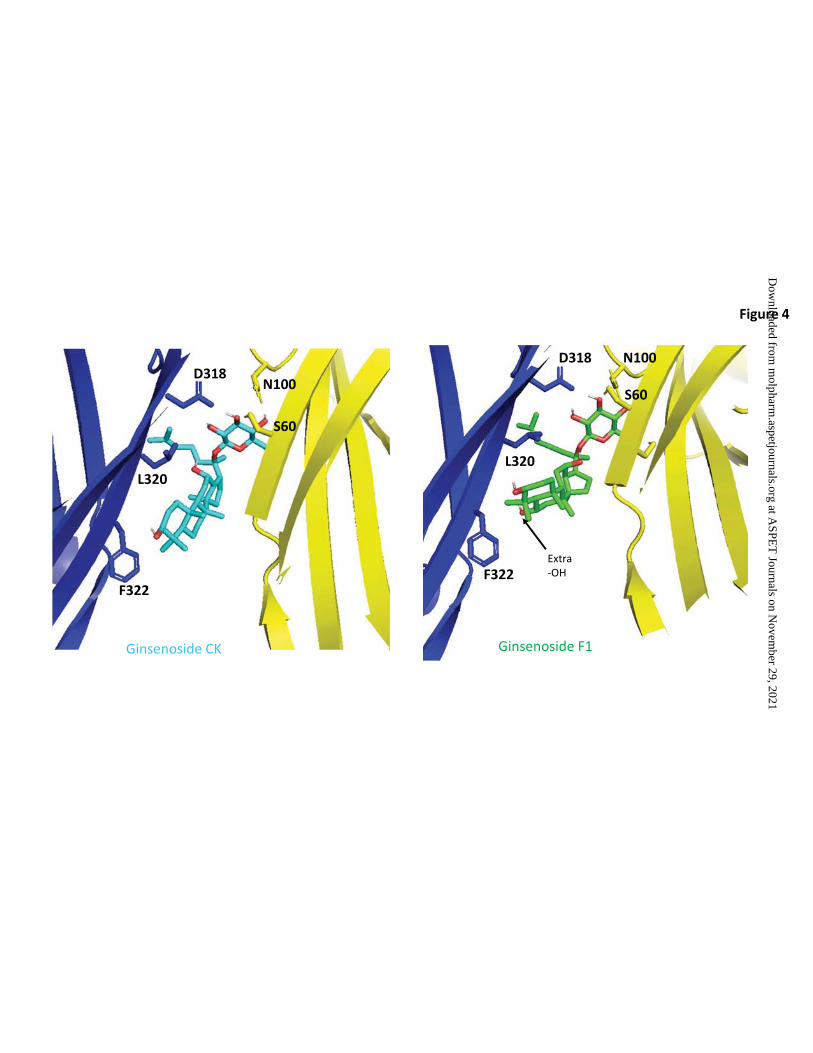
Computational docking
To investigate the predicted theoretical docking differences between ginsenoside-CK
and ginsenoside-F1 we used computational docking to a homology model of human P2X7
(Bidula et al., 2019b). Replacing ginsenoside-CK (Figure 4A) in the open ATP-bound model
of hP2X7 with ginsenoside-F1 resulted in an analogous predicted pose (Figure 4B) and this
would orient the additional hydroxyl group on C-6 to be facing the hydrophobic side of the
pocket close to L320 and F322. Since ginsenoside-F1 has no activity at P2X7, this purely
theoretical pose is suggestive that steric hindrance and a repulsive effect would prevent F1
from interacting with the PAM site at P2X7. Furthermore, there would likely be an energy
penalty due to poor solvation of the additional OH group within the hydrophobic pocket.
Concurrently we investigated chemicals thought to bind in an inverted mode such as
ginsenoside-Rd (Bidula et al., 2019b). Ginsenosides with sugars attached to C-3 rather than
This article has not been copyedited and formatted. The final version may differ from this version.Molecular Pharmacology Fast Forward. Published on December 17, 2020 as DOI: 10.1124/molpharm.120.000129
at ASPE
T Journals on N
ovember 29, 2021
molpharm
.aspetjournals.orgD
ownloaded from

15
C-20 are predicted to insert sugars deep into the pocket and would have the C-20 side chain
solvent-exposed. We have previously demonstrated that the monosaccharide ginsenoside-
Rh2 is able to potentiate hP2X7 responses (Helliwell et al., 2015) and have predicted a
binding model (Bidula et al., 2019b). Ginsenosides Rh2 and Rg3 are also predicted to bind
in this inverted mode and both of these ginsenosides can exist as two diasteromers with
regard to the C-20 side chain. The natural product form in Panax ginseng is believed to be
the 20(S)-diastereomer (Qi et al., 2011, Yang et al., 2014) and this is thought to have higher
bioactivity than the 20(R)-diastereomer (Wei et al., 2012). We investigated the individual
pure diastereomers to determine if this had any bearing on PAM activity at P2X7 and found
that only the 20(S)-diastereomers of Rg3 and Rh2 are active as PAMs at hP2X7 (Figure 5).
This suggests that the stereospecific positioning of the –OH group on C-20 may be critical
for mediating the potentiating effect. To investigate which part of P2X7 would be closest to
C-20, we used computational induced-fit docking using Glide as previously reported (Bidula
et al., 2019b). The most populated pose for 20(S)-Rg3 is presented in Figure 6 and this
places the C-20 hydroxyl group pointing away from the hydrophobic side of the pocket. In an
analogous theoretical pose, 20(R)-Rg3 would have this C-20 hydroxyl group in close
proximity to the hydrophobic face increasing steric hindrance and repulsive effects plus the
desolvation penalty as mentioned above, preventing 20(R)-Rg3 and 20(R)-Rh2 from
interacting with the binding pocket on P2X7.
Modulation of endogenous human P2X7 in immune cells
Finally, we verified our findings on the identified active vs inactive glycosides by
using a human THP-1 monocytic cell line known to endogenously express P2X7 (Figure 7).
Using fura-2AM loaded cells, we measured ATP-induced calcium responses (Figure 7A)
and found that there was a rapid peak increase in calcium followed by a sustained elevation
of calcium over 300 seconds of recording. Pre-treatment of THP-1 cells with commercially
available P2X7 selective antagonists such as AZ11645373 or AZ10606120 did not
This article has not been copyedited and formatted. The final version may differ from this version.Molecular Pharmacology Fast Forward. Published on December 17, 2020 as DOI: 10.1124/molpharm.120.000129
at ASPE
T Journals on N
ovember 29, 2021
molpharm
.aspetjournals.orgD
ownloaded from
16
dramatically affect the response to 500 µM ATP (Figure 7) but co-application of
ginsenoside-CK with 500 µM ATP increased the peak response and the sustained elevation
in [Ca2+]i (Figure 7). This increased response could be completely abolished in cells pre-
treated with either AZ11645373 (10 µM) or AZ10606120 (10 µM) suggesting that the
ginsenoside-CK potentiated response is solely mediated by P2X7 with no involvement of
other purinergic receptors. Using the same protocol we investigated whether we could verify
active and inactive glycosides as PAMs and confirmed that ginsenosides F2, Rd and 20(S)-
Rg3 all potentiated the ATP-induced calcium response in THP-1 cells and this potentiation
was abolished by pre-treatment with a P2X7-selective antagonist (Figure 7C).
We then determined if the identified active PAMs ginsenoside F2, 20(S)-Rg3 and
gypenoside XVII could enhance P2X7-dependent cell death in the highly expressing HEK-
hP2X7 model using ginsenoside-CK as a positive control. Both ginsenoside-F2 and 20(S)-
Rg3 could reduce cell viability over 24 hours when applied in combination with ATP and this
was prevented by pre-treatment with AZ10606120 (Figure 7D). However, neither
gypenoside XVII nor 20(R)-Rg3 in combination with ATP affected cell viability (Figure 7D).
In THP-1 cells, treatment with 500 µM ATP significantly reduced cell viability to 73.3 ± 14.4
% of control (Figure 7E) although this may not be P2X7-dependent due to the lack of effect
of AZ10606120. Only ginsenoside-CK could significantly enhance this ATP-induced cell
death in THP-1 cells (Figure 7E).
This article has not been copyedited and formatted. The final version may differ from this version.Molecular Pharmacology Fast Forward. Published on December 17, 2020 as DOI: 10.1124/molpharm.120.000129
at ASPE
T Journals on N
ovember 29, 2021
molpharm
.aspetjournals.orgD
ownloaded from

17
Discussion
In summary, we have demonstrated that the chemical requirements for positive allosteric
modulators at P2X7 appear to be quite stringent. Dose-response experiments demonstrate
that ginsenoside-CK and ginsenoside-Rd have two effects on ATP-induced responses at
hP2X7 (Figure 1B); an increase in the maximum response (a Type I PAM effect) and a shift
in the dose-response curve to the left (a Type II PAM effect) thereby enhancing the
maximum effect of the agonist and reducing the EC50 value. Our previous work has shown
ginsenoside-CK, a triterpenoid glycoside with one glucose attachment, to have the best PAM
effect at human P2X7. Here we show that ginsenoside-F2 and ginsenoside 20(S)-Rg3 have
equivalent PAM activity at P2X7. We investigated a range of glycosides with different
numbers of carbohydrate groups attached, and alternative sugar groups to glucose. Of
these, gypenoside XVII showed the best PAM activity at P2X7. This glycoside is a
dammarene glycoside found in Panax species, typically Panax notoginseng (Sakah et al.,
2013). Gypenoside XVII has a single glucose attached at C-3 and two glucose groups
attached at C-20 (a β-D-glucopyranosyl-(1-6)-β-D-glucopyranoside). It is very similar in
structure to stevenleaf, (also known as gypenoside IX, Table 1) but the sugar attachments
on C-20 are different. Stevenleaf has a glucose-xylose disaccharide attached at C-20
whereas in gypenoside XVII, this is a glucose-glucose disaccharide. The reduced activity of
stevenleaf (Supplementary Figure 1) compared to gypenoside XVII suggests that there is a
preferential requirement for glucose within the binding pocket on P2X7. Similarly
esculentoside A has a similar structure to ginsenoside 20(S)-Rg3 with a disaccharide on C-3
although this is composed of glucose-xylose whereas Rg3 has a glucose-glucose
disaccharide. Esculentoside A was inactive at P2X7 (Supplementary Figure 1).
Most useful in terms of defining a structure-activity relationship for PAMs at P2X7, was the
finding that modifications at C-6 were not tolerated. Ginsenoside-F1 showed no PAM activity
at P2X7 and computational docking suggests that this additional hydroxyl on C-6 faces the
This article has not been copyedited and formatted. The final version may differ from this version.Molecular Pharmacology Fast Forward. Published on December 17, 2020 as DOI: 10.1124/molpharm.120.000129
at ASPE
T Journals on N
ovember 29, 2021
molpharm
.aspetjournals.orgD
ownloaded from

18
hydrophobic side of the predicted binding pocket. This may be incompatible with binding to
P2X7 in order to produce effective potentiation of responses. We have not performed a
binding assay to determine if this lack of activity equates to a lack of binding to P2X7 rather
than equivalent binding and a lack of effect. Currently there are no suitably labelled
ginsenosides available as probes and this is something we shall explore in future studies.
We also discovered that the stereochemistry of 20(S) vs 20(R) in the ginsenosides Rg3 and
Rh2 have a clear effect on PAM activity with only the 20(S) diastereomers retaining good
PAM activity. Again, computational docking suggests this C-20 hydroxyl group to be close to
the highly hydrophobic side of the predicted binding pocket and we hypothesize that this is
important in correct positioning leading to effective potentiation of ATP-dependent
responses. Regarding the differences in the terpenoid backbone, several of the compounds
we selected for testing contained carbohydrate at either end of the molecule but did not
contain the dammarene scaffold. For example, daucosterol is a monosaccharide (glucose)
but contains a sitosterol-like scaffold. This includes a double bond which changes the shape
of the scaffold and has a detrimental effect on activity at P2X7. The key elements to the
structure-activity relationship for glycosides at P2X7 are summarised in Figure 8 showing
two predicted binding modes for glycosides. Figure 8A shows the CK binding mode with C-
20 glucose inserted into the binding pocket. Figure 8B shows the inverted mode for
ginsenosides with C-3 glucose inserted into the binding pocket. Substitutions are tolerated
on C-3 and C-20, but glucose is the preferred sugar attachment. Monosaccharides have
higher activity than most disaccharides, and no substitution is tolerated at C-6 (Figure 8A).
In the inverted mode, the stereochemistry of the hydroxyl on C-20 is critical for activity
(Figure 8B). In both cases the dammarene scaffold is favoured over a sitosterol scaffold.
It is important to perform full dose-response analysis to determine those compounds
that have PAM effects at P2X7. Saikosaponin A and solanine could increase the ATP-
induced dye uptake in the screen on HEK-hP2X7 cells (Figure 2A) but further investigation
showed that these compounds had a toxic effect on the cells, inducing YO-PRO-1 dye
This article has not been copyedited and formatted. The final version may differ from this version.Molecular Pharmacology Fast Forward. Published on December 17, 2020 as DOI: 10.1124/molpharm.120.000129
at ASPE
T Journals on N
ovember 29, 2021
molpharm
.aspetjournals.orgD
ownloaded from

19
uptake in the absence of ATP or the absence of the P2X7 receptor (Figure 2).
Saikosaponins have been linked to multiple biological effects including induction of apoptosis
(Li et al., 2018).
Several groups have demonstrated endogenous steroidal compounds to have
positive allosteric modulator activity at P2X receptors. Dehydroepiandrosterone and
progesterone can potentiate rodent P2X2 receptors (De Roo et al., 2010, De Roo et al.,
2003) and 17β-ester derivatives of testosterone also potentiate rat P2X2 and P2X4 (Sivcev
et al., 2019). None of these compounds had activity at P2X7 receptors. Lithocholic acid, a
bile acid, has potentiating activity at P2X7 and P2X4 (rat) but inhibits rat P2X2 (Sivcev et al.,
2020). This suggests there may exist endogenous regulators of P2X channel activity. It has
been suggested that the bile acids may share a similar binding site to ivermectin, close to
TM1 (Sivcev et al., 2020).
Positive allosteric modulation of P2X7 may be important in a number of contexts
(Stokes et al., 2020). In this study we used the human monocytic cell line, THP-1 which has
been used in other studies to investigate human P2X7 responses (Gadeock et al., 2010,
Stokes et al., 2006). Native P2X7 responses are small in this cell line, likely masked by the
multitude of other purinergic ATP-responsive receptors present. P2X7-selective antagonists
did not affect the ATP-induced calcium responses dramatically (Figure 7A) yet we did see
robust potentiation of the ATP-induced calcium response which was mediated by P2X7. The
THP-1 cell line carries a loss-of-function polymorphism in the C-terminus of P2X7,
rs3751143 encoding the Glu496>Ala substitution. Sequencing of exon 13 reveals
heterozygosity for rs3751143 (Supplementary Figure 2) confirming the work in (Gadeock et
al., 2012). Therefore, our data clearly shows that P2X7 responses in THP-1 cells can be
enhanced by CK and we can effectively rescue deficient P2X7 receptor responses in
humans carrying this loss-of-function polymorphism. Ginsenosides F2 and Rd were also
effective at increasing P2X7 responses in THP-1 cells (Figure 7) and ginsenoside-F1 was
inactive. Extending this set of data which measures immediate P2X7 responses, we looked
This article has not been copyedited and formatted. The final version may differ from this version.Molecular Pharmacology Fast Forward. Published on December 17, 2020 as DOI: 10.1124/molpharm.120.000129
at ASPE
T Journals on N
ovember 29, 2021
molpharm
.aspetjournals.orgD
ownloaded from

20
at P2X7-mediated cell death first in HEK-hP2X7 cells using an Alamar Blue viability assay.
Ginsenosides CK, Rd, 20(S)-Rg3 and F2 were very effective at enhancing cell death in
combination with ATP, however, gypenoside XVII was unable to enhance P2X7-mediated
cell death in this cell line (Figure 7D). At this stage, it is unclear why this would be the case.
Future investigations into the longevity of the potentiation may reveal that gypenoside XVII
has a shorter duration of action than the ginsenosides perhaps due to differential binding
modes. Repeating this work in THP-1 cells showed that only ginsenoside-CK was effective
at potentiation of cell death induced by ATP (Figure 7E). This may be due to the lower level
of expression of P2X7 in THP-1 cells, the expression of other purinergic receptors that may
bind ginsenosides such as P2X4, or simply due to duration of action. It is also conceivable
that monocytes may release factors such as glycosidase enzymes that can degrade
ginsenosides.
Other compounds can act as positive allosteric modulators of P2X7 (Stokes et al.,
2020) including clemastine (Norenberg et al., 2011), tenidap (Sanz et al., 1998), ivermectin
(Nörenberg et al., 2012) and polymyxin B (Ferrari et al., 2004). These bear no structural
similarities to ginsenosides although ivermectin is a glycoside, containing two oleandrose
sugars. As yet nothing is known about the potential site of action of ivermectin on P2X7
although on P2X4 the site is proposed to be close to TM1 (Asatryan et al., 2010, Samways
et al., 2012). In terms of therapeutic relevance of PAMs acting at P2X7, most exciting is the
potential to enhance the microbicidal activity of immune cells. P2X7 has been implicated in
regulation of pathogen killing, particularly those which reside in intracellular locations such
as mycobacteria and parasites (Di Virgilio et al., 2017, Savio et al., 2018). The mechanisms
by which P2X7 contributes to reduction of pathogen burden are not yet well understood but
could involve cytokine/mediator secretion, ROS generation or host cell apoptosis.
Potentiation of P2X7 responses with ginsenoside-CK changed the type of cell death in J774
macrophage cell line compared to high concentrations of ATP (Bidula et al., 2019a) and this
could be relevant within an immune response. In models of infection, most of the work has
This article has not been copyedited and formatted. The final version may differ from this version.Molecular Pharmacology Fast Forward. Published on December 17, 2020 as DOI: 10.1124/molpharm.120.000129
at ASPE
T Journals on N
ovember 29, 2021
molpharm
.aspetjournals.orgD
ownloaded from

21
been performed on P2X7 deficient mice to understand the role of this receptor within the
host immune response. However, there has not been an in vivo study to investigate
pharmacological targeting of P2X7 with a PAM. This idea has been tested in a zebrafish
whole-animal study for Mycobacterium marinum infection using clemastine to potentiate
P2X7 (Matty et al., 2019) providing evidence that targeting P2X7 may be beneficial to control
mycobacterial infections.
This article has not been copyedited and formatted. The final version may differ from this version.Molecular Pharmacology Fast Forward. Published on December 17, 2020 as DOI: 10.1124/molpharm.120.000129
at ASPE
T Journals on N
ovember 29, 2021
molpharm
.aspetjournals.orgD
ownloaded from

22
Acknowledgements:
Author contributions:
Participated in research design: Stokes, Ganesan, Bidula
Conducted experiments: Piyasirananda, Stokes, Beekman
Performed data analysis: Piyasirananda, Stokes, Beekman
Wrote, or contributed to writing of the manuscript: Piyasirananda, Stokes, Beekman,
Ganesan, Bidula.
This article has not been copyedited and formatted. The final version may differ from this version.Molecular Pharmacology Fast Forward. Published on December 17, 2020 as DOI: 10.1124/molpharm.120.000129
at ASPE
T Journals on N
ovember 29, 2021
molpharm
.aspetjournals.orgD
ownloaded from

23
References
AREESHI, M. Y., MANDAL, R. K., DAR, S., WAHID, M., KHAN, M. E., PANDA, A. K., JAWED, A. & HAQUE, S. 2015. P2X71513 A>C Polymorphism Confers Increased Risk of Extrapulmonary Tuberculosis: A Meta-analysis of Case-Control Studies. Curr Genomics, 17, 450-458.
ASATRYAN, L., POPOVA, M., PERKINS, D., TRUDELL, J. R., ALKANA, R. L. & DAVIES, D. L. 2010. Ivermectin antagonizes ethanol inhibition in purinergic P2X4 receptors. J Pharmacol Exp Ther, 334, 720-8.
BIDULA, S., DHUNA, K., HELLIWELL, R. & STOKES, L. 2019a. Positive allosteric modulation of P2X7 promotes apoptotic cell death over lytic cell death responses in macrophages. Cell Death Dis, 10, 882.
BIDULA, S. M., CROMER, B. A., WALPOLE, S., ANGULO, J. & STOKES, L. 2019b. Mapping a novel positive allosteric modulator binding site in the central vestibule region of human P2X7. Sci Rep, 9, 3231.
CHAVES, M. M., MARQUES-DA-SILVA, C., MONTEIRO, A. P., CANETTI, C. & COUTINHO-SILVA, R. 2014. Leukotriene B4 modulates P2X7 receptor-mediated Leishmania amazonensis elimination in murine macrophages. J Immunol, 192, 4765-73.
CHAVES, M. M., SINFLORIO, D. A., THORSTENBERG, M. L., MARTINS, M. D. A., MOREIRA-SOUZA, A. C. A., RANGEL, T. P., SILVA, C. L. M., BELLIO, M., CANETTI, C. & COUTINHO-SILVA, R. 2019. Non-canonical NLRP3 inflammasome activation and IL-1β signaling are necessary to L. amazonensis control mediated by P2X7 receptor and leukotriene B4. PLoS pathogens, 15, e1007887-e1007887.
DE ROO, M., BOUÉ-GRABOT, E. & SCHLICHTER, R. 2010. Selective potentiation of homomeric P2X2 ionotropic ATP receptors by a fast non-genomic action of progesterone. Neuropharmacology, 58, 569-577.
DE ROO, M., RODEAU, J. L. & SCHLICHTER, R. 2003. Dehydroepiandrosterone potentiates native ionotropic ATP receptors containing the P2X2 subunit in rat sensory neurones. J Physiol, 552, 59-71.
DI VIRGILIO, F., DAL BEN, D., SARTI, A. C., GIULIANI, A. L. & FALZONI, S. 2017. The P2X7 Receptor in Infection and Inflammation. Immunity, 47, 15-31.
FAIRBAIRN, I. P., STOBER, C. B., KUMARARATNE, D. S. & LAMMAS, D. A. 2001. ATP-mediated killing of intracellular mycobacteria by macrophages is a P2X(7)-dependent process inducing bacterial death by phagosome-lysosome fusion. Journal of immunology (Baltimore, Md. : 1950), 167, 3300-3307.
FERNANDO, S. L., SAUNDERS, B. M., SLUYTER, R., SKARRATT, K. K., GOLDBERG, H., MARKS, G. B., WILEY, J. S. & BRITTON, W. J. 2007. A polymorphism in the P2X7 gene increases susceptibility to extrapulmonary tuberculosis. Am J Respir Crit Care Med, 175, 360-6.
FERRARI, D., PIZZIRANI, C., ADINOLFI, E., FORCHAP, S., SITTA, B., TURCHET, L., FALZONI, S., MINELLI, M., BARICORDI, R. & DI VIRGILIO, F. 2004. The antibiotic polymyxin B modulates P2X7 receptor function. J Immunol, 173, 4652-60.
FRIESNER, R. A., BANKS, J. L., MURPHY, R. B., HALGREN, T. A., KLICIC, J. J., MAINZ, D. T., REPASKY, M. P., KNOLL, E. H., SHELLEY, M., PERRY, J. K., SHAW, D. E., FRANCIS, P. & SHENKIN, P. S. 2004. Glide: a new approach for rapid, accurate docking and scoring. 1. Method and assessment of docking accuracy. J Med Chem, 47, 1739-49.
GADEOCK, S., J, T., GEORGIOU, J. G., JALILIAN, I., TAYLOR, R. M., WILEY, J. S. & SLUYTER, R. 2010. TGF-β1 prevents up-regulation of the P2X7 receptor by IFN-γ and LPS in leukemic THP-1 monocytes. Biochim. Biophys. Acta, 1798, 2058-2066.
GADEOCK, S., PUPOVAC, A., SLUYTER, V., SPILDREJORDE, M. & SLUYTER, R. 2012. P2X7 receptor activation mediates organic cation uptake into human myeloid leukaemic KG-1 cells. Purinergic Signal, 8, 669-76.
This article has not been copyedited and formatted. The final version may differ from this version.Molecular Pharmacology Fast Forward. Published on December 17, 2020 as DOI: 10.1124/molpharm.120.000129
at ASPE
T Journals on N
ovember 29, 2021
molpharm
.aspetjournals.orgD
ownloaded from
24
GIULIANI, A. L., SARTI, A. C., FALZONI, S. & DI VIRGILIO, F. 2017. The P2X7 Receptor-Interleukin-1 Liaison. Frontiers in Pharmacology, 8, 123.
HACKOS, D. H. & HANSON, J. E. 2017. Diverse modes of NMDA receptor positive allosteric modulation: Mechanisms and consequences. Neuropharmacology, 112, 34-45.
HALGREN, T. 2007. New method for fast and accurate binding-site identification and analysis. Chem Biol Drug Des, 69, 146-8.
HELLIWELL, R. M., SHIOUKHUEY, C. O., DHUNA, K., MOLERO, J. C., YE, J. M., XUE, C. C. & STOKES, L. 2015. Selected ginsenosides of the protopanaxdiol series are novel positive allosteric modulators of P2X7 receptors. Br J Pharmacol, 172, 3326-40.
JACOBSON, M. P., PINCUS, D. L., RAPP, C. S., DAY, T. J., HONIG, B., SHAW, D. E. & FRIESNER, R. A. 2004. A hierarchical approach to all-atom protein loop prediction. Proteins, 55, 351-67.
LI, X., LI, X., HUANG, N., LIU, R. & SUN, R. 2018. A comprehensive review and perspectives on pharmacology and toxicology of saikosaponins. Phytomedicine, 50, 73-87.
MATTY, M. A., KNUDSEN, D. R., WALTON, E. M., BEERMAN, R. W., CRONAN, M. R., PYLE, C. J., HERNANDEZ, R. E. & TOBIN, D. M. 2019. Potentiation of P2RX7 as a host-directed strategy for control of mycobacterial infection. eLife, 8, e39123.
MILLER, C. M., ZAKRZEWSKI, A. M., ROBINSON, D. P., FULLER, S. J., WALKER, R. A., IKIN, R. J., BAO, S. J., GRIGG, M. E., WILEY, J. S. & SMITH, N. C. 2015. Lack of a Functioning P2X7 Receptor Leads to Increased Susceptibility to Toxoplasmic Ileitis. PLoS One, 10, e0129048.
MOREIRA-SOUZA, A. C. A., ALMEIDA-DA-SILVA, C. L. C., RANGEL, T. P., ROCHA, G. D. C., BELLIO, M., ZAMBONI, D. S., VOMMARO, R. C. & COUTINHO-SILVA, R. 2017. The P2X7 Receptor Mediates Toxoplasma gondii Control in Macrophages through Canonical NLRP3 Inflammasome Activation and Reactive Oxygen Species Production. Front Immunol, 8, 1257.
NORENBERG, W., HEMPEL, C., URBAN, N., SOBOTTKA, H., ILLES, P. & SCHAEFER, M. 2011. Clemastine potentiates the human P2X7 receptor by sensitizing it to lower ATP concentrations. J Biol Chem, 286, 11067-81.
NÖRENBERG, W., SOBOTTKA, H., HEMPEL, C., PLÖTZ, T., FISCHER, W., SCHMALZING, G. & SCHAEFER, M. 2012. Positive allosteric modulation by ivermectin of human but not murine P2X7 receptors. British Journal of Pharmacology, 167, 48-66.
PLACIDO, R., AURICCHIO, G., FALZONI, S., BATTISTINI, L., COLIZZI, V., BRUNETTI, E., DI VIRGILIO, F. & MANCINO, G. 2006. P2X(7) purinergic receptors and extracellular ATP mediate apoptosis of human monocytes/macrophages infected with Mycobacterium tuberculosis reducing the intracellular bacterial viability. Cellular immunology, 244, 10-18.
QI, L. W., WANG, C. Z. & YUAN, C. S. 2011. Ginsenosides from American ginseng: chemical and pharmacological diversity. Phytochemistry, 72, 689-99.
SAKAH, K. J., WANG, T., LIU, L., CHEN, Y., HAN, L. & ZHANG, Y. 2013. Eight darmarane-type saponins isolated from the roots of Panax notoginseng. Acta Pharmaceutica Sinica B, 3, 381-384.
SAMWAYS, D. S., KHAKH, B. S. & EGAN, T. M. 2012. Allosteric modulation of Ca2+ flux in ligand-gated cation channel (P2X4) by actions on lateral portals. J Biol Chem, 287, 7594-602.
SANZ, J. M., CHIOZZI, P. & DI VIRGILIO, F. 1998. Tenidap enhances P2Z/P2X7 receptor signalling in macrophages. Eur J Pharmacol, 355, 235-44.
SAVIO, L. E. B., DE ANDRADE MELLO, P., DA SILVA, C. G. & COUTINHO-SILVA, R. 2018. The P2X7 Receptor in Inflammatory Diseases: Angel or Demon? Front Pharmacol, 9, 52.
SIVCEV, S., SLAVIKOVA, B., IVETIC, M., KNEZU, M., KUDOVA, E. & ZEMKOVA, H. 2020. Lithocholic acid inhibits P2X2 and potentiates P2X4 receptor channel gating. The Journal of Steroid Biochemistry and Molecular Biology, 105725.
SIVCEV, S., SLAVIKOVA, B., RUPERT, M., IVETIC, M., NEKARDOVA, M., KUDOVA, E. & ZEMKOVA, H. 2019. Synthetic testosterone derivatives modulate rat P2X2 and P2X4 receptor channel gating. Journal of Neurochemistry, 150, 28-43.
STOKES, L., BIDULA, S., BIBIC, L. & ALLUM, E. 2020. To inhibit or enhance? Is there a benefit to positive allosteric modulation of P2X receptors? . Frontiers in Pharmacology, 11.
This article has not been copyedited and formatted. The final version may differ from this version.Molecular Pharmacology Fast Forward. Published on December 17, 2020 as DOI: 10.1124/molpharm.120.000129
at ASPE
T Journals on N
ovember 29, 2021
molpharm
.aspetjournals.orgD
ownloaded from

25
STOKES, L., JIANG, L. H., ALCARAZ, L., BENT, J., BOWERS, K., FAGURA, M., FURBER, M., MORTIMORE, M., LAWSON, M., THEAKER, J., LAURENT, C., BRADDOCK, M. & SURPRENANT, A. 2006. Characterization of a selective and potent antagonist of human P2X(7) receptors, AZ11645373. Br J Pharmacol, 149, 880-7.
WEI, X., CHEN, J., SU, F., SU, X., HU, T. & HU, S. 2012. Stereospecificity of ginsenoside Rg3 in promotion of the immune response to ovalbumin in mice. Int Immunol, 24, 465-71.
YANG, W. Z., HU, Y., WU, W. Y., YE, M. & GUO, D. A. 2014. Saponins in the genus Panax L. (Araliaceae): a systematic review of their chemical diversity. Phytochemistry, 106, 7-24.
This article has not been copyedited and formatted. The final version may differ from this version.Molecular Pharmacology Fast Forward. Published on December 17, 2020 as DOI: 10.1124/molpharm.120.000129
at ASPE
T Journals on N
ovember 29, 2021
molpharm
.aspetjournals.orgD
ownloaded from

26
Footnotes:
This work was supported by a Royal Thai government funded PhD scholarship to WP and a
BBSRC project grant to LS (BB/N018427/1).
This article has not been copyedited and formatted. The final version may differ from this version.Molecular Pharmacology Fast Forward. Published on December 17, 2020 as DOI: 10.1124/molpharm.120.000129
at ASPE
T Journals on N
ovember 29, 2021
molpharm
.aspetjournals.orgD
ownloaded from
27
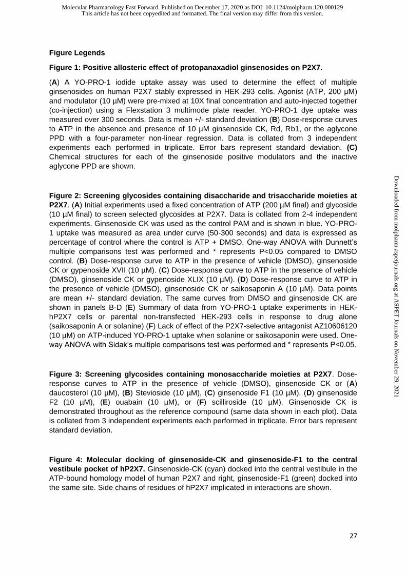
Figure Legends
Figure 1: Positive allosteric effect of protopanaxadiol ginsenosides on P2X7.
(A) A YO-PRO-1 iodide uptake assay was used to determine the effect of multiple
ginsenosides on human P2X7 stably expressed in HEK-293 cells. Agonist (ATP, 200 μM)
and modulator (10 µM) were pre-mixed at 10X final concentration and auto-injected together
(co-injection) using a Flexstation 3 multimode plate reader. YO-PRO-1 dye uptake was
measured over 300 seconds. Data is mean +/- standard deviation (B) Dose-response curves
to ATP in the absence and presence of 10 µM ginsenoside CK, Rd, Rb1, or the aglycone
PPD with a four-parameter non-linear regression. Data is collated from 3 independent
experiments each performed in triplicate. Error bars represent standard deviation. (C)
Chemical structures for each of the ginsenoside positive modulators and the inactive
aglycone PPD are shown.
Figure 2: Screening glycosides containing disaccharide and trisaccharide moieties at
P2X7. (A) Initial experiments used a fixed concentration of ATP (200 µM final) and glycoside
(10 µM final) to screen selected glycosides at P2X7. Data is collated from 2-4 independent
experiments. Ginsenoside CK was used as the control PAM and is shown in blue. YO-PRO-
1 uptake was measured as area under curve (50-300 seconds) and data is expressed as
percentage of control where the control is ATP + DMSO. One-way ANOVA with Dunnett’s
multiple comparisons test was performed and * represents P<0.05 compared to DMSO
control. (B) Dose-response curve to ATP in the presence of vehicle (DMSO), ginsenoside
CK or gypenoside XVII (10 µM). (C) Dose-response curve to ATP in the presence of vehicle
(DMSO), ginsenoside CK or gypenoside XLIX (10 µM). (D) Dose-response curve to ATP in
the presence of vehicle (DMSO), ginsenoside CK or saikosaponin A (10 µM). Data points
are mean +/- standard deviation. The same curves from DMSO and ginsenoside CK are
shown in panels B-D (E) Summary of data from YO-PRO-1 uptake experiments in HEK-
hP2X7 cells or parental non-transfected HEK-293 cells in response to drug alone
(saikosaponin A or solanine) (F) Lack of effect of the P2X7-selective antagonist AZ10606120
(10 µM) on ATP-induced YO-PRO-1 uptake when solanine or saikosaponin were used. One-
way ANOVA with Sidak’s multiple comparisons test was performed and * represents P<0.05.
Figure 3: Screening glycosides containing monosaccharide moieties at P2X7. Dose-
response curves to ATP in the presence of vehicle (DMSO), ginsenoside CK or (A)
daucosterol (10 µM), (B) Stevioside (10 µM), (C) ginsenoside F1 (10 µM), (D) ginsenoside
F2 (10 µM), (E) ouabain (10 µM), or (F) scilliroside (10 µM). Ginsenoside CK is
demonstrated throughout as the reference compound (same data shown in each plot). Data
is collated from 3 independent experiments each performed in triplicate. Error bars represent
standard deviation.
Figure 4: Molecular docking of ginsenoside-CK and ginsenoside-F1 to the central
vestibule pocket of hP2X7. Ginsenoside-CK (cyan) docked into the central vestibule in the
ATP-bound homology model of human P2X7 and right, ginsenoside-F1 (green) docked into
the same site. Side chains of residues of hP2X7 implicated in interactions are shown.
This article has not been copyedited and formatted. The final version may differ from this version.Molecular Pharmacology Fast Forward. Published on December 17, 2020 as DOI: 10.1124/molpharm.120.000129
at ASPE
T Journals on N
ovember 29, 2021
molpharm
.aspetjournals.orgD
ownloaded from
28
Figure 5: Diastereoisomers of ginsenosides have different activity at hP2X7.
(A) Dose-response curves to ATP in the presence of vehicle (DMSO), 20(S)-ginsenoside
Rg3 or 20(R)-ginsenoside Rg3 (10 µM), (B) ginsenoside 20(S)-Rh2 or 20(R)-Rh2 (10 µM).
Data is collated from 3 independent experiments and is mean +/- standard deviation. The
same DMSO data is shown in both plots. Chemical structures of ginsenosides Rg3 and Rh2
are shown with the stereo-centres highlighted in red.
Figure 6: Induced-fit docking of 20S-Rg3 at human P2X7
Ginsenoside-20(S)-Rg3 (green) docked into the central vestibule site in a homology model of
ATP-bound human P2X7 (open state). The predicted orientation of the stereocentre C-20 is
such that the –OH is pointing away from the hydrophobic face of the binding site thus
minimising any repulsive interactions. Key side chains of residues D318, L320 and F322 are
indicated.
Figure 7: The PAM effects of glycosides in human THP-1 monocyte cell line.
(A) ATP-induced calcium responses were measured in fura-2AM loaded THP-1 cells in
suspension using a Flexstation 3 plate reader. Agonist (500 µM ATP) and PAM (ginsenoside
CK 10 µM) were co-injected following establishment of a baseline for 40 seconds. Cells were
pre-incubated with various P2X7 antagonists for 10 minutes prior to start of plate recordings.
(B) Summary of collated data from calcium measurements. Fura-2 responses were
calculated as area under curve. Data was analysed using one-way ANOVA with Tukey’s
multiple comparisons test to assess the effect of antagonists. * represents P<0.05. (C)
Investigating the P2X7 dependence of glycoside effects on ATP-induced calcium responses.
AZ10606120 (10 µM) was added to cells to block P2X7 receptors prior to measuring calcium
responses. Data was analysed using one-way ANOVA with Dunnett’s multiple comparison
test comparing each column against the control (500 µM ATP+DMSO). * represents P<0.05.
(D) Cell viability experiments were performed over 24 hours using HEK-hP2X7 cell line.
Alamar blue fluorescence was measured and data was normalised to % of control (DMSO).
Data was collated from 5 independent experiments. One-way ANOVA was used to analyse
the data with Sidak’s multiple comparisons test to compare selected pairs of columns
(DMSO+ATP vs ginsenoside+ATP). * represents P<0.05. (E) Cell viability experiments were
performed over 24 hours using THP-1 cells. Alamar blue fluorescence was measured and
data was normalised to % of control (DMSO). Data was collated from 5 independent
experiments. One-way ANOVA was used to analyse the data with Sidak’s multiple
comparisons test to compare selected pairs of columns (DMSO+ATP vs ginsenoside+ATP).
* represents P<0.05.
Figure 8: Proposed structure-activity relationship for glycosides acting as PAMs at
P2X7. (A) The chemical structure of ginsenoside CK is shown with important groups
highlighted. Glucose attachment (cyan) is critical for activity at P2X7. C-6 substitutions are
not tolerated (yellow). (B) The chemical structure of ginsenoside 20(S)-Rg3 is demonstrated
with positioning relative to the P2X7 binding mode (inverted). The C-3 glucose attachments
(cyan) face up into the binding pocket. The C-20 hydroxyl group shows stereochemistry and
positioning is critical for activity at P2X7.
This article has not been copyedited and formatted. The final version may differ from this version.Molecular Pharmacology Fast Forward. Published on December 17, 2020 as DOI: 10.1124/molpharm.120.000129
at ASPE
T Journals on N
ovember 29, 2021
molpharm
.aspetjournals.orgD
ownloaded from

29
This article has not been copyedited and formatted. The final version may differ from this version.Molecular Pharmacology Fast Forward. Published on December 17, 2020 as DOI: 10.1124/molpharm.120.000129
at ASPE
T Journals on N
ovember 29, 2021
molpharm
.aspetjournals.orgD
ownloaded from
30
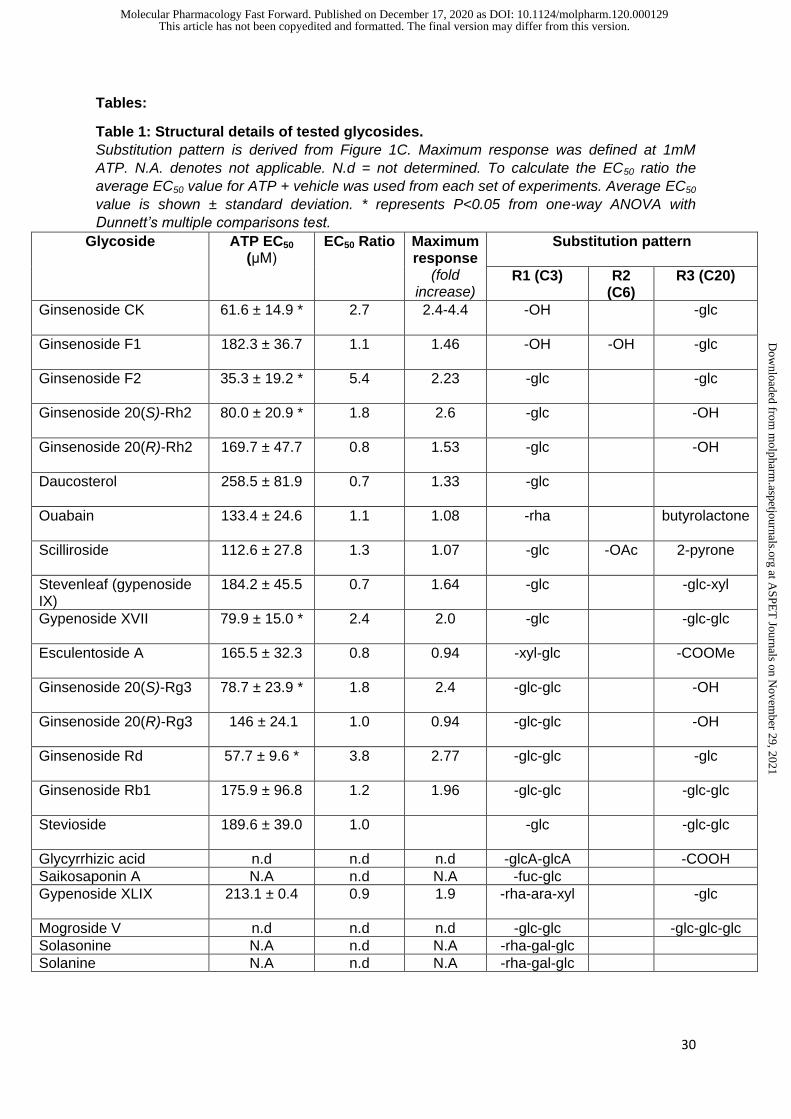
Tables:
Table 1: Structural details of tested glycosides.
Substitution pattern is derived from Figure 1C. Maximum response was defined at 1mM
ATP. N.A. denotes not applicable. N.d = not determined. To calculate the EC50 ratio the
average EC50 value for ATP + vehicle was used from each set of experiments. Average EC50
value is shown ± standard deviation. * represents P<0.05 from one-way ANOVA with
Dunnett’s multiple comparisons test.
Glycoside
ATP EC50
(μM) EC50 Ratio Maximum
response (fold
increase)
Substitution pattern
R1 (C3) R2 (C6)
R3 (C20)
Ginsenoside CK
61.6 ± 14.9 *
2.7 2.4-4.4 -OH -glc
Ginsenoside F1 182.3 ± 36.7
1.1 1.46 -OH -OH -glc
Ginsenoside F2
35.3 ± 19.2 *
5.4 2.23 -glc -glc
Ginsenoside 20(S)-Rh2
80.0 ± 20.9 * 1.8 2.6 -glc -OH
Ginsenoside 20(R)-Rh2
169.7 ± 47.7 0.8 1.53 -glc -OH
Daucosterol
258.5 ± 81.9
0.7 1.33 -glc
Ouabain
133.4 ± 24.6
1.1 1.08 -rha butyrolactone
Scilliroside 112.6 ± 27.8
1.3 1.07 -glc -OAc 2-pyrone
Stevenleaf (gypenoside IX)
184.2 ± 45.5 0.7 1.64 -glc -glc-xyl
Gypenoside XVII
79.9 ± 15.0 * 2.4 2.0 -glc -glc-glc
Esculentoside A
165.5 ± 32.3 0.8 0.94 -xyl-glc -COOMe
Ginsenoside 20(S)-Rg3
78.7 ± 23.9 * 1.8 2.4 -glc-glc
-OH
Ginsenoside 20(R)-Rg3
146 ± 24.1 1.0 0.94 -glc-glc -OH
Ginsenoside Rd 57.7 ± 9.6 *
3.8 2.77 -glc-glc -glc
Ginsenoside Rb1 175.9 ± 96.8
1.2 1.96 -glc-glc -glc-glc
Stevioside 189.6 ± 39.0
1.0 -glc -glc-glc
Glycyrrhizic acid n.d n.d n.d -glcA-glcA -COOH
Saikosaponin A N.A n.d N.A -fuc-glc
Gypenoside XLIX 213.1 ± 0.4
0.9 1.9 -rha-ara-xyl -glc
Mogroside V n.d n.d n.d -glc-glc -glc-glc-glc
Solasonine N.A n.d N.A -rha-gal-glc
Solanine N.A n.d N.A -rha-gal-glc
This article has not been copyedited and formatted. The final version may differ from this version.Molecular Pharmacology Fast Forward. Published on December 17, 2020 as DOI: 10.1124/molpharm.120.000129
at ASPE
T Journals on N
ovember 29, 2021
molpharm
.aspetjournals.orgD
ownloaded from

100 200 300
-50
0
50
100
150
Time (seconds)YO-P
RO-1
fluor
esce
nce
(RFU
)
ATP+vehATP+CK ATP+PPD
ATP+Rd
ATP+Drug
-5 -4 -3
-20000
0
20000
40000
60000
log [ATP] M
YO-P
RO
-1dy
eup
take
(Are
aun
derc
urve
50-3
00s)
ATP+vehATP+CK
ATP+RdATP+PPDATP+Rb1
Figure 1A B
C
H
H
HOH
H
20
17
6
1
3
R1R2
R3
PPDCKRdRb1
R1 R2 R3
-OH -H -OH-OH -H -glc-glcglc -H -glc-glcglc -H -glcglc
This article has not been copyedited and formatted. The final version may differ from this version.Molecular Pharmacology Fast Forward. Published on December 17, 2020 as DOI: 10.1124/molpharm.120.000129
at ASPE
T Journals on N
ovember 29, 2021
molpharm
.aspetjournals.orgD
ownloaded from

-6 -5 -4 -3 -20
25000
50000
75000
log[ATP], M
YO-P
RO
-1up
take
(Are
aun
derc
urve
50-3
00s)
DMSOCKGypenoside XLIX
-6 -5 -4 -3 -20
25000
50000
75000
100000
125000
log[ATP], M
YO-P
RO
-1up
take
(Are
aun
derc
urve
50-3
00s)
DMSOCKSaikosaponin A
0
20000
40000
60000
80000
100000
YO-P
RO
-1up
take
(Are
aun
derc
urve
50-3
00s)
ATP + + + + - + + - + +CK - - + + - - - - - -AZ106 - + - + - - + - - +Saikosaponin - - - - + + + - - -Solanine - - - - - - - + + +
**
ns ns
DMSO CK
Saikosa
poninA
solan
ineDMSO CK
Saikosa
poninA
solan
ine0
20000
40000
60000
80000
100000
YO-P
RO
-1up
take
(Are
aun
derc
urve
50-3
00s)
HEK-hP2X7 HEK-293
Figure 2
DMSO CK RdRg3
Gypen
oside XLIX
Esculen
toside A
mogroside
steve
nleaf
glycyrr
hizin
stevio
side
solas
onine
saiko
saponin
A
solan
ine0
400
800
1200
YO-P
RO
-1up
take
(Are
aun
derc
urve
;%
ofco
ntro
lres
pons
e)
A B
C D
E F
-6 -5 -4 -3 -20
10000
20000
30000
40000
log[ATP], M
YO-P
RO
-1dy
eup
take
(Are
aun
derc
urve
50-3
00s)
DMSOCKGypenoside XVII
*
**
This article has not been copyedited and formatted. The final version may differ from this version.Molecular Pharmacology Fast Forward. Published on December 17, 2020 as DOI: 10.1124/molpharm.120.000129
at ASPE
T Journals on N
ovember 29, 2021
molpharm
.aspetjournals.orgD
ownloaded from

-5.5 -5.0 -4.5 -4.0 -3.5 -3.0 -2.50
10000
20000
30000
40000
log [ATP] M
YO-P
RO
-1up
take
(Are
aun
derc
urve
50-3
00s)
DMSOCKF1
-5.5 -5.0 -4.5 -4.0 -3.5 -3.0 -2.50
10000
20000
30000
40000
log[ATP], M
YO-P
RO
-1up
take
(Are
aun
derc
urve
50-3
00s)
DMSOCKF2
-5.5 -5.0 -4.5 -4.0 -3.5 -3.0 -2.50
10000
20000
30000
40000
log[ATP], M
YO-P
RO
-1up
take
(Are
aun
derc
urve
50-3
00s)
DMSOCKOuabain
-5.5 -5.0 -4.5 -4.0 -3.5 -3.0 -2.50
10000
20000
30000
40000
log[ATP], M
YO-P
RO
-1up
take
(Are
aun
derc
urve
50-3
00s)
DMSO
Scilliroside
CK
A
Figure 3
-5.5 -5.0 -4.5 -4.0 -3.5 -3.0 -2.50
10000
20000
30000
40000
50000
log[ATP], M
YO-P
RO
-1up
take
(Are
aun
derc
urve
50-3
00s)
DMSO
DaucosterolCK
-5.5 -5.0 -4.5 -4.0 -3.5 -3.0 -2.50
10000
20000
30000
40000
50000
log [ATP] MYO
-PR
O-1
upta
ke(A
rea
unde
rcur
ve50
-300
s)
DMSOCKSV
B
C D
E F
This article has not been copyedited and formatted. The final version may differ from this version.Molecular Pharmacology Fast Forward. Published on December 17, 2020 as DOI: 10.1124/molpharm.120.000129
at ASPE
T Journals on N
ovember 29, 2021
molpharm
.aspetjournals.orgD
ownloaded from
F322
D318
L320
N100
S60
Extra-OH
Ginsenoside F1
S60
N100D318
L320
F322
Ginsenoside CK
Figure 4
This article has not been copyedited and formatted. The final version may differ from this version.Molecular Pharmacology Fast Forward. Published on December 17, 2020 as DOI: 10.1124/molpharm.120.000129
at ASPE
T Journals on N
ovember 29, 2021
molpharm
.aspetjournals.orgD
ownloaded from

OH
H
HOH
OH
H
O
OO
HO
HO
HO
HO
HO
OH
OH
20
63
OH
H
HOH
H
O
OO
HO
HO
HO
HO
HO
OH
OH
OH
63
20
Figure 5
20S-Rg3
20R-Rg3
-5 -4 -30
10000
20000
30000
40000
log [ATP], MYO
-PR
O-1
upta
ke(A
rea
unde
rcur
ve50
-300
s)
DMSO
20R-Rh220S-Rh2
-5 -4 -30
10000
20000
30000
40000
log[ATP], M
YO-P
RO
-1up
take
(Are
aun
derc
urve
50-3
00s)
DMSO
20R-Rg3
20S-Rg3
OH
H
HOH
OH
H
O
HO
HO
HO
OH
63
20
20R-Rh2
A B
OH
H
HOH
OH
H
O
HO
HO
HO
OH
20
63
20S-Rh2
C
This article has not been copyedited and formatted. The final version may differ from this version.Molecular Pharmacology Fast Forward. Published on December 17, 2020 as DOI: 10.1124/molpharm.120.000129
at ASPE
T Journals on N
ovember 29, 2021
molpharm
.aspetjournals.orgD
ownloaded from

ATPATP
D318
F322
L320
Figure 6
This article has not been copyedited and formatted. The final version may differ from this version.Molecular Pharmacology Fast Forward. Published on December 17, 2020 as DOI: 10.1124/molpharm.120.000129
at ASPE
T Journals on N
ovember 29, 2021
molpharm
.aspetjournals.orgD
ownloaded from

100 200 300-0.2
0.0
0.2
0.4
0.6
0.8
1.0
Time (sec)
Fura
-2Ra
tio(3
40/3
80nm
)
ATPAZ116 ATP
100 200 300-0.2
0.0
0.2
0.4
0.6
0.8
1.0
Time (seconds)
ATPCK and ATPAZ116 ATP and CK
buffer
500m
MATP
AZ116 + ATP
AZ106 + ATP
CK + ATP
AZ116+
CK+ATP
AZ106+
CK+ATP
0
100
200
300
Fura
-2Ra
tio(3
40/3
80nm
)A
rea
unde
rcur
ve
** **
100 200 300-0.2
0.0
0.2
0.4
0.6
0.8
1.0
Time (seconds)
ATPCK and ATPAZ106 CK and ATP
buffer
500m
MATP+D
MSO
CK+ATP
AZ106+
CK+ATP
F2+ATP
AZ106+
F2 ATP
Rd+ATP
AZ106+
Rd+ATP
20(S)R
g3 + ATP
AZ106+
20(S)R
g3 + ATP
GPS XVII +ATP
AZ106 + GPS XVII +
ATP
F1+ATP
AZ106+
F1 ATP0
100
200
300
Fura
-2Ra
tio(3
40/3
80nm
)A
rea
unde
rcur
ve
**
* *
A
B C
Figure 7
D
DMSOco
ntrol
DMSO+ATP
AZ106+
ATP CK
CK+ATP
AZ106+
CK ATP Rd
Rd+ATP
AZ106+
Rd ATP F2
F2+ATP
AZ106+
F2 ATP
GPS XVII
GPS XVII +ATP
AZ106 + GPS XVII +
ATP
20(S)R
g3
20(S)R
g3 + ATP
AZ106 + 20
(S)R
g3 + ATP
20(R
)Rg3
20(R
)Rg3 + ATP
AZ106 + 20
(R)R
g3 + ATP0
50
100
150
200
Cel
lvia
bilit
y%
ofD
MSO
cont
rol
* * * *
E
DMSOco
ntrol
DMSO+ATP
AZ106+
ATP CK
CK+ATP
AZ106+
CK+ATP Rd
Rd+ATP
AZ106 + Rd+ATP F2
F2+ATP
AZ106 + F2+
ATP
GPS XVII
GPS XVII+ATP
AZ106 + GPS XVII+
ATP
20(S)R
g3
20(S)R
g3+ATP
AZ106 + 20
(S)Rg3+
ATP
20(R
)Rg3
20(R
)Rg3+
ATP
AZ106 + 20
(R)R
g3 ATP0
50
100
150
Cel
lvia
bilit
y(%
ofco
ntro
l)
*
This article has not been copyedited and formatted. The final version may differ from this version.Molecular Pharmacology Fast Forward. Published on December 17, 2020 as DOI: 10.1124/molpharm.120.000129
at ASPE
T Journals on N
ovember 29, 2021
molpharm
.aspetjournals.orgD
ownloaded from

This article has not been copyedited and formatted. The final version may differ from this version.Molecular Pharmacology Fast Forward. Published on December 17, 2020 as DOI: 10.1124/molpharm.120.000129
at ASPE
T Journals on N
ovember 29, 2021
molpharm
.aspetjournals.orgD
ownloaded from