Embed Size (px)
Citation preview

10/30/2016
1
Viruses, viroids, and prions
Chapter 13
BIO 220
Fig. 13.1
Characteristics of viruses
• Very, very small (filterable)
• Obligatory intracellular parasite
• They have no ribosomes, so must use host cell machinery to translate viral mRNA into viral proteins
• Do not store or generate ATP, so energy is derived from the host cell
• Parasitize host cell for building materials like amino acids, lipids, and nucleotides
• Without the host cell, viruses can not carry out “life”-sustaining processes
10/30/2016
2
Host range of virus
• Spectrum of cells virus can invade
• Most viruses can only infect specific types of
cells of only one host species
• Range determined by
– Virus must be able to interact with specific
receptor sites on host cell surface
– Availability within the specific host of cellular
factors necessary for viral multiplication
Viral structure
• Viruses are composed of a nucleic acid surrounded by a protein coat called a capsid
• Some viruses have a lipid/protein/CHO envelope surrounding the capsid
• A virion is a complete, fully developed, infectious viral particle located outside a host cell
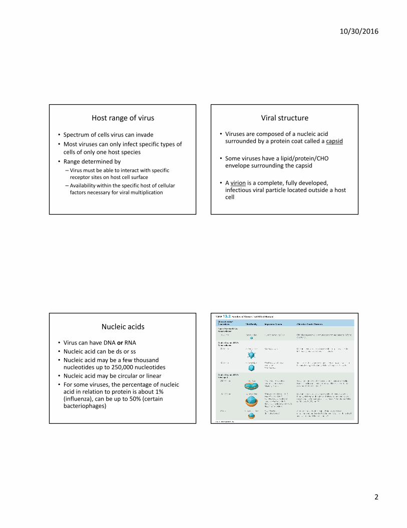
Nucleic acids
• Virus can have DNA or RNA
• Nucleic acid can be ds or ss
• Nucleic acid may be a few thousand nucleotides up to 250,000 nucleotides
• Nucleic acid may be circular or linear
• For some viruses, the percentage of nucleic acid in relation to protein is about 1% (influenza), can be up to 50% (certain bacteriophages)
10/30/2016
3
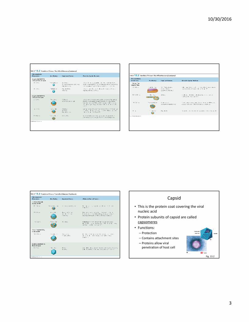
Capsid
• This is the protein coat covering the viral
nucleic acid
• Protein subunits of capsid are called
capsomeres
• Functions:
– Protection
– Contains attachment sites
– Proteins allow viral
penetration of host cell
Fig. 13.2
10/30/2016
4
Envelopes
• Nonenveloped viruses lack an envelope
• Enveloped viruses do have an envelope
• Some viral capsids are covered by envelopes which may be made of lipids, proteins, and CHOs
– May be a result of extrusion from host cell
– Viral nucleic acid codes for envelope proteins, other components derived from the host cell
• Some envelopes may be covered in spikes (CHO/protein complexes)
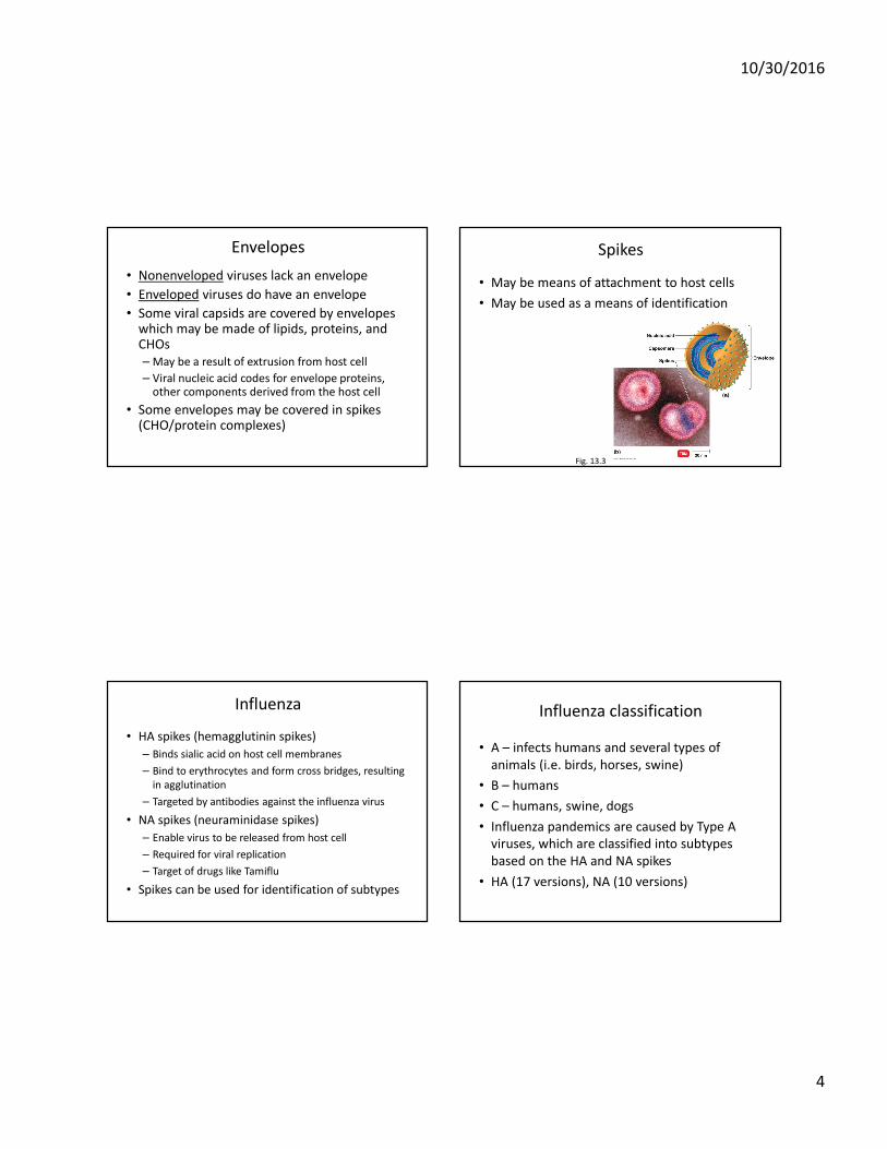
Spikes
• May be means of attachment to host cells
• May be used as a means of identification
Fig. 13.3
Influenza
• HA spikes (hemagglutinin spikes)
– Binds sialic acid on host cell membranes
– Bind to erythrocytes and form cross bridges, resulting
in agglutination
– Targeted by antibodies against the influenza virus
• NA spikes (neuraminidase spikes)
– Enable virus to be released from host cell
– Required for viral replication
– Target of drugs like Tamiflu
• Spikes can be used for identification of subtypes
Influenza classification
• A – infects humans and several types of
animals (i.e. birds, horses, swine)
• B – humans
• C – humans, swine, dogs
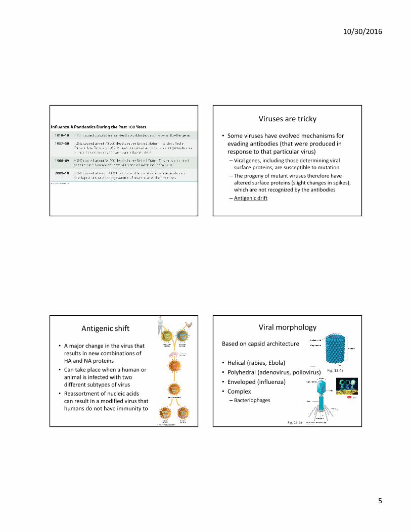
• Influenza pandemics are caused by Type A
viruses, which are classified into subtypes
based on the HA and NA spikes
• HA (17 versions), NA (10 versions)
10/30/2016
5
Viruses are tricky
• Some viruses have evolved mechanisms for
evading antibodies (that were produced in
response to that particular virus)
– Viral genes, including those determining viral
surface proteins, are susceptible to mutation
– The progeny of mutant viruses therefore have
altered surface proteins (slight changes in spikes),
which are not recognized by the antibodies
– Antigenic drift
Antigenic shift
• A major change in the virus that
results in new combinations of
HA and NA proteins
• Can take place when a human or
animal is infected with two
different subtypes of virus
• Reassortment of nucleic acids
can result in a modified virus that
humans do not have immunity to
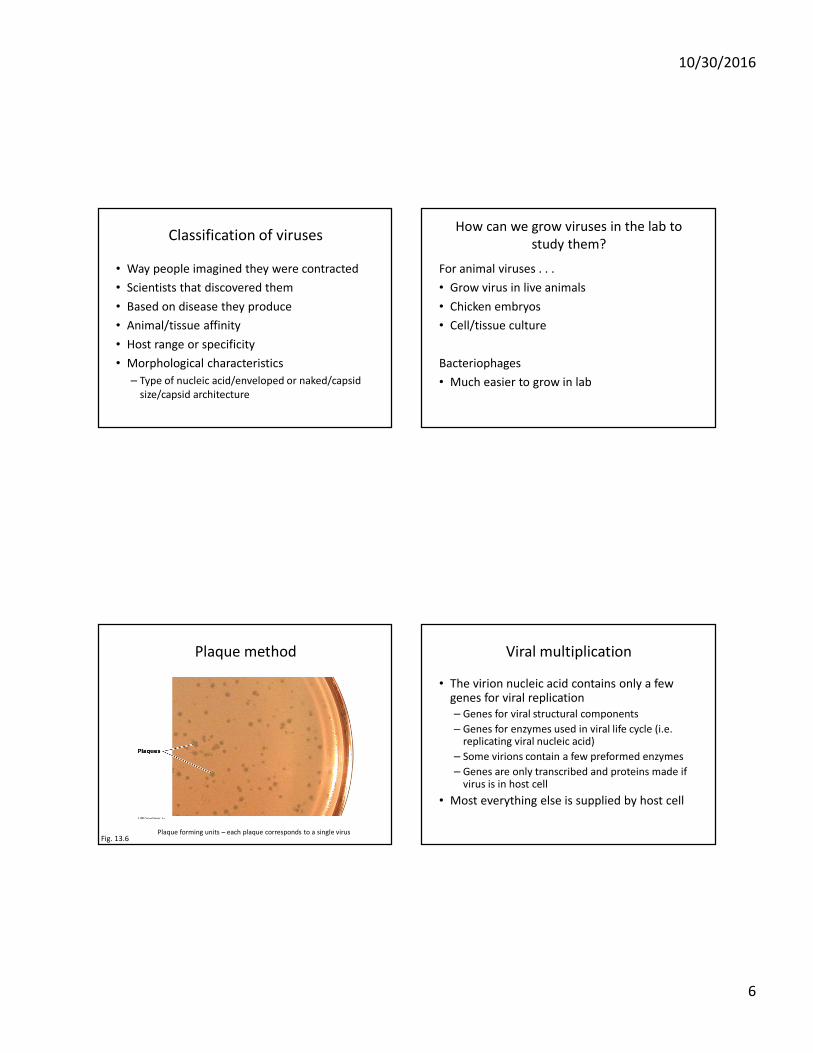
Viral morphology
Based on capsid architecture
• Helical (rabies, Ebola)
• Polyhedral (adenovirus, poliovirus)
• Enveloped (influenza)
• Complex
– Bacteriophages
Fig. 13.5a
Fig. 13.4a
10/30/2016
6
Classification of viruses
• Way people imagined they were contracted
• Scientists that discovered them
• Based on disease they produce
• Animal/tissue affinity
• Host range or specificity
• Morphological characteristics
– Type of nucleic acid/enveloped or naked/capsid
size/capsid architecture
How can we grow viruses in the lab to
study them?
For animal viruses . . .
• Grow virus in live animals
• Chicken embryos
• Cell/tissue culture
Bacteriophages
• Much easier to grow in lab
Plaque method
Fig. 13.6Plaque forming units – each plaque corresponds to a single virus
Viral multiplication
• The virion nucleic acid contains only a few genes for viral replication
– Genes for viral structural components
– Genes for enzymes used in viral life cycle (i.e. replicating viral nucleic acid)
– Some virions contain a few preformed enzymes
– Genes are only transcribed and proteins made if virus is in host cell
• Most everything else is supplied by host cell

10/30/2016
7
Viral one-step growth curve
Fig. 13.10
Bacteriophage multiplication
• The lytic cycle (T-even bacteriophage)
– Ends with the lysis and death of host cell
• The lysogenic cycle (Bacteriophage λ)
– Host cell lives
Virulent phages
• Undergoes the lytic cycle
• The result of the lytic cycle is viral replication
and death of the host cell as mature virions
are released
Phage lysozyme
Degradation host DNA
Viral mRNA transcribed/translated
Phage components synthesized
Lysozyme
Fig. 13.11
10/30/2016
8
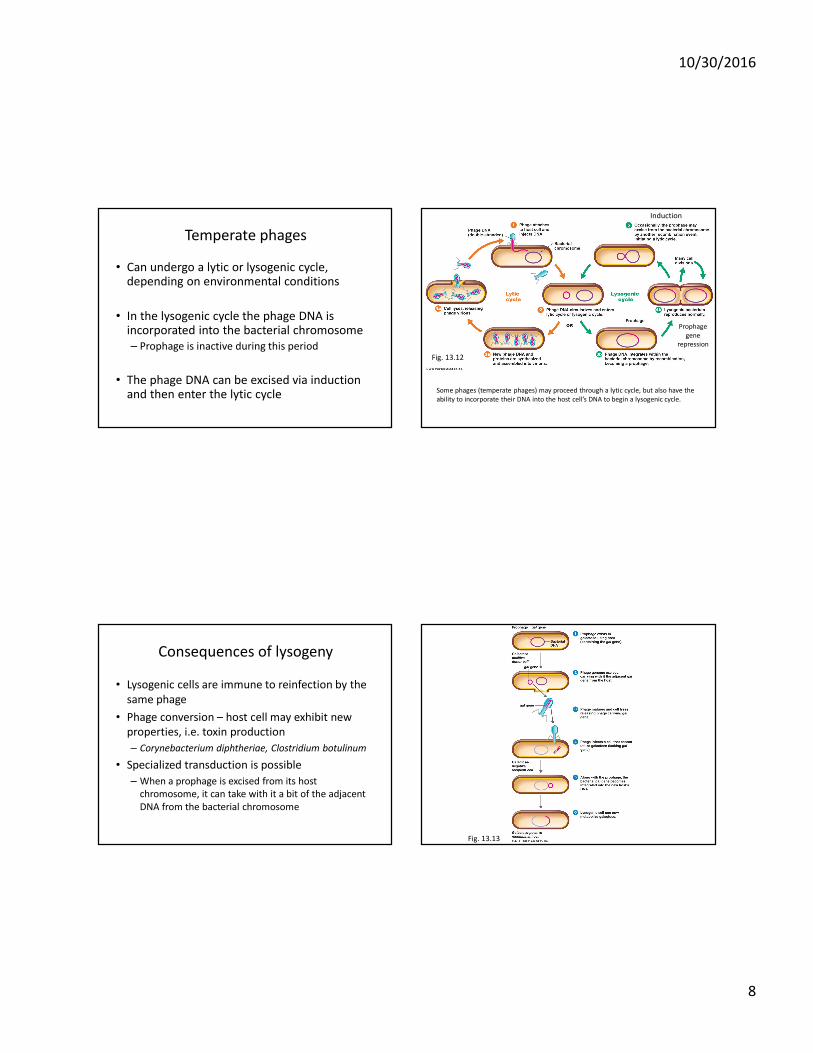
Temperate phages
• Can undergo a lytic or lysogenic cycle, depending on environmental conditions
• In the lysogenic cycle the phage DNA is incorporated into the bacterial chromosome
– Prophage is inactive during this period
• The phage DNA can be excised via induction and then enter the lytic cycle Some phages (temperate phages) may proceed through a lytic cycle, but also have the
ability to incorporate their DNA into the host cell’s DNA to begin a lysogenic cycle.
Prophage
gene
repression
Fig. 13.12
Induction
Consequences of lysogeny
• Lysogenic cells are immune to reinfection by the
same phage
• Phage conversion – host cell may exhibit new
properties, i.e. toxin production
– Corynebacterium diphtheriae, Clostridium botulinum
• Specialized transduction is possible
– When a prophage is excised from its host
chromosome, it can take with it a bit of the adjacent
DNA from the bacterial chromosome
Fig. 13.13
10/30/2016
9
The type of nucleic acid as well as whether or not the virus has an envelope
will determine the life cycle of an animal virus.
Multiplication of animal viruses
• Attachment
• Entry
• Uncoating
• Biosynthesis of virus
• Maturation and release
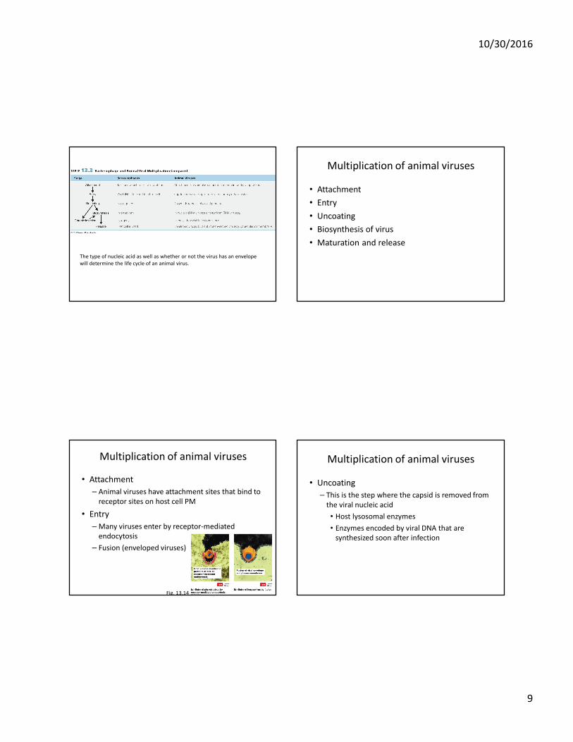
Multiplication of animal viruses
• Attachment
– Animal viruses have attachment sites that bind to
receptor sites on host cell PM
• Entry
– Many viruses enter by receptor-mediated
endocytosis
– Fusion (enveloped viruses)
Fig. 13.14
Multiplication of animal viruses
• Uncoating
– This is the step where the capsid is removed from
the viral nucleic acid
• Host lysosomal enzymes
• Enzymes encoded by viral DNA that are
synthesized soon after infection
10/30/2016
10
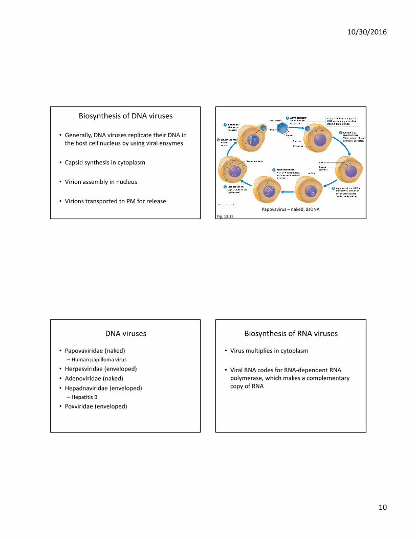
Biosynthesis of DNA viruses
• Generally, DNA viruses replicate their DNA in
the host cell nucleus by using viral enzymes
• Capsid synthesis in cytoplasm
• Virion assembly in nucleus
• Virions transported to PM for release
Fig. 13.15
Papovavirus – naked, dsDNA
DNA viruses
• Papovaviridae (naked)
– Human papilloma virus
• Herpesviridae (enveloped)
• Adenoviridae (naked)
• Hepadnaviridae (enveloped)
– Hepatitis B
• Poxviridae (enveloped)
Biosynthesis of RNA viruses
• Virus multiplies in cytoplasm
• Viral RNA codes for RNA-dependent RNA
polymerase, which makes a complementary
copy of RNA
10/30/2016
11
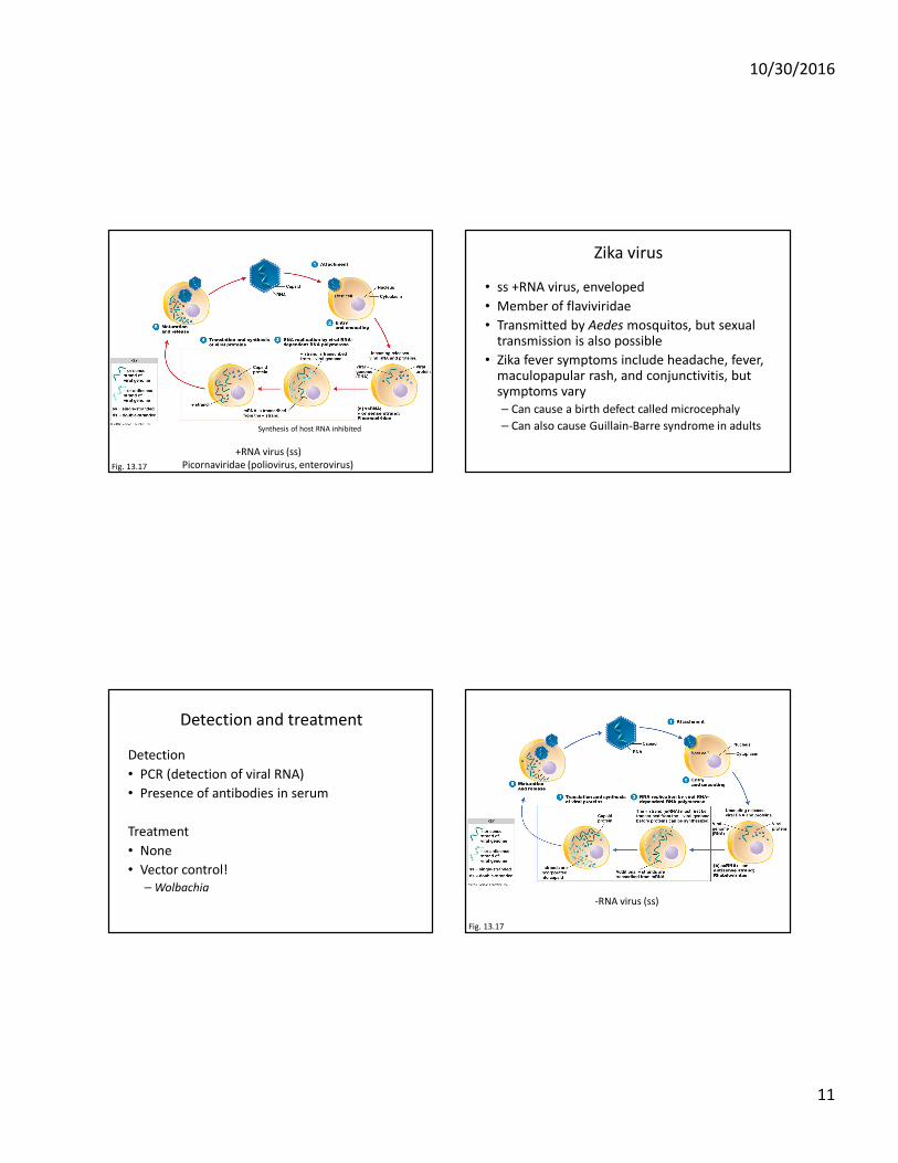
Fig. 13.17
+RNA virus (ss)
Picornaviridae (poliovirus, enterovirus)
Synthesis of host RNA inhibited
Zika virus
• ss +RNA virus, enveloped
• Member of flaviviridae
• Transmitted by Aedes mosquitos, but sexual transmission is also possible
• Zika fever symptoms include headache, fever, maculopapular rash, and conjunctivitis, but symptoms vary
– Can cause a birth defect called microcephaly
– Can also cause Guillain-Barre syndrome in adults
Detection and treatment
Detection
• PCR (detection of viral RNA)
• Presence of antibodies in serum
Treatment
• None
• Vector control!
– Wolbachia
Fig. 13.17
-RNA virus (ss)
10/30/2016
12
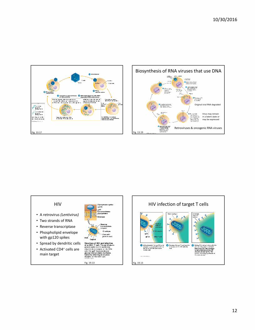
Fig. 13.17
Biosynthesis of RNA viruses that use DNA
Fig. 13.19
Retroviruses & oncogenic RNA viruses
Original viral RNA degraded
Virus may remain
in a latent state or
may be expressed
HIV
• A retrovirus (Lentivirus)
• Two strands of RNA
• Reverse transcriptase
• Phospholipid envelope
with gp120 spikes
• Spread by dendritic cells
• Activated CD4+ cells are
main target
Fig. 19.13
HIV infection of target T cells
Fig. 19.13
10/30/2016
13
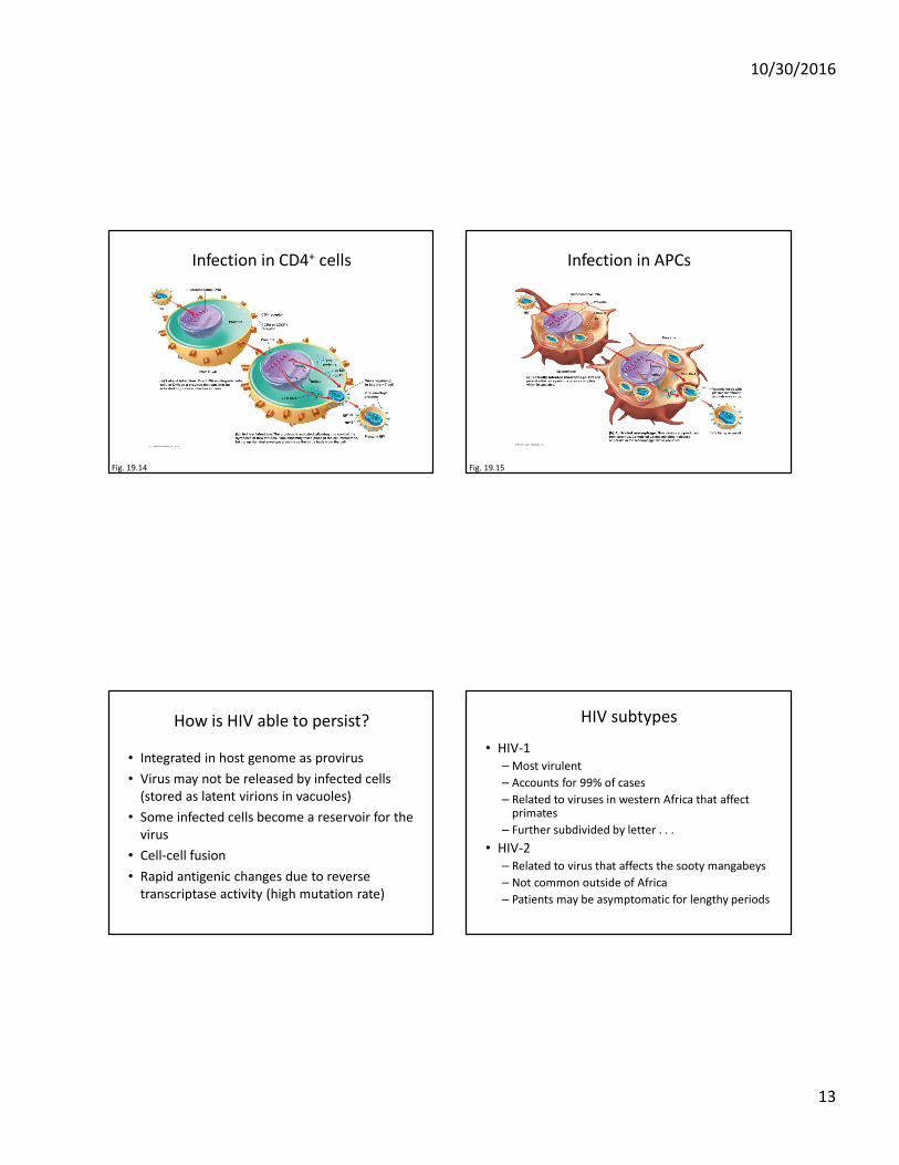
Infection in CD4+ cells
Fig. 19.14
Infection in APCs
Fig. 19.15
How is HIV able to persist?
• Integrated in host genome as provirus
• Virus may not be released by infected cells
(stored as latent virions in vacuoles)
• Some infected cells become a reservoir for the
virus
• Cell-cell fusion
• Rapid antigenic changes due to reverse
transcriptase activity (high mutation rate)
HIV subtypes
• HIV-1
– Most virulent
– Accounts for 99% of cases
– Related to viruses in western Africa that affect primates
– Further subdivided by letter . . .
• HIV-2
– Related to virus that affects the sooty mangabeys
– Not common outside of Africa
– Patients may be asymptomatic for lengthy periods
10/30/2016
14
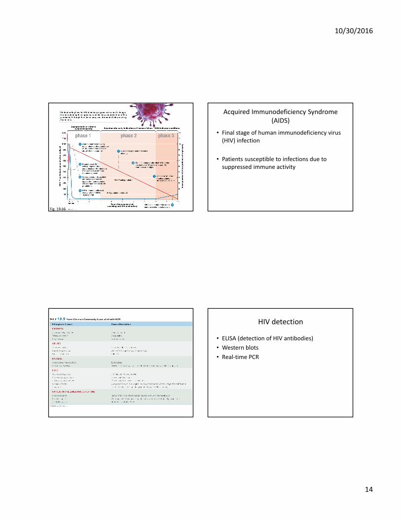
Fig. 19.16
Acquired Immunodeficiency Syndrome
(AIDS)
• Final stage of human immunodeficiency virus
(HIV) infection
• Patients susceptible to infections due to
suppressed immune activity
HIV detection
• ELISA (detection of HIV antibodies)
• Western blots
• Real-time PCR
10/30/2016
15
HIV transmission
• Blood
• Semen
• Intimate sexual contact
• Breast milk
• Transplacental
• Blood-contaminated needles
• Organ transplants
• Artificial insemination
• Blood transfusion
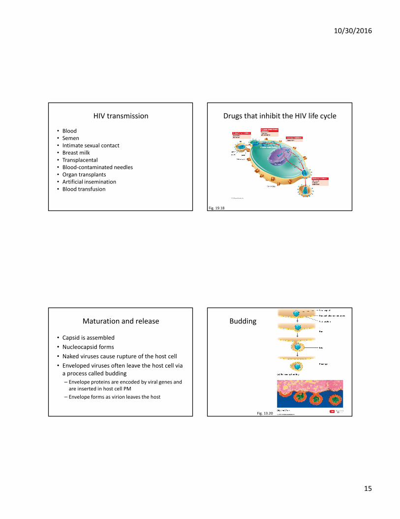
Drugs that inhibit the HIV life cycle
Fig. 19.18
Maturation and release
• Capsid is assembled
• Nucleocapsid forms
• Naked viruses cause rupture of the host cell
• Enveloped viruses often leave the host cell via
a process called budding
– Envelope proteins are encoded by viral genes and
are inserted in host cell PM
– Envelope forms as virion leaves the host
Budding
Fig. 13.20
10/30/2016
16
Transformation of normal cells into cancer
cells
• Can be due to viruses
• Cancer-inducing genes (oncogenes) carried by viruses are actually derived from animal cells
• Oncogenes can be activated to abnormal functioning by a variety of factors
• Oncogenic viruses can induce tumor formation
– Virus integrates into host cell DNA and replicates along with the host cell DNA, ultimately transforming host cell
• After being transformed by viruses, tumor cells contain a virus-specific antigen on their cell surface (tumor-specific transplantation antigen (TSTA) or in the nucleus (T antigen)
DNA oncogenic viruses
• Adenoviridae
• Herpesviridae
– Epstein-Barr virus
• Poxviridae
• Papovaviridae
– Human papillomaviruses
• Hepadnaviridae
– Hepatitis B
RNA oncogenic viruses
• Retroviridae
– Leukemia virus
Viruses to treat cancer
• Adenovirus (H101)
• Talimogene laherparepvec (T-VEC)
• Reolysin
• Delta 24 cold virus
• Modified measles
• Modified herpesvirus
• Modified HIV
10/30/2016
17
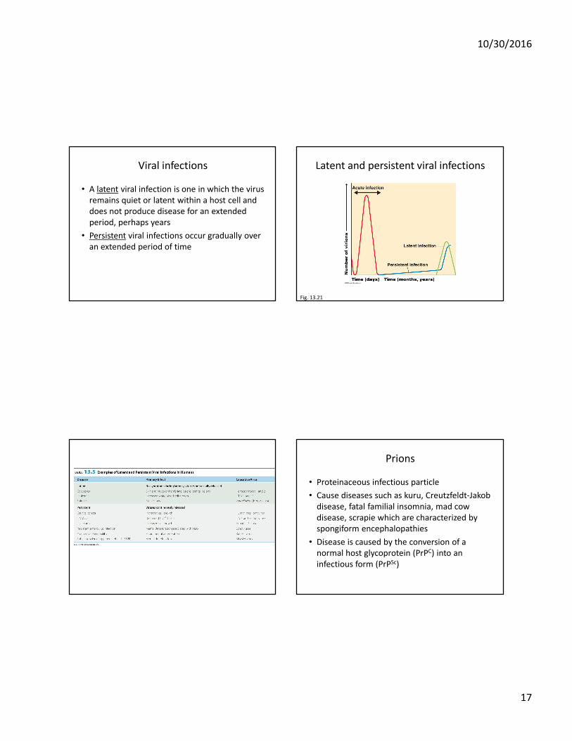
Viral infections
• A latent viral infection is one in which the virus
remains quiet or latent within a host cell and
does not produce disease for an extended
period, perhaps years
• Persistent viral infections occur gradually over
an extended period of time
Latent and persistent viral infections
Fig. 13.21
Prions
• Proteinaceous infectious particle
• Cause diseases such as kuru, Creutzfeldt-Jakob
disease, fatal familial insomnia, mad cow
disease, scrapie which are characterized by
spongiform encephalopathies
• Disease is caused by the conversion of a
normal host glycoprotein (PrPC) into an
infectious form (PrPSc)

10/30/2016
18
Fig. 13.22
Plant viruses and viroids
• Plant viruses are morphologically similar to animal viruses and have similar types of nucleic acids
• Because of the presence of the plant cell walls, viruses typically gain access through wounds or are assisted by other parasites (nematodes, fungi, insects)
• Some plant diseases are caused by viroids, which consist of naked RNA