Embed Size (px)
Citation preview

Virus-Based Nanoparticles for Tumor Selective Targeting and Oncolysis
By
Vrushali Chavan
Thesis submitted to the faculty of the Virginia Polytechnic Institute and
State University in partial fulfillment of the requirement for the degree of
Master of Science
In
Biomedical and Veterinary Sciences
Elankumaran Subbiah, Chairman
Xiang-Jin Meng
William Rupert Huckle
December 8, 2010
Blacksburg, VA
Keywords: nanoparticles, virus-based nanoparticles, Newcastle disease
virus, virotherapy, cancer virotherapy
Copyright: Vrushali Chavan, 2010

Virus-based Nanoparticles for Tumor Selective Targeting and
Oncolysis
Vrushali Chavan
Abstract
Many oncolytic virotherapies have shown great advantages for rapid,
rational design through recombinant DNA technology to facilitate the
targeting of a broad spectrum of malignancies. Newcastle disease virus
(NDV), an avian paramyxovirus, is naturally tumor-selective and inherently
oncolytic. Our approach is to develop NDV-based nanoparticles (VBNP) for
oncolytic virotherapy. VBNPs are non-infectious and non-replicating and are
relatively safe. We obtained VBNPs by co-expressing matrix (M),
hemagglutinin (HN), and fusion (F) proteins of NDV in avian/ mammalian
cells. The budding characteristics, size and morphology of VBNPs were
similar to authentic virions. As a proof of concept, we engineered the
apoptin (VP3) gene of chicken anemia virus in VBNPs and specifically
targeted them to folate-receptor bearing tumor cells by surface conjugation
to folate. The VBNPs killed tumor cells by apoptosis and induced
proinflammatory and chemotactic cytokines. The VBNPs, although not
curative, were able to limit the progression of xenotransplanted fibrosarcoma

iii
and malignant glioma tumors and provided a survival advantage in nude
mice. We also engineered NDV M based particles with nipah virus surface
glycorporteins to target ephrin B receptors. NDV based nipah Virus BNPs
(NiV-ndBNP) were morphologically similar to authentic NiV virions. NiV
glycoproteins were incorporated into the NDV M based particles, despite
poor sequence homology in the transmembrane domain and cytoplasmic
tails of glycoproteins. Our results suggest that VBNPs could be used to
deliver small molecules, tumor antigens, anti-tumor/ reporter genes and also
aid in generating tumor specific immunity by rational design.

iv
Dedication
I would like to dedicate my thesis to my family and friends. To my parents, Mr. Pramod
Chavan and Mrs. Shalaka Chavan for their love, support and encouragement, and to my pet dog,
Sophie, her presence in my life pushed me to do my best for animals. This work is a result of the
trust and faith they always had in me.

v
Acknowledgements
This thesis was a conjoint effort from a lot of people and lab members, over the years of
research since I joined Dr. Subbiah’s lab. The contribution and advice from them in different
ways helped in the research and made my experience great and unforgettable.
Beforehand, I would like to thank Virginia-Maryland Regional College of Veterinary
Medicine, especially Dr. Roger Avery, Cindy Booth and Becky Jones, for giving me the
opportunity to come to this school and achieve graduate experience and to thank them for
providing financial support throughout my graduate career. I would also like to thank Institute for
Critical Technology and Applied Sciences (ICTAS) for funding the project.
I gratefully thank my mentor, Dr. Elankumaran Subbiah, for his supervision and guidance
since the beginning and giving me the chance to attain research excellence. Under his leadership,
my scientific knowledge and curiosity has flourished which I will always benefit me in my future
endeavors. Because of his scientific passion and ideas, at all times inspired me to work hard and
thrive to achieve my goals.
I would also like to express my gratitude towards both my committee members, Dr. X.J.
Meng and Dr. William Huckle who always gave their valuable advice, helped me improve my
work and encouraged me to believe in myself. I truly believe I was able to get this far only with
the help of my mentor and all my committee members and made my graduate career a success. I
would like to thank Dr. Judy Riffle and her team for their collaboration.
I would collectively like to thank and acknowledge all my colleagues in Dr. Subbiah’s
lab, Moanaro Biswas, Shobana Raghunath, Adria Allen, Jagadeeswaran D., Sandeep Kumar,
Abhilash P.S. and Wang Yong as well as everyone in CMMID and ILSB, whose presence
eternally revived and helped me and made my experience memorable.
I would like to take this opportunity to especially thank Moanaro Biswas for her help
towards completing my project and thesis. I would also like to thank Gopakumar Moorkanat and

vi
Laure Deflube, for the continued help not only in a lot of research techniques but also providing
me with a great attitude towards research.
I would like to thank Nanoscale Characterization and Fabrication Laboratory (NCFL);
John McIntosh, NCFL, ICTAS; Melissa Makris, Flow Cytometry Supervisor
at VMRCVM; Kathy Lowe, Laboratory specialist, VMRCVM; and Dr. Tanya LeRoith,
VMRCVM for their help. I would like to thank Dr. Griffith Parks; Dr. John Johnson and Dr. Ken
Grant from Wake Forest University, School of Medicine for microscopy analysis. I would also
like to thank Dr. Ronald Iorio, University of Massachusetts Medical School for the reagents
needed to complete this work; Dr. Paul Rota, CDC for Nipah-F and -G plasmids; Dr. Benhur Lee,
University of California, Los Angeles, for the pcMV-3tag1 plasmid and Nipah virus antibodies.
I am truly thankful to Ameya Redkar for always being supportive of me. He always
encouraged me to keep trying and bore an optimistic attitude towards life. Without your support, I
do not believe I would have been able to get this far and successfully complete this work.
I would like to take this opportunity to also thank all my friends I met at VMRVCM
during my graduate career. Their presence made my time in Blacksburg memorable and I cannot
thank them for being such wonderful friends to me.
Lastly, I would like to thank my parents and my siblings, Geyata Fernandez and Vidyesh
Chavan, from the bottom of my heart, for their faith in me, and being the inspiration in my life.

vii
Attribution
Several colleagues and coworkers aided in the writing and research behind several of the
chapters of this thesis. A brief description of their background and their contributions are included
here.
Chapter 3: Virus-Based Nanoparticles for Tumor Selective Targeting and Oncolysis
Dr. Elankumaran Subbiah- Ph.D. (Department of Biomedical Sciences and Pathobiology,
Virginia Tech) is the Major Advisor and Committee Chair. Dr. Subbiah and I conceived, designed
the experiments, performed statistical analysis, data analysis and processing and wrote the paper.
Shobana Raghunath - Ph.D. student (Department of Biomedical and Pathobiology, Virginia
Tech) in Dr. Subbiah’s Lab and contributed reagents, materials and analysis tools.
Adria Allen- Ph.D. student (Department of Biomedical and Pathobiology, Virginia Tech) in Dr.
Subbiah’s Lab. Adria contributed towards my research by helping with experiments.
Moanaro Biswas - Ph.D. (Department of Biomedical and Pathobiology, Virginia Tech) post-
doctoral fellow in Dr. Subbiah’s Lab. Moanaro helped me perfom experiments.
Abilash P.S - Ph.D. student (Department of Biomedical and Pathobiology, Virginia Tech) in Dr.
Subbiah’s lab. Abilash contributed in performing some of the experiments.
Nikorn Pothayee - Ph.D. student (Department of Chemistry, Virginia Tech) in Dr. Judy Riffle’s
lab and contributed by providing reagents and materials for the experiments.
Dr. Judy Riffle - Ph.D. (Professor, Department of Chemistry, Virginia Tech) collaborated with
Dr. Subbiah on this project and provided technical consultancy and reagents
Dr. Tanya LeRoith - Ph.D (Department of Biomedical and Pathobiology, Virginia Tech) is a

viii
professor of Anatomic Pathology and she analyzed histopathology sections for the experiments.
Chapter 4: Surface Modification of Newcastle disease Virus-Based Nanoparticles with Nipah
Virus Glycoproteins
Dr. Elankumaran Subbiah- Ph.D. (Department of Biomedical Sciences and Pathobiology,
Virginia Tech) is the Major Advisor and Committee Chair. Dr. Subbiah and I conceived, designed
the experiments, performed statistical analysis, data analysis and processing and wrote the paper.
Moanaro Biswas - Ph.D. (Department of Biomedical and Pathobiology, Virginia Tech) post-
doctoral fellow in Dr. Subbiah’s Lab. Moanaro assisted with some experiments.
Abilash P.S - Ph.D. student (Department of Biomedical and Pathobiology, Virginia Tech) in Dr.
Subbiah’s lab. Abilash contributed reagents, materials and analysis tools.
Wang Yong - Ph.D. (Department of Biomedical and Pathobiology, Virginia Tech) in Dr.
Subbiah’s Lab and contributed reagents, materials and analysis tools.

ix
Table of contents
Title Page
Abstract...........................................................................................................................................ii
Dedication.......................................................................................................................................iv
Acknowledgements.........................................................................................................................v
Attribution.....................................................................................................................................vii
Table of contents............................................................................................................................ix
List of Figures...............................................................................................................................xii
List of Tables................................................................................................................................xiv
List of Abbreviations...................................................................................................................xv
Chapter 1: Introduction....................................................................................................1
1.1 Background.............................................................................................................1
1.2 Justification.............................................................................................................2
1.3 Hypothesis..............................................................................................................3
1.4 Objectives...............................................................................................................3
1.5 Thesis Outline.........................................................................................................4
1.6 References...............................................................................................................5
Chapter 2: Literature Review...........................................................................................7
2.1 Introduction.............................................................................................................7
2.2 Newcastle disease virus..........................................................................................8
2.3 Oncolytic Viruses...................................................................................................9
2.3.1 Targeting OV’s.....................................................................................................10
2.3.2 Shielding OV’s.....................................................................................................12
2.3.3 Arming OV’s........................................................................................................12

x
2.3.4 Tumor Selectivity.................................................................................................13
2.4 Oncolytic viruses in clinical trials........................................................................13
2.5 Virus like Particles................................................................................................15
2.5.1 Virus-based nanoparticles for cancer therapy.......................................................17
2.6 References.............................................................................................................21
2.7 Figure....................................................................................................................30
Chapter 3: Virus-Based nanoparticles for Tumor Selective Targeting and
Oncolysis...........................................................................................................................31
3.1 Abstract.................................................................................................................31
3.2 Introduction...........................................................................................................33
3.3 Materials and Methods......................................................................................…36
3.4 Results...................................................................................................................45
3.4.1 Morphology, budding and release of VBNP.........................................................45
3.4.2 Folate conjugated VBNP uptake in Folate receptor (FR) positive cell line..........46
3.4.3 Cytoplasmic distribution of VBNP in cells............................................................46
3.4.4 VBNPs are cytotoxic to tumor cells......................................................................47
3.4.5 VBNP and VBNP-VP3 causes early apoptosis in tumor cell lines.......................47
3.4.6 VBNP elicits pro-inflammatory and chemotactic cytokines.................................48
3.4.7 VBNP dosage is insufficient to effect cures in fibrosarcoma and malignant
glioma models.......................................................................................................48
3.4.8 VBNP therapy prolonged survival in fibrosarcoma model...................................49
3.4.9 VBNP treatment failed to prolong survival in malignant glioma...........................49
3.4.10 Histology of implanted tumors indicates VBNPs induced inflammatory
response and necrosis..............................................................................................49

xi
3.5 Discussion................................................................................................................50
3.6 Tables.......................................................................................................................52
3.7 Figures......................................................................................................................55
3.8 References................................................................................................................73
3.9 Acknowledgements..................................................................................................76
Chapter 4: Surface Modification of Newcastle Disease Virus-based Nanoparticles
with Nipah Virus Glycoproteins.....................................................................................77
4.1 Abstract.................................................................................................................77
4.2 Introduction...........................................................................................................79
4.3 Materials and Methods..........................................................................................82
4.4 Results...................................................................................................................86
4.4.1 Fusogenecity of NiV-ndBNP................................................................................86
4.4.2 Morphology and strutcure of NiV-ndBNP...........................................................86
4.4.3 Composition of NiV-ndBNP................................................................................86
4.4.4 Poor sequence homology of NDV and NiV glycoproteins’ CT and TM.............87
4.5 Discussion.............................................................................................................88
4.6 Figures..................................................................................................................90
4.7 References.............................................................................................................96
4.8 Acknowledgements.............................................................................................100
Chapter 5: General Conclusions and Future Research..............................................101

xii
List of Figures
Chapter 2
Figure 2.7: Diagrammatic representation of Newcastle disease virus...........................................30
Chapter 3
FIGURE 1. A schematic representation of construction of VBNP-VP3 plasmid.........................55
FIGURE 2. Fusogenicity with VBNP in DF-1 cells......................................................................56
FIGURE 3. Fusogenicity with VBNP-VP3 in DF-1 cells.............................................................57
FIGURE 4. Electron micrograph of VBNP budding and release in 293T cells............................58
FIGURE 5. Transmission electron microscopy (TEM) of negatively stained NDV and NDV-
BNPs..........................................................................................................................59
FIGURE 6. Immunogold transmission electron microscopy (iTEM) of VBNP...........................60
FIGURE 7. VBNPs incorporated NDV structural proteins and cellular proteins.........................61
FIGURE 8. VBNP uptake in cells expressing folate receptor.......................................................62
FIGURE 9. Cell entry and cytoplasmic distribution of NDV VBNPs.........................................63
FIGURE 10. VBNPs induce cytotoxicity in treated transformed and tumor cells........................64
FIGURE 11: VBNP treated cells undergo intranucleosomal DNA fragmentatio.........................65
FIGURE 12: NDV VBNPS induce early apoptosis......................................................................67
FIGURE 13: VBNPs induce proinflammatory cytokines and chemokines in tumor cells...........68
FIGURE 14. Mean tumor volume in VBNP treated xenotransplanted tumors.............................69
FIGURE 15. VBNP treatment in both HT1080 and U87MG failed to prolong the
survival times in tumor bearing mice.......................................................................70
FIGURE 16. VBNP therapy induced a proinflammatory response and cell death in implanted
tumors.......................................................................................................................71

xiii
FIGURE 17. VBNP therapy kills tumor cells by necrosis and causes spleenomegaly in U87MG
xenotransplanted mice..............................................................................................72
Chapter 4
FIGURE 1. Diagrammatic representation of Henipavirus.............................................................90
FIGURE 2. Fusogenicity of NiV-ndBNP in 293T cells................................................................91
FIGURE 3. Transmission Electron Microscopy (TEM) of NiV-ndBNPs.....................................92
FIGURE 4. Immunoblot of NiV-BNP and NiV-ndBNP...............................................................93
FIGURE 5. Poor sequence homology between NDV and NiV glycoproteins’ CT and TM.........94
FIGURE 6. Poor sequence homology between CT and TM of NDV HN and NiV glycoprotein,
G...............................................................................................................................95

xiv
List of Tables
Table 1. Mean tumor volume of xenotransplanted HT1080 fibrosarcoma in nude
mice............................................................................................................................52
Table 2. Mean tumor volume of xenotransplanted U87MG malignant glioma in nude
mice............................................................................................................................53
Table 3. Histological lesion scores of xenotransplanted tumors in nude mice........................54

xv
List of Abbreviations
ACS American Cancer society
CNS Central Nervous System
CT Cytoplasmic tail
CTL Cytotoxic T Lymphocytes
DAPI 4', 6-diamidino-2-phenylindole
DMEM Dulbecco’s modified Eagle’s medium
EtBr Ethidium Bromide
FCS Fetal calf serum
Fo Folate
FR Folate Receptor
FA Folic Acid
F Fusion
GM-CSF Granulocyte-Macrophage Colony Stimulating Factor
GPI Glycosylphosphatidylinositol
HN Hemagglutinin- neuraminidase
H/E Hematoxylin and Eosin
HeV Hepatitis E virus
HSV-1 Herpes Simplex Virus 1
IF Immunofluorescence
IFN Interferon
L Large polymerase
MHC-1 Major Histocompatiblity type I
M Matrix

xvi
MMP Matrix metallo-proteases
MeV Measles virus
miRNA MicroRNA
MuV Mumps virus
NHS N-hydroxysuccinimide ester
NDV Newcastle disease Virus
NVNDV Neurotropic velogenic NDV
NiV Nipah Virus
NP Nucleoprotein
Nt Nucleotide
OV Oncolytic Virus
OVT Oncolytic virotherapy
PIV Parainfluenza virus
PNS Peripheral nervous system
PPRV Peste des Petits ruminants virus
PBS Phosphate Buffered Saline
P Phosphoprotein
PTA Phosphotunstic acid
PEG Polyethylene glycol
PNP Purine Nucleoside Phosphorylase
Rb Retinoblastoma
RSV Respiratory syncytial virus
RNA Ribonucleic acid
RNP Ribonucleoprotein core
RPV Rinderpest virus
SDS-PAGE Sodium-dodecyl sulphate polyacrylamide gel electrophoresis

xvii
TEM Transmission Electron Microscopy
TM Transmembrane
UTR Untranslated Region
VBNP Virus based nanoparticles
VLP Virus like particles
VP3 Apoptin protein
VVNDV Viscerotropic velogenic NDV
VSV Vesicular Stomatitis Virus

1
Chapter 1: Introduction
1.1 Background
Cancer is a disease wherein a cell, or a group of cells exhibit uncontrolled growth,
invasion and sometimes metastasis. These properties of cancer differentiate it from benign
tumors, which are self-limited, and do not invade or metastasize. Cancer affects people at all ages
but the risk of cancer increases with age (Cancer Research, January 2007). It is responsible for
causing the highest number of human deaths every year. Cancer resulted in about 13% of all
human deaths in 2007 (American Cancer Society, 2007; WHO, February 2006). Many factors
contribute towards the etiology of cancer in humans. About 90-95% of cancers are caused due to
lifestyle and environmental factors like diet and obesity, infections, tobacco, radiation, stress, lack
of physical activity, environmental pollutants, which cause abnormality and mutations in the
genetic material of cells (Anand et al., 2008; Kinzler and Vogelstein, 2002).
Over the past decade, several attempts have been made toward understanding cancer
biology and discoveries emerging from cancer genomics that can be made beneficial for cancer
patients along with radiation and chemotherapy as standard of care (Ladanyi and Hogendoorn,
2011). In view of the deleterious effects of cancer therapy with radiation and chemicals,
alternative treatments like the use of viruses for cancer therapy is becoming popular. Certain
viruses have tumor killing (oncolytic) property and this channels a new pathway for cancer
therapeutics (Kelly and Russell, 2007). Gene therapy is another technology which aims at the
correction of inherited genetic diseases by providing to the targeted cells a functional copy of the
deficient gene responsible for the disease, in particular cancers (Touchefeu et al., 2010).

2
1.2 Justification
Oncolytic virotherapy (OVT) is the deliberate use of lytic viruses to infect and kill cancer
cells (Adrienne et al., 2010). The field of OVT gradually developed through the use of promising
animal tumor models. It was reported that both vaccinia virus and herpes virus could be
propagated in mouse and rat skin tumors and sarcomas (Levaditi and Nicolau, 1922; Moore,
1952). This gave a hint in the use virus infection of cancer cells might show promise as a cancer
therapy. More viruses were discovered and small animal tumor models were created over the next
several years. There have been multiple clinical trials using OVs and in combination with
immunosuppressive drugs like cyclophosphamide; retargeting of viruses to specific tumor types;
using infected cell carriers to protect and deliver the virus to tumors; and genetic manipulation of
the virus to increase viral spread and/or express transgenes during viral replication (Lech and
Russell, 2010).
OVs are appealing as cancer therapeutics since they can be genetically manipulated and
they exhibit multiple discrete anticancer mechanisms. OVs cause direct tumor cell killing,
independent of conventional drug-resistance mechanisms (Coukos et al., 2000). Notably, they
also have the ability for self-propagation, thereby effectively spreading throughout a tumor and
being active against massive and potentially metastatic disease. Viruses can also be used to
deliver therapeutic genes by arming the viruses, which further enhances the antitumor capabilities
of these viruses (Hermiston, 2000), and can cause substantial modification in the tumor
microenvironment (Kaur et al., 2009). They can possibly sensitize the host immune system to
tumor-specific antigens that in other instances would not have been immunogenic, hence acting
as an in situ cancer vaccine (Benencia et al., 2008; Toda et al., 1999). In the current epoch of
molecular biology, OVs are a potentially useful therapy to reduce bulky disease, alone or in
combination with conventional therapies, and as an immunotherapy to prevent cancer (Hammill

3
et al., 2010). However, in instances where the cancer patients are immunocompromised and the
host antiviral immunity restrict OV replication, other OV-based options that would retain the
inherent ability of OV to kill cancer cells and evoke antitumor immunity, are necessary. The use
of non-replicating viruses or virus-like particles that could be engineered for oncolytic efficacy is
an attractive strategy.
1.3 Hypothesis
We hypothesize that virus-based nanoparticles from a promising OV such as avian
Newcastle disease virus (NDV) could be engineered for selective retargeting and arming for
cancer therapy.
1.4 Objectives
1. To study the requirements for the formation of NDV-based nanoparticles
2. To characterize the budding and morphology of NDV-based nanoparticles in human cells
3. To determine whether NDV-based nanoparticles will be cytotoxic to cancer cells
4. To explore selective retargeting of NDV-based nanoparticles to folate receptors
5. To determine whether it would be possible to arm NDV-based nanoparticles with a
model proapoptotic gene
6. To examine whether NDV-based nanoparticles will induce anti-tumor immune response
in select cancer cell populations in vitro
7. To determine whether it would be possible to surface modify NDV-based nanoparticles
with envelope glycoproteins from other paramyxoviruses for efficient targeting
8. To determine whether NDV-based nanoparticles can cure or prolong survival in
xenotransplanted pre-clinical mouse models of fibrosarcoma and malignant glioma.

4
1.5 Thesis Outline
This thesis consists of five chapters. Chapter one introduces the research topic, while
chapter two reviews current research being done in related areas. Subjects discussed include use
of viruses for cancer therapy, Newcastle disease virus as an oncolytic virus, need for better OV
platforms and methods for shielding, arming, and targeting oncolytic viruses or virus-based
platforms. Chapter three presents the research completed to determine the efficacy of virus-based
nanoparticles for cancer therapy. Additionally, this chapter will discuss the ability to engineer
foreign antigens on NDV envelope and folate targeting for cancer therapy. Chapter four will
present the results of surface modification of NDV-based nanoparticles with Nipah virus
envelope glycoproteins and the options available for efficient targeting to select cell types using
this approach. Finally, chapter 5 will summarize the results and provide conclusions and areas for
future research..

5
1.6 References
Adrienne, M., M.D. Hammill, J. Conner;, and T.P. Cripe. 2010. Oncolytic Virotherapy reaches
Adolescence. Pediatric Blood & Cancer. 55: 1253–1263.
American Cancer Society. 2007. Report sees 7.6 million global 2007 cancer deaths. Retrieved
2008-08-07.
Anand, P., A.B. Kunnumakkara, C. Sundaram, K.B. Harikumar, S.T. Tharakan, O.S. Lai, B.
Sung, and B.B. Aggarwal. 2008. Cancer is a preventable disease that requires major
lifestyle changes. Pharm Res. 25: 2097-2116.
Benencia, F., M.C. Courreges, N.W. Fraser, and G. Coukos. 2008. Herpes virus oncolytic therapy
reverses tumor immune dysfunction and facilitates tumor antigen presentation. Cancer
Biol Ther. 7:1194-1205.
Cancer Research, 2007. "UK cancer incidence statistics by age". Retrieved 2007-06-25.
Coukos, G., A. Makrigiannakis, E.H. Kang, S.C. Rubin, S.M. Albelda, and K.L. Molnar-Kimber.
2000. Oncolytic herpes simplex virus-1 lacking ICP34.5 induces p53-independent death
and is efficacious against chemotherapy-resistant ovarian cancer. Clin Cancer Res. 6:
3342-3353.
Hammill, A.M., J. Conner, and T.P. Cripe. 2010. Oncolytic virotherapy reaches adolescence.
Pediatr Blood Cancer. 55: 1253-1263.
Hermiston, T. 2000. Gene delivery from replication-selective viruses: arming guided missiles in
the war against cancer. J Clin Invest. 105: 1169-1172.
Kaur, B., T.P. Cripe, and E.A. Chiocca. 2009. "Buy one get one free": armed viruses for the
treatment of cancer cells and their microenvironment. Curr Gene Ther. 9: 341-355.
Kelly, E., and S.J. Russell. 2007. History of oncolytic viruses: genesis to genetic engineering. Mol
Ther. 15:651-659.

6
Kinzler, K.W., and B. Vogelstein. 2002. "Introduction". The genetic basis of human cancer (2nd,
illustrated, revised ed.). New York: McGraw-Hill, Medical Pub. Division: 3-6.
Ladanyi, M., and P.C. Hogendoorn. 2011. Cancer biology and genomics: translating discoveries,
transforming pathology. J Pathol 223: 99-101.
Lech, P.J., and S.J. Russell. 2010. Use of attenuated paramyxoviruses for cancer therapy. Expert
Rev Vaccines. 9:1275-1302.
Levaditi, C., and S. Nicolau. 1922. Sur la culture de virus vaccinal dans les neoplasmes
epitheliaux. . CR Soc Biol. 1922; 86: 928.
Moore, A.E. 1952. Viruses with oncolytic properties and their adaptation to tumors. Ann N Y
Acad Sci. 54: 945-952.
Toda, M., S.D. Rabkin, H. Kojima, and R.L. Martuza. 1999. Herpes simplex virus as an in situ
cancer vaccine for the induction of specific antitumor immunity. Hum Gene Ther. 10:
385-393.
Touchefeu, Y., K.J. Harrington, J.P. Galmiche, and G. Vassaux. 2010. Gene therapy, recent
developments and future prospects in gastrointestinal oncology. Aliment Pharmacol Ther.
32: 953-968.
WHO. February 2006. "Cancer". Retrieved 2007-06-25.

7
Chapter 2: Literature Review
2.1 Introduction
In the last few decades, many novel paramyxoviruses have emerged causing catastrophic
diseases in different species of animals and some of them also made the species jump to humans.
Members of the family Paramyxoviridae have been isolated from diverse host species and they
are found to be associated with central nervous and respiratory system diseases (Lamb and Parks,
2007). There are many important viruses in this family, including animal pathogens that are
economically important in poultry and livestock industry, such as Newcastle disease virus (NDV)
(Alexander, 2009), Rinderpest virus (RPV) (Roeder and Taylor, 2002) and Peste des Petits
ruminants virus (PPRV) and human pathogens such as measles virus (MeV), mumps virus
(MuV), respiratory syncytial virus (RSV), and parainfluenza viruses (PIV) (Lamb and Parks,
2007). Some members of the Paramyxoviridae family such as MeV and NDV are also being
extensively studied as a vaccine vector or for use as anticancer agents (Cassel and Garrett, 1965;
Elankumaran et al., 2006; Russell and Peng, 2009).
Paramyxoviruses are enveloped RNA viruses possessing non-segmented, negative strand
genomes in the order Mononegavirales (Loney et al., 2009). Family Paramyxoviridae can be
divided into two subfamilies, Paramyxovirinae and Pnuemovirinae, based on virion morphology,
genome organization, and structure and sequence relatedness. Currently, there are five genera
within the subfamily Paramyxovirinae: Rubulavirus, Avulavirus, Respirovirus, Morbillivirus, and
Henipavirus, and two genera within the subfamily Pneumovirinae: Pneumovirus and
Metapneumovirus (Lamb and Parks, 2007). Respiroviruses and rubulaviruses demonstrate both
hemagglutinating (HA) and neuraminidase (NA) activity. Morbilliviruses and henipaviruses
exhibit only HA but lack NA activity. Pneumoviruses are morphologically differentiated by their
narrower nucleocapsids from Paramyxovirinae. Pneumovirus does not cause substantial HA in

8
mammalian and avian erythrocytes (Lamb and Parks, 2007).
They have a close relationship with two other negative strand RNA viruses: Rhabdoviruses
(for their unique non-segmented genome organization and its expression) and Orthomyxoviruses
(for the biological properties of the envelope glycoproteins (Lamb and Parks, 2007).
2.2 Newcastle Disease Virus
Newcastle disease virus (NDV) is responsible for causing a highly contagious disease in
many species of domestic and wild birds. The disease was first reported in Java in 1926 and was
brought to international attention in Newcastle on Tyne, England, in 1926 (Alexander, 2001;
Shope, 1964). The disease is characterized by the presence of respiratory, digestive and
neurological signs in many avian species. The severity of clinical signs ranges from an inapparent
infection to a rapidly fatal condition (Alexander, 2001). Standard pathotyping assays utilize
inoculation of embryonated eggs and live chickens to determine the virulence amongst different
strains of NDV. NDV strains are classified as velogenic (highly virulent), mesogenic (moderately
virulent) and lentogenic (avirulent) depending on the severity of the disease they cause
(Alexander, 2000; Beard and Hanson, 1984). The velogenic strain is further divided into two
pathotypes, the viscerotropic velogenic (VVNDV), which cause acute lethal disease with visceral
hemorrhages and the neurotropic velogenic (NVNDV) responsible for neurological and
respiratory signs.
NDV genome is about 15,186 nucleotides long and encodes 6 genes, placed 3’-NP-P-M-F-
HN-L-5’, with short untranslated regions (UTRs) at either end of each gene. The six NDV genes
encode structural and non-structural proteins that are classified into membrane and core
components. The membrane components consist of two transmembrane glycoproteins, the Fusion

9
(F) and hemagglutinin (HN) proteins, and the matrix (M) protein (Fig. 2.7). The virus envelope
encases the ribonucleoprotein (RNP) core, which is formed by the RNA genome associated with
the nucleocapsid protein (NP) and the polymerase complex composed of phosphoprotein (P) and
large polymerase (L) proteins. The inner surface of the virion is lined by the M protein which
probably mediates the interaction between the RNP complex and lipid-bilayer, as well as the
cytoplasmic tails of the spike glycoproteins (Cathomen et al., 1998a). The structure of NDV is
presented as a schematic cartoon in Fig. 2.7.
2.3 Oncolytic Viruses
Cancer is a major cause of deaths in humans. Though there has been significant progress
in cancer therapy, the limited efficacy and toxicities of current chemo- and radiotherapies have
provided an impetus for the search of novel therapeutics. A therapeutic approach, which uses
viruses for the treatment of cancer termed, oncolytic virotherapy has recently emerged (Kelly and
Russell, 2007). Oncolytic virus (OV) is a virus that preferentially infects and lyses cancer cells.
OVs have achieved a high recognition in cancer therapy, both by directly destroying the tumor
cells, and by having the capacity to be modified as vectors. They can carry genes that express
anticancer proteins and deliver them specifically to the tumor site. Most current OVs are
engineered for tumor selectivity, but there are a few naturally occurring OVs. The safety and
efficacy of OVs were widely debated, and studied in both subjective and official clinical trials
since 1950s. Early virotherapy clinical trials based on natural viruses were poorly directed, but
recently, modified viruses have been subjected to extensive screening of viral replication, gene
expression, and host immunity (Liu et al., 2007). Several strategies can be applied to many
viruses to augment therapy (Cattaneo, 2010; Cattaneo et al., 2008).
Over the past few years, there have been new insights on the molecular mechanisms of
viral cytotoxicity, which provided the scientific rationale to design more effective OVs. Several

10
recent clinical trials have used genetically engineered viral strains, such as adenovirus and herpes
simplex virus 1 (HSV-1) as well as wild-type Newcastle disease virus, which show promising
results with these viruses being relatively non-toxic and tumor specific. But how do OVs
specifically target cancer cells, and what is the potential for using OVs as cancer therapeutics?
Replicative selectivity of viral genes can be modified for proficient replication, so that the virus
can only replicate in cells that have interruptions in normal homeostatic pathways, such as tumor-
suppressor defects or activation of oncogenic pathways (Chiocca, 2002). OVs, along with their
positive use in virotherapy come with a few disadvantages. Certain OVs such as the laboratory
engineered HSV-1, adenovirus, can easily be manipulated genetically but induce side effects
which include serious or potentially fatal disease in immunocompromised individuals. Also, the
engineered OVs like reovirus and VSV are not easy to manipulate genetically. Most OVs are
also limited by the host antiviral response.
Arming oncolytic viruses with transgenes that are capable of inducing apoptosis is a
striking strategy for improving anti-cancer activity (Cattaneo et al., 2008). Three principles for
developing more specific and potent OVs have been employed, for future clinical trials, namely:
targeting, shielding, and arming (Cattaneo et al., 2008).
2.3.1.Targeting OVs:
During infection, viruses bind to one or more host cell surface proteins, and the viral
tropism can be determined by tissue-specific expression of these proteins. A range of chemical
and genetic engineering strategies have been tested to retarget the cell entry of both enveloped
and non-enveloped viruses through specific cancer-cell-specific receptors (Waehler et al., 2007).
Paramyxoviruses have contributed to the development of the next generation of cancer
therapeutics and in particular on targeting viral entry to cancer cells. The paramyxovirus

11
envelopes can target substrate by receptor attachment and fusion, which functions on two separate
proteins. In contrast, a single protein in retroviruses performs both functions, which has
complicated retargeting strategies. The two-protein entry system of paramyxoviruses is also
simpler than those of large DNA viruses that use several proteins. Among paramyxoviruses,
targeting of the measles virus (MeV) envelope is the most advanced (Cattaneo, 2010).
OVs are cytotoxic and target cancers via multiple mechanisms of action and at the same
time they utilize authenticated genetic pathways, which are dysregulated in nearly all cancers. In
tumor cells, cell growth is unregulated, with many human cancers harboring mutations in p53 or
Retinoblastoma (Rb). A vast majority of cancers arise as a result of the accumulation of multiple
genetic mutations. But only in very specific cases, the detection and consequent targeting of
tumors is feasible. Subsequently, a plethora of therapeutic approaches targeting the related
pathways/key players that support or are crucial for tumor development are being developed and
explored for effective targeting and virotherapy (Ferrara et al., 2004; Pavet et al., 2010; Tennant
et al., 2010). Some tumors exhibit certain common characteristics that are at the basis of
anticancer drug development (Bianco et al., 2007; Desgrosellier and Cheresh, 2010; Granchi et
al., 2010; Tennant et al., 2010). Current approaches target three features of tumor growth, i.e.
self-sufficiency in growth signals, sustained angiogenesis and resistance to apoptotic stimuli, and
have been shown to be successful in some cases (Desgrosellier and Cheresh, 2010; Pavet et al.,
2010; Tennant et al., 2010).
Virus reprogramming has been achieved with several targeting strategies that exploit
either receptor-mediated entry or by particle activation (eg. via proteases), which was based on
mutating the F-protein in paramyxoviruses that required protease cleavage for activation. Both
Sendai virus and MeV were used for this targeting strategy (Cattaneo, 2010; Springfeld et al.,
12
2006; Wolfe et al., 2004b). Also targeting the transcriptional and replication steps (eg. promoters,
cancer cell defects) was found to be effective.
2.3.2 Shielding the OVs
Once the entry of the OVs is achieved, the body’s immune mechanism kicks in to
eliminate any foreign body from the system. In order to accomplish a successful effect of OVs
after entry, a strategy to shield the OVs before it gets degraded by remodeling the viral or
serotype envelope, coating a polymer by chemicals, biological cell carriers or simply by
intratumoral/specific injections is successful in the recent years. These methods have been used to
shield icosahedral viruses, in particular adenovirus. The envelopes of non-human
paramyxoviruses can be modeled into MeV nucleocapsids to produce chimeric viruses that would
help to evade the pre-existing MeV immunity for a short period of time till the desired effect has
been achieved (Cattaneo et al., 2008).
2.3.3 Arming the OVs
Arming OVs with pro-apoptotic genes, pro-drug convertases like Purine Nucleoside
Phosphorylase (PNP) or fludarabine, or selective disarming in normal cells like interferons,
granulocyte-macrophage colony stimulating factor (GM-CSF) are being explored (Cattaneo,
2010; Cattaneo et al., 2008). Suppressing the host immune system with cyclophosphamide before
virus administration may likely enhance the oncolytic efficacy, possibly by suppressing host
innate and adaptive immunity and momentarily supporting virus replication. Arming of OVs
enhances the efficacy of virotherapy by directly integrating it into a chemotherapy treatment,
locally thereby amplifying its efficacy (Cattaneo, 2010).

13
2.3.4 Tumor Selectivity
A strategy of OVs to target replication of the attenuated virus is achieved by mutating
viral functions that are necessary for replication in normal cells. This gives the virus the
attenuated phenotype that leads to replication only in cells that are permissive such as dividing
tumor cells or cells with defects in specific cancer pathway and sparing normal cells (Manservigi
et al., 2010; Martuza et al., 1991; Meignier et al., 1990). Recent research highlighted another
strategy to target viral replication by interfering the expression of essential viral genes that are
controlled by tumor or tissue-specific promoters, which are preferentially active in tumor cells
(Chung et al., 1999; MacLean et al., 1991; Mullen et al., 2002). Tumor cells with a defective or
truncated Interferon (IFN) pathway escape the antitumor activity of IFNs and are selected (Vigil
et al., 2007). Normal cells have an effective antiviral response and hence they are capable of
inhibiting viral replication before a major damage is prompted. This characteristic defects of the
IFN pathway on tumor cells elucidate the tumor-selective replication of some IFN-sensitive RNA
viruses such as Reovirus, NDV and VSV, thereby providing a mechanism for using these OVs as
safe and effective cancer therapeutic agents (Vigil et al., 2007).
2.4 Oncolytic Viruses in Clinical trials
In recent years, OVs have been genetically engineered to target malignant cancer cells
selectively. Adenovirus is an OV, which is being tested in clinical trials for the treatment of
cancer. The cells that are infected with replication-competent adenoviruses undergo autophagy,
which has granted new opportunities for investigating the mechanism of adenovirus-induced cell
death (Gomez-Manzano and Fueyo, 2010). Several studies have established that inserting
microRNA (miRNA)-targeted sequences into the adenoviral genome can transform adenoviral
protein expression for tissue and tumor selectivity (Gomez-Manzano and Fueyo, 2010). Many
HSV-1 vectors have been developed and studied so far for specific gene therapy treatments

14
involving the central (CNS) and peripheral (PNS) nervous systems using different routes of
inoculation in order to efficiently deliver genes into the CNS and PNS for treating
neurodegenerative diseases (Manservigi et al., 2010; Poliani et al., 2001; Wolfe et al., 2004a).
An adenovirus mutant has recently been approved in China for use in patients with head
and neck squamous cell carcinoma (Crompton and Kirn, 2007). In the United States, nearly 20
ongoing or completed phase I and II clinical trials of oncolytic virotherapy using derivatives of at
least seven virus types including HSV, vaccinia, Seneca Valley virus, Coxsackie virus, reovirus,
measles virus, and NDV have been documented (clinicaltrials.gov, March 7, 2010; Hammill et
al., 2010).
NDV is one such virus with an inherent oncolytic property. In humans, it is reported to
have oncolytic and immunostimulatory effects (Dembinski et al., 2010). It specifically replicates
in tumor cells while sparing normal cells and causes oncolysis (Ravindra et al., 2009). The
oncolytic efficacy of NDV is mediated through its ability to selectively lyse tumor cells by
apoptosis (Elankumaran et al, 2006) and the apoptotic ability is mediated through its HN protein
(Ravindra et al., 2008). Tumor response and prolongation of survival have been demonstrated in
several models after single or multiple injections, with complete tumor regression in some cases
by using HSV-1 (Harland et al., 2002; Randazzo et al., 1995). A variety of RNA viruses have
been studied as possible cancer therapies including reovirus. Reovirus is intrinsically oncolytic
without any genetic exploitation. The inherent oncolytic properties of this virus are consequent
from the fact that it specifically targets cells with an activated Ras pathway which is found in
many cancer cells (Kapadia and Coffey, 2010).

15
Despite the advantages of replicating OVs, their use in virotherapy is debatable in cancer
patients with respect to their possible disease inducing capabilities. Hence, the consensus is to use
non-human viruses and non-replicating OVs that can be relatively safer to use in
immunocompetent and immunocompromised patients. Different types of defective recombinant
vectors have been developed in the past years and a few factors have to be taken into
consideration for their design. The following criteria have been followed in order to create an
efficient replication defective OV: By eliminating the lytic viral gene expression and also the
innate immune responses that are toxic to the host; by engineering the promoter systems so as to
accomplish most suitable, long-term transgene expression; by synchronized expression of various
genes (Burton et al., 2002; Manservigi et al., 2010). Non-replicating OVs can also be engineered
in the laboratory as gene delivery vehicles that have anticancer activity (Aghi and Chiocca, 2006).
Amongst the OVs used for virotherapy; paramyxoviruses are capable of forming non-replicating
OVs more efficiently and have been used in several clinical trials (Hammill et al., 2010).
2.5 Virus Like Particles
Virus-like particles (VLPs) resemble viruses, but are non-infectious because they do not
contain any viral genetic material. The expression of viral structural proteins, such as envelope
and capsid proteins can result in the self-assembly of VLPs which mimic the conformation of a
native virus. VLPs are incapable of multiple rounds of infection, yet they retain the superb
antigenicity of virus particles. VLPs are commonly used in studies to identify protein components
required for viral assembly and also as a useful tool for the development of vaccines. VLP
vaccines can be produced relatively rapidly; within weeks as compared to months for egg-
produced vaccines, which make them particularly useful when new pandemic strains emerge as in
influenza viruses. VLPs contain viral surface proteins, which offer conformational viral epitopes
that can elicit strong T and B cell immune responses (Akahata et al., 2010).

16
Most paramyxoviruses are able to self-assemble as VLPs. The requirements for budding
and release of non-infectious, non-replicating VLPs for many paramyxoviruses have been
documented (Alexander, 2000; McGinnes et al., 2010; Pantua et al., 2006). The M protein of
most paramyxoviruses is sufficient for the release of VLPs from transfected cells even in the
absence of other proteins. Membrane deformation and vesicle budding have been reconstituted in
vitro using purified M protein and unilamellar vesicles in NDV (Shnyrova et al., 2007),
representing that all of the actions required for inducing curvature and fission of a membrane are
enclosed within M protein. It has been shown in NDV VLP studies, that only M protein was
sufficient for NDV like particle release with an efficiency that was similar to that observed when
all four structural proteins were expressed, implying that no other protein is required for efficient
release of VLPs (Pantua et al., 2006).
The critical role of M protein in paramyxovirus assembly has been obtained through
study of viruses with mutations in M protein (Harrison et al., 2010). Early studies were dependant
on Sendai virus temperature-sensitive mutants in which M protein was unsuccessful in
accumulating to threshold levels at non-permissive temperatures. This failed to produce virus
particles (Kondo et al., 1993; Yoshida et al., 1979). Studies have demonstrated compelling
efficient paramyxovirus particle formation only in the presence of a threshold level of functioning
M protein (Cathomen et al., 1998b; Harrison et al., 2010; Inoue et al., 2003). VLP production
from transfected cells with M protein becomes more efficient when the M proteins are co-
expressed with other viral components, such as glycoproteins, nucleocapsid proteins, and C
proteins (Harrison et al., 2010). Some paramyxovirus M proteins lack the capacity for directing
efficient VLP production when expressed alone in cells. Hence, the requirements for efficient
VLP production differ among paramyxoviruses. However, for paramyxovirus VLP production, M
protein appears to be the major requirement whether expressed alone or in combination with other
glycoproteins (Li et al., 2009; Schmitt et al., 2002; Teng and Collins, 1998).

17
VSV budding depends on a functional cellular VPS4 protein whereas influenza virus
VLP formation occurs independent of a functional VPS4 protein (Bruce et al., 2009). M protein
appears to be the major requirement for most paramyxovirus VLP production. VLPs failed to
form in the absence of M protein (Li et al., 2009; Schmitt et al., 2002; Teng and Collins, 1998)
whereas for hemagglutinin (HA) glycoprotein is the major requirement for influenza A VLP
budding (Chen et al., 2007). Hepatitis E Virus like particles are not yet attainable by optimal cell
culture system, but there has been a good progress with the in vitro expression of HeV-like
particles that required ORF3 domain for efficient budding of HeV particles (Bihl et al., 2010;
Bihl and Negro, 2010; Chandra et al., 2010). For Ebola virus (Jasenosky and Kawaoka, 2004;
Jasenosky et al., 2001), VSV (Jayakar et al., 2004), MeV, hPIV-1, NiV, Sendai virus (Sugahara et
al., 2004; Takimoto et al., 2001) and NDV (Pantua et al., 2006), M protein is sufficient for VLP
production. In PIV5, M protein expressed alone does not result in VLP production (Coronel et al.,
1999); F and HN proteins are necessary but redundant for VLP production whereas in MuV (Li et
al., 2009), M protein expressed alone does not result n efficient VLP formation but along with F it
is enhanced (Schmitt et al., 2002; Subhashri and Shaila, 2007).
2.5.1 Virus-based nanoparticles for cancer therapy
With the beginning of nanotechnology, nanoparticles have emerged as effective agents to
carry drug payloads to specific sites, permitting localized activity as well as payload protection
(Farokhzad and Langer, 2009; Narayanan et al., 2010). The term “nanoparticles” was coined due
to particle sizes being extremely small and covering a range between 100 and 2500 nanometers in
diameter. They have been extensively used for antigen delivery and efficient activation of cells in
the immune system owing to their ease of manufacture, to administer, ability to be well tolerated,
and dispense with the need for exogenous adjuvants. Nanoparticles are immunogenic and when
injected into animals, they induce the production of antiviral antibodies that can block infection.

18
Nanoparticles have also gained considerable recognition as new-generation vaccines that are safe
and effective.
Targeting of tumors by conjugating nanoparticles with targeting ligands against tumor-cell-
specific receptors for receptor mediated endocytosis, is possible (Narayanan et al., 2010). Ideally,
specific delivery of drug-loaded nanocarriers to the area of interest would grant the maximum
therapeutic efficacy (Torchilin, 2007). There have been several attempts in using folic acid (FA)
and other cell-type specific ligands for exploring the advantages of targeted delivery of
chemotherapeutic phytochemicals on multiple cancer cells without any known lethal effects on
normal cells. The FA owing to its smaller size, lack of immunogenicity, high stability, ready
availability and low cost has proven itself as a proficient targeting moiety. Importantly, folate
receptor (FR) is highly expressed in several types of solid tumors such as ovarian, uterine, lung,
breast, and head and neck cancers (Narayanan et al., 2010; Wang et al., 2009). In contrast, normal
tissues lack FR expression, making folate an exceptional tumor-targeting moiety. It has been
reported that FA is taken up by FRs which are glycosyl–phosphatidylinositol linked membrane
proteins, by a theorized process known as “potocytosis” (Farokhzad and Langer, 2009).
The exogenous administration of viral-derived proteins are used to stimulate tumor-
selective cell death on virus based therapies as demonstrated for the adenovirus-derived E4orf4,
the parvovirus H1 protein NS1 and chicken anemia virus-derived apoptin (Guelen et al., 2004;
Maddika et al., 2005; Maddika et al., 2006; Rohn and Noteborn, 2004). Chicken anemia virus-
derived apoptin induces apoptosis in a wide variety of human cancer cell lines via classical
apoptotic pathways (Backendorf et al., 2008; Maddika et al., 2005; Maddika et al., 2006; Rohn
and Noteborn, 2004).
19
A few prophylactic VLP-based vaccines like Glaxo SmithKline's Engerix (hepatitis B
virus) and Cervarix (human papillomavirus), and Merck and Co., Inc.'s Recombivax HB (hepatitis
B virus) and Gardasil (human papillomavirus) are at present commercialized globally (Roldao et
al., 2010). Other VLP-based vaccine candidates such as influenza virus, parvovirus, Norwalk and
various chimeric VLPs are currently in clinical trials or undergoing preclinical evaluation. Many
other VLPs are still controlled to small-scale fundamental research, despite their accomplishment
in several preclinical tests (Barra et al., 2010; Roldao et al., 2010).
VLPs can be targeted by employing strategies that can cause particle activation by
cancer-specific proteases; targeting by entry through cancer-specific cell-surface molecules and
by exploiting cancer-specific molecular defects as described for OVs (Cattaneo et al., 2008).
Viral particles can be modified by targeting cancer cells expressing proteases like matrix
metalloproteases (MMP); by conjugating to cell-targeting ligands by antibody–virus interactions,
or by molecular bridges like biotin–avidin and chemically by polyethylene glycol (PEG)
(Cattaneo et al., 2008). Cancer cells expressing folate receptors can also be targeted by VLPs
conjugated with FA. VLPs are capable of incorporating therapeutic payloads effectively and
hence remodeling of VLPs using critical proteins and by combination with other proteins, drugs,
apoptotic genes, etc., makes them more popular to use in vaccine therapy and cancer therapy.
Many OVs can efficiently form VLPs when cultivated experimentally in vitro, therefore,
combining the oncolytic properties of OVs with their ability to incorporate various therapeutic
payloads in VLPs makes their use a success in most cases. Also, genetic manipulation of most
OVs has restrictions for the size of the therapeutic cargo that they can hold.
NDV VLPs have the ability to fuse and modulate their surface with the same effect in
host cells as the authentic virus particles (Singh et al., 2010). The cytoplasmic tail (CT) and
transmembrane (TM) domain of NDV HN and F proteins are easily modifiable and they have the

20
ability to incorporate foreign genes or proteins in the CT and TM (McGinnes et al., 2010) and
there has been evidence that the CT and TM of HN and F interact with M protein of NDV
(McGinnes et al., 2010; Murawski et al., 2010). This ability of NDV envelope glycoproteins CT
and TM affords a great advantage to incorporate foreign antigens such as therapeutic transgenes
(pro-apoptotic genes, cytokines); tumor antigens etc., besides the capability to surface decorate
for ligand-directed targeting. Besides, the inherent ability of the NDV HN protein to induce
apoptosis independent of virus replication (Ravindra et al., 2008) offers a novel approach to
deliver HN through the VLP platform dispensing with the need for virus replication in the host
cell and associated toxicity issues in immunocompromised hosts. Further, the cell specific
delivery of NDV glycoproteins in cancer cells would usurp the host immune system to redirect
the antiviral immunity toward VLP-infected tumor cells. These novel NDV-based non-replicating
platforms could serve both as non-replicating OVs and as immunotherapeutic cancer vaccines.
In general, OVs hold great potential as strong, self-amplifying cancer therapeutics
(Cattaneo et al., 2008). Virotherapy is a very promising and attractive strategy because there is no
cross-resistance with chemotherapy and radiation therapies and with the advent of VLP based
platforms for cancer vaccines and virotherapy, it assures the use in immunocompromised patients.
The ease with which the VLPs can be manipulated has drawn a number of groups to exploit their
capabilities in the world of virotherapy for cancer research (Cattaneo, 2010; Kelly and Russell,
2007).

21
2.6 References
Aghi, M., and E.A. Chiocca. 2006. Gene therapy for glioblastoma. Neurosurg Focus. 20:E18.
Akahata, W., Z.Y. Yang, H. Andersen, S. Sun, H.A. Holdaway, W.P. Kong, M.G. Lewis, S.
Higgs, M.G. Rossmann, S. Rao, and G.J. Nabel. 2010. A virus-like particle vaccine for
epidemic Chikungunya virus protects nonhuman primates against infection. Nat Med.
16:334-338.
Alexander, D.J. 2000. Newcastle disease and other avian paramyxoviruses. Rev Sci Tech. 19:443-
462.
Alexander, D.J. 2001. Gordon Memorial Lecture. Newcastle disease. Br Poult Sci. 42:5-22.
Alexander, D.J. 2009. Newcastle disease. In OIE Manual of Diagnostic Tests and Vaccines for
Terrestrial Animals. . Office of International Des Epizooties., Paris.
Backendorf, C., A.E. Visser, A.G. de Boer, R. Zimmerman, M. Visser, P. Voskamp, Y.H. Zhang,
and M. Noteborn. 2008. Apoptin: therapeutic potential of an early sensor of carcinogenic
transformation. Annu Rev Pharmacol Toxicol. 48:143-169.
Barra, N.G., A. Gillgrass, and A.A. Ashkar. 2010. Effective control of viral infections by the
adaptive immune system requires assistance from innate immunity. Expert Rev Vaccines.
9:1143-1147.
Beard, C., and R. Hanson. 1984. Newcastle Disease. In Disease of Poulty. H.J.B. M. S. Hofstad,
B. W. Calnek, W. M. Reid and W. Yoder., editor. Iowa state University Press, Ames.
452-470.
Bianco, R., T. Gelardi, V. Damiano, F. Ciardiello, and G. Tortora. 2007. Rational bases for the
development of EGFR inhibitors for cancer treatment. Int J Biochem Cell Biol. 39:1416-
1431.

22
Bihl, F., F. Janssens, F. Boehlen, L. Rubbia-Brandt, A. Hadengue, and L. Spahr. 2010.
Anticoagulant therapy for nodular regenerative hyperplasia in a HIV-infected patient.
BMC Gastroenterol. 10:6.
Bihl, F., and F. Negro. 2010. Hepatitis E virus: a zoonosis adapting to humans. J Antimicrob
Chemother. 65:817-821.
Bruce, E.A., L. Medcalf, C.M. Crump, S.L. Noton, A.D. Stuart, H.M. Wise, D. Elton, K. Bowers,
and P. Digard. 2009. Budding of filamentous and non-filamentous influenza A virus
occurs via a VPS4 and VPS28-independent pathway. Virology. 390:268-278.
Burton, E.A., Q. Bai, W.F. Goins, and J.C. Glorioso. 2002. Replication-defective genomic herpes
simplex vectors: design and production. Curr Opin Biotechnol. 13:424-428.
Cassel, W.A., and R.E. Garrett. 1965. Newcastle Disease Virus as an Antineoplastic Agent.
Cancer. 18:863-868.
Cathomen, T., B. Mrkic, D. Spehner, R. Drillien, R. Naef, J. Pavlovic, A. Aguzzi, M.A. Billeter,
and R. Cattaneo. 1998a. A matrix-less measles virus is infectious and elicits extensive
cell fusion: consequences for propagation in the brain. EMBO J. 17:3899-3908.
Cathomen, T., H.Y. Naim, and R. Cattaneo. 1998b. Measles viruses with altered envelope protein
cytoplasmic tails gain cell fusion competence. J Virol. 72:1224-1234.
Cattaneo, R. 2010. Paramyxovirus entry and targeted vectors for cancer therapy. PLoS Pathog.
6:e1000973.
Cattaneo, R., T. Miest, E.V. Shashkova, and M.A. Barry. 2008. Reprogrammed viruses as cancer
therapeutics: targeted, armed and shielded. Nat Rev Microbiol. 6:529-540.
Chandra, V., M. Kalia, K. Hajela, and S. Jameel. 2010. The ORF3 protein of hepatitis E virus
delays degradation of activated growth factor receptors by interacting with CIN85 and
blocking formation of the Cbl-CIN85 complex. J Virol. 84:3857-3867.

23
Chen, B.J., G.P. Leser, E. Morita, and R.A. Lamb. 2007. Influenza virus hemagglutinin and
neuraminidase, but not the matrix protein, are required for assembly and budding of
plasmid-derived virus-like particles. J Virol. 81:7111-7123.
Chiocca, E.A. 2002. Oncolytic viruses. Nat Rev Cancer. 2:938-950.
Chung, R.Y., Y. Saeki, and E.A. Chiocca. 1999. B-myb promoter retargeting of herpes simplex
virus gamma34.5 gene-mediated virulence toward tumor and cycling cells. J Virol.
73:7556-7564.
clinicaltrials.gov. March 7, 2010.
Coronel, E.C., K.G. Murti, T. Takimoto, and A. Portner. 1999. Human parainfluenza virus type 1
matrix and nucleoprotein genes transiently expressed in mammalian cells induce the
release of virus-like particles containing nucleocapsid-like structures. J Virol. 73:7035-
7038.
Crompton, A.M., and D.H. Kirn. 2007. From ONYX-015 to armed vaccinia viruses: the
education and evolution of oncolytic virus development. Curr Cancer Drug Targets.
7:133-139.
Dembinski, J.L., E.L. Spaeth, J. Fueyo, C. Gomez-Manzano, M. Studeny, M. Andreeff, and F.C.
Marini. 2010. Reduction of nontarget infection and systemic toxicity by targeted delivery
of conditionally replicating viruses transported in mesenchymal stem cells. Cancer Gene
Ther. 17:289-297.
Desgrosellier, J.S., and D.A. Cheresh. 2010. Integrins in cancer: biological implications and
therapeutic opportunities. Nat Rev Cancer. 10:9-22.
Elankumaran, S., D. Rockemann, and S.K. Samal. 2006. Newcastle disease virus exerts oncolysis
by both intrinsic and extrinsic caspase-dependent pathways of cell death. J Virol.
80:7522-7534.
Farokhzad, O.C., and R. Langer. 2009. Impact of nanotechnology on drug delivery. ACS Nano.
3:16-20.

24
Ferrara, N., K.J. Hillan, H.P. Gerber, and W. Novotny. 2004. Discovery and development of
bevacizumab, an anti-VEGF antibody for treating cancer. Nat Rev Drug Discov. 3:391-
400.
Gomez-Manzano, C., and J. Fueyo. 2010. Oncolytic adenoviruses for the treatment of brain
tumors. Curr Opin Mol Ther. 12:530-537.
Granchi, C., S. Bertini, M. Macchia, and F. Minutolo. 2010. Inhibitors of lactate dehydrogenase
isoforms and their therapeutic potentials. Curr Med Chem. 17:672-697.
Guelen, L., H. Paterson, J. Gaken, M. Meyers, F. Farzaneh, and M. Tavassoli. 2004. TAT-apoptin
is efficiently delivered and induces apoptosis in cancer cells. Oncogene. 23:1153-1165.
Hammill, A.M., J. Conner, and T.P. Cripe. 2010. Oncolytic virotherapy reaches adolescence.
Pediatr Blood Cancer. 55:1253-1263.
Harland, J., V. Papanastassiou, and S.M. Brown. 2002. HSV1716 persistence in primary human
glioma cells in vitro. Gene Ther. 9:1194-1198.
Harrison, M.S., T. Sakaguchi, and A.P. Schmitt. 2010. Paramyxovirus assembly and budding:
building particles that transmit infections. Int J Biochem Cell Biol. 42:1416-1429.
Inoue, M., Y. Tokusumi, H. Ban, T. Kanaya, M. Shirakura, T. Tokusumi, T. Hirata, Y. Nagai, A.
Iida, and M. Hasegawa. 2003. A new Sendai virus vector deficient in the matrix gene
does not form virus particles and shows extensive cell-to-cell spreading. J Virol. 77:6419-
6429.
Jasenosky, L.D., and Y. Kawaoka. 2004. Filovirus budding. Virus Res. 106:181-188.
Jasenosky, L.D., G. Neumann, I. Lukashevich, and Y. Kawaoka. 2001. Ebola virus VP40-induced
particle formation and association with the lipid bilayer. J Virol. 75:5205-5214.
Jayakar, H.R., E. Jeetendra, and M.A. Whitt. 2004. Rhabdovirus assembly and budding. Virus
Res. 106:117-132.
Kapadia, R., and M.C. Coffey. 2010. The use of immunohistochemistry to determine oncolytic
reovirus distribution and replication in-human tumors. Methods.

25
Kelly, E., and S.J. Russell. 2007. History of oncolytic viruses: genesis to genetic engineering. Mol
Ther. 15:651-659.
Kondo, T., T. Yoshida, N. Miura, and M. Nakanishi. 1993. Temperature-sensitive phenotype of a
mutant Sendai virus strain is caused by its insufficient accumulation of the M protein. J
Biol Chem. 268:21924-21930.
Lamb, R.A., and G.D. Parks. 2007. Paramyxoviridae: the viruses and their replication. Lippincott
Williams & Wilkins,, Philadelphia.
Li, M., P.T. Schmitt, Z. Li, T.S. McCrory, B. He, and A.P. Schmitt. 2009. Mumps virus matrix,
fusion, and nucleocapsid proteins cooperate for efficient production of virus-like
particles. J Virol. 83:7261-7272.
Liu, T.C., E. Galanis, and D. Kirn. 2007. Clinical trial results with oncolytic virotherapy: a
century of promise, a decade of progress. Nat Clin Pract Oncol. 4:101-117.
Loney, C., G. Mottet-Osman, L. Roux, and D. Bhella. 2009. Paramyxovirus ultrastructure and
genome packaging: cryo-electron tomography of sendai virus. J Virol. 83:8191-8197.
MacLean, A.R., M. ul-Fareed, L. Robertson, J. Harland, and S.M. Brown. 1991. Herpes simplex
virus type 1 deletion variants 1714 and 1716 pinpoint neurovirulence-related sequences
in Glasgow strain 17+ between immediate early gene 1 and the 'a' sequence. J Gen Virol.
72 ( Pt 3):631-639.
Maddika, S., E.P. Booy, D. Johar, S.B. Gibson, S. Ghavami, and M. Los. 2005. Cancer-specific
toxicity of apoptin is independent of death receptors but involves the loss of
mitochondrial membrane potential and the release of mitochondrial cell-death mediators
by a Nur77-dependent pathway. J Cell Sci. 118:4485-4493.
Maddika, S., F.J. Mendoza, K. Hauff, C.R. Zamzow, T. Paranjothy, and M. Los. 2006. Cancer-
selective therapy of the future: apoptin and its mechanism of action. Cancer Biol Ther.
5:10-19.

26
Manservigi, R., R. Argnani, and P. Marconi. 2010. HSV Recombinant Vectors for Gene Therapy.
Open Virol J. 4:123-156.
Martuza, R.L., A. Malick, J.M. Markert, K.L. Ruffner, and D.M. Coen. 1991. Experimental
therapy of human glioma by means of a genetically engineered virus mutant. Science.
252:854-856.
McGinnes, L.W., H. Pantua, J.P. Laliberte, K.A. Gravel, S. Jain, and T.G. Morrison. 2010.
Assembly and biological and immunological properties of Newcastle disease virus-like
particles. J Virol. 84:4513-4523.
Meignier, B., B. Martin, R.J. Whitley, and B. Roizman. 1990. In vivo behavior of genetically
engineered herpes simplex viruses R7017 and R7020. II. Studies in immunocompetent
and immunosuppressed owl monkeys (Aotus trivirgatus). J Infect Dis. 162:313-321.
Mullen, J.T., H. Kasuya, S.S. Yoon, N.M. Carroll, T.M. Pawlik, S. Chandrasekhar, H. Nakamura,
J.M. Donahue, and K.K. Tanabe. 2002. Regulation of herpes simplex virus 1 replication
using tumor-associated promoters. Ann Surg. 236:502-512; discussion 512-503.
Murawski, M.R., L.W. McGinnes, R.W. Finberg, E.A. Kurt-Jones, M.J. Massare, G. Smith, P.M.
Heaton, A.E. Fraire, and T.G. Morrison. 2010. Newcastle disease virus-like particles
containing respiratory syncytial virus G protein induced protection in BALB/c mice, with
no evidence of immunopathology. J Virol. 84:1110-1123.
Narayanan, S., N.S. Binulal, U. Mony, K. Manzoor, S. Nair, and D. Menon. 2010. Folate targeted
polymeric 'green' nanotherapy for cancer. Nanotechnology. 21:285107.
Pantua, H.D., L.W. McGinnes, M.E. Peeples, and T.G. Morrison. 2006. Requirements for the
assembly and release of Newcastle disease virus-like particles. J Virol. 80:11062-11073.
Pavet, V., M.M. Portal, J.C. Moulin, R. Herbrecht, and H. Gronemeyer. 2010. Towards novel
paradigms for cancer therapy. Oncogene.
Poliani, P.L., H. Brok, R. Furlan, F. Ruffini, A. Bergami, G. Desina, P.C. Marconi, M. Rovaris,
A. Uccelli, J.C. Glorioso, G. Penna, L. Adorini, G. Comi, B. t Hart, and G. Martino.

27
2001. Delivery to the central nervous system of a nonreplicative herpes simplex type 1
vector engineered with the interleukin 4 gene protects rhesus monkeys from hyperacute
autoimmune encephalomyelitis. Hum Gene Ther. 12:905-920.
Randazzo, B.P., S. Kesari, R.M. Gesser, D. Alsop, J.C. Ford, S.M. Brown, A. Maclean, and N.W.
Fraser. 1995. Treatment of experimental intracranial murine melanoma with a
neuroattenuated herpes simplex virus 1 mutant. Virology. 211:94-101.
Ravindra, P.V., A.K. Tiwari, B. Sharma, and R.S. Chauhan. 2009. Newcastle disease virus as an
oncolytic agent. Indian J Med Res. 130:507-513.
Ravindra, P.V., A.K. Tiwari, B. Sharma, Y.S. Rajawat, B. Ratta, S. Palia, N.R. Sundaresan, U.
Chaturvedi, G.B. Kumar, K. Chindera, M. Saxena, P.K. Subudhi, A. Rai, and R.S.
Chauhan. 2008. HN protein of Newcastle disease virus causes apoptosis in chicken
embryo fibroblast cells. Arch Virol. 153:749-754.
Roeder, P.L., and W.P. Taylor. 2002. Rinderpest. Vet Clin North Am Food Anim Pract. 18:515-
547, ix.
Rohn, J.L., and M.H. Noteborn. 2004. The viral death effector Apoptin reveals tumor-specific
processes. Apoptosis. 9:315-322.
Roldao, A., M.C. Mellado, L.R. Castilho, M.J. Carrondo, and P.M. Alves. 2010. Virus-like
particles in vaccine development. Expert Rev Vaccines. 9:1149-1176.
Russell, S.J., and K.W. Peng. 2009. Measles virus for cancer therapy. Curr Top Microbiol
Immunol. 330:213-241.
Schmitt, A.P., G.P. Leser, D.L. Waning, and R.A. Lamb. 2002. Requirements for budding of
paramyxovirus simian virus 5 virus-like particles. J Virol. 76:3952-3964.
Shnyrova, A.V., J. Ayllon, Mikhalyov, II, E. Villar, J. Zimmerberg, and V.A. Frolov. 2007.
Vesicle formation by self-assembly of membrane-bound matrix proteins into a fluidlike
budding domain. J Cell Biol. 179:627-633.

28
Shope, R.E. 1964. Introduction: The Birth of a New Disease. In Newcastle Disease Virus, an
evolving pathogen. R.P. Hanson, editor. University of Wisconsin Press, Madison. 3-22.
Singh, R., P.C. Verma, and S. Singh. 2010. Immunogenicity and protective efficacy of virosome
based vaccines against Newcastle disease. Trop Anim Health Prod. 42:465-471.
Springfeld, C., V. von Messling, M. Frenzke, G. Ungerechts, C.J. Buchholz, and R. Cattaneo.
2006. Oncolytic efficacy and enhanced safety of measles virus activated by tumor-
secreted matrix metalloproteinases. Cancer Res. 66:7694-7700.
Subhashri, R., and M.S. Shaila. 2007. Characterization of membrane association of Rinderpest
virus matrix protein. Biochem Biophys Res Commun. 355:1096-1101.
Sugahara, F., T. Uchiyama, H. Watanabe, Y. Shimazu, M. Kuwayama, Y. Fujii, K. Kiyotani, A.
Adachi, N. Kohno, T. Yoshida, and T. Sakaguchi. 2004. Paramyxovirus Sendai virus-like
particle formation by expression of multiple viral proteins and acceleration of its release
by C protein. Virology. 325:1-10.
Takimoto, T., K.G. Murti, T. Bousse, R.A. Scroggs, and A. Portner. 2001. Role of matrix and
fusion proteins in budding of Sendai virus. J Virol. 75:11384-11391.
Teng, M.N., and P.L. Collins. 1998. Identification of the respiratory syncytial virus proteins
required for formation and passage of helper-dependent infectious particles. J Virol.
72:5707-5716.
Tennant, D.A., R.V. Duran, and E. Gottlieb. 2010. Targeting metabolic transformation for cancer
therapy. Nat Rev Cancer. 10:267-277.
Torchilin, V.P. 2007. Targeted pharmaceutical nanocarriers for cancer therapy and imaging.
AAPS J. 9:E128-147.
Vigil, A., M.S. Park, O. Martinez, M.A. Chua, S. Xiao, J.F. Cros, L. Martinez-Sobrido, S.L.
Woo, and A. Garcia-Sastre. 2007. Use of reverse genetics to enhance the oncolytic
properties of Newcastle disease virus. Cancer Res. 67:8285-8292.

29
Waehler, R., S.J. Russell, and D.T. Curiel. 2007. Engineering targeted viral vectors for gene
therapy. Nat Rev Genet. 8:573-587.
Wang, X., J. Li, Y. Wang, K.J. Cho, G. Kim, A. Gjyrezi, L. Koenig, P. Giannakakou, H.J. Shin,
M. Tighiouart, S. Nie, Z.G. Chen, and D.M. Shin. 2009. HFT-T, a targeting nanoparticle,
enhances specific delivery of paclitaxel to folate receptor-positive tumors. ACS Nano.
3:3165-3174.
Wolfe, D., A. Niranjan, A. Trichel, C. Wiley, A. Ozuer, E. Kanal, D. Kondziolka, D. Krisky, J.
Goss, N. Deluca, M. Murphey-Corb, and J.C. Glorioso. 2004a. Safety and biodistribution
studies of an HSV multigene vector following intracranial delivery to non-human
primates. Gene Ther. 11:1675-1684.
Wolfe, D.A., C. Wekerle, K. Scott, A.L. Straatman, and C. Grasley. 2004b. Predicting abuse in
adolescent dating relationships over 1 year: the role of child maltreatment and trauma. J
Abnorm Psychol. 113:406-415.
Yoshida, T., Y. Nagai, K. Maeno, M. Iinuma, M. Hamaguchi, T. Matsumoto, S. Nagayoshi, and
M. Hoshino. 1979. Studies on the role of M protein in virus assembly using a ts mutant of
HVJ (Sendai virus). Virology. 92:139-154.

30
2.7 Figure
Figure 2.7. Diagrammatic representation of Newcastle disease virus: Fusion protein (F) and
attachment (HN) proteins appear as spikes on the virion surface. Matrix protein (M) inside the envelope
stabilizes virus structure. The ribonucleocapsid core is composed of the genomic RNA, nucleocapsid
protein (NP), phosphoprotein (P) and Large polymerase protein (L) forming the transcriptase complex
(Source: Dr. Elankumaran Subbiah).

31
Chapter 3: Virus-based Nanoparticles for Tumor Selective Targeting
and Oncolysis
Vrushali Chavan1, Shobana Raghunath
1, Adria Allen
1, Moanaro Biswas
1, Abilash
P.S.1, Nikorn Pothayee
2, Judy Riffle
2, Tanya LeRoith
1 and Subbiah Elankumaran
1
1Department of Biomedical Sciences and Pathobiology, Virginia-Maryland Regional
College of Veterinary Medicine, and 2Department of Chemistry, Virginia Polytechnic
Institute and State University, Blacksburg, VA.
3.1 Abstract
Many oncolytic virotherapies have shown great advantages for rapid, rational design
through recombinant DNA technology to facilitate the targeting of a broad spectrum of
malignancies. Ligand-directed, receptor-mediated targeting is a common platform for targeting
oncolytic viruses (OV) to specific cell types. Our approach is to develop folate-receptor targeted
Newcastle disease virus (NDV) based nanoparticles (VBNP) for oncolytic virotherapy. NDV, an
avian paramyxovirus, is naturally tumor-selective and inherently oncolytic. We have devised a
technique for obtaining VBNPs by co-expressing matrix (M), hemagglutinin (HN), and fusion (F)
proteins of the virus in avian/ mammalian cells. VBNPs are known to be non-infectious, lacking
in viral genome, non-replicating and kill a variety of tumor cells by apoptosis. We found that the
budding characteristics and morphology of VBNPs were similar to authentic NDV virions. The
VBNPs ranged in size from 50nm to 300nm. We also engineered VBNPs to express the apoptin
(VP3) gene of chicken anemia virus, which confirmed to be cytotoxic to tumor cells. VBNPs
were engineered to specifically target folate-receptor bearing tumor cells by surface conjugation
to folate. Analysis of cytokine profiles induced by VBNP and VBNP-VP3 in tumor cells showed

32
an induction of proinflammatory and chemotactic cytokines. Regression studies of
xenotransplanted human HT1080 fibrosarcoma and U87MG malignant glioma in nude mice
using multiple doses of VBNP, VBNP-VP3 and VBNP-VP3-Fo on days 0, 6, 18, 28 after tumor
induction showed a propensity for limiting tumor growth during the time of treatment in HT1080
injected tumors. The VBNP treated animals also had a slight survival advantage over the control
group in HT1080 injected tumors although no statistical significance was seen. Our results
suggest that VBNPs could be used to deliver small molecules, anti-tumor/reporter genes and also
aid in generating tumor specific immunity by rational design.

33
3.2 Introduction
Oncolytic virotherapy for cancer treatment has gained increasing recognition by the use
of oncolytic viruses that can be engineered to selectively replicate within cancer cells and thus
kill tumors. The cells that were transformed with oncogenes or which were in active cell-cycling
state allowed replication of some viruses (Farassati et al., 2001; Imperiale et al., 1984). These
viruses are tumor-specific, self-replicating, lysis-inducing cancer killers that give hope and in the
future it may be possible to completely eliminate certain types of tumors at local, regional, and
distant sites using a single treatment (Davis and Fang, 2005).
The efficacy of OVs may be limited by their immunogenicity. Although
immunosuppression can improve viral oncolysis (Bridle et al., 2009), the host's immune system
responds by efficiently neutralizing and inhibiting viral vectors from having any prolonged
persistence or widespread distribution. OVs for replication must produce viral proteins
intracellularly, which are processed and presented on major histocompatiblity type I (MHC-I)
proteins as viral antigens. These complexes naturally attract cytotoxic T lymphocytes (CTL),
which efficiently clears intracellular pathogens, especially viruses (Neff-LaFord et al., 2003) by
randomly destroying any cells exhibiting these antigens, thus eliminating cell populations that
harbor therapeutic OVs. Viral infection interferes with the immune tolerance some tumors induce
(Fuchs and Matzinger, 1996; Ochsenbein et al., 1999). As a result, attracted CTLs specificity
against the tumor antigen will support proliferation and attack cells that present the tumor
antigen. Theoretically, this same outcome could be reconciled with non-replicating, transgene-
expressing vectors (Asemissen et al., 2005; Stevenson et al., 2004; Wodarz, 2001).
Like with any clinical treatment, questions have and will persist in oncolytic virotherapy
(OVT). Nevertheless, some efforts have been made to prevent or thwart the effects of viral

34
infection and improve the efficacy, safety and applicability of OVT. Immunosuppressed or
immunodeficient patients are highly susceptible to uncontainable viral infections. Even when
restricted by several mechanisms, no gene therapy, especially replicating viruses, can be
completely guaranteed to adhere to its restrictions. Hence, in order to counteract this problem in
virotherapy, the use of non-replicating OVs has gained considerable popularity. Owing to their
relatively safer use in immunocompromised patients and ease of manipulation to carry therapeutic
payloads on their surface, non-replicating OVs can be used in conjunction with radiation and
chemotherapy, reducing their doses considerably (Davis and Fang, 2005).
Virus-like particles (VLP), which are non-infectious and non-replicating, are progressively
getting accepted as safe, effective vaccines (Jennings and Bachmann, 2008). VLPs are virus-sized
particles composed of recurring structures on their surfaces and in their cores, which impersonate
those of infectious viruses that accounts for the very effective immunogenicity of viruses
(Jennings and Bachmann, 2008; Rice et al., 2004; Stevenson et al., 2004). VLPs are formed by
the assembly of the structural proteins and at times lipids without the integration of the viral
genome. Thus, VLPs are inept at several rounds of infection, but preserve the superb antigenicity
of virus particles (Wodarz, 2001). VLPs permit the insertion or fusion of foreign antigenic
sequences, resulting in chimeric particles that can deliver foreign antigens on their surface
including non-protein antigens via chemical conjugation (Buonaguro et al., 2010). This could be a
safety net, to provide control over the use and effect of VLPs in cancer patients.
Newcastle disease virus (NDV), an avian paramyxovirus, is a single-stranded, negative
sense RNA virus. NDV is naturally tumor-selective and inherently oncolytic and has shown
promise in several preclinical models and in human clinical trials (Lech and Russell, 2010; Wu et
al., 2010b). Avian cells that express viral NP, M, HN and F proteins are capable of releasing

35
VLPs which have similar protein ratios to infectious virus with homogeneous densities which
differ only slightly from the authentic virus (Pantua et al., 2006).
The folate receptor (FR) is a glycosylphosphatidylinositol (GPI)-anchored, high-affinity,
membrane binding protein, which is over-expressed in a wide variety of human tumors (Ross et
al., 1994). In the interim, FR distribution in normal tissue is highly restricted, making it a useful
marker for targeted drug delivery to tumors. Folic acid (FA), a high-affinity ligand for the FR,
retains its receptor binding property when covalently derivatized via its gamma-carboxyl terminal
(Cheng et al., 2009). Conjugation of targeting ligands onto nanocarriers enables selective delivery
of OVs to improve therapeutic responses both in vitro and in vivo (Yu et al., 2010). Folate
conjugated poly-L-lysine coupled with replication-defective adenovirus has been shown to
mediate receptor-specific transfection in vitro in tumor cells (Gottschalk et al., 1994).
We hypothesized that non-replicating, non-infectious virus-based nanoparticles (VBNPs)
that are selectively targeted and armed could be engineered using NDV. To obtain proof-of-
concept for our hypothesis, we generated VBNPs using NDV structural proteins and evaluated
their oncolytic efficacy in vitro and in vivo. The VBNPs were further engineered to express the
apoptin (VP3) gene of chicken anemia virus and subsets of them were targeted to folate-receptor
bearing tumor cells by surface conjugation to folate. Targeted and non-targeted (Fo+, Fo-), armed
or unarmed (VP3+, VP3-) VBNPs were found to be cytotoxic to tumor cells by apoptosis. Folate-
targeted VBNPs were capable of infecting only folate-receptor (FR+) bearing cells. The VBNPs
were able to elicit pro-inflammatory cytokine and chemotactic chemokine response in tumor
cells. Although the VBNPs failed to completely regress xenotransplanted human fibrosarcoma
and malignant glioma in Balb/c nude mice, they were able to initiate tumor cell death and
marginally prolong survival in treated animals, suggesting the need for dose optimization. Our
results favor the development of NDV VBNP platform for oncolysis.

36
3.3 Materials and Methods
3.3.1 Cells: DF1 chicken embryo fibroblast cells, HT1080 fibrosarcoma cells and 293T human
epithelial kidney cells were grown in Dulbecco’s modified Eagle’s medium (DMEM)
supplemented with penicillin-streptomycin (Gibco Inc.) and 10% fetal calf serum (FCS) (Thermo
Scientific). U87MG, human glioma and A549, human lung carcinoma cell lines required MEM
(Gibco Inc.) and RPMI 1640 medium (Gibco Inc.) respectively, with 10% FCS and penicillin-
streptomycin (Gibco Inc.). SVHUC, SV40 Immortalized Human Uroepithelial Cells were grown
and maintained in DMEM/F12 (Gibco Inc.) + 10% FCS and penicillin-streptomycin. Cells were
incubated at 37C in a humidified incubator with 5% CO2. For infectivity assays, a maintenance
medium containing the specific medium for the cell lines with 2% FCS and antibiotics was used.
3.3.2 Plasmids: NDV cDNA sequences encoding M, HN, and uncleaved F proteins were
subcloned into the expression vector pCAGGS (Pantua et al., 2006) to generate pCAGGS-M,
pCAGGS-HN, and pCAGGS-F, respectively.
3.3.3 Generation of VBNPs: Transfection of sub-confluent DF-1 cells was accomplished
using Lipofectamine (Invitrogen Inc.) as recommended by the manufacturer. The following
amounts of plasmid DNA were used per 150-mm dish: 10 µg pCAGGS-M, 10 µg pCAGGS-F,
and 10 µg pCAGGS- HN. For each transfection, a mixture of each of the three plasmids above
with 2 mL OptiMEM medium (Invitrogen Inc.) and 10 µl of Plus reagent (Invitrogen Inc.) in tube
A and 2 mL OptiMEM medium with 20 µl Lipofectamine 2000 in Tube B were incubated at
room temperature for 20 min. Tube A and B were mixed and further incubated for 20 min at
room temperature. Then, 4 mL of this mixture was added to each 150-mm dish after washing it 3

37
times with phosphate buffered saline (PBS, pH 7.4) and further incubated at 37°C, 5% CO2 for 4-
6 h. After incubation, 16 mL of DMEM with 2% FCS and antibiotics was added to each dish. At
72 h post-transfection, cells were harvested with a cell scraper and clarified at 5000g for 30min.
The supernatant was collected and subjected to ultracentrifugation.
3.3.4 Generation of VBNP with Apoptin: To generate VBNP with chicken anemia virus
apoptin (VP3) gene, apoptin was fused to NDV-F gene transmembrane (TM) and cytoplasmic
domains (CT) and cloned into pCAGGS expression vector (Fig. 1). The primers were designed
for a 2-step PCR reaction and the resulting clone (VP3) was inserted into pCAGGS using Nhe1
and ECoR1 restriction sites. The primers used were: 5’- GCTAGCTCACATTTTTGTAGTGGC-
3’ (forward) and 5’- GAATTCGCCACCATGAACGCTCTCCAAGAAGATACTC- 3’ (reverse)
(letters in boldface represent Nhe1 and ECoR1 restriction enzyme sites, respectively). The
VBNP-VP3 was obtained by transfecting the pCAGGS-VP3 and pCAGGS-F, pCAGGS-HN and
pCAGGS-M plasmids as above. The VBNP-VP3 containing supernatants were clarified and
subjected to purification by ultracentrifugation.
3.3.5 VBNP purification: The clarified supernatants were pelleted by centrifugation in a
SW28 rotor in a Beckman ultracentrifuge (Beckman Inc., USA) at 25,000 rpm for 2 h at 4°C. The
pellet was re-suspended in TNE buffer (25 mM Tris- HCl, pH 7.4, 150 mM NaCl, 5 mM EDTA)
(Pantua et al., 2006) and overlaid on top of a step gradient consisting of 5 mL 20% and 3 mL 55%
sucrose solutions (g/mL) in Ca/Mg free PBS and centrifuged using a SW41 rotor at 22,000 rpm
for 2 h at 4°C. The band at the interface (containing concentrated particles) was collected and was
diluted in TNE buffer and centrifuged in a SW28 rotor at 25,000 rpm for 2 h at 4°C for

38
concentrating the VBNPs. The pellet was then re-suspended in 500 µl of TNE buffer and stored at
4°C until use. The purified VBNPs were further filtered through Amicon Ultra-15 centrifugal
filter (Millipore Inc.) at 5000 xg for 15 minutes. The retentate was collected containing the
concentrated and purified VBNPs and was later dialyzed overnight with constant stirring using a
Slide-A-Lyzer Dialysis Cassette (Pierce, USA) in PBS, pH 7.4.
3.3.6 Folate conjugation of VBNPs: The VBNPs were conjugated to N-hydroxysuccinimide
ester of folate (NHS-folate) and NHS-fluorescein (Thermo Scientific, USA) (Molar ratio 1:1) and
then dialyzed using a Slide-A-Lyzer Dialysis Cassette (Pierce, USA) in PBS, pH 7.4 overnight.
For in vivo experiments, the VBNPs were conjugated only to folate using NHS-folate. The
protein concentration of folate and folate/fluorescein conjugated VBNPs was measured by Micro
BCA protein assay kit (Thermo Scientific, USA).
3.3. 7 Cell entry and subcellular localization of VBNPs: To determine the cell entry of
VBNPs, DF-1 cells were grown in chamber slides (Lab-Tek) and layered with 100 µl (0.25
ng/mL) of VBNPs. After 72 h at 37°C, the nuclei were stained with 5 μg/mL of Syto-60
(Invitrogen Inc.) for 10 min at 37°C. Cells were then washed two times with ice-cold
immunofluorescence (IF) buffer (PBS containing 1% bovine serum albumin, 0.02 % sodium
azide, and 5 mM CaCl2), fixed with 2% paraformaldehyde, blocked with IF buffer for 2 h, and
incubated for 1 h at 4°C in IF buffer containing polyclonal chicken antiserum against NDV
(SPAFAS, Inc, USA) at 1:500 dilution. Cells were then washed two times with ice-cold IF buffer,
followed by incubation for 1 h at 4°C in IF buffer containing fluorescein conjugated goat anti-
chicken secondary antibodies (KPL). The cells were then washed two times with ice-cold IF

39
buffer and stained with Syto-60 (Molecular Probes, Invirotgen inc.) as a nuclear stain for 5 min.
The cells were washed and mounted with 1:1 glycerol: methanol for immunofluorescence
microscopy. Fluorescence images were acquired using a Nikon eclipse TE 2000-E confocal
microscope (Nikon, Inc., USA).
For demonstrating FR-mediated uptake of VBNPs, chamber slides (Lab-Tek) were plated
with HT1080 and 293T cells and layered with 100 µl (0.25 ng/mL) of FA and fluorescein
conjugated VBNP. After 2 h incubation at 37°C, 5% CO2, the cells were washed once with PBS,
pH 7.4 and fixed using 1:1 methanol-acetone for 45 min. The cells were washed again with PBS,
pH 7.4 three times before staining the nucleus with DAPI (4', 6-diamidino-2-phenylindole)
(Molecular Probes, Invitrogen Inc.). The slides were mounted on 1:1 glycerol-PBS and visualized
under a Zeiss LSM 490 confocal microscope.
3.3.8 Transmission Electron Microscopy: 293T cells were plated in 6 well plates and
transfected using 1 g of each pCAGGS-M, -F, -HN plasmids. The transfected cells were
harvested and centrifuged at 3000 rpm in a microcentrifuge (ThermoScientific Inc.) for 5 min to
form a loose pellet. Pellet was fixed overnight in 2.5% glutaraldehyde, 2% paraformaldehyde in
0.1 M sodium cacodylate buffer (pH 7.3). The pellet was washed using cacodylate buffer, and
incubated in 2% OsO4 for 2 h. After washing with water, the sample was stained overnight in
0.5% uranyl acetate, washed again and dehydrated through a graded acetone (50%, 70%, 90%
and 100% acetone, 5 min each) series. The sample was embedded in formvar resin and ultrathin
sections (80 nm) were stained with uranyl acetate and lead citrate. The sections were overlaid on
200 mesh carbonated nickel grids (Electron Microscopy Sciences, PA). The morphologic
characteristics and budding of VBNPs were examined using a Philips 420 transmission electron
microscope.

40
3.3.9 Immunogold Transmission Electron Microscopy: Purified VBNPs (10 µl) was
adsorbed on to carbon coated 200 mesh gold grids (Electron Microscopy Sciences, PA) and
incubated for 1 h at 4°C in a humidified chamber to enrich the VBNPs on the grid. The grids were
blocked with PBS, pH 7.4 containing 1% BSA. Adsorbed VBNPs were then stained with
polyclonal anti-NDV chicken serum (SPAFAS, Inc.,) at a dilution of 1:10. The bound antibody
was probed with 12 nm colloidal gold with donkey anti-chicken IgY antibody (Jackson
Immunoresearch), followed by negative staining with 2% phosphotungstic acid (PTA), pH 6.6.
The stained grids were examined under a Philips TEM 400 transmission electron microscope at
55,000 x magnification.
To visualize the structure of VBNPs, purified VBNPs were adsorbed onto carbon coated
200 mesh gold grids (Electron Microscopy Sciences, PA) and were subjected to negative staining
with 2% PTA as above. The stained grids were examined for VBNPs under a Philips TEM 400
transmission electron microscope at 55,000 x magnification.
3.3.10 Immunoblotting: The sucrose purified VBNPs were subjected to immunoblotting using
standard procedures. Proteins were separated in a 4-20% sodium-dodecyl sulphate
polyacrylamide gel electrophoresis (SDS-PAGE) and transferred to Immobilon-P (Millipore)
membrane. The membrane was blocked overnight in 5% skimmed milk and 0.05% Tween 20
solution at 4°C. The membrane was washed in PBS, pH 7.4 with 0.05% Tween 20 (PBST) and
incubated at 4°C with 1:500 dilution polyclonal anti-NDV chicken serum (SPAFAS, Inc) for 1 h.
The membrane was washed 3 times on a shaker plate with PBST for 10 min each before labeling

41
the membrane with 1:5000 dilution of horseradish peroxidase labeled anti-chicken antibody
(KPL) for 1 h. The membrane was washed 3 times with PBST for 30min. Then, 2mL of
chemiluminesence TMB substrate (KPL) was added and allowed to react with the membrane. The
enzyme-substrate reaction was stopped with water before visualizing the protein bands under
ChemDoc XRS (Thermo Scientific Inc.).
3.3.11 Coomassie blue staining for VBNP composition: The proteins from sucrose
purified VBNPs were separated in a 4-20% sodium-dodecyl sulphate polyacrylamide gel
electrophoresis (SDS-PAGE) and later the gel was fixed in 10 volumes of 1X fixer for 30 min at
room temperature. Fixer (1X) was prepared by mixing 40% methanol and 10% acetic acid. After
discarding the fixer, gel was stained in 10 volumes of Coomassie Blue Stain for 1 h at room
temperature or when the bands appear distinct. Destaining of the gel was done by using 10
volumes of 1X destain containing 50% methanol in water with 10% acetic acid for 30 minutes at
room temperature. Gel was soaked in distilled water for 15 min before visualizing the gel under
Chemdoc XRS (ThermoScientific).
3.3.12 Cytometric bead array (CBA): Human tumor cells such as HT1080 fibrosarcoma and
U87MG malignant glioma cells were grown in 6 well plates (Corning Inc.) and infected with 100
µl (0.25 ng/mL) of VBNP. Cell supernatant was collected at 72 h post infection and clarified by
centrifugation. Using the human CBA flexi kit (BD Biosciences Inc.), the cells were then
processed and analysed for different cytokines and chemokines like IFN, IL-6, IL-8, MIP-1,
MCP-1 and RANTES by flow cytometry following manufacturer’s guidelines.

42
3.3.13 WST-1 assay: It is a colorimetric assay for analyzing cell viability and is based on the
cleavage of the tetrazolium salt WST-1 by mitochondrial dehydrogenases. WST-1 assay was
performed essentially as described (Elankumaran et al., 2006). In each experiment, the tumor cell
line was seeded into 96-well plates at 1 x 10⁴
cells/well in DMEM containing 10% FCS and
antibiotics (Invitrogen, USA). Following overnight incubation (37°C and 5% CO2), the medium
was removed by aspiration, and washed with PBS, pH 7.4 once before adding 50 µl of 0.25
ng/mL and 0.025 ng/mL of VBNP or VBNP-VP3 to the wells. After 2 h incubation, 100 µl of
medium was added and further incubated for 72 h. The cell viability was measured by adding 10
µl of WST-1 reagent (Roche, USA) in each well. An expansion of the number of viable cells
increases the overall activity of mitochondrial dehydrogenases. The formazan dye
produced by
metabolically active cells was quantified 2 h after the addition of WST-1 reagent by measurement
in a multiwell spectrophotometer at 450 nm (Tecan Sapphire 2) and Magellan 6 software.
Background absorbance was subtracted using the control medium and WST-1 reagent. The
control cells were normalized to 100% cell viability.
3.3.14 Flow cytometry: Tumor cells were grown in vitro in tissue culture treated 6 well plates
(Corning Inc.) and infected with 100 µl (0.25 ng/mL) of VBNP or VBNP-VP3. The infected cells
were incubated for 48 h and were trypsinized, centrifuged and resuspended in 500μl 1X binding
buffer. From each cell suspension, 2 × 10⁵ cells were centrifuged and resuspended in 500 μl 1X
FACS binding buffer and then stained with 5 µl Annexin V conjugated to FITC or Pacific Blue
(BD Biosciences), respectively and 1 µl Propidium Iodide (PI, 1mg/mL) to each well (BD
Biosciences) and incubated for 5 min in dark. The percentage of cells undergoing apoptosis was

43
determined by flow cytometry (FACS Calibur System, BD Biosciences, Franklin Lakes, NJ). The
annexin-V-FITC positive and PI-negative cells were enumerated as early apoptotic cells.
3.3.15 Apoptotic DNA laddering: DF-1 cells (2 x 10⁶ cells) were infected with 100 µl (0.25
ng/mL) of VBNP and trypsinized using 0.025% Trypsin EDTA (Gibco Inc.) and washed with
PBS, pH 7.4. Intranucleosomal DNA fragmentation in VBNP infected cells was detected by using
an apoptotic DNA laddering kit (Roche), following the manufacturer’s instructions.
3.3.16 In vivo tumor models: All procedures involving animals were in accordance with NIH
protocols and were executed according to the guidelines of the Institutional Animal Care and Use
committee (IACUC) of the Virginia Polytechnic Institute and State University. Six-week-old
Balb/c nude mice (Harlan) were housed in groups of 3 in micro-isolator cages under pathogen-
free conditions at the Integrated Life Sciences Building (ILSB) vivarium. HT1080 human
fibrosarcoma (5 x 10⁶ cells/ 100 µl) and U87MG human malignant glioma cells (5 x 10
6 cells/ 100
µl) were implanted subcutaneously into the flank of mice. Tumors reaching more than 5 mm in
diameter were intratumorally inoculated with 50 µl (6µg/100 µl, 0.6µg/100 µl concentrations) of
VBNP, VBNP-VP3 or VBNP-VP3-Fo on day 0, followed by 100 µl of each on days 6, 18, and
28. These two concentrations of VBNPs used are referred to as VBNP, VBNP 1/10, VBNP-VP3,
VBNP-VP3 1/10, VBNP-VP3-Fo, VBNP-VP3-Fo 1/10 hereafter in the text. Tumor volumes
were measured in two planes and body weight was monitored every 2 days. The tumor volume
was calculated using the formula: Tumor volume= (L x B2)/2, where “L” is the length of the
longest diameter in cm, “B” is the length of the shortest diameter in cm. Animals were euthanized

44
when tumor volume reached >20 % body weight or at defined experimental time point (Day 60)
following the IACUC protocol.
3.3.17 Histopathology: For histological analyses, tumor tissue and spleen were collected from
each mouse at necropsy and fixed in 10% formalin and processed in paraffin, sectioned, and
stained with hematoxylin and eosin (H/E) at the Histopathology unit, Virginia-Maryland Regional
College of Veterinary Medicine. The tumor sections were examined and scored based on the
degree of necrosis by a board-certified veterinary pathologist (T. LeRoth) blinded to the
experimental groups.
3.3.18 Statistical analysis: The tumor volume was analyzed by one-way analysis of variance
(ANOVA), Student’s t- test and Tukey’s test in JMP 8.0 software. The tumor volume of each
mouse in a group was calculated on treatment days until Day 42 post treatment. Significance of
tumor volumes between the treatment groups was analyzed by Dunnett’s test. A P value of ≤0.05
was considered to be significant. Survival curves were plotted by Kaplan-Meier method and
differences between groups were investigated using the log-rank test in JMP 8.0.

45
3.4 Results
3.4.1 Morphology, budding and release of VBNP: The cells expressing M, F and HN
proteins of NDV were able to produce VBNPs with morphological characteristics reminiscent of
authentic NDV virions. DF-1 cells were transfected with M, F and HN plasmids and were
observed every 24 h. Multinucleated cells in the form of syncytia were evident in DF-1 cells 48 h
post-transfection (Fig. 2). Similarly, VBNP-VP3 plasmid was transfected into DF-1 cells with M,
F, HN and VP3 plasmids and fusogenicity was observed 48 h post transfection (Fig. 3). The co-
expression of M, F and HN proteins resulted in the formation of VBNPs reminiscent of the
infectious virus but lacking the nuclear core material. The VBNPs displayed generally spherical
morphologies and densely stained cores. The spike projections on the surface of VBNPs were no
different in appearance to those on authentic NDV virions.
To study the assembly and budding of VBNPs, 293T cells were transfected with M, F and
HN plasmids. From TEM images, it was confirmed that the VBNPs assembled in the cytoplasm
of the cell and budded out from the cell membrane. Pleomorphic VBNPs were also observed after
budding and release from the cell membrane, having a distinct viral envelope and had dense
material packed in the core (Fig. 4A to C). It appears that the VBNPs packed the cellular material
during assembly and budding from the cell wall. Recently, it has been shown influenza A virus
VLPs incorporated several cellular proteins. The resemblance between VLP proteome and virus
cellular proteins suggested that the budding behaviors and constituents of mammalian VLPs are
very similar to those of authentic viruses (Wu et al., 2010a) (Fig. 5A to C).
The presence of NDV F and HN glycoproteins on the surface spikes of these VBNPs
were confirmed by using immunogold labeled with polyclonal anti-NDV chicken antibody and
counterstained with 12 nm gold spheres coupled to secondary antibodies (Fig. 6).

46
To determine if the purified VBNPs were composed of HN, M and F proteins, the purified
VBNPs were subjected to SDS-PAGE and confirmed by immunoblot which detected the HN at
~75K Da., M at ~37K Da., and F at ~50K Da (Fig. 7A to B). To determine whether the VBNPs
packed the cellular material during assembly to form a particle, purified VBNPs and VBNP-VP3
were subjected to SDS-PAGE followed by Commassie Blue staining of the gel. It was found that
several non-viral and possibly cellular proteins were packed into the VBNPs (Fig. 7C), before
budding out from the cell. Immunoblot of the corresponding gel confimed the presence of NDV
glycoproteins and M protein in the VBNPs (Fig. 7A to B).
3.4.2 Folate conjugated VBNP uptake in Folate receptor (FR) positive cell line: To
determine whether folate conjugated VBNPs are taken up only in cells that expressed FR on their
surface, HT1080, fibrosarcoma cells and 293T, human epithelial kidney cells were used to
represent the FR negative (FR-) and FR positive (FR+) cell lines, respectively. Confocal
microscopic analysis confirmed the uptake of folate conjugated VBNPs in FR+ cell line (293T)
and not in FR- cell line (HT1080), showing selective uptake and targeting of FR (Fig. 8A to B).
3.4.3 Cytoplasmic distribution of VBNP in cells: To determine the cell entry and selective
uptake of VBNPs in cell cytoplasm, DF-1 chicken embryo fibroblast cells were infected with
VBNPs and visualized by confocal microscopy post immunological staining with anti-NDV
polyclonal chicken serum. The overlay shows VBNP uptake and subcellular localization only in
the cytoplasm (green fluorescence) (Fig. 9), thus, confirming the ability of VBNPs to enter and
get distributed in the cytoplasm of cells.

47
3.4.4 VBNPs are cytotoxic to tumor cells: To assess the cytotoxicity of VBNPs, 293T
(human kidney epithelial cells) and different tumor cell lines; HT1080 (human fibrosarcoma),
A549 (human non-small cell lung cancer), J3T (canine glioma) were infected with different
concentrations of VBNPs (0.25 ng and 0.025 ng). The WST-1 assay indicated that VBNP and
VBNP-VP3 iterated cells showed approximately 60% cytotoxicity in HT1080 and J3T (Fig.10A
to B) tumor cell lines and also in 293T cells (Fig. 10C) but not in A549 cells (Fig. 10D). This
indicated that VBNP and VBNP-VP3 were cytotoxic to tumor cells.
3.4.5 VBNP and VBNP- VP3 causes early apoptosis in tumor cell lines: To check for
apoptotic ability of VBNPs, DF-1 cells treated with VBNP and VBNP-VP3 were processed for
the detection of DNA fragmentation, using a DNA laddering kit. Intranucleosomal DNA
fragmentation indicating apoptosis was observed in both the VBNP and VBNP-VP3 treated cells
(Fig. 11).
Additionally, to understand the kinetics of apoptosis by VBNP and VBNP-VP3, we
evaluated their apoptotic ability in different cell lines. VBNP treatment in DF-1 cells resulted in
about 23% early apoptosis at 24 h post-infection and progressed to late apoptosis (about 42%) by
72 h (Fig. 12A). U87MG cells showed VBNP treatment induced early apoptosis whereas VBNP-
VP3 treatment caused all the cells to die by 48 h (Fig. 12B). J3T cells had ~20% apoptosis when
treated with VBNP whereas only about 14% early apoptotic cells were noticed with VBNP-VP3
treatment (Fig. 12C). Hence, it was concluded that both VBNP and VBNP-VP3 induced early
apoptosis in tumor cell lines validating the potential oncolytic efficacy in vivo in tumor cells.

48
3.4.6 VBNPs elicit pro-inflammatory and chemotactic cytokines: To evaluate whether
the VBNPs elicit an inflammatory and chemotactic response in tumor cells, HT1080 and U87MG
cells were treated with VBNPs and assessed for pro-inflammatory and chemotactic cytokines.
VBNP treatment in HT1080 cells induced a high IL-6 and IL-8 response and a lower MCP-1
response as compared to VBNP-VP3 and control cells whereas VBNP-VP3 treatment in U87MG
cells was more significant than VBNP treatment and elicited a higher MCP-1 response compared
to IL-6, IL-8 and RANTES (Fig. 13A to B). Other cytokines (IFNα, IL-10) and chemokines (IP-
10, MIP-1α) were not induced (data not shown) in any of these cells by different VBNPs.
3.4.7 VBNP dosage is insufficient to effect cures in fibrosarcoma and malignant
glioma models: It has been shown earlier, that recombinant NDV strains like rBC-EGFP and
rBC-Edit can effectively induce complete tumor regression in nude mice (Elankumaran et al.,
2010). In order to determine the efficacy of VBNPs in effecting tumor regression, growth of
implanted HT1080 or U87MG flank tumors in 6 week old Balb/c nude mice were followed in
mock infected control groups or groups treated with VBNP, VBNP-VP3, and VBNP-VP3-fo on
Days 0, 6, 18 and 28 (Table 1 and Table 2) at different doses.
There was no statistically significant reduction in tumor volumes in both tumor models
when compared to the control group (Fig. 14A to B), although tumor growth was seen to be
limited until the time of the last treatment (Day 28), suggesting that the doses tested were
inefficient to effect a cure in these preclinical models. Further dose optimization studies are
needed.

49
3.4.8 VBNP therapy prolonged survival in fibrosarcoma model: The Kaplan-Meier survival
curves indicated that VBNP, VBNP-VP3 and VBNP-VP3-Fo had longer survival time compared
to the control group (data not shown) in HT1080 tumor bearing mice. But, they were not
statistically significant by the Kaplan-Meier survival analysis and the Log rank test (Fig. 15A).
3.4.9 VBNP treatment failed to prolong survival in malignant glioma: The Kaplan-Meier
survival curve showed no difference in the VBNP-treated U87MG xenotransplant groups when
compared to the mock-treated control group (Fig. 15B). The U87MG is generally a very
aggressive tumor and at tested doses, the VBNPs were inefficient in prolonging the survival of
these mice. Further, many mice had to be euthanized for humane reasons following the tumor
policy at Virginia Polytechnic Institiute and State University.
3.4.10 Histology of implanted tumors indicates VBNPs induced inflammatory response and
necrosis: The H/E staining of histological tumor sections showed infiltration of megakaryocytes,
mainly band neutrophils, in both HT1080 (Fig. 16A to D) and U87MG tumor implants (Fig. 17A
to H) indicating a proinflammatory response. Similarly, the spleen sections showed
extramedullary hematopoietic changes in U87MG induced tumors (Fig. 17A to H) compared to a
normal spleen. Gross examination showed that mice with tumor sizes of > 0.5 cm in U87MG
glioma had enlarged spleens without any correlation to the treatment. Tumor sections were scored
based on the amount of visible microscopic necrosis. It was found that the VBNP-VP3 and the
VBNP-VP3-fo groups had more evidence of necrosis compared to the other groups in both tumor
types (Table 3), suggesting the ability of VBNPs to induce cell death.

50
3.5 Discussion
NDV based virus-like particles can be effective, very safe anti-tumor agents, although their
potential has not been fully exploited. In this study, we have established a platform to generate
oncolytic VBNPs by the directed expression of only three structural proteins of NDV. We have
also demonstrated the flexibility of this approach to incorporate therapeutic transgenes by using a
model proapoptotic protein (apoptin) and for ligand-directed targeting using folate conjugation on
the surface. When examined by TEM, these VBNPs were reminiscent of authentic NDV virions
in terms of size, particle morphology, and the fine structure of surface glycoprotein spikes.
Further, the VBNPs contained the major surface glycoproteins and retained the biological
properties such as fusion and apoptosis. We found that the VBNPs appeared to incorporate
cellular proteins in the core by TEM which implied that the interactions between host and viral
proteins involved in the biosynthesis of VBNPs probably reflected similar processes during virus
assembly and budding as the authentic virions. Future studies are needed to identify the nature of
cellular proteins incorporated by VBNPs.
We were successful in generating NDV VBNPs expressing apoptin (VP3) from chicken
anemia virus and conjugating them with NHS-Folate. We were able to develop a proof of concept
for the cytotoxicity of VBNP and VBNP-VP3 in tumor cells. We also found that VBNP and
VBNP-VP3 were able to elicit IL-6, IL-8 and MCP-1 response in HT1080 and to some extent in
U87MG tumor cell lines indicating that the VBNPs can elicit an effective antitumor response in
the host. We have also successfully demonstrated that VBNPs were capable of entering and
getting selectively distributed in the cytoplasm of tumor cells. We were able to confirm that
VBNPs conjugated to folate specifically targeted tumor cells expressing FR on their surface.

51
From our in vivo studies, we found that VBNP, VBNP-VP3 and VBNP-VP3-fo were
unsuccessful in effecting a complete tumor regression (p>0.05), although the tumor volumes over
time indicated a controlled growth of tumor mass as long as the VBNPs were being administered.
The antitumor immune response of the VBNPs, VBNP-VP3 and the folate conjugated VBNP has
to be further investigated in immunocompetent and nude mouse tumor models for dose
optimization (the frequency and quantity of VBNP).
In summary, we have established a VBNP construction system for NDV and demonstrated
that they are capable of inducing apoptosis in tumor cells. We were able to surface modify the
VBNPs by incorporating foreign genes/proteins using only the structural core M protein and CT
and TM of HN protein, thus increasing their capacity to carry multiple or long transgenes. Our
results present a novel and flexible platform for developing NDV VBNP as a powerful vaccine
vector or cancer therapeutic agent. Besides, the VBNPs could be effectively used for studying the
basic paramyxoviral assembly and budding processes.

52
3.6 Tables
Treatment
group
Number of
mice/group
Mean tumor volume (cm3)
Day 0 (start of Rx) Day 28 (end of Rx)
PBS 4 0.2275 1.2632
VBNP 5 0.1326 1.7635
VBNP 1/10 6 0.1861 0.9312
VP3 6 0.1843 0.8811
VP3 1/10 6 0.0720 0.9300
VP3-FO 6 0.0950 1.0762
VP3-FO 1/10 6 0.1381 1.0939
Table 1. Mean tumor volume of xenotransplanted HT1080 fibrosarcoma in nude
mice: Mice injected with HT1080 tumor cells were subjected to multiple VBNP, VBNP-VP3,
VBNP-VP3-Fo or PBS treatments at various concentrations as described in material and methods.
The mean tumor volumes at the beginning of treatment (Day 0) and the last day of treatment (Day
28) are indicated for each group. Rx: treatment.

53
Treatment
group
Number of
mice/group
Mean tumor volume (cm3)
Day 0 (start of Rx) Day 28 (end of Rx)
PBS 4 0.0905 0.3912
VBNP 5 0.1008 1.0727
VBNP 1/10 4 0.0893 0.4832
VP3 5 0.0743 1.1790
VP3 1/10 5 0.0836 1.2367
VP3-FO 3 0.0833 1.7250
VP3-FO 1/10 5 0.0808 0.7332
Table 2. Mean tumor volume of xenotransplanted U87MG malignant glioma in
nude mice: Mice implanted with U87MG tumor cells were subjected to multiple VBNP,
VBNP-VP3, VBNP-VP3-Fo or PBS treatments at various concentrations as described in
materials and methods. The mean tumor volumes at the beginning of treatment (Day 0) and the
last day of treatment (Day 28) are indicated for each group. Rx: treatment.

54
Treatment groups Mean histological lesion scores (percent necrosis)
HT1080 implants U87MG implants
VBNP 20 45
VBNP-VP3 40 90
VBNP-VP3-fo 70 85
Mock-infected 20 60
Table 3. Histological lesion scores of xenotransplanted tumors in nude mice: H&E
staining of paraffin-embedded tissue sections of tumor mass from each group of treated and
control mice from HT1080 and U87MG tumors were scored based on the amount of visible
microscopic necrosis evident in the tissue sections. The data shows mean percent tumor necrosis
(n=5) for each group.

55
3.7 Figures
FIGURE 1. A schematic representation of construction of VBNP-VP3 plasmid: The
VP3, apoptin gene from chicken anemia virus was fused with the transmembrane (TM) and
cytoplasmic tail (CT) sequences from the NDV-F gene and the resulting VP3/F-tm/ct fusion gene
was cloned into pCAGGS expression plasmid to obtain the pCAGGS-VP3 plasmid.

56
FIGURE 2. Fusogenicity of VBNPs in DF-1 cells: DF-1 cells were transfected with
pCAGGS- HN, -M and -F plasmids as described in materials and methods (section 3.3) and
syncytia were observed at 48 and 72 h post-transfection by phase contrast microscopy (Nikon
Eclipse) at 20 x magnification.

57
FIGURE 3. Fusogenicity of VBNP-VP3 in DF-1 cells: DF-1 cells were transfected with
pCAGGS -HN, -M, -F and -VP3 plasmids as described in materials and methods (section 3.3) and
syncytia were observed at 48 and 72 h post-transfection by phase contrast microscopy (Nikon
Eclipse) at 20 x magnification.

58
A B
C
FIGURE 4. Electron micrograph of VBNP budding and release in 293T cells: The
assembly of VBNPs in the cytoplasm of the cells is evident (A and B) and these particles bud out
from the cell cytoplasm acquiring the cellular membrane revealing structural similarities to
authentic NDV virus particles. The particles were pleomorphic in shape and the particle sizes
ranged from 50- 300nm (C).

59
A.
B. C.
FIGURE 5. Transmission electron microscopy (TEM) of negatively stained NDV
and NDV-VBNPs: The NDV (A) or VBNPs (B and C) were visualized after adsorption on
nickel grids and negative staining with 2% phosophotungstic acid. The particles were examined at
55,000 x magnification under a Phillips 400 TEM. Note prominent and fine glycoprotein spikes
on the surface of the virus (A) and VBNPs (B and C) giving them a rugged end look. Both
authentic virus particles and VBNPs show evidence of cellular proteins in the core. Note the
absence of nucleocapsid in the VBNPs.

60
FIGURE 6. Immunogold transmission electron microscopy (iTEM) of VBNP:
VBNPs were immunogold labeled using chicken anti-NDV serum bound to 12 nm colloidal gold
donkey anti-chicken IgY antibody, followed by negative staining and visualized under a Philips
400 TEM at a magnification of 55,000 x. Note specific immunogold labeling of the surface
projections on the NDV-VBNPs.

61
A. B
C.
FIGURE 7. VBNPs incorporated NDV structural proteins and cellular proteins:
NDV glycoproteins that were employed to generate VBNPs (HN, M and F proteins) were
detected by SDS-PAGE and confirmed on Immunoblot using anti-NDV chicken serum (A, B).
Commassie blue stained gels revealed the presence of cellular proteins in purified VBNPs in
addition to viral proteins (C).

62
FIGURE 8. VBNP uptake in cells expressing folate receptor: Folate-FITC conjugated
VBNP uptake (green) was observed only in Folate receptor (FR) positive cells (293T) (A) and
undetectable in FR negative cells (B) by confocal microscopy. Cell nuclei were stained with
DAPI (blue).
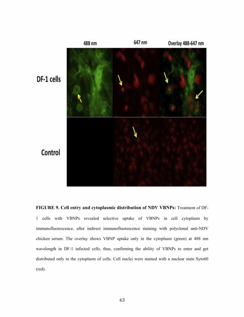
63
FIGURE 9. Cell entry and cytoplasmic distribution of NDV VBNPs: Treatment of DF-
1 cells with VBNPs revealed selective uptake of VBNPs in cell cytoplasm by
immunofluorescence, after indirect immunofluorescence staining with polyclonal anti-NDV
chicken serum. The overlay shows VBNP uptake only in the cytoplasm (green) at 488 nm
wavelength in DF-1 infected cells, thus, confirming the ability of VBNPs to enter and get
distributed only in the cytoplasm of cells. Cell nuclei were stained with a nuclear stain Syto60
(red).

64
A. B.
C. D.
FIGURE 10. VBNPs induce cytotoxicity in treated transformed and tumor cells: J3T
(A) and HT1080 (B), 293T (C), and A549 (D) cells were treated with VBNP and VBNP-VP3 in a
96 well plate (Corning). After 72 h incubation, 10 μl WST-1 reagent was added to each well and
incubated for additional 2 h. The resulting number of viable cells was calculated by measuring the
absorbance produced in each well compared to mock-infected control cells. VBNP-VP3 showed
higher cytotoxicity than VBNP in treated cells. VBNP-treated 293T cells showed significant
cytotoxicity compared to the control but no appreciable cytotoxicity was detected in A549 cells.
0
20
40
60
80
100
120
0.25 0.025 0
Pe
rce
nta
ge o
f ce
ll v
iab
ilit
y (%
)
BNP concentration (ng/mL)
NDV BNP
NDV Apoptin
CONTROL
0
20
40
60
80
100
120
0.25 0.025 0
Pe
rce
nta
ge o
f C
ell
Via
bil
ity
(%)
BNP concentration (ng/mL)
NDV BNP
NDV Apoptin
CONTROL
0
10
20
30
40
50
60
70
80
90
100
110
0.25 0.025
cell
viab
ility
, %
BNP protein concentration, µg/ml
CONTROL
NDV VLP
NDV VLP-VP3
0
10
20
30
40
50
60
70
80
90
100
110
0.25 0.025
cell
viab
ility
, %
BNP protein concentration, µg/ml
CONTROL
NDV VLP
NDV VLP-VP3

65
LANES 1 2 3 4
FIGURE 11: VBNP treated cells undergo intranucleosomal DNA fragmentation.
VBNP-VP3 (lanes 1, 2) and VBNP (lanes 3, 4) treated cells were processed using Apoptosis
DNA Laddering kit (Roche) as described in materials methods (section 3.3). Nuclear
fragmentation was observed as formation of laddering DNA bands and smearing in VBNP and
VBNP-VP3 lanes.

66
A.
B.

67
C.
FIGURE 12. NDV VBNPs induce early apoptosis: Flow cytometric analysis of VBNP and
VBNP-VP3 treated cells after staining with propidium iodide and FITC-Annexin V (U87MG and
J3T) or PacBlue-Annexin V (Df-1 cells) to detect dead and apoptotic cells. DF-1 cells showed
percent early apoptotic cells (lower right quadrant) at 24 h post-infection and late apoptosis
(upper right quadrant) by 72 h were indicated (A). VBNPs elicited early apoptosis in U87MG
cells whereas VBNP-VP3 induced all the cells to die by 48 h (B). Whereas J3T cells had ~20%
apoptosis when treated with VBNP but only about 14% early apoptotic cells (lower right
quadrant) were noticed with VBNP-VP3 treatment (C).

68
A.
B.
FIGURE 13. VBNPs induce proinflammatory cytokines and chemokines in tumor
cells: HT1080 and U87MG cells were treated with VBNPs and VBNP-VP3 and analyzed for
proinflammatory and chemokine response by cytometric bead assy. In HT1080 cells, VBNPs
elicited a comparatively higher IL-6 and IL-8 response to VBNP-VP3 and control groups (A)
whereas MCP-1 and to some extent IL-8 response was higher in VBNP-VP3 infected U87MG
cells (B).

69
A.
B.
FIGURE 14. Mean tumor volume in VBNP treated xenotransplanted tumors: The
tumor dimensions were observed every other day and the tumor size was calculated for tumor
volume and average of each group was plotted in a graph over a period of time in days post
treatment for each group. Although no statistical significance was observed for tumor regression
in both HT1080 (A) and U87MG (B) tumor implants, graphical representation of the tumor
volume indicated a controlled growth of tumor mass until end of treatment (Day 28).
0
1
2
3
0 6 14 28 36 42 50 60
Tu
mo
r v
olu
me
(cm
3)
Days post -inoculation
U87MG VBNP
VBNP 1:10
VBNP-VP3
VBNP-VP3 1:10
VBNP-VP3-fo
VBNP-VP3-fo 1:10
PBS
0
1
2
3
4
0 6 14 28 36 42 50 60
Tu
mo
r V
olu
me
(cm
3)
Days post -inoculation
HT1080VBNP
VBNP 1:10
VBNP-VP3
VBNP-VP3 1:10
VBNP-VP3-fo
VBNP-VP3-fo 1:10
PBS

70
A.
B.
FIGURE 15. VBNP treatment in both HT1080 and U87MG failed to prolong the
survival times in tumor bearing mice: Surviving mice from each group was plotted to
determine the percent survival for each group in both HT1080 (A) and U87MG (B) using Kaplan
Meier method. No statistical significance was observed.

71
FIGURE 16. VBNP therpay induced a proinflammatory response and cell death in
implanted tumors: The H&E stained histological sections of HT1080 implants showed
infiltration of megakaryocytes, mainly band neutrophils. The pink streaking indicates areas of
necrosis containing fragmented neutrophic infiltration. VBNP (A), VBNP-VP3 (B), VBNP-VP3-
Fo (C) and mock-infected control (D).
.

72
FIGURE 17. VBNP therapy kills tumor cells by necrosis and causes spleenomegaly
in U87MG xenotransplanted mice: Representative H&E stained histological tumor sections
showed infiltration of megakaryocytes, mainly band neutrophils as indicated with arrows (blue),
irrespective of treatment. The pink streaking indicates areas of necrosis containing fragmented
neutrophilic infiltration (A, C, E). Gross examination showed splenomegaly. The spleen sections
showed extramedullary hematopoietic changes (red arrows) in U87MG induced tumors (B, D, F).

73
3.8 References
Asemissen, A.M., C. Scheibenbogen, A. Letsch, K. Hellstrand, F. Thoren, K. Gehlsen, A.
Schmittel, E. Thiel, and U. Keilholz. 2005. Addition of histamine to interleukin 2
treatment augments type 1 T-cell responses in patients with melanoma in vivo:
immunologic results from a randomized clinical trial of interleukin 2 with or without
histamine (MP 104). Clin Cancer Res. 11:290-297.
Bridle, B.W., J.E. Boudreau, B.D. Lichty, J. Brunelliere, K. Stephenson, S. Koshy, J.L. Bramson,
and Y. Wan. 2009. Vesicular stomatitis virus as a novel cancer vaccine vector to prime
antitumor immunity amenable to rapid boosting with adenovirus. Mol Ther. 17:1814-
1821.
Buonaguro, L., M.L. Tornesello, and F.M. Buonaguro. 2010. Virus-like particles as particulate
vaccines. Curr HIV Res. 8:299-309.
Cheng, H., J.L. Zhu, X. Zeng, Y. Jing, X.Z. Zhang, and R.X. Zhuo. 2009. Targeted gene delivery
mediated by folate-polyethylenimine-block-poly(ethylene glycol) with receptor
selectivity. Bioconjug Chem. 20:481-487.
Davis, J.J., and B. Fang. 2005. Oncolytic virotherapy for cancer treatment: challenges and
solutions. J Gene Med. 7:1380-1389.
Elankumaran, S., V. Chavan, D. Qiao, R. Shobana, G. Moorkanat, M. Biswas, and S.K. Samal.
2010. Type I interferon-sensitive recombinant newcastle disease virus for oncolytic
virotherapy. J Virol. 84:3835-3844.
Elankumaran, S., D. Rockemann, and S.K. Samal. 2006. Newcastle disease virus exerts oncolysis
by both intrinsic and extrinsic caspase-dependent pathways of cell death. J Virol.
80:7522-7534.

74
Farassati, F., A.D. Yang, and P.W. Lee. 2001. Oncogenes in Ras signalling pathway dictate host-
cell permissiveness to herpes simplex virus 1. Nat Cell Biol. 3:745-750.
Fuchs, E.J., and P. Matzinger. 1996. Is cancer dangerous to the immune system? Semin Immunol.
8:271-280.
Gottschalk, S., R.J. Cristiano, L.C. Smith, and S.L. Woo. 1994. Folate receptor mediated DNA
delivery into tumor cells: potosomal disruption results in enhanced gene expression. Gene
Ther. 1:185-191.
Imperiale, M.J., H.T. Kao, L.T. Feldman, J.R. Nevins, and S. Strickland. 1984. Common control
of the heat shock gene and early adenovirus genes: evidence for a cellular E1A-like
activity. Mol Cell Biol. 4:867-874.
Jennings, G.T., and M.F. Bachmann. 2008. The coming of age of virus-like particle vaccines.
Biol Chem. 389:521-536.
Lech, P.J., and S.J. Russell. 2010. Use of attenuated paramyxoviruses for cancer therapy. Expert
Rev Vaccines. 9:1275-1302.
Neff-LaFord, H.D., B.A. Vorderstrasse, and B.P. Lawrence. 2003. Fewer CTL, not enhanced NK
cells, are sufficient for viral clearance from the lungs of immunocompromised mice. Cell
Immunol. 226:54-64.
Ochsenbein, A.F., P. Klenerman, U. Karrer, B. Ludewig, M. Pericin, H. Hengartner, and R.M.
Zinkernagel. 1999. Immune surveillance against a solid tumor fails because of
immunological ignorance. Proc Natl Acad Sci U S A. 96:2233-2238.
Pantua, H.D., L.W. McGinnes, M.E. Peeples, and T.G. Morrison. 2006. Requirements for the
assembly and release of Newcastle disease virus-like particles. J Virol. 80:11062-11073.
Rice, J., S. Buchan, H. Dewchand, E. Simpson, and F.K. Stevenson. 2004. DNA fusion vaccines
induce targeted epitope-specific CTLs against minor histocompatibility antigens from a
normal or tolerized repertoire. J Immunol. 173:4492-4499.

75
Ross, J.F., P.K. Chaudhuri, and M. Ratnam. 1994. Differential regulation of folate receptor
isoforms in normal and malignant tissues in vivo and in established cell lines. Physiologic
and clinical implications. Cancer. 73:2432-2443.
Stevenson, F.K., C.H. Ottensmeier, P. Johnson, D. Zhu, S.L. Buchan, K.J. McCann, J.S. Roddick,
A.T. King, F. McNicholl, N. Savelyeva, and J. Rice. 2004. DNA vaccines to attack
cancer. Proc Natl Acad Sci U S A. 101 Suppl 2:14646-14652.
Wodarz, D. 2001. Viruses as antitumor weapons: defining conditions for tumor remission.
Cancer Res. 61:3501-3507.
Wu, C.Y., Y.C. Yeh, Y.C. Yang, C. Chou, M.T. Liu, H.S. Wu, J.T. Chan, and P.W. Hsiao. 2010a.
Mammalian expression of virus-like particles for advanced mimicry of authentic
influenza virus. PLoS One. 5:e9784.
Wu, Y., J. Hao, and D. Li. 2010b. [Progress in using Newcastle disease virus for tumor therapy: a
review]. Sheng Wu Gong Cheng Xue Bao. 26:1031-1036.
Yu, B., H.C. Tai, W. Xue, L.J. Lee, and R.J. Lee. 2010. Receptor-targeted nanocarriers for
therapeutic delivery to cancer. Mol Membr Biol. 27:286-298.

76
3.9 Acknowledgements
We thank VMRCVM for providing the opportunity to perform this project, Ms. Kathy
Lowe, Biomedical Sciences and Pathobiology and Nanomaterial characterization and fabrication
laboratory, Virginia Tech, Dr. Griffith Parks and Dr. John B. Johnson, Wake Forest University
for transmission electron microscopy. This work is funded in part by the Institute of Critical
Technology and Applied Science (ICTAS), Virginia Tech, Blacksburg.

77
Chapter 4: Surface Modification of Newcastle disease Virus-Based
Nanoparticles with Nipah Virus Glycoproteins
Vrushali Chavan, Abilash P. S, Wang Yong, Moanaro Biswas and Subbiah
Elankumaran
Department of Biomedical Science and Pathobiology, Virginia-Maryland Regional
College of Veterinary Medicine, Virginia Polytechnic Institute and State University,
Blacksburg, VA 24060.
4.1 Abstract
Extensive advancements have been made in the past few years towards understanding
both unique and shared processes between paramyxoviruses during virus particle formation.
Nipah (NiV) and Hendra viruses are novel viruses which fall under the genus henipavirus, in the
family Paramyxoviridae. They are responsible for causing deadly infections in humans and
domestic animals with severe vasculitis and encephalitis. There are presently no successful
treatments or vaccines approved for henipavirus infections. NiV is a single-stranded, negative-
sense RNA virus that encodes six structural proteins including two envelope glycoproteins: the G
and F protein. G protein is responsible for binding to the cellular receptors, ephrin B2 and the F
protein mediates membrane fusion. It has been known that high expression of ephrin-B is
associated with increased tumor growth, tumorigenicity and metastasis. For paramyxoviruses,
viral M proteins are the organizers of this assembly process and are highly abundant that bind
directly to cellular membranes and occupy a central position allowing interaction both with viral
RNP cores and also with viral glycoproteins via the cytoplasmic tail. Therefore, we hypothesized
that the core structural M protein of NDV independently could incorporate surface glycoproteins
from other paramyxoviruses without the help of transmembrane cytoplasmic tail sequences of

78
NDV glycoproteins. We tested this hypothesis by co-expressing NDV M gene with NiV F and G
glycoproteins to form virus-based nanoparticles (NiV-ndBNP) in 293T cells and studied the
morphology and biological characteristics of authentic NiV virus-like particles and NiV-ndBNPs.
Our results demonstrated that NDV M successfully incorporated NiV glycoproteins on the
envelope, and the resulting NiV-ndBNPs were fusogenic, and were reminiscent of authentic
NDV/NiV virions. These novel NiV-ndBNPs could be developed for efficient targeting to
ephrinB2/B3 receptor bearing tumor cell types and as vaccines/diagnostics for NiV.

79
4.2. Introduction
Nipah and Hendra viruses are novel viruses which fall under the genus henipavirus, in the
family Paramyxoviridae. They are responsible for causing deadly infections in humans and
domestic animals with severe vasculitis and encephalitis resulting in about 40% mortality rate
(Bishop and Broder, 2008). Fruit bats, such as flying foxes are the natural hosts for these zoonotic
viruses. Pigs are intermediary hosts for Nipah virus (NiV) infections and horses in the case of
hendra virus (Eaton et al., 2006). There are presently no successful treatments or vaccines
approved for henipavirus infections. Outbreaks of hendravirus have potential agricultural
significance; NiV epidemic in Malaysia was responsible for culling 1.1 million pigs after which
the epidemic was under control (Mohd Nor et al., 2000).
NiV is a single-stranded, negative-sense RNA virus and consists of six genes (N-P-M-F-
G-L) flanked by a 3′ leader and 5′ trailer region with a genome length of 18246 nucleotide (nt) in
Malaysian isolate or 18252 nt in Bangladesh isolate (Mungall et al., 2007) (Fig. 1). NiV has a
pleomorphic structure ranging from 50 nm to greater than 600 nm in diameter (Hyatt et al., 2001).
It has six transcription units that encode six structural proteins including two envelope
glycoproteins: the G and F protein. G protein is responsible for binding to the cellular receptors,
ephrin B2 and B3 (Bonaparte et al., 2005; Negrete et al., 2005) and the F protein mediates
membrane fusion (Bossart et al., 2002). M protein is located below and interacts with both the
glycoproteins and the rinobnuceloprotein (RNP) complex (Lamb and Parks, 2007; Schmitt and
Lamb, 2004). Like other paramyxoviruses, the P gene of NiV expresses four proteins, namely P,
V, W and C (Harcourt et al., 2000; Wang et al., 2001).
For some paramyxoviruses, such as Sendai virus, NDV and MuV, attachment is mediated
by HN proteins that bind to sialic acid receptors (Lamb et al., 2006). Other paramyxovirus
attachment proteins mediate binding to protein receptors like the H protein of MeV and the G

80
proteins of the henipaviruses (Russell and Luque, 2006). The receptor for the attachment
glycoprotein (G) of NiV is ephrin B2/B3 (Ciancanelli and Basler, 2006; Wild, 2009). The Ephrin
B2/B3, binding site for NiV G resides in a similar position as HN protein sialic acid binding sites
(Xu et al., 2008). NiV interacts with ephrin-B2 and -B3 via an overlapping site on the virus G
attachment protein (Negrete et al., 2006). NiV forms particles by budding from the cellular
membranes.
Ephrins have been recognized to be differentially expressed in various human tumors
such as malignant melanoma, neuroblastoma and cancers of the prostate, breast, lungs,
esophagus, gastrium and colorectum (Kiyokawa et al., 1994). Evidence has shown that ephrin-B
signaling is associated with cancer progression (Campbell and Robbins, 2008; Merlos-Suarez and
Batlle, 2008; Pasquale, 2008). An increased activity and expression of ephrin-B receptors have
been interrelated with the growth of solid tumors (Alam et al., 2009; Heroult et al., 2006; Irie et
al., 2008; Merlos-Suarez and Batlle, 2008). Besides, high expression of ephrin-B is associated
with increased tumor growth, tumorigenicity and metastasis (Kiyokawa et al., 1994; Nakada et
al., 2009; Tang et al., 1999).
Paramyxovirus assembly can be viewed as a three-part process. First the genomic RNA
associate with NP subunits to form the helical nucleocapsid structure, and then the P and L
proteins are added (Harrison et al., 2010) to form the RNP. The membrane proteins (HN, F, and
M) then accumulate at the plasma membrane. The nucleocapsid is enveloped during the budding
process and progeny virions are produced. Viral M proteins are the organizers of this assembly
process. These highly abundant viral proteins bind directly to cellular membranes and occupy a
central position that allows interaction both with viral RNP cores and also with viral
glycoproteins via the cytoplasmic tail (CT). For paramyxoviruses, viral M proteins are key
drivers of virus assembly and budding (Harrison et al, 2010). At present, the protein-protein

81
interaction that regulates the incorporation of paramyxovirus glycoproteins into virions and the
detailed sequence specificity involved in these processes are not known.
The M protein is one of the abundant proteins in the virion and plays a multifaceted role
in paramyxovirus replication. Expression of NiV M protein alone leads to the budding of VLP’s.
A late domain sequence (YMYL) is important for this property (Ciancanelli and Basler, 2006;
Wild, 2009). Similarly, M protein of NDV occupies a central position that allows interaction
both with viral RNP cores and also with viral glycoproteins via the CTs incorporating them into
viral particles (Harrison et al., 2010). Recently, it has been shown that NDV structural core
proteins, NP and M protein were able to incorporate the ectodomain of respiratory syncytial virus
G protein fused to the CT and transmembrane (TM) sequences of the NDV HN protein
(McGinnes et al, 2010). We have also incorporated the chicken anemia virus VP3 protein on the
surface of NDV in addition to the NDV HN and F proteins but used only the M protein of NDV
for the structure (Chapter 3).
We hypothesize based on the above findings that the core structural M protein of NDV
independently could incorporate surface glycoproteins from other paramyxoviruses without the
help of TM and CT sequences of NDV glycoproteins. As a proof of concept, we tested this
hypothesis by co-expressing NDV M gene with NiV F and G glycoproteins (NiV-ndBNP) in
293T cells. The morphology and biological characteristics of authentic NiV virus-like particles
and NiV-ndBNPs were compared. Our results demonstrated that NDV M successfully
incorporated NiV glycoproteins on the envelope, and the resulting NiV-ndBNPs were fusogenic,
and were reminiscent of authentic NDV/NiV virions. These novel NiV-ndBNPs could be
exploited for efficient targeting to ephrinB2/B3 receptor bearing tumor cell types and as
vaccines/diagnostics for NiV.

82
4.3. Materials and Methods
4.3.1. Cells: 293T human embryonic kidney cells and DF1 cells were grown in Dulbecco’s
modified Eagle’s medium (DMEM) (Gibco Inc.) supplemented with penicillin-streptomycin
(Gibco Inc.) and 10% fetal calf serum (FCS) (Fisher Scientific).
4.3.2 Plasmids and antibodies: NDV cDNA sequences encoding the M protein were cloned
into the expression vector pCAGGS to generate pCAGGS-M. NiV cDNA sequences encoding M,
F and G proteins were cloned into the expression vector pCMV3Tag1 and pCAGGS to generate
pPCMV-3Tag1-M (Wang et al., 2010), pCAGGS-F and pCAGGS-G plasmids, respectively. The
plasmid pCMV-3tag1-M was a kind gift from Dr. Benhur Lee from University of California, Los
Angeles (UCLA). The plasmids pCAGGS-F and pCAGGS-G were obtained from Dr. Paul Rota,
Centers for Disease Control, Atlanta. Chicken polyclonal anti-NDV serum (SPAFAS) was used
to probe for NDV structural proteins. Monospecific serum against NiV F and G structural
proteins, raised in rabbits were gifts from Dr. Benhur Lee, UCLA.
4.3.3 Generation of NiV-BNP and NiV-ndBNP: Transfections of subconfluent 293T cells
were accomplished using Lipofectamine 2000 (Invitrogen Inc.). The following amounts of
plasmid DNA were used per 150-mm dish (Corning Inc.): 10µg each of pPCMV-3Tag1-M,
pCAGGS-F NiV, and pCAGGS-G NiV for NiV VBNP and pCAGGS-M NDV, pCAGGS-F NiV,
and pCAGGS-G NiV for NiV-ndBNP. For each transfection, a mixture of each of the plasmid
DNA with 2 mL OptiMEM medium (Gibco Inc.) and 10 µl of Plus reagent (Invitrogen Inc.) in
tube A and 2 mL OptiMEM medium with 20 µl Lipofectamine 2000 (Invitrogen Inc.) in tube B
were incubated at room temperature for 20 min. Tube A and B were mixed and further incubated

83
for 20 min at room temperature. The mixture was then added to each plate after washing it 3
times with phosphate buffered saline (PBS), pH 7.4 and further incubated at 37°C, 5% CO2 for 4-
6 h. DMEM with 2% FCS and Penicillin-Streptomicin (Gibco Inc.) was then added to make up
the volume to 20 mL. At 72 h post-transfection, cells were harvested with a cell scraper (Corning
Inc.) and clarified at 5000g for 30 min.
4.3.4 NiV-BNP and NiV-ndBNP purification: The clarified supernatants were pelleted by
centrifugation in a SW 28 rotor in a Beckman ultracentrifuge (Beckman Inc., USA) at 25,000 rpm
for 2 h at 4°C. The pellet was re-suspended in TNE buffer (25 mM Tris- HCl, pH 7.4, 150 mM
NaCl, 5 mM EDTA) (Pantua et al., 2006) and overlaid on top of a step gradient consisting of 5 ml
20% and 3 ml 55% sucrose solutions (g/ml) in Ca/Mg free PBS, pH 7.4, and centrifuged using a
SW 41 rotor at 22,000 rpm for 2 h at 4°C. The band at the interface (containing concentrated
particles) was collected and was diluted in TNE buffer and centrifuged in a SW 28 rotor at 25,000
rpm for 2 h at 4°C for concentrating the NiV-BNP and NiV-ndBNP. The pellet was then re-
suspended in 500 µl of TNE buffer and stored at 4°C until use. The purified BNPs were further
filtered through Amicon Ultra-15 centrifugal filter (Millipore Inc.) at 5000g for 15 min. The
retentate was collected containing the filtered and concentrated BNPs and was stored at 4°C.
4.3.5 Transmission Electron microscopy (TEM): The purified NiV-ndBNPs were
adsorbed to formvar carbon-coated 200 mesh nickel grids (Electron Microscopy systems). The
grids were then washed in PBS, pH 7.4 and fixed with 2% paraformaldehyde (10 min) and
extensively washed with PBS, pH 7.4 again before negatively staining with 1% phosphotungstic
acid. The morphologic characteristics were examined using a Philips 420 transmission electron

84
microscope at a magnification of 55,000 x, operating at 80 kV.
4.3.6 Immunogold Transmission Electron Microscopy: NiV-ndBNP suspension (10 l)
was layered on carbon coated 200 mesh gold grids (Electron Microscopy systems) and incubated
for 1h at 4OC, after which the remaining suspension was removed. The grids were blocked with
PBS, pH 7.4 containing 1% BSA. Adsorbed NiV-ndBNPs were then stained with a mixture of
both anti-F and anti-G rabbit polyclonal antibodies against NiV at a dilution of 1:25. Bound
antibody was detected with 12 nm colloidal gold, goat anti-rabbit antibodies (Jackson
Immunoresearch), followed by negative staining and analysis under Philips TEM 400
transmission electron microscope at a magnification of 55,000 x, operating at 80 kV.
4.3.7 ImmunoBlot: The gradient purified NiV-BNPs and NiV-ndBNPs were subjected to
immunoblotting using standard procedures. The proteins were separated in a 4-20% sodium-
dodecyl sulphate polyacrylamide gel electrophoresis (SDS-PAGE) and transferred to Immobilon-
P (Millipore) membranes. The membranes were blocked overnight in 5% skim milk and 0.05%
Tween 20 solution at 4°C and washed in PBS, pH 7.4 with 0.05% Tween 20 (PBST) and
incubated at 4°C with a cocktail containing 1:1000 dilution of NDV polyclonal chicken serum,
Nipah F and Nipah G antibodies for 1 h for detecting NiV-ndBNP. The membranes were washed
again 3 times with PBST for 30 min. The membranes were allowed to react with 2 mL of
chemiluminesence TMB substrate (KPL Inc.). The reaction was stopped with water before
visualizing the protein bands under ChemDoc XRS (Thermo Scientific Inc.).

85
4.3.8 Sequence Comparison: The CT and TM domain amino acid sequences of NDV HN-
NiV G and NDV F-NiV F were aligned in MegAlign (DNASTAR). NDV HN (GenBank
accession #. P32884), NDV F (GenBank Accession #. P06156), NiV F (GenBank Accession #.
Q9IH63) and the NiV G (GenBank Accession %. Q9IH62) were used for sequence comparisons
of the CT and TM.

86
4.4. Results
4.4.1. Fusogenicity of NiV-ndBNP: 293T cells were transfected with pCAGGS-M (NDV),
pCAGGS-F NiV and pCAGGS-G NiV plasmids and was observed for fusion every 24 h. Syncitia
were evident in the cells 48 h post-transfection (Fig. 2A). Similarly, DF-1 cells transfected with
NDV pCAGGS-HN, -M and -F plasmids induced syncytia appearing as multinucleated cells (Fig.
2B). It appeared that NiV-ndBNPs were more fusogenic than the NDV BNPs, which should be
confirmed by future quantitative fusion studies.
4.4.2. Morphology and structure of NiV-ndBNP: 293T cells expressing M protein of NDV
and F, G proteins of NiV were able to produce NiV-ndBNP with morphological characteristics
similar to that of the authentic infectious virus, which was confirmed by TEM (Fig. 3A). The
secreted NiV-ndBNPs were pleomorphic but generally displayed either a spherical or oval
morphology (Fig. 3B to C). The spike projections on the surface of NDV M based virions were
no different than authentic NiV virions (Fig. 3D). To further verify the presence of NiV F and G
glycoproteins on the surface of NiV-ndBNPs, the particles were labeled with individual specific
antibodies and counterstained with 12nm gold spheres conjugated to seconday antibodies (Fig.
3E).
4.4.3. Composition of NiV-ndBNPs: To determine if the purified NiV-ndBNP’s were
composed entirely of NDV-M and NiV-G, and NiV-F proteins, the proteins were subjected to
SDS-PAGE and confirmed by immunoblot (Fig. 4, lane 2) which detected G at ~70K Da., M at
~35K Da., and F at ~40K Da along with NiV-BNP (Fig. 4, lane 1).

87
4.4.4. Poor sequence homology of NDV and NiV glycoproteins’ CT and TM: We
compared the amino acid sequences of the CT and TM of NDV (Beaudette C strain)
glycoproteins (F and HN) to that of NiV (Bangladesh strain) glycoproteins (F and G) and found
that only two residues in the CT (Fig. 5A to B) and seven residues in the TM (Fig. 5C to D) were
similar in the F protein while six residues in CT (Fig. 6A to B) and one residue in TM (Fig. 6C to
D) were similar in the attachment proteins.

88
4.5. Discussion
Extensive advancements have been made in the past few years towards understanding
both unique and shared processes between paramyxoviruses during virus particle formation. The
functions of paramyxovirus M proteins, glycoproteins, nucleocapsid proteins, and accessory
proteins during virus assembly have been elucidated. We were successful in developing a proof
of concept for the assembly and structural properties of NDV-based NiV BNPs (NiV-ndBNP)
using the NDV M protein and NiV F and G glycoproteins. Electron microscopy revealed that the
NiV-ndBNP were morphologically similar to authentic NiV virions and by immunoglod labeling,
we were able to demonstrate that NiV glycoproteins were incorporated into the NDV M based
particles.
Paramyxovirus glycoproteins display conserved amino acid motifs in their CT and TM of
glycoproteins. For example, human and bovine parainflenzavirus 3 (hPIV3 and bPIV3,
respectively) share a 5-residue sequence (PYVLT) in their F cytoplasmic tails and an 8-residue
sequence (MEYWKHTN) in their HN cytoplasmic domains. Further comparison of these motifs
revealed that 2 amino acids are conserved between the HNs of Sendai, hPIV1, hPIV3, and bPIV3,
as are 2 amino acids (YXL) in the F protein (Takimoto et al., 1998). In addition, HN and F
proteins derived from hPIV1 were incorporated into hPIV3 virions (Tao et al., 1998). Our results
demonstrate that NDV M can essentially incorporate henipavirus and possibly other
paramyxovirus envelope glycoproteins, despite poor sequence homology in the CT and TM
domains. The mechanism(s) by which NDV M protein incorporates NiV glycoproteins need to be
further explored.
Further investigations into the biological properties of the NiV-ndBNP are required to
provide a clearer understanding of the events that are essential for viral particle formation and

89
may contribute to the development of effective antiviral strategies designed to intervene the late
steps of paramyxovirus life cycles.
This report focused on the development and characterization of a flexible NDV M protein
based platform to display the surface antigens of an unrelated paramyxovirus. We, therefore
propose that this may serve as a promising platform for cancer therapy and vaccine/diagnostics
development because 1) we have demonstrated by a simple exchange of surface antigens that our
technology is flexible and can be developed to display a plethora of viral or tumor antigens on the
envelope including other paramyxoviruses for cell-type specific targeting for tumor therapy or
vaccine/diagnostic purposes, 2) this approach will dispense with the biosafety level-4 restrictions
and bottlenecks associated with the use of live NiV.

90
4.6. Figures
FIGURE 1. Diagrammatic representation of a henipavirus: The surface glycoproteins
(G, F), Matrix (M) protein and RNP complex consisting of Nucleocapsid protein (N), Large
protein (L) and Phosphoprotein (P) is shown (adapted from
http://en.wikipedia.org/wiki/Henipavirus).

91
A.
B.
FIGURE 2. Fusogenecity of NiV-ndBNP in 293T cells: Human 293T embryonic kidney
cells were transfected with pCAGGS- M (NDV), -G (NiV) and -F (NiV) plasmids. Syncytia were
observed at 48 h and 72 h post-transfection by phase contrast microscopy, under 40 x
magnification (A). Transfected DF-1 cells with pCAGGS-HN, -M and -F plasmids (NDV) shows
syncytia at 48 h and 72 h (B). Note NiV-ndBNPs were highly fusogenic than NDV-BNPs.
Arrowheads point to the syncytia.

92
A B C
D E
FIGURE 3. Transmission Electron Microscopy (TEM) of NiV-ndBNPs: The
morphology of NiV-ndBNP (A, B, C), and the authentic NiV (Ciancanelli and Basler, 2006)
virions (D) by TEM. Arrowheads indicate the NiV glycoproteins, G and/or F at the surface of the
VBNPs. Immunogold labeled NiV-ndBNP (E) detected by rabbit anti-F (NiV) and anti-G (NiV)
bound to 12 nm colloidal gold donkey anti-rabbit IgY antibodies.

93
FIGURE 4. Immunoblot of NiV-BNP and NiV-ndBNP: NiV-BNP (Lane 1) and NiV-
ndBNP (Lane 2) proteins were detected by SDS-PAGE and confirmed by Immunoblot using anti-
NDV chicken serum and anti- NiV F, G, and M rabbit antibody which detected G at ~70K Da., M
at ~35K Da., and F at ~40K Da.

94
FIGURE 5. Poor sequence homology between NDV and NiV glycoproteins’ CT and
TM: The amino acid sequences of the CT and TM of NDV (Beaudette C strain) glycoprotein, F
were compared to that of NiV (Bangladesh strain) glycoprotein, F and only two residues in the
CT (A to B) and seven residues in the TM (C to D) were homologous in the F protein. These
results indicated that although the CT and TM of NDV and NiV glycoproteins had a high
variability in the sequences, yet NDV M was able to incorporate F protein of NiV successfully.
Homologous amino acids are highlighted in red, the numbers to the far right corner of each row
indicate the amino acid sequence number and dots indicate the amino acid homology.

95
FIGURE 6. Poor sequence homology between CT and TM of NDV HN and NiV
glycoprotein, G: The amino acid sequences of the CT and TM of NDV (Beaudette C strain)
glycoprotein, HN were compared to that of NiV (Bangladesh strain) glycoprotein, G and only six
residues in CT (A to B) and one residue in TM (C to D) were homologous in the attachment
proteins. These results indicated that the CT and TM of NDV and NiV glycoproteins had a high
variability in the sequences; yet, NDV M was able to incorporate G protein of NiV successfully.
Homologous amino acids are highlighted in red, the numbers to the far right corner of each row
indicate the amino acid sequence number and dots indicate the amino acid homology.

96
4.7. References
Alam, S.M., J. Fujimoto, I. Jahan, E. Sato, and T. Tamaya. 2009. Coexpression of EphB4 and
ephrinB2 in tumor advancement of uterine cervical cancers. Gynecol Oncol. 114:84-88.
Bishop, and C.C. Broder. 2008. Hendra and Nipah: lethal zoonotic paramyxoviruses. In Emerging
infections. S.M.H.a.J.M.H. W.M. Scheld, editor. American Society for Microbiology,
Washington, DC. pp. 155–187.
Bonaparte, M.I., A.S. Dimitrov, K.N. Bossart, G. Crameri, B.A. Mungall, K.A. Bishop, V.
Choudhry, D.S. Dimitrov, L.F. Wang, B.T. Eaton, and C.C. Broder. 2005. Ephrin-B2
ligand is a functional receptor for Hendra virus and Nipah virus. Proc Natl Acad Sci U S
A. 102:10652-10657.
Bossart, K.N., L.F. Wang, M.N. Flora, K.B. Chua, S.K. Lam, B.T. Eaton, and C.C. Broder. 2002.
Membrane fusion tropism and heterotypic functional activities of the Nipah virus and
Hendra virus envelope glycoproteins. J Virol. 76:11186-11198.
Campbell, T.N., and S.M. Robbins. 2008. The Eph receptor/ephrin system: an emerging player in
the invasion game. Curr Issues Mol Biol. 10:61-66.
Ciancanelli, M.J., and C.F. Basler. 2006. Mutation of YMYL in the Nipah virus matrix protein
abrogates budding and alters subcellular localization. J Virol. 80:12070-12078.
Eaton, B.T., C.C. Broder, D. Middleton, and L.F. Wang. 2006. Hendra and Nipah viruses:
different and dangerous. Nat Rev Microbiol. 4:23-35.
Harcourt, B.H., A. Tamin, T.G. Ksiazek, P.E. Rollin, L.J. Anderson, W.J. Bellini, and P.A. Rota.
2000. Molecular characterization of Nipah virus, a newly emergent paramyxovirus.
Virology. 271:334-349.
Harrison, M.S., T. Sakaguchi, and A.P. Schmitt. 2010. Paramyxovirus assembly and budding:
building particles that transmit infections. Int J Biochem Cell Biol. 42:1416-1429.

97
Heroult, M., F. Schaffner, and H.G. Augustin. 2006. Eph receptor and ephrin ligand-mediated
interactions during angiogenesis and tumor progression. Exp Cell Res. 312:642-650.
Hyatt, A.D., S.R. Zaki, C.S. Goldsmith, T.G. Wise, and S.G. Hengstberger. 2001. Ultrastructure
of Hendra virus and Nipah virus within cultured cells and host animals. Microbes Infect.
3:297-306.
Irie, F., M. Okuno, K. Matsumoto, E.B. Pasquale, and Y. Yamaguchi. 2008. Heparan sulfate
regulates ephrin-A3/EphA receptor signaling. Proc Natl Acad Sci U S A. 105:12307-
12312.
Kiyokawa, E., S. Takai, M. Tanaka, T. Iwase, M. Suzuki, Y.Y. Xiang, Y. Naito, K. Yamada, H.
Sugimura, and I. Kino. 1994. Overexpression of ERK, an EPH family receptor protein
tyrosine kinase, in various human tumors. Cancer Res. 54:3645-3650.
Lamb, R.A., and G.D. Parks. 2007. Paramyxoviridae: the viruses and their replication. Lippincott
Williams & Wilkins,, Philadelphia.
Lamb, R.A., R.G. Paterson, and T.S. Jardetzky. 2006. Paramyxovirus membrane fusion: lessons
from the F and HN atomic structures. Virology. 344:30-37.
Merlos-Suarez, A., and E. Batlle. 2008. Eph-ephrin signalling in adult tissues and cancer. Curr
Opin Cell Biol. 20:194-200.
Mohd Nor, M.N., C.H. Gan, and B.L. Ong. 2000. Nipah virus infection of pigs in peninsular
Malaysia. Rev Sci Tech. 19:160-165.
Mungall, B.A., D. Middleton, G. Crameri, K. Halpin, J. Bingham, B.T. Eaton, and C.C. Broder.
2007. Vertical transmission and fetal replication of Nipah virus in an experimentally
infected cat. J Infect Dis. 196:812-816.
Nakada, M., Eric M. Anderson, Tim Demuth, Satoko Nakada, Linsey B. Reavie, Kelsey L.
Drake, Dominique B. Hoelzinger, and M.E. Berens. 2009. The phosphorylation of
ephrin-B2 ligand promotes glioma cell migration and invasion. International Journal of
Cancer. Voleume 126:pages 1155-1165.

98
Negrete, O.A., E.L. Levroney, H.C. Aguilar, A. Bertolotti-Ciarlet, R. Nazarian, S. Tajyar, and B.
Lee. 2005. EphrinB2 is the entry receptor for Nipah virus, an emergent deadly
paramyxovirus. Nature. 436:401-405.
Negrete, O.A., M.C. Wolf, H.C. Aguilar, S. Enterlein, W. Wang, E. Muhlberger, S.V. Su, A.
Bertolotti-Ciarlet, R. Flick, and B. Lee. 2006. Two key residues in ephrinB3 are critical
for its use as an alternative receptor for Nipah virus. PLoS Pathog. 2:e7.
Pantua, H.D., L.W. McGinnes, M.E. Peeples, and T.G. Morrison. 2006. Requirements for the
assembly and release of Newcastle disease virus-like particles. J Virol. 80:11062-11073.
Pasquale, E.B. 2008. Eph-ephrin bidirectional signaling in physiology and disease. Cell. 133:38-
52.
Russell, C.J., and L.E. Luque. 2006. The structural basis of paramyxovirus invasion. Trends
Microbiol. 14:243-246.
Schmitt, A.P., and R.A. Lamb. 2004. Escaping from the cell: assembly and budding of negative-
strand RNA viruses. Curr Top Microbiol Immunol. 283:145-196.
Takimoto, T., T. Bousse, E.C. Coronel, R.A. Scroggs, and A. Portner. 1998. Cytoplasmic domain
of Sendai virus HN protein contains a specific sequence required for its incorporation
into virions. J Virol. 72:9747-9754.
Tang, X.X., A.E. Evans, H. Zhao, A. Cnaan, W. London, S.L. Cohn, G.M. Brodeur, and N.
Ikegaki. 1999. High-level expression of EPHB6, EFNB2, and EFNB3 is associated with
low tumor stage and high TrkA expression in human neuroblastomas. Clin Cancer Res.
5:1491-1496.
Tao, T., A.P. Durbin, S.S. Whitehead, F. Davoodi, P.L. Collins, and B.R. Murphy. 1998.
Recovery of a fully viable chimeric human parainfluenza virus (PIV) type 3 in which the
hemagglutinin-neuraminidase and fusion glycoproteins have been replaced by those of
PIV type 1. J Virol. 72:2955-2961.

99
Wang, L., B.H. Harcourt, M. Yu, A. Tamin, P.A. Rota, W.J. Bellini, and B.T. Eaton. 2001.
Molecular biology of Hendra and Nipah viruses. Microbes Infect. 3:279-287.
Wang, Y.E., A. Park, M. Lake, M. Pentecost, B. Torres, T.E. Yun, M.C. Wolf, M.R. Holbrook,
A.N. Freiberg, and B. Lee. 2010. Ubiquitin-regulated nuclear-cytoplasmic trafficking of
the nipah virus matrix protein is important for viral budding. PLoS Pathog. 6:e1001186.
Wild, T.F. 2009. Henipaviruses: a new family of emerging Paramyxoviruses. Pathol Biol (Paris).
57:188-196.
Xu, K., K.R. Rajashankar, Y.P. Chan, J.P. Himanen, C.C. Broder, and D.B. Nikolov. 2008. Host
cell recognition by the henipaviruses: crystal structures of the Nipah G attachment
glycoprotein and its complex with ephrin-B3. Proc Natl Acad Sci U S A. 105:9953-9958.

100
4.8. Acknowledgments
We thank Dr. John Johnson and Dr. Griffith Parks, Wake Forest University, School of Medicine
for transmission electron microscopy, Dr. Paul Rota (CDC) for the nipah virus plasmids and Dr.
Benhur Lee (UCLA) for the pCMV-3tag1-M plasmid and anti-nipah F and G antibodies.

101
Chapter 5: General Conclusions and Future Research
The following general conclusions could be drawn from this study:
In this study, we have established a platform by creating different NDV based
nanoparticles, VBNPs, through directed expression of only three structural proteins
(M, F and HN) of NDV.
The flexibility of this platform to incorporate therapeutic transgenes was demonstrated
by using a model proapoptotic protein and for ligand-directed targeting using folate
conjugation on the surface.
The VBNPs were reminiscent of authentic NDV virions in terms of size, particle
morphology, and the fine structure of surface glycoprotein spikes.
The VBNPs incorporated cellular proteins in the core.
The VBNPs retained the biological properties of NDV virions such as infectivity,
cytoplasmic distribution and fusogenicity.
Fusion of NDV F protein transmembrane and cytoplasmic tails to foreign gene
sequences will incorporate these fusion genes on the envelope of NDV.
The VBNPs of NDV could be surface modified by conjugating them with folate for
folate receptor (FR) mediated targeting.
The proof of concept for cytotoxicity of VBNP and VBNP-VP3 in different tumor cell
lines.
Surface conjugation of folate to VBNPs targeted them specifically FR positive tumor
cell lines.
VBNPs induced killed tumor cells by apoptosis.
VBNPs elicited proinflammatory cytokines and chemokines in tumor cells indicating
they may serve as effective cancer immunotherapeutics by careful and rational design.

102
Although the VBNPs were not curative in nude mouse models of human tumors, the
tumor volumes over time indicated a controlled growth of tumor mass as long as the
VBNPs were being administered.
The antitumor ability of the VBNPs has to be further investigated in
immunocompetent and nude mouse tumor models for dose optimization (the
frequency and quantity of VBNP).
The ability to surface modify the VBNPs by incorporating foreign genes/proteins
using only the structural core M protein and CT and TM of HN protein offers for a
flexible platform to carry multiple or long transgenes that would not have been
possible in recombinant NDV. Our results present a novel and flexible platform for
developing NDV VBNP as a powerful vaccine vector or cancer therapy agent.
For the first time, the ability of a paramyxoviral matrix (M) protein to incorporate
envelope glycoproteins from unrelated paramyxoviruses is demonstrated.
The M protein of NDV was shown to assemble the unrelated Biosafety level-4 nipah
virus (NiV) envelope glycoproteins in an unmodified form.
NiV-ndBNP was morphologically similar to authentic NiV virions and by
immunogold labeling demonstrated that NiV glycoproteins were incorporated into the
NDV M based particles.
This novel approach will dispense with the biosafety level-4 restrictions and
bottlenecks associated with the use of live NiV.

103
Future directions:
This platform VBNP technology is very promising and flexible to engineer various
therapeutic transgenes and receptor specific targeting.
It would be interesting to study the ability of these VBNPs to carry fusion products of
tumor -associated antigens to elicit effective antitumor immunity.
The ability to engineer the fusion protein of NDV for protease specific targeting offers
an attractive target for cell type specific delivery of VBNPs.
Dose optimization studies in nude mouse models of xenotransplanted tumors.
The ability of VBNPs to elicit NK-cell mediated antitumor immunity could be studied
in nude mouse models of xenotransplanted human tumors.
The efficacy of VBNPs in immunocompetent preclinical tumor models
The nature and composition of cellular proteins in the VBNPs need to be identified
which would provide insights into the assembly and sorting of cellular proteins.
The VBNP platform provides an opportunity to understand the assembly and budding
of paramyxoviruses and afford development of intervention strategies to intervene in
the late stages of paramyxovirus life cycle.
The mechanism by which NiV glycoproteins is able to get incorporated into NDV M
based particles needs to be further explored.
Further research is needed to study the biological properties of the NiV-ndBNP and to
understand their properties to provide a clearer understanding of the events that are
essential for viral particle formation and may contribute to the development of
effective antiviral strategies for this BSL-4 agent.
Future research may be directed to receptor-mediated targeting to ephrin B receptors
and oncolytic efficacy of NiV-ndBNP and NiV BNPs using in vitro and in vivo tumor
models.