Embed Size (px)
Citation preview

1
Virtual Human Model CASIMIR - A Chance and a Challenge for the Aetiology Understanding of
Pressure Injury Development Alexander Siefert, Martin Beiersdörfer, Aravinda R. Veeraraghavan
Wölfel Engineering GmbH + Co. KG, Max-Planck-Str. 15, 97204 Höchberg, Germany
Abstract: The human body model CASIMIR was developed at the TU of Darmstadt [Buck 1997]. The goal was the risk evaluation of a possible lumbar spine degeneration due to vibrations exposed in working environments. Currently it is used by OEM and Tier-1 suppliers for the virtual analysis of static and dynamic seating comfort in passenger cars.
Within the last years, the model setup was improved step-by-step to apply it into the medical field. This covered creating a detailed tissue model, determining the properties by test data and finally validating the model via MRI data. So far, the main application involves investigations on the aetiology understanding of pressure injury development. Pressure injuries are one of the most common health conditions worldwide. The Agency for Healthcare Research & Quality (AHRQ) estimates more than 2.5 million individuals in the US develop pressure injuries annually, see AHRQ 2014.
Based on the injury stages defined by the EPUAP (European Pressure Ulcer Advisory Panel) one of the highest risk are deep tissue injuries (DTI), see NPUAP 2014. There the cell necrosis starts inside the body. Typical measurements using pressure maps have here a limitation as they are only evaluating the force at the surface of the human body. Simulations with human body models provide here the possibility to increase the knowledge as internal stresses and strains can be analyzed. For the final goal of aetiology understanding of DTI it is important to implement damage schemes.
Finally, a case study of an intensive care bed is presented showing the application of the simulation method with the human body model CASIMIR. In the study, relevant mechanical quantities for the risk evaluation of pressure injuries are analyzed for different bed designs. Further, a comparison to a real measurement procedure via motion capture is presented.
Keywords: Virtual Human Body Mode, Automotive, Seating Comfort, Tissue, Pressure Sore, Pressure Ulcer, Pressure Injury, Pressure Map, Bed, Wheelchair, Foam, Hyperfoam, Hyperelasticity.

2
1. Introduction
The development of software packages like Abaqus in combination with steadily improving hardware enable today the modelling of the human body in detail and the usage within industrial product development. Here the automotive sector leads the way by using human body models in different scenarios. Beside crash analysis and ergonomic studies, the investigation of the seating comfort is one of the main applications. OEM and Tier-1 suppliers worldwide use the tool CASIMIR/Automotive, developed by Wölfel engineering, to evaluate seat designs with respect to static, dynamic and ride comfort. The main part of the software is the human body model CASIMIR. The human body model was developed in the 1990s at Darmstadt University of Technology in cooperation with BAUA (Federal Institute of Occupational Health, Germany) in Berlin, see [Buck 1997].
Figure 1. CASIMIR Model of 1998 and 2017 and cross section of lumbar spine.
The initial goal for the modelling was to compute internal forces acting on the lumbar spine due to vibrations, which occur in working environments. Within the last 15 years, the model has been improved step-by-step (see Figure 1). Nowadays seat development departments use the tool to compute relevant comfort quantities as the pressure map, the compression of the foam pad or the seat transmissibility, describing the isolation behavior in the frequency range, (see Figure 2).
Figure 2. Pressure Map (left) and Seat Transmissibility (right).

3
The goal is the optimization of cushion geometry or material properties for foam, padding and trim. The material behavior is analyzed via uniaxial compression, tension or shear tests. In the second step optimization algorithms programmed in Matlab, [Mathworks 2012], are used to determine numerical parameters for adequate approaches in Abaqus as e.g. HYPERFOAM or FABRICS. The optimization is carried out via the strain energy formulation. The key benefits using a numerical approach in the seat development are the reduction of costs and time. In times of increasing number of car design variants, the application of numerical approaches is a prerequisite to be competitive. Further the knowledge to improve the design increases via the evaluation of non-measurable quantities as e.g. stresses and strains in the foam pad. Typically, the application of numerical approaches is not standard in the development process of medical devices. The main problems are on the one side the required level of modelling for a realistic representation of the human body and medical devices. On the other side, the acceptance of numerical approaches by the medical society is still on a lower level. Regarding seating, a related medical area is the analysis of pressure injuries. Based on studies of the AHRQ more than 2.5 million people in the United States develop pressure injuries each year. These skin lesions bring pain, associated risk for serious infection, and increased health care utilization. The international system, see NPUAP 2014, classifies pressure injuries as follows:
Table 1. Classification of pressure injuries [NPUAP 2014].
Stage I II III
Definition Nonblanchable Erythema
Partial Thickness Skin Loss
Full Thickness Skin Loss
Setup
Stage IV Unstageable Deep Tissue Injury
Definition Full Thickness Tissue Loss
Depth Unknown Depth Unknown
Setup

4
Manufactures improve steadily the design of wheelchair cushions or intensive care beds to prevent pressure injuries. The main quantity for the assessment is the pressure map showing the interface force acting on the human body. The risk for pressure injuries of the classes 1 to 5 is evaluated via the pressure map result. Thereby the detection of DTI is difficult, as the cell necrosis starts internally showing no direct effect on the pressure map. Accordingly, the prevalence of DTI is low, compared to the other classifications. This represents a severe problem, because tissue damage at the skin surface becomes apparent at an advanced stage, by which treatment becomes problematic and several complications can occur ([Thomas 2001], [Brem 2010]). For evaluating product designs regarding DTI the internal tissue strains and stress state must be analyzed. The application of the numerical simulation using a human body model enable this and is the motivation for this work. The goal is an enhanced numerical method, validated via test data and finally applied for the risk evaluation of wheelchair cushions and beds.
2. Human Body Model: Geometry and Properties
2.1 Model Geometry Starting point for the enhancement of the simulation method is the current setup of the human body model CASIMIR. Scans of test persons in combination with anthropometric standards as e.g. the DIN 33402 define the model geometry of CASIMIR, i.e. the outer skin surface. Further CT scan data are the input for the bones. The different type of tissue, i.e. skin, adipose and muscles are modelled via a homogeneous approach, as the focus for the seat comfort analysis is on the global human behavior and not on internal tissue results. For the analysis of DTI, the setup is improved via a separate representation of the different tissue types [Beiersdörfer 2014]. The input are MRI images of a real test person of the 50th percentile. Starting point are the MRI of the test person lying on the stomach. For the generation of the detailed tissue geometry images with a distance of 3 mm are used (see Figure 3).
Figure 3. Distance of used MRI and image example in the buttock area.

5
Using the software ScanIP, [Simpleware 2010], the main muscles are separated in each MRI and the outer boundary of each section is defined via a polygon. Further, another polygon determines the outer surface of the body. The user generates in the next step the volumes along the buttock and the thigh via the triangulation algorithm. Thereby the polygons of each cross section are combined via triangles. In the next step, the surfaces are smoothed iteratively to eliminate artefacts due to section definition. The bony geometries of CASIMIR are used for the modelling and adapted with respect to the MRI of the test person. Then all model parts, i.e. muscles, bones and outer surface are combined in one model and overlapping of volumes are manually eliminated. The adipose tissue is defined via the volumes between muscles, bones and outer surface. Finally, all volumes are meshed using first order tetrahedron elements (C3D4) in Abaqus (see Figure 4).
Figure 4. Modelling steps: Surface input, smoothed volumes, final cross section The generated model setup represents the test person under gravity load. For the final analysis a undeformed model state is required. A procedure to generate the determined model state is an analysis where a gravity load in the opposite direction is applied. The nodal displacement are then mapped without stresses and strains on the model. Finally, an undeformed state is generated as starting point for the simulation.
2.2 Tissue Properties – Material Behavior In the next step, the material properties are optimized. Starting point are the material definitions developed by Siefert [Siefert 2013]. There published data of animal tissue tests is used to define the material parameters for skin, adipose and muscle tissue. The following Table 2 shows an

6
overview about the publication providing the test data and the continuum mechanical approach applied in Abaqus.
Table 2. Material approach and test data [Siefert 2013].
Tissue Type Material Model in Abaqus Input data
Skin Reduced Polynomial Form [Annaidh 2012]
Adipose Polynomial Form [Comley 2009]
Muscle Generalized Fung Form [Van Loocke 2006]
A validation of the chosen material models and identified parameters was only carried out via global test data of test persons as e.g. pressure maps. Consequently, the evaluation of internal tissue stresses and strains is limited. For the application on the issue of DTI a validation of the internal tissue behavior is required. Therefore, a second MRI measurement is carried out with a defined loading of the buttock. The test person was lying on its stomach and a coquina plate with a weight of about 9 kg and a size of 300 x 300 x 40 mm³ was positioned on the buttock. The load state was simulated with the generated detailed tissue model. Then a comparison was carried out via contour lines of the deformed tissue at three different cross sections (see Figure 5).
Figure 5. Position Cross Section - Example Contour line for position B
The difference between the contour line of the MRI and the simulation was used as target function for an optimization algorithm defined in Isight [Isight 2016]. Here Isight is applied as the optimization requires the results of an Abaqus analysis. The material parameters are varied for the adipose and the muscle tissue in the optimization to reduce the deviation of the simulation. Further, some general boundary conditions are defined as e.g. the range of the Poisson’s ratio between 0.45 and 0.49. It must be mentioned that already the initial material definition shows a good correlation with the measurement. Nevertheless, the optimization results in a significant improvement see Figure 6. Especially the overall deformation below the coquina plate correlates very well after the
Area of the contour lines Outside 1 Outside 2
Inside M.gluteus max. 1 M.gluteus max. 2 M.gluteus max. 3

7
optimization. Greater differences are still observed for the outer surface. A possible reason is the pre-tension state of the skin, which was not analyzed in detail.
Figure 6. Optimization process and comparison between simulation / measurement In a second defined load case, where the person was lying on its back, the optimized material parameters have finally been validated. Due to the good correlation, the usage of the model for the analysis of internal stresses and strains is now reliable.
3. Multi-Scale Approach and Damage Scheme
For improving the aetiology understanding of pressure injury development the initial model setup is too coarse. Based on different experiments, see e.g. [Ceelen 2008] and [Loerakker 2011], it is known that DTI starts on the cell level. Consequently, the model setup must be refined in critical areas, like the ischial tuberosity (IT) or the sacrum. A refinement to a required element size of about 0.2 mm is impractical in the complete human body model, as the computational times will increase significantly. A multi-scale approach, using sub models with smaller elements for the critical areas, is a possible solution (see Figure 7).
Figure 7. Multi-Scale approach for tissue analysis
Buttock Model: 15 mm IT Model: 2 mm Cell Model: 0.2 mm
A – Simulation B – Export coordinates C – Import coordinates D1-4 – Comparison contours E – Optimizer
Initial / Optimized
8
The chosen element size of 0.2 mm is based on a sensitivity analysis of [Loerakker 2011], where elements between 0.3 and 0.7 mm were found to be adequate. There different factors and their influence on the simulation results are analyzed and compared to MRI measurement. The most sophisticated work in the generation of the multi-scale approach is the definition of the interfaces. On the one side, they are used to apply the deformations from upper to lower scale. On the other side, the material state is transferred from lower to upper scale. A general test of the defined interface regarding the transfer of deformations was carried out by comparing the stress results of two model setups. While the first one is without an interface the second one includes a kinematic coupling interface around the fine model setup. Based on the stress results it can be concluded that the important information is transferred via the interface (see Figure 8).
Figure 8. Multi-Scale approach for tissue analysis
In the next step, a damage scheme is developed, to improve the aetiology understanding of DTI. The development of DTI is, on the one hand, strongly related to the change in material properties of the muscle tissues, and, on the other hand, depends on a threshold deformation that initiates cell damage, [Loerakker 2011]. In the developed damage scheme, these two concepts are combined to change the material properties of the muscle tissue in response to the cell damage. The definition of muscle viability V is typically used to define the stiffness of the muscle based on the current deformation and the time interval. As reported by [Loerakker 2011] the deformation level influences the probability for damage. Thereby the greatest effect was observed for the shear deformation. Accordingly, the idea of viability is extended by [Veeraraghavan 2016] using the additional deformation factor of γcrit / γmax. There γcrit defines a critical shear strain threshold defined via cell tests and γmax is the maximum shear strain in the current setup. A new variable denoted as integrity I is introduced by [Veeraraghavan 2016], defining a modified form of the tissue viability, see Equation 1. Integrity is in general used to define the health state of the tissue. In the equation, d is a constant and Δt is the total time of the simulation.
( )
max
e d tcritI gg
- ×D= ×
Stress [N/mm²] Simple Average
Interface

9
The discussed material and damage parameters have to be defined and then validated. Therefore, the data from indenter tests on the tibialis anterior muscle of Brown Norway rats is used [Loerakker 2011]. The data was provided by Eindhoven University of Technology, Netherlands (with courtesy Prof. Dr. Ir. C. W. J. Oomens and Dr. S. Loerakker). In the tests, three different loading regimes are applied: 10-minutes continuous loading, 120-minutes continuous loading and 12 x 10-minutes intermittent loading. The evaluation is carried out at a 2D cross section of the muscle. Via the MRI the deformation under the specified displacement of the indenter is determined. Further T2-weighted MRI are used to define the damaging of the tissue cells. In a first step, the material properties of the model are optimized to the rat muscle tested, through a comparison of the external contour of the muscle tissue of the MRI with the simulation. Although effect of the optimization is not visual, the results of the optimization process showed a reduction in the value of the penalty function from 10.7 to 8.6, representing an improvement of about 20 %. Hence, the optimized parameter values will be considered in the following (see Figure 9).
Figure 9. Comparison of outer contour and flow diagram optimization process In the next step, an Isight [Isight 2016] process for the optimization of the damage parameters is defined. As measurement input the T2-weighted MRI are used. In these measurements, areas with increased T2 values are indicating a cell damage.
10 min
120 min
12 x 10 min
Initial Optimized
Variables:
γmax: maximal shear strain I: integrity d: rate of damage Δt: total time

10
The variables to be optimized here are the strain threshold γcrit, the rate of deformation d and the critical integrity Icrit defining the onset of damage. The purpose of this optimization is to match the damaged regions between the simulation and MRI data. The algorithm for the damage computation is illustrated in the flow diagram (see Figure 9). The goal of the optimization was to reach a similar area of damage as measured in the images. Thereby, the damage variables are calibrated to be further used in the human body model setup. The following figure shows the result of the optimization process.
Figure 10. Comparison of damage distribution of measurement and simulation
In general, a good correlation between simulation and measurement is achieved. Nevertheless, it can observed that the damage distribution in the simulation is smoother than in the measurement. A possible reason is that in reality the muscle tissue is not homogeneous due to the architecture of the tissue including e.g. fibers.
4. Case Study
The developed human body model is applied for a case study of an intensive care bed of the company Hill-Rom. In the carried out project three different designs of bed systems have been investigated: a foam mattress and a continuous low pressure air mattress with and without the kinematic feature StayInPlaceTM (activation of backrest extension while inclination). First, the material properties of the foams, bladders and cover materials of the mattresses are determined via uniaxial compression, tension and shear tests. In Abaqus the nonlinear material behaviors are implemented using the approaches HYPERFOAM or FABRICS.
Figure 11. Cross section of model setup with low pressure air mattress
10 min 12 x 10 min
11
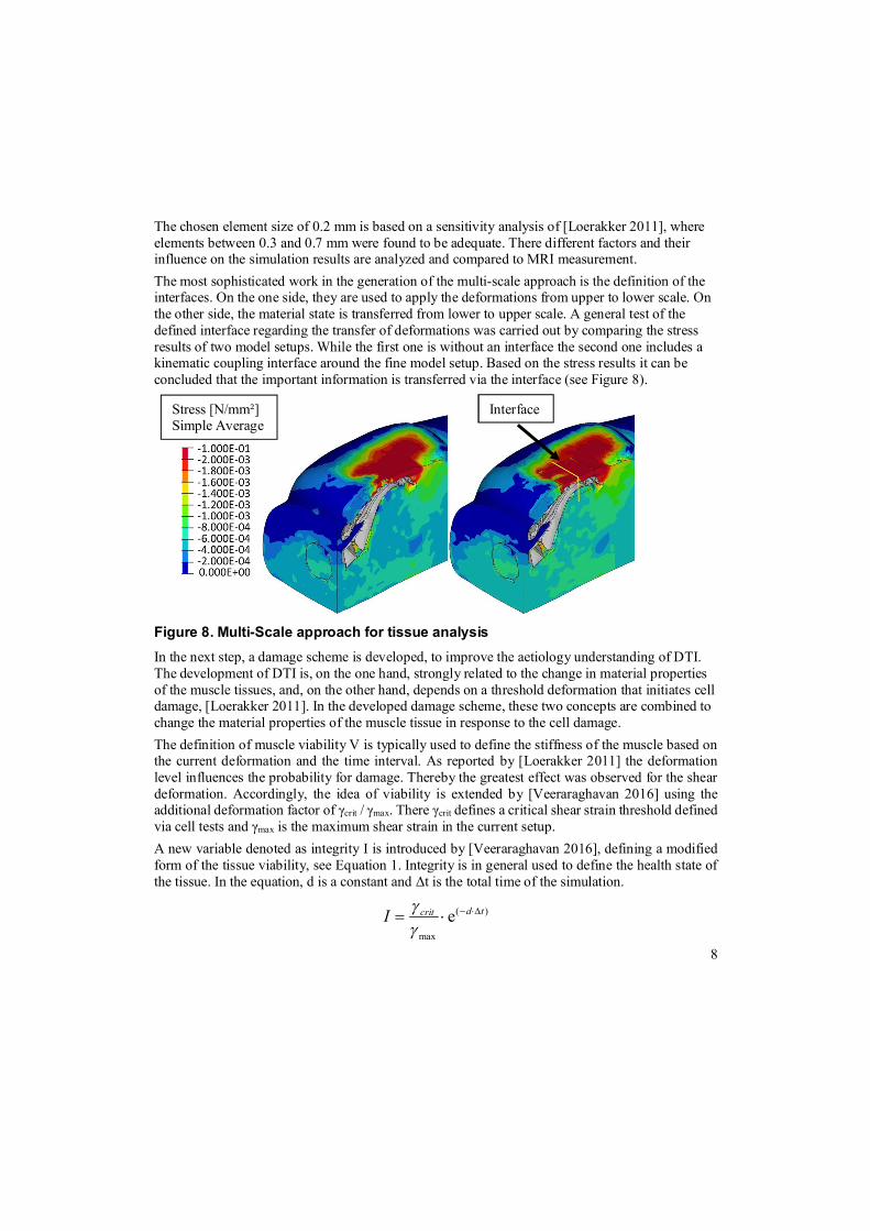
The models are generated for the explicit solver scheme. The bladders of the low pressure air mattress are modelled via cavities. The frame is modelled via rigid bodies and the kinematic mechanism is idealized and implemented via connector elements, see Figure 11. All model parts are in contact using the General Contact definition. Next, the human body model is positioned on top of the bed. At the beginning of the simulation, the human body model is hovering above the surface. The simulation is defined by two steps:
1. Application of gravity load 2. Alignment of the headrest up to an inclination of 40°
The model setup consists of 240,000 nodes, 360,000 elements and 1,000,000 degrees of freedom. The computation is carried out on a server with 16 cpus and takes about 5 hours. The following Figure 12 show the general simulation process.
Figure 12. Simulation Steps: Initial (top), Gravity load (medium) and Inclined (bot) One of the result values is the migration of the patient while the bed is inclined. In reality, it is a benefit if the migration is decreased. On the one side migration increases the risk for pressure injury of the patient. On the other side the nursing staff has to reposition the patient, which is

12
physically stressful. The following figure shows the horizontal downwards displacement of the patient for each setup. It can be observed that the greatest migration appears in the standard foam mattress design. The other design with low air pressure systems show a significant decrease of the migration, where the StayInPlaceTM provides overall the best results.
Figure 13. Migration result for different bed designs
A similar experimental study using motion capture date was carried out by [Wiggermann 2015]. In the study, twelve participants (6 female and 6 male) were recruited from different weight classes. In the setup the migration initiated by an inclination up to 30 and 45° was analyzed. The outcome was very similar to the simulation, not only with respect to the overall behavior of the designs but also with respect to the differences in migration.
Bed A Low pressure
with StayInPlaceTM
Low pressure without
StayInPlaceTM
Foam without
StayInPlaceTM

13
In the experimental study, an evaluation of the shear forces at the interface or the tissue strains is not possible. Here the application of numerical approaches with validated human body models have a great benefit for the evaluation of designs but also for increasing the knowledge with respect to the aetiology development of pressure injuries.
The following figure show as an example the shear stresses of all three beds at the interface of human body and bed surface. It can be observed that the increased migration of the foam mattress goes along with an increased shear stress in the area of the buttock. Based on general observations that higher shear forces are critical it can be concluded that the risk for a pressure sore is higher for the foam mattress compared to the two other setups.
Figure 14. Shear stress [N/mm²] for all bed designs at the end of the simulation. A further analysis via the detailed tissue model in combination with damage scheme will be carried out in the future. The goal is a detailed evaluation on DTI in the area of the sacrum and the IT.
5. Summary and Conclusion
Human body models are used in many industries to compute ergonomic and mechanical quantities. The results are used for product development as e.g. the usage of the software CASIMIR/Automotive for the comfort assessment of car seats. Typically, human body models are not applied in the development process of medical devices. The problem is the required level of modelling for a realistic representation the acceptance of numerical approaches by the medical society. The work here presented showed the enhancement of the human body model CASIMIR for the analysis of the aetiology understanding of pressure injuries. In a first step, a detailed tissue model

14
is generated and the material parameters for the skin, fat and muscle tissue are validated based on MRI images. In the second step, a damage scheme was developed and its application on cell level via a defined multi-scale approach is introduced. Finally, a case study of an intensive care bed was presented. Different bed designs are evaluated in the study. Via the simulation, the influence of a kinematic feature and the mattress on the migration of the patient are determined. Similar conclusions are the results of a comprehensive measurements campaign using motion capture data. In addition, the simulation enabled the analysis of the shear forces at the surface of the human body and the tissue strains in the IT region, which cannot be measured. Based on the simulation the differences of the bed designs regarding the risk for DTI are determined in detail, which was impossible using standard measurements like pressure maps. The study showed in detail the benefits of using the numerical simulation for the aetiology understanding of pressure injury development. In the future, the numerical method with human body models should be applied for development of other medical devices enabling a digitalization of this industry.
6. Acknowledgement
The setup of the detailed tissue model was carried out in the research project UDASim. Therefore, we thank the German Federal Ministry of Education and Research (BMBF) for funding this project. The data for the validation of the damage parameters is courtesy of the Eindhoven University of Technology, Netherlands. We would like to thank Prof. Cees Oomens and Dr. Sandra Loerakker for providing the test data. Finally, we want to thank HillRom, who funded the presented case study on their bed design.
7. References
1. Abaqus, Simulia 3DS, “Abaqus – Documentation, Version 6.14”, 2014 2. Agency for Healthcare Research and Quality, “Preventing Pressure Ulcers in Hospitals.”,
http://www.ahrq.gov/professionals/systems/hospital/pressureulcertoolkit/index.html, 2014 3. Nί Annaidh, A. et al., “Characterization of the anisotropic mechanical properties of excised
human skin.” Journal of the Mechanical Behavior of Biomedical Materials, Volume 5, 2012 4. Beiersdörfer, M., “Aufbau und Validierung eines detaillierten Gewebemodells für CASIMIR
für Anwendungen im Bereich der Medizintechnik“, Master thesis, TU Chemnitz 2014 5. Brem, H. et al., “High cost of stage IV pressure ulcers.”, The American Journal of Surgery,
Volume 188, Issue 1, Supplement 1, pp 1-8, 2010 6. Buck, B., “Ein Modell für das Schwingungsverhalten des sitzenden Menschen mit detaillierter
Abbildung der Wirbelsäule und Muskulatur im Lendenbereich“, phd thesis TU Darmstadt, Shaker, ISBN 3-8265-2970-7, 1997

15
7. Ceelen, K., K., et al., “Validation of a numerical model of skeletal muscle compression with MR tagging: a contribution to pressure ulcer research.”, Journal of Biomechanical Engineering, Volume 130, 2008
8. Comley, K., Fleck, N., “The mechanical response of porcine adipose tissue.” ASME Journal of Biomechanical Engineering, ISSN 0148-0731, 2009
9. DIN-33402, “Ergonomie – Körpermaße des Menschen – Teil 2: Werte.”, Deutsches Institut für Normung, 2005
10. Isight, Simulia 3DS, “Isight – Documentation, Version 5.9”, (2016) 11. Loerakker, S., “The relative contributions of muscle deformation and ischaemia to pressure
ulcer development.”, phd thesis TU Eindhoven, ISBN 978-90-386-2550-8, 2001 12. Mathworks®, “Matlab® - The Language of Technical Computing.”, Version R2012a, 2012 13. National Pressure Ulcer Advisory Panel (NPUAP), European Pressure Ulcer Advisory Panel
(EPUAP) and Pan Pacific Pressure Injury Alliance. “Prevention and Treatment of Pressure Ulcers: Quick Reference Guide”, Cambridge Media: Perth, Australia; ISBN 978-0-9579343-6-8, 2014.
14. Pankoke, S., et al., “Determination of vibration-related spinal loads by numerical simulation”, Journal Clinical Biomechanics, Vol. 16, pp. 45-56, 2001
15. Siefert A., et al., “Virtual optimisation of car passenger seats: Simulation of static and dynamic effects on drivers seating comfort”. International Journal of Industrial Ergonomics, Volume 38, p. 410-424, 2008
16. Siefert, A., “Numerische Modellierung und experimentelle Validierung der passive und aktiven mechanischen Eigenschaften des menschlichen Gewebes und dessen Implementierung in ein Ganzkörpermodell”, Phd thesis TU Darmstadt, Shaker, ISBN 978-3-8440-1824-0, 2013.
17. Simpleware, “ScanIP – Reference Guide”, Simpleware, 2010, http://www.simpleware.com/ 18. Thomas, D., R., “Prevention and treatment of pressure ulcers: What works? What doesn’t?”,
Cleveland Clinical Journal of Medicine, Vol 68(8), pp 704-722, 2001 19. Van Loocke, M., et al., “A validated model of passive muscle in compression.”, Journal of
Biomechanics, Volume 39, 2006 20. Veeraraghavan, A., R., “Multiscale Modelling for Risk Evaluation of Deep Tissue Injuries”,
Master thesis COMMAS, Universität Stuttgart, 2016 21. Wiggermann, N., et al., “The effect of patient migration in bed on torso elevation”, Nurse
Researcher, Volume 64, 2015


![The Dynamical Casimir Effect · 2012. 8. 9. · The Casimir effect The static Casimir effect Vacuum fluctuations [2] Casimir force between two metal plates [2] Two static mirrors](https://img.dokumen.tips/doc/110x75/60fba485759e576738445374/the-dynamical-casimir-effect-2012-8-9-the-casimir-effect-the-static-casimir.jpg)