Embed Size (px)
Citation preview

VILNIUS UNIVERSITY
AUDRIUS GEGECKAS
CHARACTERIZATION AND APPLICATION OF
KERATINOLYTIC PEPTIDASES FROM GEOBACILLUS SP. AND
BACILLUS SPP.
Summary of doctoral dissertation
Biomedical sciences, biology (01B)
Vilnius, 2016

The work presented in this doctoral dissertation has been carried out at the Department
of Microbiology and Biotechnology, Faculty of Natural Sciences, Vilnius University
during 2012-2016.
Scientific supervisors:
Prof. dr. Donaldas Jonas Čitavičius (Vilnius University, biomedical sciences, biology
– 01B); 2012-10-01 – 2016-01-31;
Prof. dr. Nomeda Kuisienė (Vilnius University, biomedical sciences, biology – 01B);
2012-02-01 – 2016-09-30.
The dissertation is defended at the Vilnius University Council of Biological Science:
Chairman:
Doc. dr. Saulius Serva (Vilnius University, biomedical sciences, biology – 01B).
Members:
Dr. Vida Časaitė (Vilnius University, biomedical sciences, biology – 01B);
Dr. Giedrius Gasiūnas (Vilnius University, physical sciences, biochemistry – 04P);
Dr. Saulius Kulakauskas (Micalis Institute, French National Institute for Agricultural
Research, biomedical sciences, biology – 01B);
Prof. dr. Jolanta Sereikaitė (Vilnius Gediminas Technical University, technological
sciences, chemical engineering – 05T).
The dissertation will be defended at the public session of the Council of Biological
Science at 12 p.m. on 5th of September in the R 401 auditorium at the Joint Life Sciences
Center, Vilnius University (Saulėtekio ave. 7, LT-10257, Vilnius, Lithuania).
The summary of doctoral dissertation was sent on 5th of July, 2016.
The thesis is available at the Library of Vilnius University and at website of Vilnius
University: www.vu.lt/lt/naujienos/ivykiu-kalendorius
2

VILNIAUS UNIVERSITETAS
AUDRIUS GEGECKAS
GEOBACILLUS SP. IR BACILLUS SPP. SEKRETUOJAMŲ
KERATINOLIZINIU AKTYVUMU PASIŽYMINČIŲ PEPTIDAZIŲ
CHARAKTERIZAVIMAS IR PRITAIKYMAS
Daktaro disertacijos santrauka
Biomedicinos mokslai, biologija (01B)
Vilnius, 2016 metai 3

Disertacija rengta 2012-2016 metais Vilniaus universiteto Gamtos mokslų fakultete,
Mikrobiologijos ir biotechnologijos katedroje.
Moksliniai vadovai:
Prof. dr. Donaldas Jonas Čitavičius (Vilniaus universitetas, biomedicinos mokslai,
biologija – 01B); 2012-10-01 – 2016-01-31;
Prof. dr. Nomeda Kuisienė (Vilniaus universitetas, biomedicinos mokslai, biologija –
01B); 2016-02-01 – 2016-09-30.
Disertacija ginama Vilniaus universiteto Biologijos mokslų krypties taryboje:
Pirmininkas:
Doc. dr. Saulius Serva (Vilniaus universitetas, biomedicinos mokslai, biologija – 01B).
Nariai:
Dr. Vida Časaitė (Vilniaus universitetas, biomedicinos mokslai, biologija – 01B);
Dr. Giedrius Gasiūnas (Vilniaus universitetas, fiziniai mokslai, biochemija – 04P);
Dr. Saulius Kulakauskas (Prancūzijos Nacionalinis žemės ūkio tyrimų institutas,
biomedicinos mokslai, biologija – 01B);
Prof. dr. Jolanta Sereikaitė (Vilniaus Gedimino technikos universitetas, technologijos
mokslai, chemijos inžinerija – 05T).
Disertacija bus ginama viešame Biologijos mokslų krypties tarybos posėdyje 2016 m.
rugsėjo 5 d. 12 val. Vilniaus universiteto Jungtiniame gyvybės mokslų centre, R 401
auditorijoje (Saulėtekio al. 7, LT-10257, Vilnius, Lietuva).
Disertacijos santrauka išsiuntinėta 2016 m. liepos 05 d.
Disertaciją galima peržiūrėti Vilniaus universiteto bibliotekoje ir VU interneto svetainėje
adresu: www.vu.lt/lt/naujienos/ivykiu-kalendorius
4

Table of contents INTRODUCTION .............................................................................................................. 6
MATERIAL AND METHODS .......................................................................................... 8
RESULTS AND DISCUSSION ....................................................................................... 15
1. Keratinolytic peptidases produced by thermophilic and mesophilic microorganisms ...................................................................................................................................... 15
1.1. Isolation and identification of keratinolytic bacterial strains ............................ 15
1.2. Production and mass spectrometry analysis of peptidase from Geobacillus sp. AD-11 ....................................................................................................................... 16
1.3. Cloning and bioinformatic analysis of GEOker gene ........................................ 17
1.4. Expression and purification of the GEOker proteins ........................................ 19
1.5. Biochemical characterization of RecGEOker ................................................... 20
1.6. Production and purification of keratinolytic peptidases from mesophilic microorganisms......................................................................................................... 25
1.7. Biochemical characterization of BtKER, BPKer and BAKer ........................... 26
2. Chimeric keratinolytic peptidases ............................................................................ 31
2.1. Synthetic homodimer SynKer-TT ..................................................................... 31
2.2. Synthetic heterodimer SynKer-TM ................................................................... 34
3. Lab-scale keratin biodegradation process................................................................. 36
CONCLUSIONS .............................................................................................................. 39
LIST OF PUBLICATIONS .............................................................................................. 40
CONFERENCE PRESENTATIONS ............................................................................... 40
FINANCIAL SUPPORT .................................................................................................. 42
CURRICULUM VITAE ................................................................................................... 43
ACKNOWLEDGEMENTS .............................................................................................. 44
REZIUMĖ ......................................................................................................................... 45
REFERENCES ................................................................................................................. 48
5

INTRODUCTION
Solid keratin-rich waste management is one of essential research area in
nowadays. Conventional chemical and high thermal keratin waste decomposition
methods are fully explored and not enough effective for future biotechnology
perspectives 1. However, traditional keratin-rich waste decomposition methods could be
replaced by environmentally-friendly and economical microbial keratin waste
biodegradation methods without energy wastage and essential amino acids and nutrition
elements loss 2. Therefore, microbial bio-decomposition are attractive approach to
keratin or keratin-like waste manage without any nutrition loss in eco-friendly process
environment.
Keratinolytic peptidases or keratinases (EC 3.4.21/24/99.11) are a particular class
of proteolytic enzymes that displays the capability of degrading insoluble substrates such
as fibrin, keratin, elastin, collagen and soluble substrates such as sodium caseinate,
albumin and gelatin or other keratin-like substrates with different efficiency 3.
Keratinolytic peptidases are produced by various microorganisms including bacteria,
actinomycetes and fungi and belongs to metallo and serine or metallo-serine proteases
based on their catalytic type with precedence for aromatic and hydrophobic residues at
the P1 position 1, 4, 5.
Keratin is the insoluble fibrous hard-to-degrade protein of feathers, wool, hair
including other epidermal appendages which can be degraded by keratinolytic peptidases 6, 7. The recalcitrance of keratins derives from compact packing of α-helices (α-keratins,
40-60 kDa) or β-sheets (β-keratin, 10 kDa) into a supercoiled polypeptide chains.
Compact structure is stabilized by high degree of cross-linking disulfide bonds,
hydrogen bonds and hydrophobic interactions. These proteins show high stability and
resistance to proteolytic enzymes such as trypsin, pepsin or papain 6, 8.
Increased attention has been diverted to these keratinolytic peptidases because of
their important potential uses in biomedicine, pharmaceutics, cosmetics and keratin
waste bioconversion industries associated to the hydrolysis of keratin 1. Keratinolytic
proteinases are next generation molecular tools for keratin hydrolysis and production of
small value-added peptides. It is important to find suitable biocatalysts with proper
6

physical and chemical properties for effective keratin-rich waste biodegradation and
replace current not enough efficient biocatalysts.
Unfortunately, most naturally produced enzymes are not effective or suitable for
biotechnology processes. For decades, protein engineering has been utilized to modify
natural enzymes to meet the needs of different industrial biotechnology. It is therefore a
powerful tool in synthetic biology through the altering of enzyme properties to suit the
requirements of any hydrolysis process. Enzyme modification or construction of
chimeric proteins open up new possibilities for industrial application 9, 10.
The aim of the dissertation work is to evaluate application possibilities of a newly
identified keratinolytic peptidases in biodegradation processes of proteinaceous
materials.
The following tasks have been formulated to achieve this aim:
• To evaluate physico-chemical properties and enzymatic activity of keratinolytic
peptidases from thermophilic and mesophilic microorganisms;
• To evaluate the hydrolysis efficiency of proteinaceous waste by keratinolytic
peptidases from thermophilic and mesophilic microorganisms;
• To design chimeric keratinolytic biocatalysts by protein engineering methods;
• To create laboratory-scale raw keratin waste biodegradation system.
Scientific novelty:
For the first time, we identified, purified and characterized native keratinolytic
peptidase from Bacillus thuringiensis AD-12 (BtKER). Similarly, we analyzed and
evaluated features of three unique enzymes from Geobacillus sp. AD-11 (RecGEOker),
Bacillus sp. AD-W (BPKer) and Bacillus sp. AD-AA3 (BAKer) for industrial
application. For the first time, we created chimeric (SynKer-TT and SynKer-TM)
keratinolytic peptidases with improved physicochemical properties. Eventually, we
demonstrated that metabolically active secretomes from keratinolytic microorganisms
can be used for eco-friendly enzymatic keratin waste management.
7
Practical value:
Our findings and results expand scientific knowledge about keratinolytic
peptidases produced by thermophilic and mesophilic microoganisms. Our created keratin
and other proteinaceous material biodegradation system is particularly relevant to
industrial biotechnology associated with conversion of large scale wastes to value-added
products.
MATERIAL AND METHODS
Chemicals. All chemicals were of analytical reagent grade or higher quality and
were purchased from Merck, Sigma-Aldrich and AppliChem, unless otherwise stated.
All enzymes were purchased from Thermo Fisher Scientific, unless otherwise stated.
Strains. AD-11, AD-12, AD-W and AD-AA3 bacterial strains were identified by
author. E. coli DH5α, E. coli BL21 (DE3) and E. coli Rosetta (DE3) pLysS were
purchased from Novagen. E. coli BW25113 was obtained from prof. dr. Edita
Sužiedėlienė.
Plasmids. Cloning plasmid vector used in this work: pTZ57R/T (Ampr).
Expression vectors used in this work: pET-28c(+) (Kanr), pET-21c(+) (Ampr) and
pBAD30 (Ampr, gift from prof. dr. Edita Sužiedėlienė).
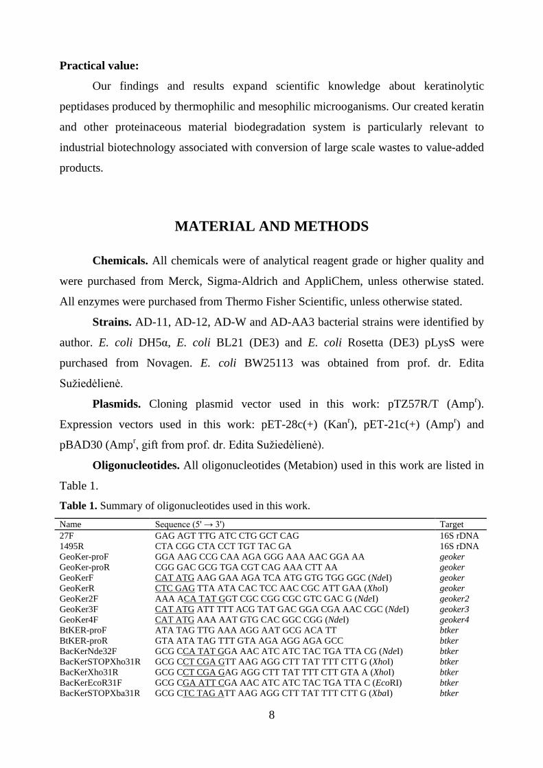
Oligonucleotides. All oligonucleotides (Metabion) used in this work are listed in
Table 1.
Table 1. Summary of oligonucleotides used in this work.
Name Sequence (5' → 3') Target 27F GAG AGT TTG ATC CTG GCT CAG 16S rDNA 1495R CTA CGG CTA CCT TGT TAC GA 16S rDNA GeoKer-proF GGA AAG CCG CAA AGA GGG AAA AAC GGA AA geoker GeoKer-proR CGG GAC GCG TGA CGT CAG AAA CTT AA geoker GeoKerF CAT ATG AAG GAA AGA TCA ATG GTG TGG GGC (NdeI) geoker GeoKerR CTC GAG TTA ATA CAC TCC AAC CGC ATT GAA (XhoI) geoker GeoKer2F AAA ACA TAT GGT CGC CGG CGC GTC GAC G (NdeI) geoker2 GeoKer3F CAT ATG ATT TTT ACG TAT GAC GGA CGA AAC CGC (NdeI) geoker3 GeoKer4F CAT ATG AAA AAT GTG CAC GGC CGG (NdeI) geoker4 BtKER-proF ATA TAG TTG AAA AGG AAT GCG ACA TT btker BtKER-proR GTA ATA TAG TTT GTA AGA AGG AGA GCC btker BacKerNde32F GCG CCA TAT GGA AAC ATC ATC TAC TGA TTA CG (NdeI) btker BacKerSTOPXho31R GCG CCT CGA GTT AAG AGG CTT TAT TTT CTT G (XhoI) btker BacKerXho31R GCG CCT CGA GAG AGG CTT TAT TTT CTT GTA A (XhoI) btker BacKerEcoR31F GCG CGA ATT CGA AAC ATC ATC TAC TGA TTA C (EcoRI) btker BacKerSTOPXba31R GCG CTC TAG ATT AAG AGG CTT TAT TTT CTT G (XbaI) btker
8

TT2 GCG CGA GCT CAC TTC CTC CAT ACA CTC CAA GCG CAT T (SacI)
geoker
TT3 GCG CGA GCT CAA GGA AAG ATC AAT GGT GTG G (SacI) geoker TM1 GCG CGA GCT CGA AAC ATC ATC TAC TGA TTA C (XhoI) btker Underlined sequences indicate the restriction sites.
Bacterial growth conditions. Solid keratin medium (B) containing NH4Cl, 0.05
%; NaCl, 0.05 %; K2HPO4, 0.03 %; KH2PO4, 0.03 %; yeast extract, 0.01 %; keratin from
wool (TCI Europe), 0.1 %; MgSO4, 2 mM; agar 1.8 %, pH 7.8 was used for isolation of
keratinolytic peptidase producing bacterial strains. The bacterial isolates were purified
and checked for keratinase production. Stock culture of the bacterial isolates were
maintained in 15 % glycerol stored at -70 ºC.
Inoculum (preculture) preparation:
Geobacillus sp. AD-11 strain: cells from solid medium are inoculated to 5 mL of
the A medium containing K2HPO4, 0.3 %; NaCl, 0.05 %; NH4Cl, 0.1 %; glucose, 0.25
%; Tris, 0.6 %; yeast extract, 0.5 %; peptone, 1.5 %; MgSO4, 2 mM; CaCl2, 0.1 mM; pH
7.8. Culture was incubated for 14-16 h at 55 ºC with agitation 200 rpm.
Bacillus sp. AD-12 strain: cells from solid medium are inoculated to 50 mL of the
LB medium. Culture was incubated for 12-16 h at 30 ºC with agitation 150 rpm.
Bacillus sp. AD-W strain: cells from solid medium are inoculated to 50 mL of the
LB medium. Culture was incubated for 12-16 h at 30 ºC with agitation 180 rpm.
Bacillus sp. AD-AA3 strain: cells from solid medium are inoculated to 50 mL of
the LB medium. Culture was incubated for 12-16 h at 30 ºC with agitation 180 rpm.
Cells cultivation:
Geobacillus sp. AD-11 strain: keratinolytic peptidase production was carried out
in 2 L of previously described liquid medium A and distributed in 500 mL Erlenmeyer
flasks. Each flask contained 100 mL of the A medium and inoculated with 1 % of the
preculture (3 × 10⁵ cfu mL-1). Flasks were incubated for 12-14 h at 60 ºC with agitation
180 rpm. The culture supernatant was recovered by centrifugation two times at 8500 × g
for 15 min at 4 °C.
Geobacillus sp. AD-11 strain: peptidase production was carried out in 2 L of
previously described liquid medium B and distributed in 500 mL Erlenmeyer flasks.
Each flask contained 100 mL of the B medium and inoculated with 1 % of the preculture
(3 × 10⁵ cfu mL-1). Flasks were incubated for 80-92 h at 60 ºC with agitation 180 rpm.
9

The culture supernatant was recovered by centrifugation two times at 8500 × g for 15
min at 4 °C.
Bacillus sp. AD-12 strain: peptidase production was carried out in 2 L of
previously described liquid medium B (pH 7) and distributed in 500 mL Erlenmeyer
flasks. Each flask contained 100 mL of the B medium and inoculated with 1 % of the
preculture. Flasks were incubated for 80-96 h at 30 ºC with agitation 180 rpm. The
culture supernatant was recovered by centrifugation at 8500 × g for 15 min at 4 °C.
Bacillus sp. AD-W strain: peptidase production was carried out in 2 L of
previously described liquid medium B (pH 8) and distributed in 500 mL Erlenmeyer
flasks. Each flask contained 100 mL of the B medium and inoculated with 1 % of the
preculture. Flasks were incubated for 48 h at 30 ºC with agitation 150 rpm. The culture
supernatant was recovered by centrifugation at 8500 × g for 15 min at 4 °C.
Bacillus sp. AD-AA3 strain: peptidase production was carried out in 2 L of
previously described liquid medium B (pH 7) and distributed in 500 mL Erlenmeyer
flasks. Each flask contained 100 mL of the B medium and inoculated with 1 % of the
preculture. Flasks were incubated for 48 h at 30 ºC with agitation 150 rpm. The culture
supernatant was recovered by centrifugation at 8500 × g for 15 min at 4 °C.
Protein precipitation. All steps were carried out at 4 ºC. After all strains
cultivation in previously described liquid medium A or B the culture medium was
centrifuged at 8500 × g for 15 min and the supernatant was filtered through filter paper
to obtain the crude enzyme filtrate. The filtrate containing the keratinolytic activity was
collected. Proteins present in the crude enzyme filtrate were precipitated by using solid
ammonium sulfate at desired saturation level. Precipitated proteins were collected by
centrifugation at 20000 × g for 20 min. The pellet was dissolved in a 10 mL buffer (50
mM Tris-HCl, pH 7.8) and dialyzed in dialysis tubes (3.5K MWCO, Thermo Fisher
Scientific) overnight against the same buffer for 24 h and the buffer was changed at 8 h
intervals. After dialysis crude extract was concentrated with PEG 35000 to obtain ~2 mL
final protein extract.
DNA manipulation. Genomic DNA of AD-11, AD-12, AD-W and AD-AA3 was
extracted according to the method described by Sambrook and Rusell 11. Plasmid DNA
from E. coli transformants was isolated with the GeneJETTM Plasmid Miniprep Kit
(Thermo Fisher Scientific). Gene JETTM Gel Extraction Kit (Thermo Fisher Scientific) 10

was used to recover DNA fragments from agarose gels. Electrocompetent E. coli strains
preparation, ligation of DNA and all molecular methods were performed using standard
molecular biology techniques 11.
Gene and protein sequence analysis. Chromosomal DNA of investigated
organisms was used as template for amplifying the 16S rRNA gene. The 16S rRNA gene
was amplified by PCR using primers 27F and 1495R. The PCR process was carried out
according to Kuisiene et al 12. The purified amplicons were sequenced at the Institute of
Biotechnology (Vilnius, Lithuania). The DNA and protein sequences similarities were
assessed by BLASTn (Basic Local Alignment Search Tool for Nucleotides) and
BLASTp (Basic Local Alignment Search Tool for Protein), respectively 13, 14. Multiple
sequence alignments were performed with ESPript 3.0 15. A phylogenetic tree was
constructed by using the neighbor-joining (NJ) algorithm in MEGA 7.0 16-19. Each
dataset was bootstrapped 1000 times. The signal peptide sequence was analyzed by
SignalP (www.cbs.dtu.dk/services/SignalP/) 20. Possible transcription promoters were
analysed with Neural Network Promoter Prediction program
(http://www.fruitfly.org/seq_tools/promoter.html) 21.
SDS-PAGE analysis. Molecular weight (MW) and protein analyses were
determined by 12 % SDS-PAGE using the Laemmli method 22. A broad range of protein
standards PageRulerTM Unstained Protein Ladder (Pierce) and PageRulerTM Unstained
Low Range Protein Ladder (Pierce) were used as a molecular mass markers. After
electrophoresis gels were stained with PageBlueTM Protein Staining Solution (Pierce).
Low and ultra low molecular weight SDS-PAGE (LMW-T-EG-PAGE and uLMW-T-
EG-PAGE) were prepared and performed according to Thermo Fisher Scientific
recommendations. Tricine SDS-PAGE was performed according to Schagger 23.
Zymography. Keratinolytic activity was determined on SDS-PAGE gels using
keratin from wool as a substrate. After electrophoresis renaturation procedure was
carried out following Fakhfakh-Zouari et al 24. The renaturated gel was placed into Petri
dish and covered with 50 mL B-agar medium. The gel was incubated overnight at
desired temperature. The molecular weight of hypothetical keratinolytic peptidase was
determined by the comparison of stained gel and the obtained zymogram.
Mass spectrometry and protein identification. Proteomic analysis was carried
out at the Mass Spectrometry Laboratory, Institute of Biochemistry and Biophysics, 11

Polish Academy of Sciences, Warsaw. Gel bands of interest were excised, followed by
in-gel trypsin digestion. Resulting peptides were separated on nano-LC, directly coupled
with ESI-LTQ-Orbitrap mass spectrometer. Proteins were identified by Mascot (Matrix
Science) searching mass spectrometry data against NCBInr database.
Assay of keratinase activity. Keratinase assay was done according to Cai et al 25
using keratin azure (Sigma-Aldrich) or keratin from wool as the substrate. The 4 mg of
substrate was suspended in a total volume of 1 mL buffer (50 mM Tris-HCl at desired
pH). The reaction was initiated by the addition of 10 U of enzyme. The reaction tubes
were incubated at desired temperature for 1 h and the reaction was stopped by the
addition of 400 μL of 10 % trichloroacetic acid (TCA). After 15 min at 4 ºC, the tubes
were centrifugated for 5 min at 16000 × g at 25 ºC. The supernatant was
spectrophotometrically measured for release of the azo dye at 595 nm or 280 nm for
keratin from wool. The control was prepared by previously described method without
addition of the enzyme. One unit (U) of keratinase activity was defined as the amount of
enzyme causing 0.001 absorbance increase between sample and control under the
standard conditions.
Biochemical characterization of keratinolytic peptidases. A range of various
temperature (20-90 ºC) was used for determining optimum temperature. Similarity,
optimum pH was determined by assaying the enzyme at different pH buffers (pH 5-6, 50
mM piperazine; pH 7-9, 50 mM Tris-HCl; pH 10-11, 50 mM sodium bicarbonate; 12
pH, 50 mM potassium chloride/NaOH). Thermostability and pH stability were measured
by incubating the enzyme at different temperatures and pH, respectively. The effects of
various metal ions, detergents, organic solvents and inhibitors on keratinolytic activity
were investigated by pre-incubating the enzyme with these chemicals for 1 h at 25 ºC.
Keratinolytic peptidase activity determined without additions was considered as 100 %.
Substrate specificity. The specificity of the keratinolytic peptidases toward
various substrates including bovine serum albumin (BSA), Na-caseinate, gelatin (Bio-
Rad), keratin from wool and collagen (Proteina) was determined on the basis of the
previously described standard assay conditions. One unit (U) of keratinase activity was
defined as described previously.
Gene cloning. Primers were constructed according to results obtained from mass
spectrometry and database analysis for Geobacillus sp. AD-11 and Bacillus sp. AD-12, 12

respectively. First set of primers (GeoKer-proF/GeoKer-proR and BtKER-proF/BtKER-
proR for AD-11 and AD-12, respectively) was used to amplify target gene with upstream
and downstream sequence fragments for further gene analysis. Second set of primers
(GeoKerF/GeoKerR and BacKerNde32F/BacKerSTOPXho31R (BacKerXho31R) for
AD-11 and AD-12, respectively) was used to amplify target gene with complete ORF
without signal sequence and incorporate NdeI and XhoI restriction sites for cloning into
expression vectors. Obtained PCR products were purified using a GeneJETTM PCR
Purification Kit and ligated with pTZ57R/T cloning vector. The ligated product was
transformed into E. coli DH5α cells using a standard protocol 11. Recombinant clones
were detected by direct blue/white screening. For target protein expression, the gene
fragment was cleaved from pTZ57R/T plasmid with NdeI and XhoI restriction enzymes
and ligated into the pET expression vector digested with the same restriction enzymes
and dephosphorylated with alkaline phosphatase (Thermo Fisher Scientific). The
resulting expression plasmid with target gene was transformed into E. coli DH5α cells
and then retransformed into E. coli BL21 (DE3) or E. coli Rosetta (DE3) pLysS cells for
protein expression. The clones were screened for the positive recombinants using colony
PCR followed by double digestion with NdeI and XhoI restriction enzymes. The
recombinant plasmid containing the target keratinase gene was confirmed by sequencing.
Gene expression. A transformant of E. coli BL21 (DE3) and E. coli Rosetta
(DE3) pLysS harboring pET plasmid with inserted target gene was incubated at 37 ºC
overnight with agitation 180 rpm in LB medium containing appropriate antibiotic. 1 %
of seed culture was transferred into fresh medium and cultured at the same conditions
until OD600 reached 0.4 (unless otherwise stated), then IPTG (to a final concentration of
0.01-5 mM) was added for recombinant protein induction. The bacterial cells were
cultivated at desired temperature for another 5 hours (unless otherwise stated) before
harvesting by centrifugation. For protein expression visualization cells samples obtained
at different times after induction were adjusted so that their OD600 value were 0.4, mixed
with 4× SDS-PAGE sample loading buffer and subjected to SDS-PAGE analysis. The
negative control was prepared by previously described method using cells without IPTG
induction.
Protein purification. Purification of recombinant proteins: the recombinant
proteins were purified by affinity chromatography using immobilized nickel ions 13

(ProfinityTM IMAC Resins, BIO-RAD). Cells were collected after 2× culture
centrifugation at 8500 × g for 15 min at 4 °C in 50 mM Tris-HCl (pH 7.8). Cells were
suspended in buffer A (50 mM Tris-HCl (pH 8)) at 1:4 buffer cells ratio. Supernatant
and cell debris with formed inclusion bodies were collected after sonication (10 s : 20 s)
of the induced cells. The column with resin was prepared and equilibrated following the
manufacturer's purification protocol with slightly modifications. The bounded target
protein was eluted from the column by passing buffer A (with 6 M urea) having a range
of 50-500 mM imidazole. The fractions were collected and analyzed on SDS-PAGE.
Purification of native proteins: obtained keratinolytic peptidase extract was
applied to DEAE-Sepharose (GE Healthcare) column (2.5 × 12 cm) equilibrated with the
buffer B (25 mM Tris-HCl, pH 8). The column was washed with same buffer and eluted
with step wise buffer B containing 100 mM, 250 mM, 500 mM, 750 mM and 1 M NaCl.
The active fractions were pooled and dialyzed against the buffer A for 24 h. Gel
filtration chromatography was performed using Bio-Gel P-60 (BIO-RAD) column (1 ×
60 cm) equilibrated with buffer B1 (50 mM Tris-HCl, pH 8). The above concentrated
sample was loaded on to the column followed by elution with same buffer with fraction
size of 1.5 ml. The eluted active fractions were pooled and concentrated with PEG 35000
to 2 mL.
Thin-layer chromatography: 10 mg of keratin from wool was incubated with 10
U of peptidase in a total volume of 1 mL buffer (50 mM Tris-HCl) at desired
temperature and pH for 12 h. The samples were collected and analyzed on silica plate
(ProteoChrom® HPTLC plates, Merck) by Thin-layer chromatography (TLC) according
to manufacturer's protocol and recommendations using solvent system: 2-
butanol:pyridine:acetic acid:water (30:20:6:24). The documentation and evaluation of
the TLC plate was done using UV visualizer under UV, using fluorescamine.
Statistical analysis. All data were analyzed using GraphPad Prism 5.0. Values
are expressed as means ± standard deviation of results from three independent
experiments. Data were considered as statistically significant for P values of 0.05 or less.
14

RESULTS AND DISCUSSION
1. Keratinolytic peptidases produced by thermophilic and mesophilic
microorganisms
1.1. Isolation and identification of keratinolytic bacterial strains
57 thermophilic bacterial isolates were selected from collection of
microorganisms (Department of Microbiology and Biotechnology, Faculty of Natural
Sciences, Vilnius university, Lithuania). 19 mesophilic bacterial isolates were recovered
from soil in Lithuania (Vilnius and Palanga). Their keratinolytic activities were tested by
growing the isolates on keratin meal plates. The largest clearing zone on keratin was
observed for AD-11 (thermophile) and three mesophilic isolates: AD-12, AD-W and
AD-AA3.
16S rDNA phylogeny studies were carried out. The partial 16S rDNA sequences
were determined and compared to the GenBank database in the National Center for
Biotechnology Information (NCBI) using BLAST program. The AD-11 isolate was
found to belonging to the genus Geobacillus stearothermophilus with 99 % homology
(data not shown). The isolate was assigned to Geobacillus sp. AD-11.
Similarly, AD-12 isolate was found to belonging to the Bacillus cereus group
with 99 % homology (data not shown). AD-12 isolate was further identified to be
Bacillus thuringiensis based on parasporal crystal formation observed under optical and
electron microscope. The isolate was assigned to Bacillus sp. AD-12. Additionally, AD-
W and AD-AA3 isolates were found to belonging to Bacillus altitudinis DSM 21631T /
Bacillus stratosphericus MTCC 7305T and Bacillus amyloliquefaciens DSM 7T,
respectively. Therefore, AD-W and AD-AA3 were assigned to Bacillus sp. AD-W and
Bacillus sp. AD-AA3, respectively.
The amplified 16S rDNA gene sequences of all strains were sequenced and have
been submitted in GenBank under Accession Nos. listed in Table 2.
Table 2. Accession numbers of 16S rDNA sequences.
Strain Accession Nos. AD-11 KJ789443 AD-12 KJ636471.1 AD-W KU950739 AD-AA3 KU950740
15

1.2. Production and mass spectrometry analysis of peptidase from Geobacillus
sp. AD-11
Keratinolytic peptidase are largely produced in a synthetic B medium with
keratinous substrate as the sole source of carbon and nitrogen and basal A medium. The
native keratinolytic peptidase (NatGEOker) was successfully produced using previously
described induction medium B and non-induction medium A. The crude protein extract
was characterized by SDS-PAGE and zymography methods (Fig. 1.).
The bands between 30-40 kDa (Fig. 1. ; Zymo I) and 50-60 kDa (Fig. 1.; Zymo
II) from SDS-PAGE were subjected to ESI-LTQ-Orbitrap analysis. 138 (from medium
A) and 73 (from medium B) peptide mass fingerprints of the hypothetical keratinolytic
peptidase were obtained from the MS analysis. NCBInr and SwissProt protein sequences
databases were used in the MASCOT search tool to look for proteins that match the
peptide spectrum of the NatGEOker. The peptide analysis showed similarity to two
proteolytic enzymes from Geobacillus sp. strains: 99 % similarity to thermolysin
(M04.001, GeneBank: M21663.1) and 98 % simalarity to bacillolysin (M04.014,
GeneBank: CP008934.1). Thermolysin-like peptidases (TLP) are synthesized as a
precursor with a propeptide. Maturation of TLP occurs through autoprocessing and this
processing pathway is mediated by the propeptide 26-28.
Fig. 1. SDS-PAGE and zymogram analysis of NatGEOker. M – PageRulerTM Unstained Protein Ladder. I – secretome profile from B medium. II – secretome profile from A medium. Zymo I and Zymo II – peptidase analysis by zymography. Black arrows indicate zones of hydrolysis.
16

It was determined that in both medium was produced the same proteolytic enzyme
– thermolysin-like peptidase. The obtained gene product was assigned to GEOker
(Geobacillus keratinase).
1.3. Cloning and bioinformatic analysis of GEOker gene
The first set of primers (GeoKer-proF/GeoKer-proR) was constructed according
to similar sequences obtained from NCBI GenBank databases. Primers were used to
amplify target gene with upstream and downstream sequence fragments for further
bioinformatic analysis. The open reading frame (ORF) of GEOker is 1638 bp length
which encodes a protein of 546 amino acids with 59761.72 Da molecular weight and an
isoelectric point (pI) of 5.375. According to the SignalP 4.1 web server
(http://www.cbs.dtu.dk/services/SignalP/; 20), a 25 a. a. stretch with the features of the
typical Bacillus and Geobacillus signal peptides at the N- terminal region of the protein
was found. The putative signal peptide (prepeptide) cleavage site was predicted to be
between Ala25 and Lys26. This results indicates that the proenzyme is composed of 521
a. a. with 57231.60 Da molecular weight and an isoelectric point is 5.47. Mature enzyme
(after autoprocessing) is composed of 319 a. a. with 34685.03 Da and pI 5.47 (Fig. 2.).
Described GEOker has consensus zinc-binding H372ELTH376 (the positions are
indicated without the cleavage of the signal peptide) motif and Glu373 and His461
amino acids are essential form enzymatic reaction. This TLP belongs to zinc
metalloproteinases.
The required gene targets were amplified using second sets of primers constructed
according to sequence analysis (Fig. 3.). Amplicons were purified and cloned into
pTZ57R/T cloning vector. Then the target gene fragments were cleaved from pTZ57R/T
vector and ligated into pET-28c(+) expression vector. The constructed plasmids
containing GEOker gene fragments were used for target recombinant peptidase
expression.
17

Fig. 2. Amino acids sequence alignment of GEOker from Geobacillus sp. AD-11 with other Bacillus and Geobacillus peptidases. The used peptidases are: gi|696468741 – bacillolysin (G. stearothermophilus); gi|37088170 – thermolysin, thermostable neutral protease (B. caldolyticus); gi|893712617 – alkaline metalloprotease (Bacillus subtilis); gi|647687896 – bacillolysin (Geobacillus thermocatenulatus). The putative starting residues of prepeptide, propeptide and mature GEOker are indicated. The figure was produced with ESPript 3.0 15.
18

1.4. Expression and purification of the GEOker proteins
pET-28c(+) construct harboring geoker1 gene fragment was transformed into E.
coli BL21 (DE3) cells and protein expression was monitored and analyzed by SDS-
PAGE. Analysis showed the appearance of a large amount of new protein in E. coli cells
sample after induction with IPTG (Fig. 4A).
Fig. 4. SDS-PAGE analysis of expression (A) and purification (B) of GEOker1. (A) GEOker1 expression profiles at 1 h to 5 h. (--) – without IPTG addition. (+) – with 0.5 mM IPTG addition. (++) – with 1 mM IPTG addition. White arrows indicate target GEOker1 protein. M – PageRulerTM Unstained Protein Ladder. (B) purification of GEOker1 and zymogram analysis. M – PageRulerTM Unstained Protein Ladder. Lane 1 – proteins profile without IPTG induction. Lane 2 – proteins profile with 0.5 mM IPTG induction. Lane 3 – purified GEOker1 with IMAC. Lane 4 – zymogram of the purified GEOker1.
Fig. 3. GEOker sequence. Black arrows indicate different protein variants.
19

The size of this new expressed protein GEOker1 agreed well with predicted size
of GEOker approximately 57 kDa. The optimum expression level was detected at 3 h
post-induction with 0.5 mM IPTG at 30 °C. It was noted that the protein expression level
decreased 4 h post-induction. For the large-scale preparation of GEOker1 as a biocatalyst
for wide biotechnology application, expression must be performed for no longer than 3 h
after induction. Unfortunately, expression of other GEOker protein variants (GEOker2,
GEOker3 and GEOker4) was not detected.
The target recombinant N-terminus His-tagged GEOker1 protein was purified
using immobilized Ni2+ metal affinity chromatography (IMAC). The purity of protein
was analyzed by SDS-PAGE and keratinolytic activity was determined by zymography
(Fig. 4B). obtained protein band approximately at 57 kDa indicated the purification of
the target GEOker1. The recombinant protein was eluted from Ni2+ resin at 100 mM of
imidazole and observed as a single band on SDS-PAGE. The zymogram analysis further
confirmed the presence of active keratinolytic peptidase. The purified recombinant
GEOker1 (RecGEOker) protein showed specific keratinolytic activity of 1437.6±7.5 U
mg-1 with an overall yield of 61.2 % and a purification fold of 6.2.
1.5. Biochemical characterization of RecGEOker
The purified RecGEOker showed a typical bell-shaped curve with the
keratinolytic activity in the temperature range of 20-90 °C with the maximal activity at
60 °C (Fig. 5A) It was shown that thermolysin from G. stearothermophilus ATCC
31197T displayed maximal activity at 75 °C 29. The activity rapidly decreased at lower
temperatures. At 40-50 °C the activity of RecGEOker was reduced to 75 % and 33 %,
respectively. At 70-80 °C the activity was reduced to 87 % and 71 %, respectively. The
effect of temperature on the stability of purified RecGEOker was examined by
measuring the remaining activity after incubation for 1-4 h at various temperatures at pH
8 (Fig. 5B). After 4 h incubation at 60 °C the enzyme showed no loss of activity. The
purified RecGEOker retained higher than 50 % remaining activity after incubation for 2
h at 70-80 °C and retained lower than 20 % activity after 4 h incubation at the same
temperature. These results suggested that the purified RecGEOker is stable at higher
temperatures and belongs to thermostable thermolysin-like proteinases 30.
20

Fig. 5. Biochemical characterization of RecGEOker. (A) Effect of temperature on the activity. (B) Effect of temperature on the stability. (C) Effect of pH on activity and stability. Values represent the mean of three replicates.
The purified peptidase displayed keratinolytic activity within a broad pH range of
5-11 with an optimum at pH 9 (Fig. 5C). The activity rapidly decreased at pH 6 and pH
10 to 40 % and 37 %, respectively. The pH stability profile indicated that the purified
peptidase was highly stable in the pH range between 7 to 9. Generally, the commercial
microbial proteinases have pH optima in the alkaline range between 8 and 12 and the
RecGEOker fell in this range 31.
In response to various modulators, results indicate the stimulated activity by 1
mM Mg2+ and 10 mM Mn2+ ions up to 112.6±2.8 % and 116.6±1.9 %, respectively (data
not shown). The enzyme activity was decreased up to 50 % by 1 mM K+, 10 mM Mg2+, 1
mM and 10 mM Zn2+, 1 mM and 10 mM Ni2+. Among the ions tested Mn2+, Mg2+ and
Ca2+ ions positively regulated enzyme activity and stability. This phenomenon might be
attributed to these ions involvement in stabilization of the keratinolytic peptidase
molecular structure as reported Kojima 27. It is well known that tertiary structure of the
enzyme ant the substrate-enzyme complex may be stabilized and maintained by salt or
ions bridge formed by metal ions.
The stability of RecGEOker toward detergents and organic solvents was
determined by incubating the peptidase with detergents (1 % and 5 %) and organic
solvents (10 % and 25 %) for 1 h at 25 °C (data not shown). The RecGEOker produced
by Geobacillus sp. AD-11 was stable toward detergents like Tween 40 (5 %), Tween 60
(5 %), Tween 80 (5 %), Triton X-100 (1 %), Triton X-305 (1 %) and Brij 35 (5 %) and
the relative keratinolytic enzyme activities were 180.1±3.9 %, 133.5±3.6 %, 122.2±1.5
21

%, 115.3±1.9 %, 153.4±1.7 % and 105.1±2.6 %, respectively. Studies revealed that the
purified RecGEOker was slightly unstable toward tested organic solvents and relative
activities were decreased. High remaining activity of recombinant peptidase treatment
with detergents and/or organic solvents suggested that this peptidase can be a powerful
biocatalyst in various white biotechnology areas associated with small peptide
application or directly affect the surface of skin.
Peptidase can be classified based on their sensitivity to various inhibitors 32.
Accordingly, further assays were performed to evaluate the effects that various inhibitors
might have on RecGEOker activity (data not shown). The studies indicated that
RecGEOker was strongly inhibited by 1,10-phenanthroline, which are well-known
inhibitor of metalloproteinases. This suggested that metal ions were involved in the
catalytic activity. Other inhibitors, such as leupeptin and Pefabloc® SC, displayed
slightly inhibitory effects. This inhibition profiles further confirmed that the keratinolytic
peptidase belongs to the metallopeptidase family. Moreover, the thiol regents, such a 2-
mercaptoethanol and DTT showed no effect of increased activity, respectively. Several
reports have shown that keratinolytic peptidases are generally unable to hydrolyse
keratin in the absence of reducing agents which help in sulfitolysis by breaking disulfide
bonds 24. However, such effect is usually attributed to the reduction of cysteine bridges
in the keratinous substrate rather than direct effect on the enzyme. The RecGEOker
noted to retain 39.1±1.8 % and 37.8±2.4 % of its activity in the presence of EDTA and
EGTA as metallopeptidase inhibitors, respectively. This results suggests that metal
cofactors were required for peptidase function and/or stability.
The activity of the purified RecGEOker toward various substrates including
soluble substrates such as bovine serum albumine (BSA), sodium caseinate, gelatin and
insoluble substrates such as collagen and keratin from wool was determined (Table 3.).
Table 3. Relative activity of RecGEOker toward different protein substrates.
Protein substrate Relative activity (%) Keratin from wool 100 Collagen 98±1.1 Sodium caseinate 95±1.4 Gelatin 92±2.5 BSA 37±1.9
22

The purified keratinolytic peptidase showed the highest preferentially toward
keratin from wool > collagen > sodium caseinate > gelatin > and BSA in descending
order. Keratinolytic peptidases can be divided into two types based on their hydrolysis of
soluble and insoluble keratin-rich and keratin-like substrates. Purified RecGEOker
efficiently hydrolyzed both soluble and insoluble substrates.
Qualitative analysis of the hydrolysis of various soluble and insoluble protein
substrates was performed by SDS-PAGE (Fig. 6.).
Fig. 6. SDS-PAGE analysis of RecGEOker substrate specificity. (-) – samples without treatment. (+) – samples with treatment. M – PageRulerTM Unstained Protein Ladder.
The purified RecGEOker showed high hydrolysis efficiency toward analyzed
substrates and completely cleaved keratin from wool, collagen, gelatin and sodium
caseinate in descending order. Partially hydrolysis was achieved with BSA as a
substrate. Obtained result indicate high RecGEOker appliance in various biotechnology
areas associated with hydrolysis of both globular and fibrous native proteins.
The goal of RecGEOker study was to use keratinolytic peptidase from
Geobacillus sp. AD-11 in enzymatic process to obtain high value bio-active keratin
hydrolysates. Currently, the commercial keratin hydrolysates are obtained by chemical
treatment with essential amino acids and nutrition element loss. Moreover, the obtained
peptide length play most important role in various use. Protein hydrolysates with a
molecular weight lower than 5 kDa are characterized by reduced allergenicity and it is
desirable to obtain peptide fractions with molecular masses bellow 3 kDa 33.
The first step in bio-active peptide production involved transforming keratin from
wool into small peptides by RecGEOker (Fig. 7.).
23

Fig. 7. Analysis of hydrolysis products. M1 – PageRulerTM Unstained Protein Ladder. M2 – PageRulerTM Unstained Low Range Protein Ladder. (A) HMW-G-PAGE – high molecular weight glycine PAGE. (B) LMW-T-PAGE – low molecular weight tricine PAGE. (C) LMW-T-EG-PAGE – low molecular weight tricine PAGE with incorporated ethylene glycol. (D) uLMW-T-EG-PAGE – ultra low molecular weight tricine PAGE with incorporated ethylene glycol. HP/HP1 – hydrolysis products obtained by enzymatic degradation. CH – commercial hydrolysate. S – small peptides.
Firstly, obtained keratin hydrolysis products (HP/HP1) were analyzed by high
molecular weight glycine PAGE (HMW-G-PAGE) (Fig. 7A). Results showed that all
hydrolysis products are less than 15 kDa. Further, peptides were analyzed by low
molecular weight tricine PAGE (Fig. 7B) and low molecular weight PAGE with
incorporated ethylene glycol (Fig. 7C) with fractionation capability between 1 kDa and
20 kDa. LMW-T-PAGE results confirmed that all peptides molecular masses are below
15 kDa. Keratin hydrolysis products can be divided into two groups: large (between 5
kDa and 15 kDa; L) and small (about 3.4 kDa; S) according to results obtained from
LMW-T-EG-PAGE. Finally, obtained keratin hydrolysis products were analyzed in
ultra-low molecular weight tricine PAGE with incorporated ethylene glycol (Fig. 7D)
and compared with commercial peptides (CH). In comparison to commercial hydrolysate
obtained by chemical degradation of keratin S products form one group of peptides and
lack higher or lower molecular weight peptides which can be allergic or have no positive
effect, respectively. Therefore, RecGEOker is a powerful biocatalyst for eco-friendly
enzymatic biodegradation of keratin-rich substrates and produce a wide range of defined
length small peptides for biotechnological applications in broad industry areas.
24

1.6. Production and purification of keratinolytic peptidases from mesophilic
microorganisms
All mesophilic strains were growth in previously described synthetic medium B.
Proteins in secretome were partially purified by ammonium sulfate precipitation (Table
4.). Common names were given to these enzymes: BtKER (Bacillus sp. AD-12
keratinase), BPKer (Bacillus sp. AD-W keratinase) and BAKer (Bacillus sp. AD-AA3
keratinase).
Table 4. Protein precipitation by ammonium sulfate.
Saturation (%) Specific activity (U mg-1) AD-12 AD-W AD-AA3
0-20 19.67±5.03 31±3.46 15.67±3.21 20-40 43.33±6.11 160±26.06 290.33±14.05 40-60 48±5.29 255.67±11.59 209±16.82 60-80 30.33±4.16 205±13.23 160±13.75 0-75 120.4±4.1 ND ND
20-80 ND 526±15.5 520±12.01 ND – no data.
The keratinolytic peptidase BtKER was purified by ammonium sulfate
fractionation followed by DEAE-Sepharose and Bio-Gel P-60 chromatography. The
purity of protein was analyzed by SDS-PAGE and keratinolytic activity was determined
by zymography (Fig. 8.).
Fig. 8. SDS-PAGE and gel zymography of BtKER purification. M – PageRulerTM Unstained Protein Ladder. CKPE – crude keratinolytic peptidase extract. AIEX – partially purified BtKER by anion exchange chromatography. GF – purified BtKER by gel filtration. Zymo – zymogram analysis of purified BtKER.
25
DEAE-Sepharose chromatography resulted in a 2-fold purification with a 61.1 %
recovery rate. After Bio-Gel P-60 chromatography purification was 5.2-fold with a final
recovery to 28 % and 623.7±12.9 U mg -1 specific activity. The molecular weight of this
purified BtKER was determined to be 39 kDa by SDS-PAGE. An activity gel analysis
confirmed that the purified BtKER is a single monomeric protein.
Many biotechnological processes requires considerable investment and resource.
It is important to create environmentally friendly and economically attractive
biodegradation process. Keratin material is now processed by combination of mechanical
and chemical treatment methods. This approach is inefficient and uncontrolled.
Therefore, we offer to use secretome for profitable keratin-rich material biodegradation.
In that case, Bacillus sp. AD-W and Bacillus sp. AD-AA3 secretomes were used for
further experiments.
1.7. Biochemical characterization of BtKER, BPKer and BAKer
The purified BtKER showed a typical bell-shaped curve with the keratinolytic
activity in the temperatures range of 10-50 °C, with the maximal activity at 30 °C (Fig.
9A.). Optimal temperature for BPKer and BAKer – 50 °C (Fig. 9C, E). The thermal
stability of keratinolytic peptidases were evaluated by incubating the enzymes at
different temperatures (between 30 and 70 °C). The BtKER was stable at 40 °C and also
retained more than 80 % of the initial activity after 4 h of incubation at that temperature.
The half-life time of the BtKER was 2 h at 50 °C. Additionally, BtKER retained 22 %
and 13 % residual activity after 1 h incubation at 60 °C ad 70 °C, respectively. Our
observation showed that partially purified BPKer and BAKer exhibited more than 80 %
of relative activity between 47-60 °C and 43-61 °C, respectively.
The purified BtKER displayed keratinolytic activity within a broad pH range of 5-
11 with an optimal at pH 7 (Fig. 10.). BPKer showed pH optimal at 10 pH and it had
more than 80 % of relative activity at pH range 9-11. BMKer showed optimal at pH 8
and it had more than 80 % of relative activity at pH range 7-11.
26

Fig. 9. Effect of temperature on activity (A, C, E) and stability (B, D, F). A and B – BtKER. C and D – BPKer. E and F – BAKer. Grey square means more than 80 % relative activity.
Therefore, the keratinolytic peptidase BtKER from Bacillus sp. AD-12 is a new
kind of keratinase. The purified enzyme showed some novel characteristics comparing
with other authors reported results. Previous results suggested that BtKER had a
molecular weight 39 kDa and the highest enzyme activity was at pH 7 and 30 °C. BtKER
showed lowest optimal working temperature comparing with all known reported
27

keratinolytic peptidases from Bacillus. For this reason, BtKER can widely be used in
biotechnological and industrial processes where 30 °C or room temperature is required.
Fig. 10. Effect of pH on activity and stability. A – BtKER. B – BPKer. C – BAKer. Grey horizontal line means more than 80 % relative or residual activity.
Brandelli 1 declare that the presence of divalent metal ions such as Ca2+, Mg2+ or
Mn2+ often stimulate the keratinolytic peptidases. BtKER was positively stimulated only
by 10 mM Mn2+ (data not shown). Mn2+ also stimulated BPKer (120.4±16.4 %).
Obtained results suggested that Fe2+ negatively regulated enzyme activity and/or stability
of all investigated peptidases.
Keratinolytic peptidase can be used in various biotechnology areas, including
detergent and leather industries. Therefore, stability and relative activity of all peptidases
toward different detergents were determined (data not shown). Positive effect on BtKER
was obtained with detergents such as Triton X-100 (1 %), Triton X-305 (1 %) and Brij
58 (5 %); BPKer – Tween 40 (1 %); BAKer – no positive effect. Obtained results
suggested that all peptidases is detergent-stable and can be used as detergent additive or
in leather processing.
Various organic solvents were examined for their effects on the activity of the
peptidases. In the reaction mixture containing organic solvents, enzyme deactivation can
be caused by the disruption of the enzyme molecule hydrophobic core due to the change
of medium hydrophobicity. Additionally, polar solvents such as DMF, DMSO,
methanol, ethanol and propanol can penetrate into the protein and induce structural
changes for the interaction between the active site and substrate 6. Obtained results
28

suggested that all keratinolytic peptidases were unstable in the presence of polar protic
solvents (methanol, ethanol and propanol) and only BtKER was positively effected in the
presence of polar solvent DMF 25 % (up to 134.8±2.9 %).
Keratinolytic activity of studied peptidases were significantly inhibited by
Pefabloc® SC and leupeptin (data not shown). Consequently, these peptidases were
classified as serine peptidases. Meanwhile, EDTA and EGTA also showed similar
effects suggesting that peptidases are metal ion related enzymes. In fact, serine
peptidases are known to contain Ca2+ binding site and removal of this metal from the
strong binding site is associated with a significant decrease in enzyme stability 6.
Obtained results indicated that all peptidases belongs to serine keratinolytic peptidases
and require metal cofactors for hydrolysis process. Most of all known characterized
Bacillus species had also been reported as a serine peptidases 1.
The keratinolytic peptidase BtKER showed the highest preferentially toward
keratin from wool > sodium caseinate > collagen > BSA > and gelatin in descending
order (Table 5.). Similar results were obtained with BPKer and BAKer. Qualitative
analysis of the hydrolysis of various soluble and insoluble protein substrates was
performed by SDS-PAGE (Fig. 11.).
Table 5. Relative activity of peptidases toward different protein substrates.
Protein substrate Relative activity (%) BtKER BPKer BAKer Keratin from wool 100 100 61.5±4.8 Sodium caseinate 84±2.4 96.7±1.8 100 Collagen 17±1.1 91.9±5.9 52.1±0.7 BSA 12±1.2 18±5.7 34±1.7 Gelatin 7±0.8 10.9±3.5 73.7±2.9
According to Kornillowicz-Kowalska 34 all investigated peptidases can be
classified as keratinolytic peptidases with ability to hydrolyse insoluble protein
substrates in preference to keratin. Hydrolysis specificity results suggested that BtKER,
BPKer and BAKer may have potential applications in the efficient biodegradation of
keratinous waste and eco-friendly dehairing in the various white biotechnology
industries.
29

Fig. 11. SDS-PAGE analysis of BPKer (A) and BAKer (B) substrate specificity. SDS-PAGE (C) and TLC (D) analysis of hydrolysis products. M – PierceTM Unstained Protein MW Marker. (-) – samples without treatment. (+) – samples with treatment. CH – commercial hydrolysate. BPKer – hydrolysis products obtained by enzymatic degradation with BPKer. BAKer – hydrolysis products obtained by enzymatic degradation with BAKer. The goal of our study was to use keratinolytic peptidases BtKER, BPKer and
BAKer in enzymatic process to obtain high value bio-active keratin hydrolysates.
Preliminary analysis of the keratin hydrolysates and commercial peptides was done using
thin layer chromatography (Fig. 11D). Results suggested that commercial hydrolysate
products can be separate into four different peptide groups, whereas peptides produced
by BPKer or BAKer form two groups of peptides.
Obtained keratin hydrolysis products were analyzed by high molecular weight
glycine PAGE (data not shown). Results showed that all hydrolysis products are less
than 25 kDa. Further, peptides were analyzed by LMW-T-EG-PAGE (Fig. 11C). Results
confirmed that all peptides molecular weight are below 20 kDa. In comparison to
commercial hydrolysate obtained by chemical degradation of keratin, BPKer and BAKer
30

hydrolysis products formed two heterogeneous groups, whereas CH formed one peptide
group in size range 20-25 kDa.
Physical and chemical characterization of native BtKER, BPKer and BAKer
suggested that new keratinolytic peptidases are powerful biocatalysts for efficient keratin
waste biodegradation and can replace conventional insufficient non-biological hydrolysis
processes without energy, important amino acids and nutritional element loss. High
value bio-active hydrolysis products – peptides obtained from keratin waste
biodegradation are suitable for industrial applications in white and green biotechnology.
2. Chimeric keratinolytic peptidases
2.1. Synthetic homodimer SynKer-TT
Bioinformatic methods were used for analyze keratinolytic peptidase RecGEOker
gene. SynKer-TT (novel synthetic keratinase) was constructed by protein engineering
joining two protein domains (GEOker1) by a protein linker ELGGS (Fig. 12.).
Fig. 12. Strategy for construction of synthetic keratinase homodimer SynKer-TT. Prepared with SnapGene® (http://www.snapgene.com/).
31

The target recombinant N-terminus His-tagged SynKer-TT protein was purified
using immobilized Ni2+ metal affinity chromatography (IMAC). The purity of protein
was analyzed by SDS-PAGE (Fig. 13.). Obtained protein band approximately at 116 kDa
indicated the purification of the target SynKer-TT. The recombinant protein was eluted
from Ni2+ resin at 250 mM of imidazole and observed as single band on SDS-PAGE.
The purified recombinant SynKer-TT protein showed specific keratinolytic activity of
223.8±27. U mg-1 with an overall yield of 39.5 % and a purification fold of 2.58.
Fig. 13. Purification of SynKer-TT. M – PierceTM Unstained Protein MW Marker. -IPTG – proteins profile without IPTG induction. +IPTG – proteins profile with IPTG induction. IMAC – purified SynKer-TT with immobilized metal ion affinity.
Specific keratinolytic activity of recombinant SynKer-TT was reduced about 6.4
times (comparing to RecGEOker). This phenomenon could be attributed to the complex
structure of SynKer-TT. As a results, the interaction between enzyme and substrate, as
well as the subsequent hydrolysis is dramatically influenced by non-physiological
structure of GEOker1. It is well-known that selection of a suitable linker to join the
protein domains together can be complicated. Therefore, the selection or rational design
of a linker to join fusion protein domains is an important part for construction of
chimeric proteins.
Protein thermal stability is an important factor considered in pharmaceutical and
industrial applications. Synthetic linker which was used in our study hold fusion protein
32

domains in short distance. Thermal stability of SynKer-TT could be greatly improved
due to this feature (Fig. 14.).
Fig. 14. Effect of temperature (80 °C) on the stability of RecGEOker and SynKer-TT.
RecGEOker and SynKer-TT were incubated at 80 °C for 4 hours. Our results
demonstrate that thermal stability of SynKer-TT was increased up to 11 %. These results
suggested that linker provided the conformation, flexibility and stability needed for a
protein biological function in higher temperatures. Moreover, results suggested that
hydrolysis products obtained by SynKer-TT are similar to peptides obtained by
RecGEOker and are suitable for biotechnological application (Fig. 15.).
Fig. 15. Analysis of hydrolysis products obtained by RecGEOker (A) and SynKer-TT (B and C). A and B – SDS-PAGE analysis. C – TLC analysis. M1 – PageRulerTM Unstained Protein Ladder. M2 – PageRulerTM Unstained Low Range Protein Ladder. HP/HP1 – hydrolysis products obtained by enzymatic degradation with RecGEOker. HP2 – hydrolysis products obtained by enzymatic degradation with SynKer-TT. CH – commercial hydrolysate. S – small peptides.
33
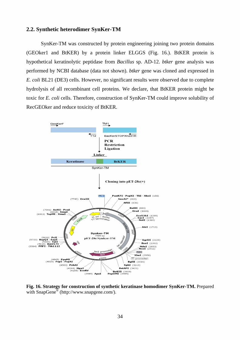
2.2. Synthetic heterodimer SynKer-TM
SynKer-TM was constructed by protein engineering joining two protein domains
(GEOker1 and BtKER) by a protein linker ELGGS (Fig. 16.). BtKER protein is
hypothetical keratinolytic peptidase from Bacillus sp. AD-12. btker gene analysis was
performed by NCBI database (data not shown). btker gene was cloned and expressed in
E. coli BL21 (DE3) cells. However, no significant results were observed due to complete
hydrolysis of all recombinant cell proteins. We declare, that BtKER protein might be
toxic for E. coli cells. Therefore, construction of SynKer-TM could improve solubility of
RecGEOker and reduce toxicity of BtKER.
Fig. 16. Strategy for construction of synthetic keratinase homodimer SynKer-TM. Prepared with SnapGene® (http://www.snapgene.com/).
34

Synthetic heterodimer SynKer-TM was expressed in E. coli BL21 (DE3) cells.
The induction of recombinant protein synthesis was performed at OD600 of 0.4 with 0.5
mM IPTG. The recombinant protein synthesis was continued for 2 h at 30 °C (Fig. 17.).
Fig. 17. Expression of synthetic heterodimer SynKer-TM. M – PierceTM Unstained Protein MW Marker. -IPTG – proteins profile without IPTG induction. +IPTG – proteins profile with IPTG induction.
After 2 h of induction obtained protein band approximately at 100 kDa indicated
the target recombinant SynKer-TM protein. However, recombinant SynKer-TM
enhanced E. coli protein degradation that occurs after induction. His-tagged SynKer-TM
was expressed as inclusion bodies. The washed inclusion bodies were solubilized by the
slow addition of 6 M urea (Fig. 18.).
Fig. 18. Solubility of RecGEOker and SynKer-TM.
35

Synthetic heterodimer SynKer-TM was successfully cloned and expressed in
heterologous system. Obtained results showed that expression of SynKer-TM result in
the formation of insoluble aggregates known as inclusion bodies. It is reliable that
protein aggregates prevented E. coli cells from toxic BtKER effect. Additionally, fusion
protein technology increased solubility of high hydrophobic RecGEOker protein up to
2.78 times (about 5 mg mL-1). Although the expression of SynKer-TM was positive,
BtKER protein functional activity was not detected.
The successful construction of a recombinant fusion proteins requires two
indispensable parts – the component protein and the linker. The selection of a suitable
linker to join the proteins together can be complicated. The reduced activity of both
SynKer-TT and SynKer-TM was only one unfavourable subsequence. However, our
results showed that either solubility or thermal stability can be improved by joining two
identical or structurally different protein domains.
3. Lab-scale keratin biodegradation process
Investigation and application of keratinolytic peptidases in biotechnology related
processes are the main tasks of this dissertation work. In our recent work we argued and
proposed to use microbial secreted proteins – secretome – as biologically active
component for efficient keratin or related waste biodegradation. Either genetically
modified organisms or recombinant proteins are suitable for many biotechnologically
important processes. However, these biological units are preparation-related expensive
and undesirable for many wide use products. It is important to create biodegradation
system in eco-friendly manner. Therefore, for this purpose we explored our keratinolytic
peptidases BtKER, BPKer and BAKer and designed lab-scale biodegradation system
(Fig. 19.). Three main factors were investigated:
• Synergistic effect of different microbial secretomes;
• Effect of process temperature;
• Effect of process pH.
36
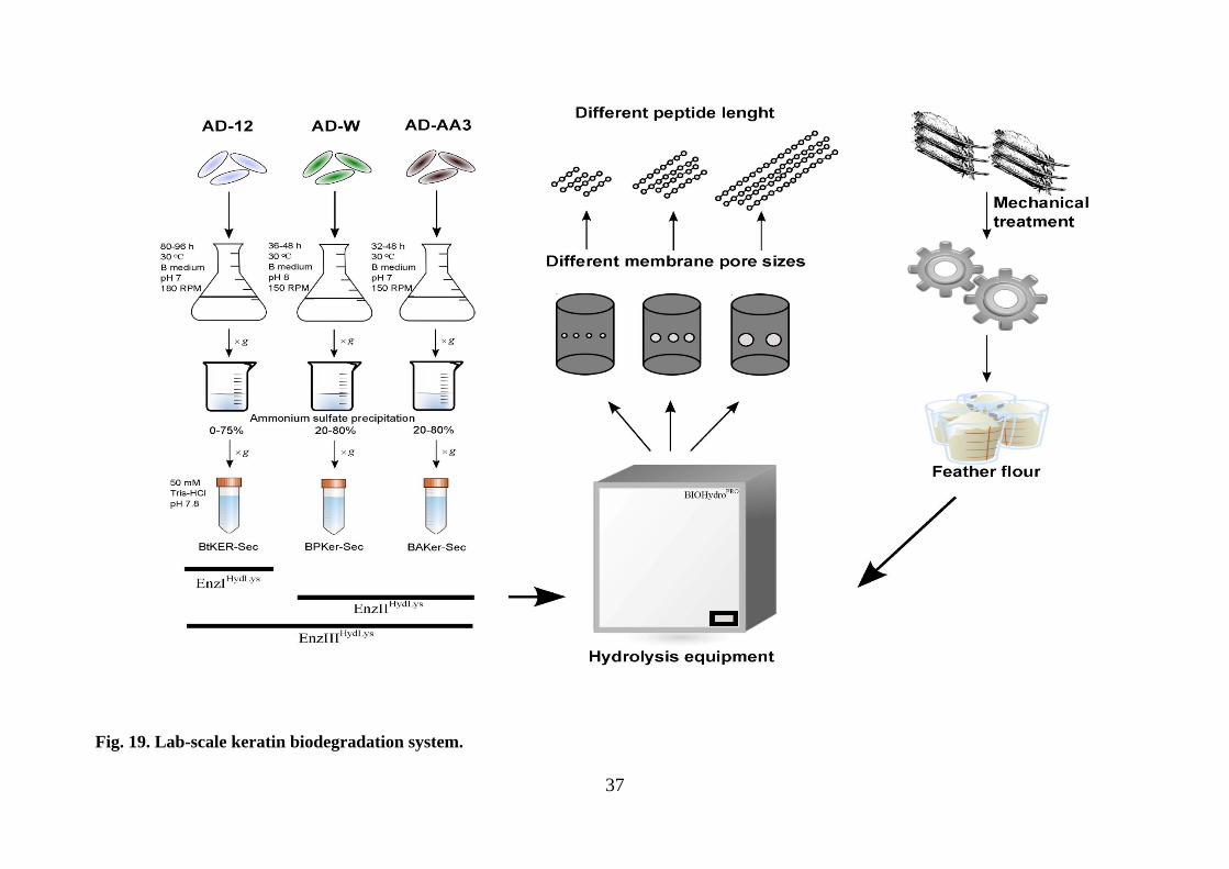
Fig. 19. Lab-scale keratin biodegradation system.
37

Recommendations: optimal conditions were chosen according to experimental
results:
• EnzIIHydLys enzyme complex;
• 30-35 °C hydrolysis temperature;
• pH 8;
• 3 h;
• 5-10 mg mL-1 proteinaceous substrate.
38

CONCLUSIONS
1) Evaluated impact of environmental conditions and various modulators on
functional activity of explored keratinolytic peptidases (RecGEOker, BtKER,
BPKer and BAKer);
2) All investigated keratinolytic peptidases are suitable for hydrolysis of various
soluble and insoluble proteinaceous materials into value-added peptides;
3) Features of thermal stability and solubility of our created chimeric (fused)
keratinolytic biocatalysts SynKer-TT and SynKer-TM were improved;
4) Designed lab-scale biological hydrolysis systems (EnzIIHydLys and EnzIIIHydLys) are
applicable for bioconversion of renewable keratin-rich waste.
39

LIST OF PUBLICATIONS
The thesis is based on the following original publications:
1) Gegeckas A., Gudiukaitė R., Debski J., Citavicius D. Keratinous waste
decomposition and peptide production by keratinase from Geobacillus
stearothermophilus AD-11. Int. J. Biol. Macromol. 2015:75, 158-165;
2) Gegeckas A., Gudiukaitė R., Citavicius D. Keratinolytic proteinase from Bacillus
thuringiensis AD-12. Int. J. Biol. Macromol. 2014:69, 46-51.
Other publications:
1. Gudiukaitė R., Gegeckas A., Sadauskas M., Citavicius D. Detection of Asp371,
Phe375, and Tyr376 influence on GD-95-10 lipase using alanine scanning
mutagenesis. Appl. Biochem. Biotechnol. 2016:178(4), 654-69;
2. Gudiukaitė R., Gegeckas A., Kazlauskas D., Citavicius D. Influence of N- and/or
C-terminal regions on activity, expression, characteristics and structure of lipase
from Geobacillus sp. 95. Extremophiles, 2014:18(1), 131-45.
CONFERENCE PRESENTATIONS
Dissertation theme was presented in 7 conferences (6 of them –
international):
1. Gegeckas A., Simkute A., Gudiukaite R., Citavicius DJ. Keratin waste
biodegradation and peptide production by keratinolytic proteinase from Bacillus
methylotrophicus AD-AA3. VAAM 2015 Annual Conference of the Association
for General and Applied Microbiology, 13-16 March 2016. Jena, Germany;
2. Simkute A., Gegeckas A., Gudiukaite R., Citavicius DJ. Keratinolytic proteinase
from Bacillus pumilus AD-W with promising keratin waste biodegradation
activity. Vita Scientia 2016, 04 January 2016. Vilnius, Lithuania;
3. Gegeckas A., Simkute A., Gudiukaite R., Citavicius DJ. Keratinolytic proteinase
from Bacillus pumilus AD-W with promising peptide-production activity.
40

BioMicroWorld 2015 6th International Conference on Environmental, Industrial
and Applied Microbiology, 28-30 October 2015. Barcelona, Spain;
4. Gegeckas A., Gudiukaitė R., Šimkutė A., ČitaviČius DJ. Keratinoliziniu
aktyvumu pasižymintys fermentai: įvairovė ir biotechnologinis potencialas. 2015
m. Jaunųjų mokslininkų konferencija BIOATEITIS: gamtos ir gyvybės mokslų
perspektyvos, Gruodžio 10 d., 2015. Vilnius, Lietuva;
5. Gegeckas A., Gudiukaite R., Citavicius DJ. Synthetic homodimer of GEOker
keratinase for efficient biodegradation of keratin by-products. BioMicroWorld
2015 6th International Conference on Environmental, Industrial and Applied
Microbiology, 28-30 October 2015. Barcelona, Spain;
6. Gegeckas A., Gudiukaite R., Čitavičius D. Keratinolytic proteinase as a powerful
biocatalyst for bio-active peptide production. CBM2014 2th Congress of Baltic
Microbiologists, 16-19 October 2014. Tartu, Estonia;
7. Gegeckas A., Gudiukaitė R., Čitavičius D. Production, purification and partial
characterization of keratinolytic serine peptidase from newly isolated Bacillus sp.
K1-2 strain. FEMS2013 5th Congress of European Microbiologists, 20-25 July
2013. Leipzig, Germany.
Other conferences (as co-author):
1. Malunavicius V., Gudiukaite R., Gegeckas A, Citavicius DJ. Construction of new
chimeric lipase using protein engineering methods. Vita Scientia 2016, 04 January
2016. Vilnius, Lithuania;
2. Gudiukaite R., Gegeckas A., Malunavicius V., Citavicius D. Functionality
analysis of structural domains from GD-95 lipase by site specific and random
mutagenesis. FEMS2015 6th Congress of European Microbiologists, 07-11 June
2015. Maastricht, The Netherlands;
3. Gudiukaite R., Gegeckas A., Čitavičius D. GD-95 lipase - new biocatalyst in
wide industry areas. CBM2014 2th Congress of Baltic Microbiologists, 16-19
October 2014. Tartu, Estonia;
4. Gudiukaitė R., Gegeckas A., Čitavičius D. Geobacillus spp. kamienų
rekombinantinių lipazių fizikinių bei cheminių savybių įvairovės įvertinimas.
41

Mokslas Gamtos Mokslų fakultete, 8-ta mokslinė konferencija, Spalio 3 d., 2014.
Vilnius, Lietuva;
5. Gudiukaitė R., Gegeckas A., Čitavičius D. Cloning, purification and esterification
capability determination of lipase produced by Geobacillus sp. 76. FEMS2013 5th
Congress of European Microbiologists, 20-25 July 2013. Leipzig, Germany.
FINANCIAL SUPPORT
1. "Scientific Research and Experimental Development (SRED) Incentive Program",
The Lithuanian Agency for Science, Innovation and Technology, 2014, 31V-106;
2. Industrial biotechnology development programme for Lithuania for 2011-2013
project “Development of completely novel approach for the cosmetics
(COSMETIZYM)”, The Lithuanian Agency for Science, Innovation and
Technology, 2011-2013, MITA 31V-18;
3. Research Council of Lithuania (RCL) doctoral scholarships (2014 – 2016);
4. Research Council of Lithuania (RCL) grant for doctoral visits (2015 – 2016).
42

CURRICULUM VITAE
PERSONAL INFORMATION AUDRIUS GEGECKAS S. Konarskio 14-6, LT-03124, Vilnius, Lithuania +370 628 05948 [email protected] 1987-11-19 WORK EXPERIENCE _____________________________________________________ 2013-09 – present LECTURER Vilnius University, Faculty of Natural Sciences, Department of Microbiology and Biotechnology 2014-05 – 2014-12 SENIOR SCIENTIFIC SPECIALIST Vilnius University, Faculty of Natural Sciences, Department of Microbiology and Biotechnology 2011-02 - 2013-12 SPECIALIST Vilnius University, Faculty of Natural Sciences, Department of Microbiology and Biotechnology 2009-01 - 2010-05 TRAINEE UAB Fermentas (now UAB Thermo Fisher Scientific Baltics) EDUCATION AND TRAINING _____________________________________________________ 20010-09 - 2012-06 MSC IN MICROBIOLOGY Vilnius University 2006-09 - 2010-06 BSC IN BIOLOGY (qualification of Molecular biology) Vilnius University PROJECTS _____________________________________________________ "Lietuvos mokinių neformaliojo švietimo centro vykdomos moksleivių stovyklos, rengiantis tarptautinei biologijos olimpiadai", Government of the Republic of Lithuania and Lithuanian centre of non-formal youth education funded project, 2015, consultant "Scientific Research and Experimental Development (SRED) Incentive Program", The Lithuanian Agency for Science, Innovation and Technology, 2014, senior scientific specialist "Mokinių jaunųjų tyrėjų atskleidimo ir ugdymo sistemos sukūrimas - II etapas", EU SF and Government of the Republic of Lithuania funded project, 2012-2015, 1,673 million EUR, scientific consultant; Industrial biotechnology development programme for Lithuania for 2011-2013 project “Development of completely novel approach for the cosmetics (COSMETIZYM)”, The Lithuanian Agency for Science, Innovation and Technology, 2011-2013, 120 000 EUR, specialist
43

ACKNOWLEDGEMENTS
I would like to thank my supervisor prof. Donaldas Čitavičius for discussions,
suggestions and support. Thank you for giving me a opportunities. Thank you for your
help through my PhD study.
I would like to give my gratitude to prof. Nomeda Kuisienė for helping me with
preparation of this dissertation.
I would like to thank our Head prof. Lilija Kalėdienė in Department of
Microbiology and Biotechnology for giving me advices. Moreover, I will never forget
countless help from all other department members.
Special thanks to prof. Rolandas Meškys and prof. Edita Sužiedėlienė for
possibility to make some important experiments.
Many thanks to my friends and colleagues: Renata Gudiukaitė, Raimonda
Petkauskaitė, Alisa Gricajeva, Aistė Šimkutė, Aida Virkšaitė, dr. Giedrius Gasiūnas,
Andrius Jasilionis, Arnoldas Kaunietis and Mikas Sadauskas.
I would like to sincerely thank my parents Rūta and Gintautas, brother Nerijus
and aunt Vida to support me to study PhD and continuous care.
Last but not least, I would like to express my gratitude to my wife Lina – for
limitless support, patience, care and comprehension. Thank You!
44

REZIUMĖ
Šiame darbe buvo siekiama identifikuoti, charakterizuoti ir pritaikyti skirtingų
mikroorganizmų – termofilinių ir mezofilinių – sekretuojamas keratinolizines peptidazes.
Mokslinių straipsnių duomenų bazėse nuolat atsiranda naujos informacijos apie iki šiol
neaprašytas keratinolizines peptidazes ir jų pritaikymo galimybes.
Nustatyta, kad pirmasis mūsų identifikuotas mikroorganizmas yra priskiriamas
termofilinių mikroorganizmų Geobacillus genčiai. Išsamesnė analizė leido sąlyginai AD-
11 izoliatą priskirti G. stearothermophilus rūšiai. Keratinolizinė peptidazė, sekretuojama
Geobacillus sp. AD-11 kamieno buvo pavadinta GEOker. Sėkminga buvo ir šios
keratinolizinės peptidazės produkcinės terpės paieška. Sukurta ir pritaikyta minimali
sintetinė terpė, kurioje vienintelis anglies, azoto, sieros ir energijos šaltinis yra vilnos
keratinas. Mikroorganizmui esant tokioje aplinkoje yra maksimaliai sintetinamas ir
sekretuojamas į aplinką fermentas, galintis vykdyti efektyvią šio vienintelio substrato
hidrolizę iki mažos molekulinės masės junginių, kurie jau gali būti panaudojami ląstelės
metabolizmui.
Gavus masių spektrometrijos rezultatus buvo galutinai įsitikinta, kad Geobacillus
sp. AD-11 kamienas sekretuoja GEOker keratinolizinę peptidazę. Detali gautų peptidų
sekų analizė duomenų bazėse leido identifikuoti hipotetinį GEOker baltymą. Nustatyta,
kad šis baltymas yra sintetinamas kaip Pre-Pro-fermentas, kuris galutinai įgyja savo
funkciškai aktyvią konformaciją tik po sėkmingos sekrecijos į užląstelinę aplinką ir po
autokatalizės, kurią vykdo paties fermento N-galinis domenas (Pro-peptidas). GEOker
baltymas savo aminorūgščių seka yra panašiausias į baciloliziną (G. stearothermophilus
ir G. thermocatenulatus), termoliziną (thermolysin-like), termostabilią neutralią
proteinazę (B. caldolyticus) ir šarminę metaloproteazę (B. subtilis).
Įvykdžius geoker geno klonavimo ir raiškos eksperimentus nustatyta, kad aktyvią
šio baltymo formą įmanoma gauti tik klonavus GEOker baltymą kartu su jo Pro-seka
(GEOker1). Tikėtina, kad Pro-seka nulemia baltymo hidrofobiškumą ir netirpių
baltyminių kūnelių susiformavimą rekombinantinių E. coli BL21 (DE3) ląstelių
citoplazmoje. Dažnu atveju netirpūs baltyminiai kūneliai yra didžiulė problema vykdant
heterologinę ar homologinę genų raišką. Mūsų tirtu atveju, šių kūnelių susidarymas
45

nulėmė funkciškai aktyvaus rekombinantinio baltymo RecGEOker gavimą. Taip pat
pademonstruota, kad neįvykus autokatalizei – baltymas yra aktyvus.
Fizikinių ir cheminių savybių įvertinimas ir gauti rezultatai papildo literatūros
duomenis apie termofilinių mikroorganizmų sekretuojamas keratinolizines peptidazes.
Nustatytos optimalios savybės (temperatūra, pH), įvairių cheminių junginių poveikis
(metalų jonai, detergentai, organiniai tirpikliai, slopikliai) ir gebėjimas hidrolizuoti
įvairius baltyminius substratus. Pademonstruota, kad RecGEOker yra efektyvus ir
tinkamas biokatalizatorius mažos molekulinės masės produktų gamybai.
Identifikuotos ir detaliai išanalizuotos 3 mezofilinių mikroorganizmų
sekretuojamos keratinolizinės peptidazės. Nustatyta, kad visi 3 izoliatai – AD-12, AD-W
ir AD-AA3 – priklauso Bacillus genties bakterijoms. Detalesnė analizė leido AD-12
izoliatą priskirti B. thuringiensis, AD-W – B. altitudinis/B. stratosphericus, o AD-AA3 –
B. amyloliquefaciens rūšims. Pasinaudojus Geobacillus sp. AD-11 kamieno augimo
terpės sudėtimi, buvo modifikuotos ir pritaikytos sintetinės terpės mezofilinių
mikroorganizmų sekretuojamų keratinolizinių peptidazių sekrecijai. Pritaikius tą pačią
produkavimo strategiją, buvo sėkmingai gauti fermentai, kurie pavadinti BtKER
(Bacillus sp. AD-12), BPKer (Bacillus sp. AD-W) ir BAKer (Bacillus sp. AD-AA3).
Atlikus BtKER baltymo gryninimą, gautas apie 39 kDa dydžio baltymas, kurio
funkcinis aktyvumas patvirtintas zimografijos metodu. Siekiant sukurti optimalų
biologinės degradacijos procesą, nuspręsta BPKer ir BAKer negryninti, o
eksperimentams ir pritaikymui naudoti šias keratinolizines peptidazes sintetinančių
mikrooganizmų sekretominius baltymus – sekretomą. Detali fizikinių ir cheminių
savybių analizė leido išskirti potencialias fermentų pritaikymo galimybes. Iki šiol
literatūroje nebuvo skelbiama informacijos apie B. thuringiensis sekretuojamas
keratinolizines peptidazes, todėl mūsų gauti rezultatai apie BtKER fermentą yra svarbus
indėlis, papildantis informaciją apie šių fermentų įvairovę.
Šiama darbe buvo sukurti du chimeriniai baltymai: homodimerinis SynKer-TT
(sintetinė keratinazė, sudaryta iš dviejų GEOker baltymų) ir heterodimerinis SynKer-TM
(sintetinė keratinazė, sudaryta iš GEOker ir BtKER baltymų). Homodimerinis SynKer-
TT baltymas pasižymėjo iki 11 % didesniu temperatūriniu stabilumu 80 °C
temperatūroje, lyginant su monomeriniu RecGEOker baltymu. Suliejus RecGEOker
baltymą su BtKER (SynKer-TM) pavyko padidinti RecGEOker baltymo tirpumą 2,78 46

karto (iki 5 mg mL-1). Taip pat pavyko sumažinti BtKER baltymo tirpumą, tačiau
nustatyti šio baltymo funkcionalumo nepavyko. Gauti rezultatai leidžia teigti, kad
baltymų inžinerija yra galingas molekulinis įrankis, leidžiantis sukurti patobulintus
biokatalizatorius. Literatūroje yra mažai duomenų apie keratinoliziniu aktyvumu
pasižyminčius chimerinius baltymus, todėl mūsų gauti rezultatai yra svarbūs moksliniu
aspektu.
Keratinolizinių peptidazių pritaikymas ir hidrolizės potencialo panaudojimas yra
vienas svarbiausių šios disertacijos uždavinių. Yra tikslinga neefektyvius mechaninius ir
cheminius keratino atliekų (žaliavų) perdirbimo procesus pakeisti į efektyvius ir aplinkai
saugius biologinius perdirbimo procesus. Buvo siekiama sukurti hidrolizės procesą,
kuriame sinergistinis identifikuotų keratinolizinių peptidazių poveikis būtų maksimaliai
išnaudojamas mažos molekulinės masės produktų gamybai. Eksperimentiškai nustatytos
baltyminės kilmės substrato (vilnos keratino) hidrolizės sąlygos (EnzIIHydLys fermentų
kompleksas, 30-35 °C temperatūra, pH 8 buferinė sistema, 3 inkubavimo valandos, 5-10
mg mL-1 vilnos keratino) leido sukurti laboratorinio lygio keratino biodegradacijos
sistemą. Mūsų sukurta aplinkai draugiška biokonversijos sistema yra tinkama peptidų
gamybai, kurie gali būti panaudojami įvairiose biotechnologijos srityse.
47

REFERENCES
1. Brandelli, A., Daroit, D.J. & Riffel, A. Biochemical features of microbial keratinases and their production and applications. Applied microbiology and biotechnology 85, 1735-1750 (2010).
2. Brandelli, A. Bacterial Keratinases: useful Enzymes for Bioprocessing Agroindustrial Wastes and Beyond. Food and bioprocess technology 1, 105-116 (2008).
3. Gupta, R. & Ramnani, P. Microbial keratinases and their prospective applications: an overview. Applied microbiology and biotechnology 70, 21-33 (2006).
4. Gupta, R., Rajput, R., Sharma, R. & Gupta, N. Biotechnological applications and prospective market of microbial keratinases. Applied microbiology and biotechnology 97, 9931-9940 (2013).
5. Kublanov, I.V. et al. Biodiversity of thermophilic prokaryotes with hydrolytic activities in hot springs of Uzon Caldera, Kamchatka (Russia). Applied and environmental microbiology 75, 286-291 (2009).
6. Jaouadi, N.Z. et al. Biochemical and molecular characterization of a serine keratinase from Brevibacillus brevis US575 with promising keratin-biodegradation and hide-dehairing activities. PloS one 8, e76722 (2013).
7. Liu, B. et al. Expression and characterization of extreme alkaline, oxidation-resistant keratinase from Bacillus licheniformis in recombinant Bacillus subtilis WB600 expression system and its application in wool fiber processing. World journal of microbiology & biotechnology 29, 825-832 (2013).
8. Daroit, D.J. & Brandelli, A. A current assessment on the production of bacterial keratinases. Critical reviews in biotechnology 34, 372-384 (2014).
9. Rizk, M., Antranikian, G. & Elleuche, S. End-to-end gene fusions and their impact on the production of multifunctional biomass degrading enzymes. Biochemical and biophysical research communications 428, 1-5 (2012).
10. Foo, J.L., Ching, C.B., Chang, M.W. & Leong, S.S. The imminent role of protein engineering in synthetic biology. Biotechnology advances 30, 541-549 (2012).
11. Sambrook, J.R. & Russell, D.W. Molecular Cloning: A Laboratory Manual, Edn. 3. (Cold Spring Harbor Laboratory Press, Cold Spring Harbor, NY, 2001).
12. Kuisienė, N. Molekulinė taksonomija. (Technologija, Kaunas, 2008). 13. Zhang, Z., Schwartz, S., Wagner, L. & Miller, W. A greedy algorithm for aligning
DNA sequences. Journal of computational biology : a journal of computational molecular cell biology 7, 203-214 (2000).
14. Morgulis, A. et al. Database indexing for production MegaBLAST searches. Bioinformatics 24, 1757-1764 (2008).
15. Robert, X. & Gouet, P. Deciphering key features in protein structures with the new ENDscript server. Nucleic acids research 42, W320-324 (2014).
16. Saitou, N. & Nei, M. The neighbor-joining method: a new method for reconstructing phylogenetic trees. Molecular biology and evolution 4, 406-425 (1987).
17. Felsenstein, J. Confidence limits on phylogenies: An approach using the bootstrap. Evolution 39, 783-791 (1985).
48

18. Nei, M. & Kumar, S. Molecular Evolution and Phylogenetics. (Oxford University Press, 2000).
19. Kumar, S., Stecher, G. & Tamura, K. MEGA7: Molecular Evolutionary Genetics Analysis version 7.0 for bigger datasets. Molecular biology and evolution (2015).
20. Petersen, T.N., Brunak, S., von Heijne, G. & Nielsen, H. SignalP 4.0: discriminating signal peptides from transmembrane regions. Nature methods 8, 785-786 (2011).
21. Reese, M.G. Application of a time-delay neural network to promoter annotation in the Drosophila melanogaster genome. Computers & chemistry 26, 51-56 (2001).
22. Laemmli, U.K. Cleavage of structural proteins during the assembly of the head of bacteriophage T4. Nature 227, 680-685 (1970).
23. Schagger, H. Tricine-SDS-PAGE. Nature protocols 1, 16-22 (2006). 24. Fakhfakh-Zouari, N., Hmidet, N., Haddar, A., Kanoun, S. & Nasri, M. A novel
serine metallokeratinase from a newly isolated Bacillus pumilus A1 grown on chicken feather meal: biochemical and molecular characterization. Applied biochemistry and biotechnology 162, 329-344 (2010).
25. Cai, C.G., Chen, J.S., Qi, J.J., Yin, Y. & Zheng, X.D. Purification and characterization of keratinase from a new Bacillus subtilis strain. Journal of Zhejiang University. Science. B 9, 713-720 (2008).
26. Mansfeld, J., Petermann, E., Durrschmidt, P. & Ulbrich-Hofmann, R. The propeptide is not required to produce catalytically active neutral protease from Bacillus stearothermophilus. Protein expression and purification 39, 219-228 (2005).
27. Kojima, K., Nakata, H. & Inouye, K. Involvement of Val 315 located in the C-terminal region of thermolysin in its expression in Escherichia coli and its thermal stability. Biochimica et biophysica acta 1844, 330-338 (2014).
28. Gao, X. et al. Structural basis for the autoprocessing of zinc metalloproteases in the thermolysin family. Proceedings of the National Academy of Sciences of the United States of America 107, 17569-17574 (2010).
29. Kim, Y.K., Bae, J.H., Oh, B.K., Lee, W.H. & Choi, J.W. Enhancement of proteolytic enzyme activity excreted from Bacillus stearothermophilus for a thermophilic aerobic digestion process. Bioresource technology 82, 157-164 (2002).
30. Eijsink, V.G., Matthews, B.W. & Vriend, G. The role of calcium ions in the stability and instability of a thermolysin-like protease. Protein science : a publication of the Protein Society 20, 1346-1355 (2011).
31. Rao, M.B., Tanksale, A.M., Ghatge, M.S. & Deshpande, V.V. Molecular and biotechnological aspects of microbial proteases. Microbiology and molecular biology reviews : MMBR 62, 597-635 (1998).
32. Rawlings, N.D., Waller, M., Barrett, A.J. & Bateman, A. MEROPS: the database of proteolytic enzymes, their substrates and inhibitors. Nucleic acids research 42, D503-509 (2014).
33. Kananen, A. et al. Influence of chemical modification of whey protein conformation on hydrolysis with pepsin and trypsin. International dairy journal 10, 691-697 (2000).
34. Kornillowicz-Kowalska, T. & Bohacz, J. Biodegradation of keratin waste: Theory and practical aspects. Waste management 31, 1689-1701 (2011).
49