Embed Size (px)
Citation preview

Victoria ZherdevaVictoria ZherdevaA.N.Bach Institute of BiochemistryA.N.Bach Institute of Biochemistry of the Russian Academy of Scienceof the Russian Academy of Science
28th Feb – 1st March 2013 New Delhi
International Seminar
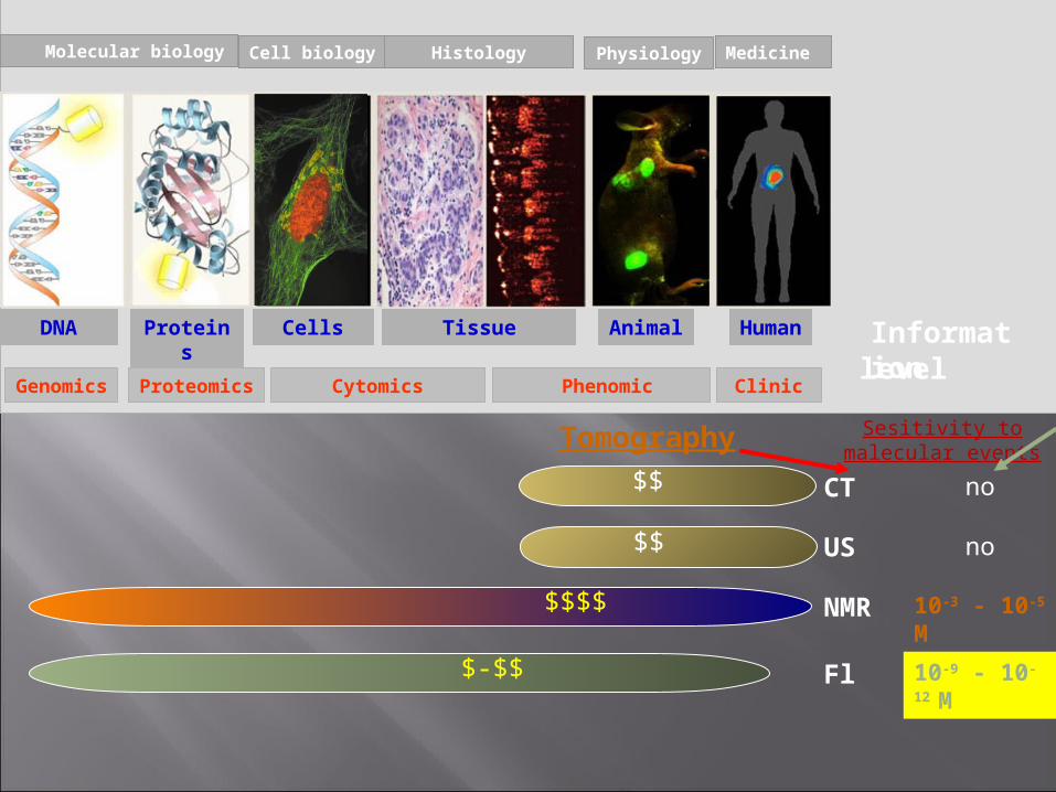
Molecular biology Cell biology Histology Physiology Medicine
Informationlevel
Proteins
Animal Human
ProteomicsGenomics Cytomics Phenomic Clinic
DNA Cells Tissue
CT no
US no
NMR 10-3 - 10-5 М
Fl 10-9 - 10-12 М
Tomography Sesitivity to malecular events
$$
$$
$$$$
$-$$
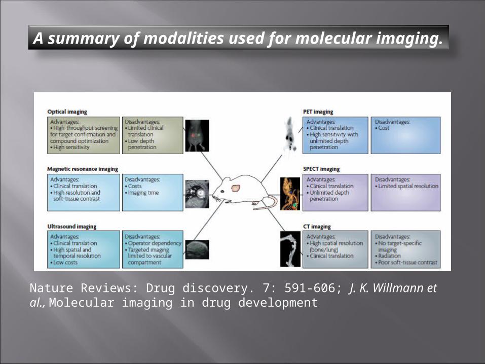
A summary of modalities used for molecular imaging.
Nature Reviews: Drug discovery. 7: 591-606; J. K. Willmann et al., Molecular imaging in drug development
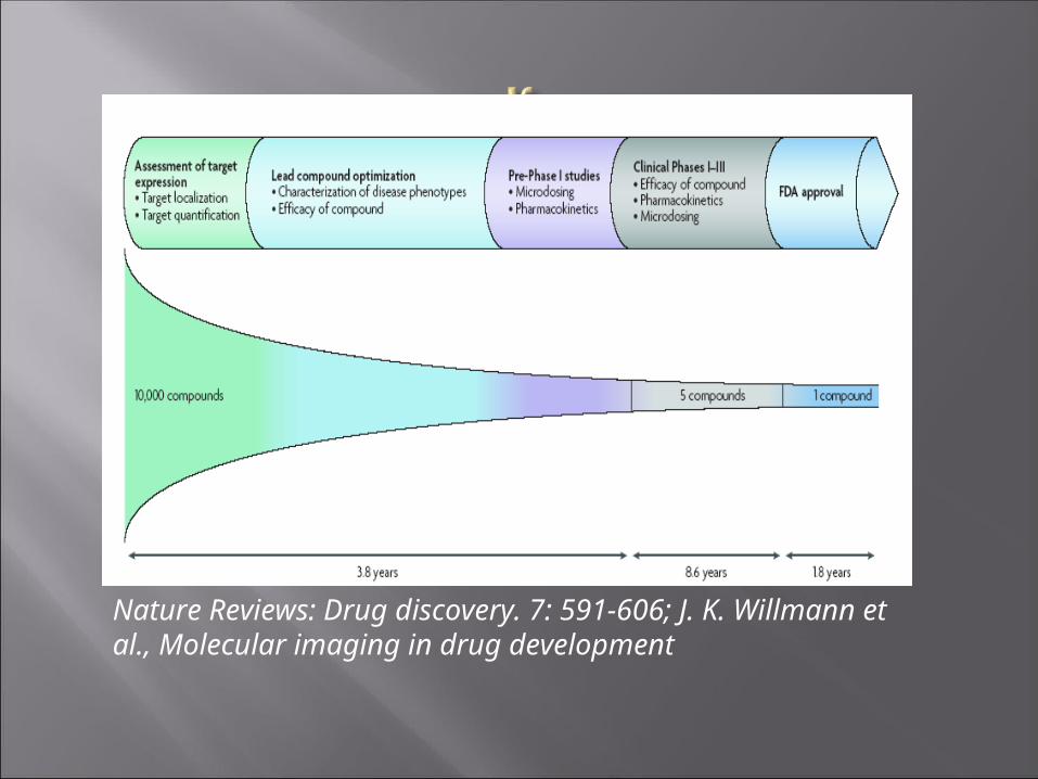
Nature Reviews: Drug discovery. 7: 591-606; J. K. Willmann et al., Molecular imaging in drug development

fusionproteins
mRNA
DNA
target
reporterstop
in vivo microscopycellular level
Whole-body imaging

• Functional studies of proteins in Functional studies of proteins in living cellsliving cells
• Construct target-GFP fusion Construct target-GFP fusion proteinprotein
• Examine at high resolution the Examine at high resolution the behaviour of the protein in living behaviour of the protein in living cellscells

2008 Nobel Prize in Chemistry "for the discovery and development of the green
fluorescent protein, GFP“.
2008 Nobel Prize in Chemistry "for the discovery and development of the green
fluorescent protein, GFP“.
Osamu ShimomuraMarine Biological Laboratory
Martin ChalfieColumbia University
Roger Y. TsienUniversity of California, San Diego

The technology of transgenic models obtainingThe technology of transgenic models obtainingThe technology of transgenic models obtainingThe technology of transgenic models obtainingTrancduced human
Tumor cell line with gene of FP
Xenotransplantation of the fluorescent cell line to the Nude mouse
Fluorescent imaging techniques
Transfection (liposomal or lentiviral) of cancer cells with fluorescent repoter gene

Fluorescence imaging techniquesFluorescence imaging techniques
Laser spectrometer with optic fiber zond
Fluorescence diffuse tomography
iBox UVP

In vivo visualization of subcutaneous transduced models of lung adenocarcinoma А549-TagRFP on iBox (USA, UVP) on the 7-th, 15-th, 20-th day after tumor cells inoculation. Ex. filter 502-547 nm, em. filter 570-640 nm. Exposure time -1s
7-th day 15-th day 20-th day


Monitoring of subcutaneous transduced models of lung adenocarcinoma А549-TagRFP (А) and А549-TRK23(B) with laser spectrometer SpectrClaster (Russia) on the 1-st, 7-th, 15-th, 20-th, 27-th, 32-d day after tumor cells inoculation.
A B
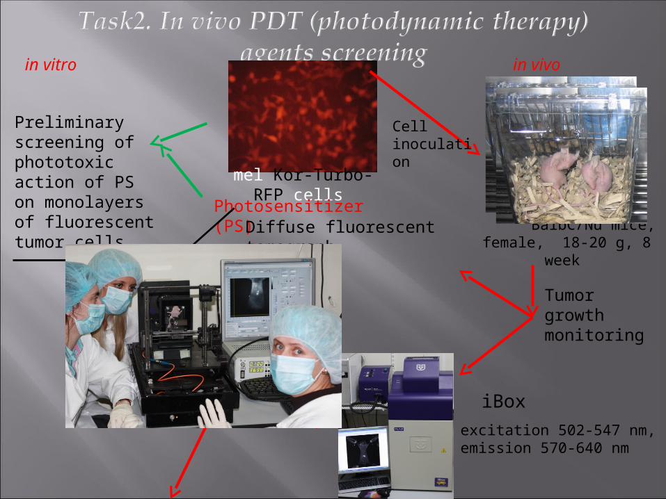
mel Kor-Turbo-RFP cellsPhotosensitizer (PS)
in vitro
Preliminary screening of phototoxic action of PS on monolayers of fluorescent tumor cells
in vivo
BalbC/Nu mice, female, 18-20 g, 8 week
Cell inoculation
Tumor growth monitoring
Diffuse fluorescent tomograph
iBox
excitation 502-547 nm,emission 570-640 nm
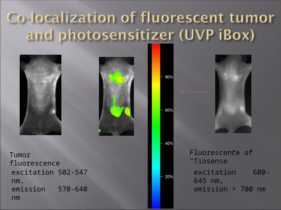
Tumor fluorescence Fluorescence of “Tiosense”
excitation 600-645 nm,emission > 700 nm
excitation 502-547 nm,emission 570-640 nm

Laser irradiation730 nm, 260 mWt/sm2, 20 min
4 mg/kg of bodyweight
0
1
2
3
0 10 20 30 40 50 60
Time after PS injection, hours
Tumor-to-normal ratio
0
10000
20000
30000
40000
50000
60000
70000
0 10 20 30 40 50 60
Fluo
resc
ence
, a.u
.
Время после введения "Тиосенса", ч Normal tissue Tumor
Laser irradiation, 20min

0
20
40
60
80
100
120
400 450 500 550 600 650 700
ин
тен
сив
но
сть
длина волны, нм
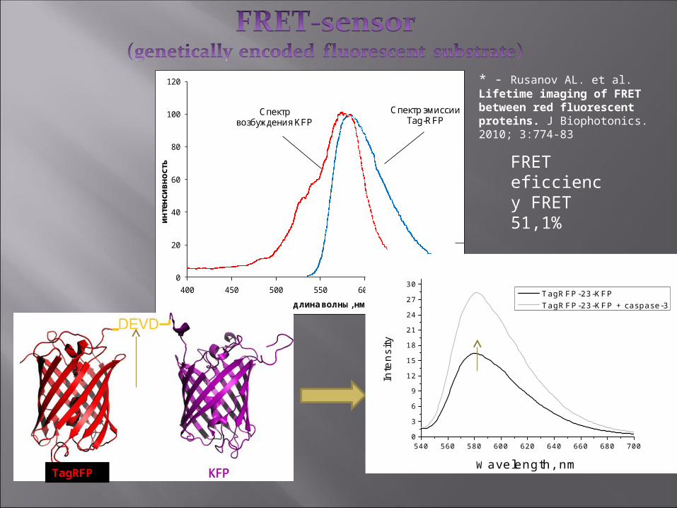
Спектр возбуждения KFP
Спектр эмиссии Tag-RFP
TagRFP
FRET eficciency FRET 51,1%
540 560 580 600 620 640 660 680 7000
3
6
9
12
15
18
21
24
27
30
Inte
nsi
ty
Wavelength, nm
TagRFP-23-KFP TagRFP-23-KFP + caspase-3
* - Rusanov AL. et al. Lifetime imaging of FRET between red fluorescent proteins. J Biophotonics. 2010; 3:774-83
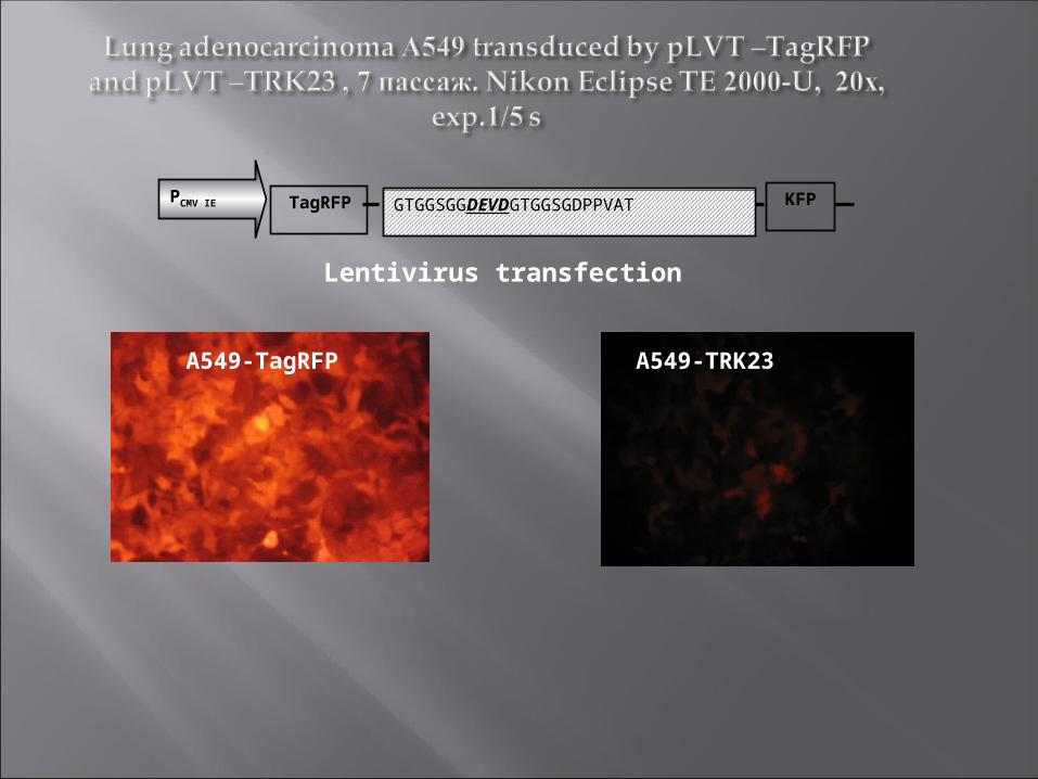
А549-ТagRFP А549-ТRK23
KFPTagRFP GTGGSGGDEVDGTGGSGDPPVAT
PCMV IE
Lentivirus transfection

FLIM
FLIM-FRET
Life-time of donor, ns
1 2
MicroTime 200, PicoQuantMicroTime 200, PicoQuant

Intact cellsA549-TRK23
A549-TRK23After apoptosis induction 800 мкм H2O2 ,after 24 ч
1.8-2.1 нс
1.8-2.1 нс 2.4-2.6
нс

Small animal FLIM-FRET whole body Small animal FLIM-FRET whole body imagingimaging
Red Fluorescent proteins (RFP)
Small animal FLIM-FRET whole body Small animal FLIM-FRET whole body imagingimaging
Red Fluorescent proteins (RFP)

Semiconducter core: Cd/Se, Cd/Te, and Ga/N
shell: Zn/S, Cd/Se
biomolecule:
polymer, protein, lipid
Quantum dots (QD) are nanometers size ( 1– 10nm) semiconductor
nanostructured materials with the tuneable size-dependent emission, high photoluminescence (PL) quantum yields, long PL lifetimes (10–50ns) and narrow
symmetric emission bands.
Task 4: biodistribution of QD

Qd applicationQd application
Michalet X, Pinaud FF, Bentolila LA, Tsay JM, Doose S, Li JJ, Sundaresan G, Wu AM, Gambhir SS, Weiss S: Quantum Dots for Live Cells, in Vivo Imaging, and Diagnositics. Science 2005 307(5709):538-544.

QDs MPA• λ em 611nм or 630 nм• d ~ 8 – 11нм•QY 10-20%
QDs PolyT• λ em 626 нм• d ~ 15 – 16 нм•QY 10-30%
QDs PolyT-APS
• λ em 678 нм• d ~ 36 нм•QY 5-20%

Excititation 502-547 nм, registration 570-640 нм, exposition 25 s.
per osper os

QDsStomach after 2 h
Intestine after 2 h
Intestine after 4 h
Intestine after 6 h
MPA + - - -
PolyT + + ± -
PolyT-APS + + + +
The relative estimation: QDs were not detected (-), low amount of QDs (±), well detectable amount of QDs (+).
Fiber optical fluorescence spectroscopyFiber optical fluorescence spectroscopy
Fluorescence spectra of feces probes 24 h after per os administration
QDs MPA
black curve - feces control, red curve - feces after administration of QDs.
QDs PolyT-APSQDs PolyT

•1. A.L. Rusanov, T.V. Ivashina, L.M. Vinokurov, I.I. Fiks, A.G. Orlova, I.V. Turchin, I.G. Meerovich, V.V. Zherdeva, and A.P. Savitsky. Lifetime imaging of FRET between red fluorescent proteins. J. Biophotonics, 2010, v. 3(12), p. 774-783.2. A.L. Rusanov, A.P. Savitsky. Fluorescence resonance energy transfer between fluorescent proteins as powerful toolkits for in vivo studies. Las. Phys. Lett., 2011, v. 8(2), p. 91-102.3. Rusanov A.L., Mironov V.A., Goryashenko A.S., Grigorenko B.L., Nemukhin A.V., Savitsky A.P. «Conformational partitioning in pH-induced fluorescence of the kindling fluorescent protein (KFP)» // J Phys Chem B. (2011);115(29):9195-201.4. Alexander L. Rusanov, Tatiana V. Ivashina, Leonid M. Vinokurov, Alexander S. Goryashenko, Victoria V. Zherdeva, Alexander P. Savitsky «FRET-sensor for imaging with lifetime resolution» // Laser Applications in Life Sciences, edited by Matti Kinnunen; Risto Myllylä. Proceedings of the SPIE, Volume 7376, pp. 737611-1-6 (2010).5. Alexander P. Savitsky, Alexander L. Rusanov, Victoria V. Zherdeva, Tatiana V. Gorodnicheva, Maria G. Khrenova and Alexander V. Nemukhin. FLIM-FRET Imaging of Caspase-3 Activity in Live Cells Using Pair of Red Fluorescent Proteins. Theranostics. (2012) v. 2, №2, pp.215-226. doi:10.7150/thno.3885
Publications:

6. Loginova Y.F., Kazachkina N.I., Zherdeva V.V., Rusanov A.L., Shirmanova M.V., Zagaynova E.V., Sergeeva E.A., Dezhurov S.V., Wakstein M.S., Savitsky A.P. Biodistribution of intact fluorescent CdSe/CdS/ZnS quantum dots coated by mercaptopropionic acid after intravenous injection into mice. – J. Biophotonics, 2012, vol. 11-12, pp. 848-859. 7. Loginova Y.F., Dezhurov S.V., Zherdeva V.V., Kazachkina N.I., Wakstein M.S., Savitsky A.P. Biodistribution and stability of CdSe core quantum dots in mouse digestive tract following per os administration: Advantages of double polymer/silica coated nanocrystals. – Biochem. Biophys. Res. Comm., 2012, vol. 419 (1), pp. 54–598. Salykina Y.F., Zherdeva V.V., Dezhurov S.V., Wakstein M.S., Shirmanova M.V., Zagaynova E.V., Martyanov A.A., Savitsky A.P. Biodistribution and clearance of quantum dots in small animals. – Proc. SPIE, 2011, vol. 7999, pp. 799908 – 799908-10.

Thank you