Embed Size (px)
Citation preview

of March 20, 2018.This information is current as
Chronic Myelogenous Leukemia CellsImatinib-Sensitive, and Imatinib-Resistantand Kill Zoledronate-Sensitized,
2 T Lymphocytes Efficiently Recognizeδ9VγV
DieliVigneri, Giorgio Stassi, Jean Jacques Fourniè and FrancescoMessina, Alfredo Salerno, Francesco Di Raimondo, Paolo Guggino, Serena Meraviglia, Nadia Caccamo, AngeloValentina Orlando, Matilde Todaro, Marisa Spina, Giuliana Matilde D'Asaro, Carmela La Mendola, Diana Di Liberto,
http://www.jimmunol.org/content/184/6/3260doi: 10.4049/jimmunol.0903454February 2010;
2010; 184:3260-3268; Prepublished online 12J Immunol
Referenceshttp://www.jimmunol.org/content/184/6/3260.full#ref-list-1
, 21 of which you can access for free at: cites 51 articlesThis article
average*
4 weeks from acceptance to publicationFast Publication! •
Every submission reviewed by practicing scientistsNo Triage! •
from submission to initial decisionRapid Reviews! 30 days* •
Submit online. ?The JIWhy
Subscriptionhttp://jimmunol.org/subscription
is online at: The Journal of ImmunologyInformation about subscribing to
Permissionshttp://www.aai.org/About/Publications/JI/copyright.htmlSubmit copyright permission requests at:
Email Alertshttp://jimmunol.org/alertsReceive free email-alerts when new articles cite this article. Sign up at:
Print ISSN: 0022-1767 Online ISSN: 1550-6606. Immunologists, Inc. All rights reserved.Copyright © 2010 by The American Association of1451 Rockville Pike, Suite 650, Rockville, MD 20852The American Association of Immunologists, Inc.,
is published twice each month byThe Journal of Immunology
by guest on March 20, 2018
http://ww
w.jim
munol.org/
Dow
nloaded from
by guest on March 20, 2018
http://ww
w.jim
munol.org/
Dow
nloaded from

The Journal of Immunology
Vg9Vd2 T Lymphocytes Efficiently Recognize andKill Zoledronate-Sensitized, Imatinib-Sensitive, andImatinib-Resistant Chronic Myelogenous Leukemia Cells
Matilde D’Asaro,*,1 Carmela La Mendola,*,1 Diana Di Liberto,* Valentina Orlando,*
Matilde Todaro,† Marisa Spina,† Giuliana Guggino,* Serena Meraviglia,*
Nadia Caccamo,* Angelo Messina,‡ Alfredo Salerno,* Francesco Di Raimondo,‡
Paolo Vigneri,‡ Giorgio Stassi,† Jean Jacques Fournie,x and Francesco Dieli*
Imatinib mesylate (imatinib), a competitive inhibitor of the BCR-ABL tyrosine kinase, is highly effective against chronic myelog-
enous leukemia (CML) cells. However, because 20–30% of patients affected by CML display either primary or secondary
resistance to imatinib, intentional activation of Vg9Vd2 T cells by phosphoantigens or by agents that cause their accumulation
within cells, such as zoledronate, may represent a promising strategy for the design of a novel and highly innovative immuno-
therapy capable to overcome imatinib resistance. In this study, we show that Vg9Vd2 T lymphocytes recognize, trogocytose, and
efficiently kill imatinib-sensitive and -resistant CML cell lines pretreated with zoledronate. Vg9Vd2 T cell cytotoxicity was largely
dependent on the granule exocytosis- and partly on TRAIL-mediated pathways, was TCR-mediated, and required isoprenoid
biosynthesis by zoledronate-treated CML cells. Importantly, Vg9Vd2 T cells from patients with CML can be induced by zoledr-
onate to develop antitumor activity against autologous and allogeneic zoledronate-treated leukemia cells, both in vitro and when
transferred into immunodeficient mice in vivo. We conclude that intentional activation of Vg9Vd2 T cells by zoledronate may
substantially increase their antileukemia activities and represent a novel strategy for CML immunotherapy. The Journal of
Immunology, 2010, 184: 3260–3268.
Chronic myelogenous leukemia (CML) is a malignanthemopoietic stem cell disorder characterized by the re-ciprocal translocation between chromosomes 9 and 22,
resulting in the BCR-ABL oncoprotein (1). Imatinib mesylate(imatinib), a competitive inhibitor of the BCR-ABL tyrosine ki-nase (TK), is highly effective in CML (2, 3), but frequent relapsehas been reported, particularly in patients with advanced-stage
disease (4). Although rescue TK inhibitors like desatinib or ni-lotinib are the preferred choice in this setting, there are patientswho are resistant to all TK inhibitors. To overcome this problem,adjunct immunotherapy may provide an opportunity to improvethe clinical outcome.gd T cells exhibit potent MHC-unrestricted lytic activity against
several tumor cells. gd T cell-based immunotherapy against dif-ferent tumors has been reported (5–8), and the efficacy of thistreatment has been demonstrated both in vivo and in vitro (9–12).The majority of gd T cells in peripheral blood expressed thevariable chain Vd2 in association with Vg9 and recognize non-peptide phosphoantigens commonly associated with metabolitesof bacterial isoprenoid biosynthesis or mevalonate pathway ineukaryotes. These compounds, such as isopentenyl pyrophosphate(IPP) and the synthetic analog bromohydrin pyrophosphate(BrHPP), activate gd T cells in vitro inducing their cytotoxic ac-tivity against tumor or infected target cells. Vg9Vd2 T cells alsorespond to nitrogen-containing bisphosphonates (N-BPs), such aszoledronate, probably via accumulation of mevalonate pathwayintermediates inside N-BP–treated cells (13).Recently, interest has emerged on the use of zoledronate in CML,
because this drug synergistically augments the anti-Ph+ leukemiaactivity of imatinib both in vitro and in vivo (14, 15) and inhibitsproliferation and induces apoptosis of imatinib-resistant CMLcells (16). Moreover, previous studies have demonstrated thatzoledronate sensitizes chemotherapy-resistant tumor target cells toVg9Vd2 T cell cytotoxicity, rendering a variety of cancer celllines highly susceptible to Vg9Vd2 T cell-mediated killing (17).Therefore, we reasoned that the intentional activation of Vg9Vd2T cells by zoledronate may represent a promising target for thedesign of novel and highly innovative immunotherapies capable toovercome imatinib resistance in patients with CML.
*Dipartimento di Biopatologia e Metodologie Biomediche and †Dipartimento diDiscipline Chirurgiche ed Oncologiche, Universita di Palermo, Palermo;‡Dipartimento di Scienze Biomediche, Universita degli Studi di Catania, Catania,Italy; and xDepartment of Oncology, Institut National de la Sante et de la RechercheMedicale Unite 563, Hospital Purpan, Toulouse Cedex 03, France
1M.D. and C.L. contributed equally to this work.
Received for publication October 22, 2009. Accepted for publication December 29,2009.
This work was supported by grants from the Ministry of University and Research, theMinistry of Health, and the University of Palermo (to G.S. and F.D.). J.J.F. wassupported by grants from Institut National de la Sante et de la Recherche Medicale,Institut National contre le Cancer, and Association pour la Recherche sur le Cancer.P.V., F.D., and A.M. were supported by grants from Regional and National Associa-zione Italiana per la Ricerca sul Cancro and the University of Catania. V.O. and M.S.are Ph.D. students of the International Ph.D. Programme in Immunopharmacology atthe University of Palermo.
Address correspondence and reprint requests to Dr. Francesco Dieli, Dipartimento diBiopatologia e Metodologie Biomediche, Universita di Palermo, Corso Tukory 211,90134 Palermo, Italy. E-mail address: [email protected]
Abbreviations used in this paper: BrHPP, bromohydrin pyrophosphate; CM, centralmemory; CMA, concanamycin A; CML, chronic myelogenous leukemia; CMTMR,5-and-6-(4-chloromethyl-benzoyl-amino-tetramethylrhodamine); EM, effector mem-ory; EMRA, terminally differentiated effector memory; FasL, Fas Ligand; HD,healthy donor; imatinib, imatinib mesylate; IPP, isopentenyl pyrophosphate; MFI,mean fluorescence intensity; NA, viable non apoptotic; N-BP, nitrogen-containingbisphosphonate; Nod, nucleotide-binding oligomerization domain; NVA, nonviableapoptotic; NVNA, nonviable nonapoptotic cell; PI, propidium iodide; TK, tyrosinekinase; VA, viable apoptotic.
Copyright� 2010 by TheAmericanAssociation of Immunologists, Inc. 0022-1767/10/$16.00
www.jimmunol.org/cgi/doi/10.4049/jimmunol.0903454
by guest on March 20, 2018
http://ww
w.jim
munol.org/
Dow
nloaded from

Promisingly, we show in this study that Vg9Vd2 T lymphocytesrecognize, trogocytose, and efficiently kill imatinib-responsive and-unresponsive CML cell lines pretreated with zoledronate. Cyto-toxicity was largely dependent on the granule exocytosis and partlyon TRAIL pathways, was TCR-mediated, and was dependent onisoprenoid production by CML cells. Importantly, Vg9Vd2 T cellsfrom patients with CML can be induced by zoledronate to developantitumor activity against autologous and allogeneic zoledronate-treated leukemia cells, both in vitro and when transferred into im-munodeficient mice in vivo.We conclude that intentional activation of Vg9Vd2 T cells by
zoledronate may substantially increase antileukemia activities andrepresent a novel strategy for CML adjunct immunotherapy.
Materials and MethodsPatients with CML and CML cell lines
Leukemia cell sampleswere obtained from13 newly diagnosed patientswithCML, followed at the Division of Haematology of the Ferrarotto Hospital inCatania, Sicily. Samples were collected at the time of diagnosis beforepatients started imatinib therapy. The study was approved by the ethicalcommittees of the University Hospital of Palermo and Ferrarotto Hospital ofCatania. Informed consent was obtained from all patients according to theDeclaration of Helsinki. To obtain CML cells, whole blood samples weretreated once or twicewithRBC lysing buffer (Sigma-Aldrich, St. Louis,MO)for 2 min at room temperature, and then centrifuged at 3503 g for 7 min torecover white cells and discard lysed red cells. The pellets were resuspendedin RPMI 1640 medium (Euroclone, Milan, Italy) supplemented with 10%FCS (Invitrogen, San Diego, CA), 2 mM L-glutamine, 20 nM HEPES, and100 U/ml penicillin/streptomycin.
Imatinib-sensitive (K562S) or -resistant (K562R, KCL22R, andLAMA84R) CML cell lines were a gift of Prof. Carlo Gambacorti-Passerini(Clinical Research Unit, University of Milano Bicocca, Milan, Italy). TheMM-1 cell line, expressing a BCR-ABL oncoprotein with two mutations inthe TK domain (E255K and T315I), was obtained from a patient with CMLin lymphoid blast crisis. All CML cell lines were grown in RPMI 1640medium supplemented with 10% FCS and 2 mM L-glutamine.
For cytotoxic experiments, primary leukemic cells or cell lines werepretreated with zoledronate (0.5 mM final concentration) or imatinib(0.25 mM final concentration), both provided by Novartis Pharma (Basel,Switzerland). Pretreatment of CML cells (either lines or freshly isolatedfrom patients) with imatinib or zoledronate, at the concentrations used inthis study, does not have cytotoxic effect (Fig. 6B).
Abs and reagents
The following unconjugated or FITC-, PE-, PE-Cy5-, or APC-conjugatedmAbs were obtained from BD Bioscience (San Diego, CA): anti-TCRVd2,anti-NKG2D, anti-Fas (CD95), anti-Fas Ligand (FasL; CD95L), anti–TNF-a, anti-MICA/B, anti-ULBP1, anti-ULBP2, anti-ULBP3, anti-CD16,anti-CD56, anti-CD161, anti-CD96, anti–DNAM-1 (CD226), and anti–nectin-2 (CD122). Additionally, the following purified mAbs were alsoused: anti-CD3 (blocking, MEM-57; a gift of Prof. Vaclav Horejsi, In-stitute of Molecular Genetics, Prague, Czech Republic), purified anti-TCRpan gd (IMMU510; a gift of Dr. Marc Bonneville, Institut de Biologie,Nantes, France), anti-NKG2D (1D11 from eBioscience, San Diego, CA;149810 from R&D Systems, Milan, Italy), anti–TNF-a (infliximab; a giftof Prof. Giovanni Triolo, Dipartimento Biomedico di Medicina Internae Specialistica, Universita di Palermo, Palermo, Italy), anti-CD95L(2C101; Vinci Biochem, Firenze, Italy), anti-TRAIL receptors TRAIL-R1(DR4), TRAIL-R2 (DR5), TRAIL-R3 (LIT, DcR1) and TRAIL-R4(TRUNDD, DcR2) (all kindly provided by Dr. Henning Walczak, TumorImmunology Unit, Division of Medicine, Imperial College, London, U.K.).Concanamycin A (CMA) was purchased from Sigma-Aldrich, and ator-vastatin was obtained from Calbiochem (Darmstadt, Germany).
Ex vivo expansion of Vg9Vd2 T cells
To expand human Vg9Vd2 T cells, PBMCs isolated from healthy adultdonors or patients with CML by density gradient centrifugation usingFicoll-Hypaque (Pharmacia Biotech, Uppsala, Sweden) were cultured inRPMI 1640 supplemented with 10% FCS and antibiotics in the presence of1 nM BrHPP (Phosphostim; a gift of Dr. Helene Sicard, Innate Pharma,Marseille, France) or 0.5 mM zoledronate (Novartis Pharma), added at day0. After 48 h, recombinant human IL-2 (Novartis Pharma; 50 IU/ml finalconcentration) was added to the cultures, and the cells were supplemented
with IL-2 every 3 d (18). Following 12–15 d culture, .90% of the cellsexpressed the Vg9Vd2 TCR, as determined by flow cytometry (FACS)analysis. The cells were harvested, and purified populations of Vg9Vd2T cells were obtained by positive selection using magnetic microbeadsconjugated to antihuman Vd2 mAb (mouse IgG1k, clone B6; Beckman-Coulter-Immunotech, Marseille, France) and immunomagnetic sorting(Miltenyi Biotec, Bergisch Gladbach, Germany). Isolated cells consisted of.98% Vg9Vd2 T cells, as determined by FACS analysis, and cell viabilityexceeded 95% as determined by trypan blue exclusion.
Proliferation assay
PBMC were labeled with CFSE (Molecular Probes, Eugene, OR) andcultured with 1 nM BrHPP or 0.5 mM zoledronate and 20 U/ml IL-2. After7 d, the number of Vg9Vd2+ T cells among CD3+ cells was determined byFACS (19). The absolute number of Vg9Vd2 T cells present in eachculture was calculated according to the following formula: % Vg9Vd2T cells 3 total number of viable cells/100. The Vg9Vd2 T cell expansionfactor was then calculated by dividing the absolute number of Vg9Vd2T cells in stimulated cultures by the number of Vg9Vd2 T cells culturedwith IL-2 alone (19).
Cytotoxic assay and blocking studies
Purified Vg9Vd2 T cells were resuspended at the final concentrations of1.5 3 106, 3 3 106, and 6 3 106 cells/ml, and 100 ml was then added toround-bottom polystyrene tubes together with CML cells (100 ml), toobtain the E:T ratios of 5:1, 10:1, and 20:1. Cytotoxicity was measured byFACS analysis using CFSE and propidium iodide (PI; both from MolecularProbes) as described in Ref. 20. Briefly, a total of 50 ml of CFSE wereadded to 1 ml target cell suspension (53 105 cells/ml) in PBS to obtain thefinal concentration of 2.5 mM CFSE. The cells were incubated for 10 minat 37˚C and gently mixed every 5 min. At the end of incubation, 1 ml FBSwas added to the cell suspension to stop the staining reaction, and the cellswere centrifuged at 600 3 g for 5 min at room temperature, washed twicewith cold PBS, and resuspended in serum-free medium. Control tubescontaining only labeled target cells and effector cells were also prepared toestablish background levels of cells death. Tubes were gently mixed,centrifuged at 3003 g for 2 min, and incubated at 37˚C in 5% CO2 for 4 h.At the end of the incubation period, the tubes were placed on ice, and 20 mlPI (1 mg/ml) were added to each tube for 10–15 min in ice. Finally, 100 mlcomplete medium were added before acquisition on an FACSCalibur cy-tometer (BD Biosciences). The calculation of cytolytic activity was basedon the degree of reduction of viable target cells (VTC) with the ability toretain CFSE and exclude PI (CFSEhigh PI2). In some experiments,blocking mAbs (all at the final concentration of 10 mg/ml) were used toevaluate the mechanisms of Vg9Vd2 T cell-mediated recognition andcytotoxicity of CML cell lines. To evaluate the contribution of zoledronate-induced accumulation of mevalonate metabolites to kill target cells, tumorcells were treated for 2 h with 5 mM atorvastatin, a selective upstreaminhibitor of the mevalonate pathway. Target cells were then incubated withzoledronate 0.5 mM for 20 h, washed, and added to Vg9Vd2 T cells. Toinhibit perforin-mediated cytotoxicity, Vg9Vd2 T cells were incubatedwith 15 nM CMA for 30 min at 37˚C prior to coculture, without furtherwashing. Pretreatment of Vg9Vd2 T cells or CML cells with CMA oratorvastatin, at the concentrations used in this study, did not have anycytotoxic effect.
Flow cytometry and flow cytometry-based measure of synaptictransfer
Expression of surface markers by CML target cells was determined by flowcytometry (FACS) analysis. Cells were incubated in U-bottom 96-wellplates, washed twice in PBS containing 1% FCS, stained for 30 min at 4˚Cwith labeled Abs according to manufacturers’ recommendations, washed,and analyzed by flow cytometry on an FACSCalibur with the use ofCellQuest software (BD Biosciences). Viable cells were gated by forwardand side scatter, and the analysis was performed on 100,000 acquiredevents for each sample.
Trogocytosiswasmeasured according toRef. 21.Briefly, CML target cellswere stained with PKH67 lipophilic fluorochrome (Sigma-Aldrich), and ef-fector Vg9Vd2 T cells were stained with Orange-5-and-6-(4-chloromethyl-benzoyl-amino-tetramethylrhodamine) (CMTMR; Molecular Probes). Cellswere then mixed together and cocultured in 96-well U-bottom tissue cultureplates at an E:T ratio of 2:1 in medium, at the final concentration of 63 106
cells/ml. Culture plates were then centrifuged at 300 3 g for 1 min and in-cubated for 3 or 60min at 37˚C. Postincubations, cellswere collected,washedtwice with PBS/EDTA, and analyzed on an FACS Calibur flow cytometer todetect trogocytosis. Data are expressed as PKH67 mean fluorescence in-tensity (MFI) of Orange-CMTMR–positive Vg9Vd2 T cells (21).
The Journal of Immunology 3261
by guest on March 20, 2018
http://ww
w.jim
munol.org/
Dow
nloaded from
Confocal microscopy analysis of synaptic transfer and viability
Confocalmicroscopyanalysiswas carriedoutasdescribed inRef. 22.PKH67-stained CML cell lines and Orange-CMTMR–stained Vg9Vd2 T cells werecoincubated for 1 h at 37˚C, as described above, at anE:Tratio of 5:1.Thecellswere then gently resuspended and plated on Lab-Tek chambered coverglass(VWR) previously coated with poly-D-lysine hydrobromide (Sigma-Aldrich)and incubated at 37˚C in a 5% CO2 atmosphere. Postadhesion, cells werewashed with PBS, fixed with PBS/2% p-formaldehyde, and slides mountedwith PBS/2% diazobicyclooctane solution. Samples were examined usinga Carl Zeiss LSM 410 confocal microscopy and AIM Imaging Software (CarlZeiss, Oberkochen, Germany).
In vivo bioluminescent imaging of leukemia progression in SCIDmice
The in vivo antitumor activity of Vg9Vd2 T cells was assessed according topreviously published methods (23, 24). Briefly, 106 MM1 CML cell linesstably expressing firefly luciferase and GFP were injected i.v. at day 0, ingroups of 8–14-wk-old nucleotide-binding oligomerization domain (Nod)/SCID mice (six mice per group; Charles River, Milan, Italy). A group ofmice received Vg9Vd2 T cells (2 3 107/mouse) previously expanded andactivated in vitro from PBMCs of patients with CML every 14 d, startingfrom day 1, together with 2 mg zoledronate i.p. (Novartis Pharma). Thesemice also received i.p. 30,000 IU IL-2 weekly. Control groups consisted ofmice receiving IL-2 and zoledronate but not Vg9Vd2 T cells, or Vg9Vd2T cells and IL-2 but not zoledronate, and Vg9Vd2 T cells and zoledronatebut not IL-2. At day 42, the experiments were terminated and mice weresacrificed. All mice were analyzed on a weekly basis by in vivo imaging(Biospace Lab, Cambridge, MA) upon i.p. injection (100 ml) of D-luciferin(40 mg/ml) (Sigma-Aldrich). Photon signals were quantified using the M3vision software for image analysis (Biospace Lab). The quantification ofsignal intensity was calculated as the sum of all detected photon fluxcounts within a uniform region of interest, manually selected during datapostprocessing. The autobioluminescence, generated by residual foodwithin the teeth and feces, was excluded and indicated by a yellow circle.
Mouse body weight was measured weekly, and animals suffering fromwasting (loss of over 20% of initial body weight) were sacrificed.
Statistics
The two-tailed Student t test was used to compare significance of differ-ences between groups. Data from experiments in Fig. 6 were comparedusing one-way ANOVA with Kruskal-Wallis multiple comparison testusing Instat software (version 3.05; GraphPad, San Diego, CA). Thep values ,0.05 were considered statistically significant.
ResultsCapture of CML cells membrane by Vg9Vd2 T lymphocytes
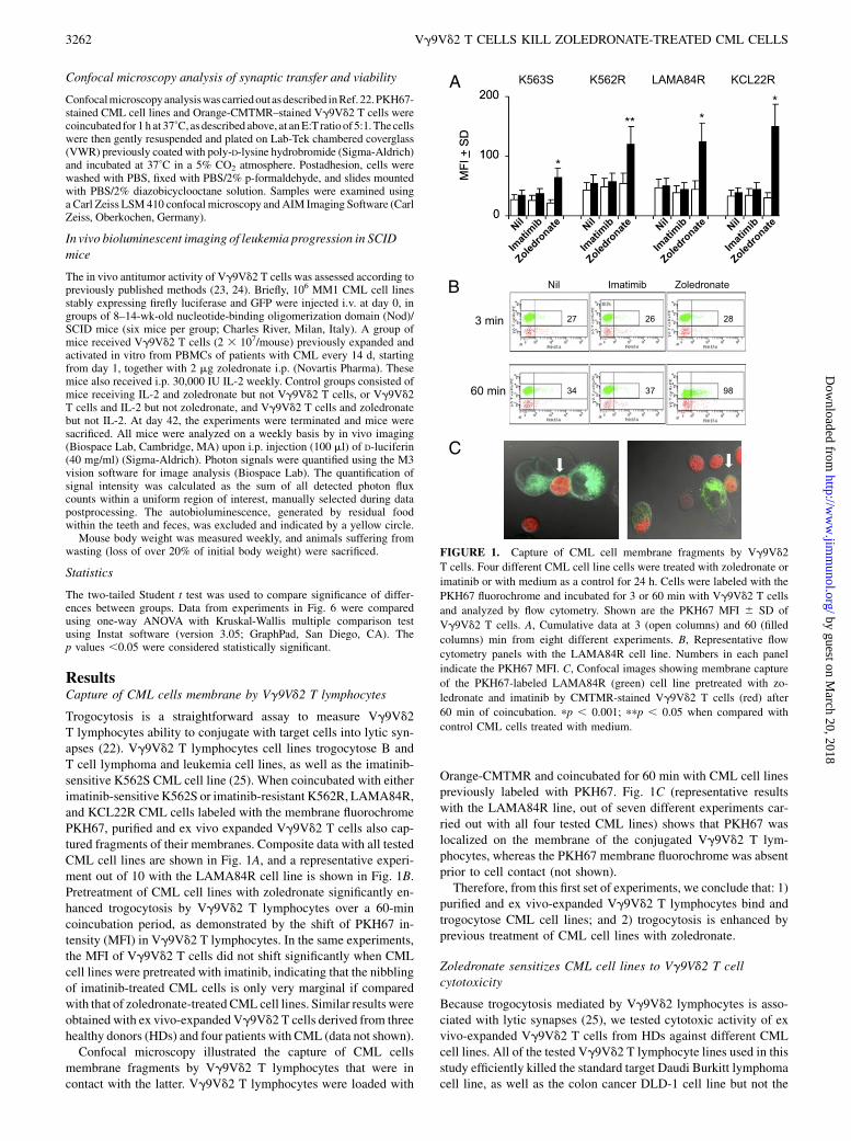
Trogocytosis is a straightforward assay to measure Vg9Vd2T lymphocytes ability to conjugate with target cells into lytic syn-apses (22). Vg9Vd2 T lymphocytes cell lines trogocytose B andT cell lymphoma and leukemia cell lines, as well as the imatinib-sensitive K562S CML cell line (25). When coincubated with eitherimatinib-sensitiveK562S or imatinib-resistant K562R, LAMA84R,and KCL22R CML cells labeled with the membrane fluorochromePKH67, purified and ex vivo expanded Vg9Vd2 T cells also cap-tured fragments of their membranes. Composite data with all testedCML cell lines are shown in Fig. 1A, and a representative experi-ment out of 10 with the LAMA84R cell line is shown in Fig. 1B.Pretreatment of CML cell lines with zoledronate significantly en-hanced trogocytosis by Vg9Vd2 T lymphocytes over a 60-mincoincubation period, as demonstrated by the shift of PKH67 in-tensity (MFI) in Vg9Vd2 T lymphocytes. In the same experiments,the MFI of Vg9Vd2 T cells did not shift significantly when CMLcell lines were pretreated with imatinib, indicating that the nibblingof imatinib-treated CML cells is only very marginal if comparedwith that of zoledronate-treated CML cell lines. Similar results wereobtained with ex vivo-expanded Vg9Vd2 T cells derived from threehealthy donors (HDs) and four patients with CML (data not shown).Confocal microscopy illustrated the capture of CML cells
membrane fragments by Vg9Vd2 T lymphocytes that were incontact with the latter. Vg9Vd2 T lymphocytes were loaded with
Orange-CMTMR and coincubated for 60 min with CML cell linespreviously labeled with PKH67. Fig. 1C (representative resultswith the LAMA84R line, out of seven different experiments car-ried out with all four tested CML lines) shows that PKH67 waslocalized on the membrane of the conjugated Vg9Vd2 T lym-phocytes, whereas the PKH67 membrane fluorochrome was absentprior to cell contact (not shown).Therefore, from this first set of experiments, we conclude that: 1)
purified and ex vivo-expanded Vg9Vd2 T lymphocytes bind andtrogocytose CML cell lines; and 2) trogocytosis is enhanced byprevious treatment of CML cell lines with zoledronate.
Zoledronate sensitizes CML cell lines to Vg9Vd2 T cellcytotoxicity
Because trogocytosis mediated by Vg9Vd2 lymphocytes is asso-ciated with lytic synapses (25), we tested cytotoxic activity of exvivo-expanded Vg9Vd2 T cells from HDs against different CMLcell lines. All of the tested Vg9Vd2 T lymphocyte lines used in thisstudy efficiently killed the standard target Daudi Burkitt lymphomacell line, as well as the colon cancer DLD-1 cell line but not the
A
B
C
FIGURE 1. Capture of CML cell membrane fragments by Vg9Vd2
T cells. Four different CML cell line cells were treated with zoledronate or
imatinib or with medium as a control for 24 h. Cells were labeled with the
PKH67 fluorochrome and incubated for 3 or 60 min with Vg9Vd2 T cells
and analyzed by flow cytometry. Shown are the PKH67 MFI 6 SD of
Vg9Vd2 T cells. A, Cumulative data at 3 (open columns) and 60 (filled
columns) min from eight different experiments. B, Representative flow
cytometry panels with the LAMA84R cell line. Numbers in each panel
indicate the PKH67 MFI. C, Confocal images showing membrane capture
of the PKH67-labeled LAMA84R (green) cell line pretreated with zo-
ledronate and imatinib by CMTMR-stained Vg9Vd2 T cells (red) after
60 min of coincubation. pp , 0.001; ppp , 0.05 when compared with
control CML cells treated with medium.
3262 Vg9Vd2 T CELLS KILL ZOLEDRONATE-TREATED CML CELLS
by guest on March 20, 2018
http://ww
w.jim
munol.org/
Dow
nloaded from

normal colon CCL-241 cell line. Fig. 2A shows representative ex-periments with Vg9Vd2 T cell from two HDs (HD 1 and HD 3).We assessed the ability of Vg9Vd2 T cells to kill imatinib-
sensitive (K562S) and -resistant (K562R, KCL22R, and MM1)CML cell lines. Untreated CML cell lines were weakly sensitive toVg9Vd2 T cell cytotoxicity, regardless of their responsiveness toimatinib. Lysis percentages ranged from 12–19%, at an E:T ratio of20:1without significant differences at other E:Tratios (Fig. 2B). Thepoor cytotoxic activity toward CML cell lines was not an intrinsicproperty of the gd T cells, because the Daudi and DLD-1 tumor celllines were recognized and killed efficiently by the same Vg9Vd2T cell lines (Fig. 2A). Pretreatment with zoledronate for 24 h wassufficient to render both imatinib-sensitive and -resistant CML celllines highly susceptible to Vg9Vd2 T cell killing, increasing levelsof cytotoxicity from 12% to 46% for K562S, from 19% to 61% forK562R, and from 13% to 56% for KCL22R, at an E:T ratio of 20:1.Treatment of target cells with imatinib alone did not increase sen-sitivity of CML cell lines to Vg9Vd2 T cell-mediated cytotoxicity.Failure to measure lysis of the imatinib-sensitve K562S cell linemight be due to the use of an imatinib concentration 5-fold lower
than that shown to inhibit 50% growth of CML cell lines over a 48-hperiod (14–16). Fig. 2B shows representative experiments withexpanded Vg9Vd2 T cells from one HD (HD 3). Notably, neitherzoledronate nor imatinib caused drug-associated cytotoxicity on alltested CML cell lines (data not shown). Confocal microscopyanalysis (Fig. 2C) illustrates apoptosis of the representative K562RCML cell line pretreated or not with zoledronate by Vg9Vd2T lymphocytes that were in contact with the latter.We also tested the cytotoxic ability of Vg9Vd2 T cells against
another peculiar CML cell line, called MM1, which expressesboth the E255K and T315I mutations, thereby exhibiting re-sistance to all available TK inhibitors. Untreated MM1 cells werepoorly sensitive to Vg9Vd2 T cell lysis (2% at a T:T ratio of 20:1)but pretreatment of these targets with zoledronate for 24 h con-sistently increased Vg9Vd2 T cell-mediated cytotoxicity, withvalues reaching almost 40% (Fig. 2B). As expected, pretreatmentwith imatinib alone did not sensitize these cells to lysis.Thus, zoledronate sensitizes CML cell lines to Vg9Vd2 T cell-
mediated cytotoxicity.
CMLcell lines constitutively expressmolecules involved in killingby Vg9Vd2 T cells
To determine possible mechanisms involved in Vg9Vd2 T cell-mediated cytotoxicity, we examined CML cell surface expressionof MICA/B molecules, ULBPs, nectin (CD122), Fas (CD95), DR4(TRAIL-R1), and DR5 (TRAIL-R2) death receptors as well asDcR1 (TRAIL-R3) and DcR2 (TRAIL-R4) decoy receptors, thatlack a functional death domain and cannot transduce a proapoptoticsignal. As depicted in Fig. 3, both imatinib-sensitive and -resistantCML cell lines constitutively express all these molecules, with theexception of DR5 receptor, which was not expressed at detectablelevels by any of the tested cell lines. Although expression of theabove-indicated molecules varied among the cell lines, no variationwas observed following exposure for 24 h to zoledronate. We onlyfound a slight, nonsignificant reduction of the expression of DcR1and DcR2 only on the imatinib-sensitive K562S cell line post-treatment with zoledronate (data not shown).
Vg9Vd2 T cells kill zoledronate-sensitized CML cell lines viaTCR-mediated recognition and the perforin and TRAILpathways
It has been proposed that human Vg9Vd2 T cells trigger severaldistinct pathways for killing tumor cells (26). These pathways in-clude secretion of proinflammatory cytokines and proapoptoticmolecules or on cell contact-dependent lysis through an NK-like orTCR-dependent signal. Mechanisms responsible for Vg9Vd2 T cellrecognition and killing of zoledronate-sensitized CML cells wereassessed by individually blocking TCR, NK receptor, perforin, Fas,and TRAIL pathways (Fig. 4). Cytotoxicity of both imatinib-sensi-tive and -resistant CML cell lines was inhibited at the greatest extentby anti-pan gd TCR (55% inhibition in K562S, 77% in K562R, and71% in KCL22R), indicating a TCR-mediated recognition and kill-ing. NKG2D seemed instead to play a minor role in Vg9Vd2 T cellcytotoxicity against CML cells, with no variation (K562R andKCL22R) or a small inhibition (15% in K562S) observed posttreat-ment with an anti-NKG2D Ab. In addition, Vg9Vd2 T cell recog-nition of zoledronate-sensitized CML cell targets was assessed in thepresence of atorvastatin, which inhibits 3-hydroxy-3-methyl-glu-taryl-CoA reductase and prevents zoledronate-mediated accumula-tion of endogenous IPP (26, 27). Atorvastatin caused significantinhibition of the killing of zoledronate pretreated CML cell lines,indicating that production ofmevalonatemetabolites is not redundantforCMLcell recognition and killing byVg9Vd2T cells. Fig. 4 showscumulative data performed in seven different experiments.
FIGURE 2. Vg9Vd2 T cell-mediated lysis of CML cell lines sensitive or
resistant to imatinib. A, Specific lysis of Daudi Burkitt lymphoma cells
(squares), colon cancer DLD-1 cells (circles), and normal colon CCL-241
cells (triangles) by Vg9Vd2 T cell line. Data shown are the mean6 SD. B,
Cytotoxic activity of Vg9Vd2 T cells against untreated (open circles), im-
atinib-treated (filled circles), or zoledronate-treated (filled squares) CML
target cells, both sensitive (K562S) and resistant (K562R, KCL22R, and
MM1) to imatinib. Percentages of specific lysis at a given E:T ratio are
shown. Data shown are the mean values of cytotoxicity 6 SD from one
representative experiment of seven independent experiments performedwith
different Vg9Vd2 T cell lines. C, Confocal image showing Vg9Vd2 T cells
(blue) coincubated for 4 h with K562R CML cells pretreated or not with
zoledronate and stained with acridine orange/ethidium bromide solution to
detect apoptosis and changes in cells morphology; four types of tumor target
cells were detected: 1) viable non apoptotic (NA), 2) viable apoptotic (VA),
3) nonviable apoptotic (NVA), and 4) nonviable nonapoptotic cell (NVNA).
The Journal of Immunology 3263
by guest on March 20, 2018
http://ww
w.jim
munol.org/
Dow
nloaded from

To further elucidate the mechanisms responsible for killing ofzoledronate-sensitized CML cell lines by Vg9Vd2 T cells, we in-dividually inhibited the granule exocytosis-, TRAIL-, TNF-a, andFasL-mediated pathways. Killing-inhibition experiments usingCMA revealed (Fig. 4) that Vg9Vd2 T cell cytotoxicity of zoledr-onate-pretreated CML targets was mainly mediated by the perforinpathway (means of 82–86% inhibition using CMA). Addition ofAbs against TRAIL-R1 and -R2 caused a 37% and a 25% inhibitionof the killing of zoledronate-treated K562S and K562R, re-spectively, but had no effect on the death of zoledronate-treatedKCL22R cells, indicating that TRAIL played a variable role in thekilling activity of Vg9Vd2 T cells toward CML cell lines. Finally,Abs to FasL and TNF-a failed to inhibit the cytotoxicity of all testedzoledronate-sensitized CML targets (Fig. 4).
Vg9Vd2 T cells from patients with CML kill primary leukemiccells freshly isolated from patients with CML
Preliminarly, we analyzed the size and functionality of Vg9Vd2T cells in the peripheral blood of patients with CML by measuringtheir ability to proliferate and differentiate, following exposure tozoledronate and IL-2. Ex vivo stimulation with zoledronate andIL-2 induced significant increase of the frequency of Vd2+ T cellswithin PBMCs from patients with CML and also promoted effi-cient Vg9Vd2 T cell expansion (9–420-fold expansion) (Fig. 5A,5B). Moreover, as shown in Fig. 5C, in cultures of PBMC frompatients with CML stimulated by zoledronate and IL-2, mostVg9Vd2 T cells displayed an effector memory (CD272CD45RA2)phenotype, like stimulated PBMC cultures from HDs.
Importantly, Vg9Vd2 T cells from patients with CML can beinduced by zoledronate to develop antitumor activity against CMLlines and autologous and allogeneic, zoledronate-treated, leukemiacells taken from patients with CML at the time of diagnosis and inthe absence of any treatment.Results show that Vg9Vd2 T cells from patients with CML failed
to kill both CML lines and their own tumor cells, but treatment oftarget cells with zoledronate significantly increased Vg9Vd2 T cellcytotoxicity in 10 out of the 13 tested patients with CML (6–63% ofcytotoxicity), while slightly increasing lysis in the other three pa-tients (2–5%, 1–6%, and 4–7%, respectively; data not shown). Fig.6A shows representative results of the cytotoxic activity of Vg9Vd2T cells from two lymphocyte-mediated cytotoxicity patients (CML7 and CML 12) toward autologous and allogeneic leukemia cells, aswell as toward CML cell lines. Notably, pretreatment of cells frompatients with CML with zoledronate did not have cytotoxic effectsper se on CML tumor cells, as demonstrated by double stainingwith CFSE and PI (Fig. 6B).Therefore, zoledronate enhances the susceptibility of both pri-
mary and immortalized CML cells to Vg9Vd2 T cell-mediatedcytotoxicity.
Vg9Vd2 T cells killing leukemia cells in vivo
To evaluate the potential of immunotherapy strategies, we useda previously published model of transplantation of human tumorsinto lymphopenic Nod/SCID mice (23, 24) and added biolumines-cent analysis of tumor development, which allows early detection oftumors and temporal evaluation throughout the course of treatmentin live animals and real-time. Four weeks postinjection of MM1cells, mice that had received activated and expanded Vg9Vd2T cells, zoledronate, and IL-2 showed significantly reduced tumorload compared with control mice (Fig. 7A). Furthermore, whereasmost controls had to be sacrificed at day 28 due to excessive bodyweight loss, Vg9Vd2 T cell-treated animals resisted wasting forlonger up to day 84 (data not shown). Fig. 7B show typical resultsobtained in two mice receiving expanded Vg9Vd2 T cells, zole-dronate, and IL-2 and in two control mice who received IL-2 andzoledronate, but not Vg9Vd2 T cells. These results attest the ca-pacity ofVg9Vd2T cells to induce anti-tumor responses invivo, andsupport their potential application in conjugation with zoledronateand IL-2 in clinical cancer settings.
DiscussionAlthough the use of imatinib has represented an important advancefor the treatment of CML, 20–30% of patients treated with thedrug fail to achieve a complete cytogenetic response, and evenpatients that exhibit an optimal response may subsequently present
FIGURE 4. Mechanisms of Vg9Vd2 T cell killing of CML target cells.
Vg9Vd2 T cell lines were cultured with zoledronate-treated CML cells at an
E:T ratio of 20:1 in the presence of blocking Abs to the gd TCR, NKG2D,
FasL, TNF-a, TRAIL-R1 or -R2, atorvastatin, or CMA. Data are mean6 SD
of seven different experiments carried out in triplicate. pp , 0.001; ppp ,0.02when comparedwith cytotoxicity carried out in the absence of inhibitors.
FIGURE 3. Phenotype of CML cell lines. Representative overlay histograms showing constitutive surface expression (open histograms) of MICA/B,
ULBP1-4, CD122 (nectin), CD95 (Fas), DR4 (TRAIL-R1) and DR5 (TRAIL-R2) molecules on CML cell lines against appropriate control Ig isotypes
(filled histograms). The numbers indicate the x-fold increase in median fluorescence intensity over the isotype control as determined on a 4-log scale.
3264 Vg9Vd2 T CELLS KILL ZOLEDRONATE-TREATED CML CELLS
by guest on March 20, 2018
http://ww
w.jim
munol.org/
Dow
nloaded from

a relapse of their disease. Moreover, ∼15% of patients treated withimatinib will only obtain a suboptimal response [i.e., a temporarystate that will likely require a higher dose of imatinib or a changein drug treatment (4)]. In this context, additional immunotherapymay provide an opportunity to either improve clinical responses inpatients who are resistant to imatinib or convert suboptimal res-ponders in optimal responders.Zoledronate is a third-generation N-BP already used in the
treatment of cancer-related bone complication (28–30). Moreover,zoledronate inhibits in vitro proliferation, induces apoptosis ofimatinib-resistant leukemia cells, and augments the anti-Ph+ leu-kemia activity of imatinib (15, 16, 31). However, zoledronate
concentrations used in these in vitro studies are in the micromolarrange (20–50 mM zoledronate was generally employed), althoughin humans treated with the drug, the maximal plasma concen-trations range from 0.5–5 mM, depending on the dosage and du-ration of infusion (32, 33). Moreover, the high micromolarconcentrations required in the in vitro experiments present a con-siderable risk for the toxicity to nonleukemic cells.
FIGURE 5. Functional analysis of Vg9Vd2
T cells from patients with CML. Kinetics of Vd2+
CD3+ cells frequency within PBMC of 13 pa-
tients with CML (A) and corresponding fold
amplifications (B) relative to day 0 were mea-
sured following specific activation with IL-2 and
zoledronate or IL-2 alone as a control. C, Anal-
ysis of the memory status of Vg9Vd2 T cells
generated in the same experiment. The percent-
age of naive (CD27+CD45RA+), central memory
(CM; CD27+CD45RA2), effector memory (EM;
CD272CD45RA2), and terminally differentiated
effector memory (EMRA; CD272CD45RA+)
cells are indicated within each subset. Data
shown are the mean percent values 6 SD of one
representative of at least three experiments per-
formed by using PBMC samples from 13 patients
with CML.
FIGURE 6. Vg9Vd2 T cells from patients with CML kill freshly iso-
lated, zoledronate-treated, autologous and allogeneic leukemia cells and
CML cell lines. Leukemia cells were purified from patients with CML at
the time of diagnosis and used for cytotoxic experiments with Vg9Vd2
T cells from patients with CML. A, Shown is specific lysis obtained with
ex vivo-expanded Vg9Vd2 T cells from two patients with CML (CML 7
and CML 12) against leukemia cells of three patients with CML (CML 7,
CML 8, and CML 12) and CML cell lines (K562S and K562R), untreated
(open bars) or pretreated with zoledronate (filled bars). Data show the
mean values6 SD of a representative experiment out of three performed in
triplicate. B, Pretreatments with imatinib or zoledronate alone did not in-
duce any cytotoxic effect per se, as indicated by CFSE versus PI staining.
Control
Treated
Day 0 Day 14 Day 28 Day 42
A
B
* ****
FIGURE 7. In vivo leukemia cell-killing activity of Vg9Vd2 T cells. Bio-
luminescent imaging of Nod/SCID mice injected with luciferase MM1 leu-
kemic cell linesand treatedwith zoledronate and IL-2, asdescribed inMaterials
and Methods (B). A, Image quantification of photon signals (tumor load)
collected at the indicated time points. Data in this figure are representative of
three independent experiments. Filled triangles indicate mice treated with zo-
ledronate, IL-2, andwith expanded and activatedVg9Vd2T cells. Open circles
indicate untreated control mice, receiving only leukemic cells. Open squares
indicate mice treated with zoledronate and IL-2. Filled circles indicate mice
treated with zoledronate and with expanded and activated Vg9Vd2 T cells.
Filled squares indicate mice treated with IL-2 with expanded and activated
Vg9Vd2 T cells. pp, 0.02; ppp, 0.001 when compared with control mice.
The Journal of Immunology 3265
by guest on March 20, 2018
http://ww
w.jim
munol.org/
Dow
nloaded from

In this study, we used purified and ex vivo-expanded Vg9Vd2T cell lines to kill CML cells, and we found that pretreatment oftarget cells with zoledronate alone or in combination with imatinibsignificantly increased their trogocytosis and killing by Vg9Vd2T lymphocytes. Similar to previous reported data (25), we foundthat trogocytosis perfectly matches cytotoxicity measured on tar-get cells, further indicating that target cell death relies much moreupon contact with Vg9Vd2 T lymphocytes and subsequent in-volvement of perforin and, at a lesser extent, TRAIL. Both imatinib-sensitive and -resistant CML cells pretreated with zoledronate for24 h were efficiently killed by Vg9Vd2 cells. Interestingly, also theleukemia cell line MM1, which carries a double mutation in theTK domain that confers resistance to any available TK inhibitor,was efficiently killed by Vg9Vd2 T cells when pretreated withzoledronate.Previous studies have demonstrated that zoledronate synergizes
with imatinib to inhibit Ph+ primary leukemic cell growth (15, 16),whereas zoledronate or imatinib, either alone or in combination,were not effective against leukemic cells harboring the E255K orT315I mutations. Conversely, in our experiments, pretreatment ofMM1 cells with zoledronate were allowed to obtain significantcytotoxic values reaching ∼40%.Human Vg9Vd2 T cells recognize phosphoantigens, which are
metabolites of isoprenoid biosynthetic pathways (34, 35), and themore recently described ATP synthase-F1/apolipoprotein A-1complex that, unlike in normal cells, is ectopically expressed onthe surface of hemopoietic and solid cancer cells (36). Moreover,Vg9Vd2 T cell activity is tightly regulated by NK-like receptors,and previous studies have indicated the importance of NKG2D-MICA/B interactions for tumor cell recognition and cytotoxicityby Vg9Vd2 T cells (9, 37, 38). It has been suggested that treat-ment of tumor cells with zoledronate leads to the intracellularaccumulation of phosphoantigens (typically IPP), favoring rec-ognition and killing of tumor cells by the reactive Vg9Vd2T lymphocytes (14, 39–41). In our study, Vg9Vd2 T cell recog-nition and killing of zoledronate-treated CML target cells wasTCR-mediated and depending on the synthesis of isoprenoid in-termediates, because preventing accumulation of IPP and/or otherendogenous phosphoantigens posttreatment with atorvastatin sig-nificantly impaired Vg9Vd2 T cell cytotoxicity, thus indicatingthat the sensitizing effect of zoledronate correlates with increasedexpression/production of mevalonate metabolites. A recent paperby Li and coworkers (40) used siRNA to provide support of theconcept that increased intracellular IPP levels are instrumental inVg9Vd2 T cell activation by tumor lines, which so far has beenbased on a correlation between IPP levels and Vg9Vd2 T cellactivation as well as by observations with enzyme inhibitors, suchas 3-hydroxy-3-methyl-glutaryl-CoA-reductase inhibitors (e.g.,mevastatin) and farnesyl pyrophosphate synthase inhibitors (e.g.,aminobisphosphonates). Although ATP synthase expression canalso be detected on CML cell lines used in this study (data notshown), because currently available anti-ATP synthase Abs are notreliable for inhibition experiments, we could not evaluate thecontribution of ATP synthase to Vg9Vd2 T cell-mediated recog-nition and killing of CML target cells. Moreover, NKG2D andother NK ligands/receptors do not appear to contribute to thecytotoxicity of zoledronate-treated CML cells, because addition ofspecific blocking Abs failed to inhibit lysis, and treatment withzoledronate did not alter MICA/B, ULBPs, and nectin expressionon the membrane of CML cells.Previous Ab-blocking studies by Wrobel and coworkers (42)
have shown three different patterns of tumor cells recognition andkilling by Vg9Vd2 T cells: preferential involvement of the TCR,preferential involvement of NKG2D, or additive involvement of
both. Moreover, and similar to the results reported in this study,the extent of inhibition of the cytolytic activity of Vg9Vd2 T cellsby anti-NKG2D Abs did not correlate directly with the level ofNKG2D ligand expression on the tumor cells or with the originand progression stage of a tumor cell (42).How far other molecules, such as the ectopically expressed F1-
ATPase, which has been claimed to serve as the Vg9Vd2 TCRligand expressed by Daudi tumor cells, are involved in IPP rec-ognition remains unclear.It is known that Vg9Vd2 T cells kill tumor targets via a number
of mechanisms including death receptor/ligand interactions withTRAIL and FasL and release of perforin/granzymes. In theory,one or more of these pathways may be involved in the killing ofCML cell lines. Although all CML cell lines evaluated in thisstudy constitutively expressed TRAIL receptors and Fas, this ex-pression did not initially translate into sensitivity to Vg9Vd2T cell killing, as documented by the failure of specific blockingAbs to consistently inhibit cytotoxicity. Additionally, exposure ofCML cell lines to zoledronate did not cause any variation ofTRAIL receptors and Fas expression. However, TRAIL-mediatedcytotoxicity may have instead a minor role because lysis of K562Swas reduced ∼35% after blocking interaction of TRAIL with itsreceptors, whereas lysis of the imatinib-resistant cell lines wasonly poorly (K562R) or not at all affected (KCL22R).Irrespective of imatinib sensitivity or resistance of CML lines,
CMA strongly inhibited cytotoxicity, indicating that zoledronate-treated targets are almost exclusively killed by perforin release byVg9Vd2 T cells, consistent with previous findings of perforin/granzyme-dominated killing (43–46).As it would be of interest to test in vivo the clinical efficacy of
a Vg9Vd2 T cell-mediated immune therapy, we assessed the cy-totoxic ability of Vg9Vd2 T cells on a limited number of cellstaken from patients with CML at the time of diagnosis and beforetherapy. The results obtained showed that Vg9Vd2 T cells killcells freshly isolated from patients with CML, but exclusivelywhen cells were pretreated with zoledronate alone or in combi-nation with imatinib.Our findings, together with the attainment that zoledronate has
antileukemic properties, firstly indicate that zoledronate-activatedVg9Vd2 T cells possess a promising cytotoxic activity againstCML cells resistant to imatinib. On this basis, we would like tosuggest the clinical utility of intentional in vivo activation ofVg9Vd2 T cells by zoledronate and low doses of IL-2 in thosepatients refractory to imatinib treatment alone, as recently per-formed in other hematological malignancies (10) and in prostatecancer (12). Even if potent cytotoxic Vg9Vd2 T cells may begenerated from blood cells of patients with myeloma and lym-phoma (8), sometimes proliferative responses of Vg9Vd2 T cellsfrom patients with cancer turned out to be suppressed (47), thusaccounting for tumor-induced anergy after chronic Vg9Vd2 T cellstimulation. This problem could be circumvented by administra-tion of in vitro-expanded allogeneic Vg9Vd2 T cells from HDs,because they are not alloreactive and have not been involved ingraft-versus-host reactions. Although clinical investigations arenecessary to test in vivo the efficacy of in vitro skilled immunecytotoxicity protocol, both the Vg9Vd2 T cell transfer and theinfusion of bisphosphonate drugs have been proven to be welltolerated (10, 12, 48–51). In this regard, data presented in thisstudy support the proposal that zoledronate may prove a novel,safe, feasible, and efficacious means to activate in vivo gd T cellsand to sensitize CML cells to their cytotoxic activity; in turn, thiscould allow us to extend the life span of patients with CML andthereby to increase the window of the patients’ availability forother more specific molecular approaches.
3266 Vg9Vd2 T CELLS KILL ZOLEDRONATE-TREATED CML CELLS
by guest on March 20, 2018
http://ww
w.jim
munol.org/
Dow
nloaded from

AcknowledgmentsWe thankMarc Bonneville, Vaclav Horejsi, Helene Sicard, Giovanni Triolo,
Henning Walczak, and Carlo Gambacorti-Passerini for the generous gift of
reagents and for reading the manuscript.
DisclosuresF.D. is a founding member of a University of Palermo spinout company
(TetraPharm S.r.l.), in which he has a share of equity and for which he acts
as scientific advisor in a nonexecutive capacity. F.D. is named inventor for
several patents filed by TetraPharm S.r.l. on products that are related to
those studied in this work.
References1. Goldman, J. M., and J. V. Melo. 2003. Chronic myeloid leukemia—advances in
biology and new approaches to treatment. N. Engl. J. Med. 349: 1451–1464.2. Sawyers, C. L., A. Hochhaus, E. Feldman, J. M. Goldman, C. B. Miller,
O. G. Ottmann, C. A. Schiffer, M. Talpaz, F. Guilhot, M. W. Deininger, et al.2002. Imatinib induces hematologic and cytogenetic responses in patients withchronic myelogenous leukemia in myeloid blast crisis: results of a phase II study.Blood 99: 3530–3539.
3. Kantarjian, H., C. Sawyers, A. Hochhaus, F. Guilhot, C. Schiffer, C. Gambacorti-Passerini, D. Niederwieser, D. Resta, R. Capdeville, U. Zoellner, et al; In-ternational STI571 CML Study Group. 2002. Hematologic and cytogenetic re-sponses to imatinib mesylate in chronic myelogenous leukemia. N. Engl. J. Med.346: 645–652.
4. Baccarani, M., G. Saglio, J. Goldman, A. Hochhaus, B. Simonsson, F. Appelbaum,J. Apperley, F. Cervantes, J. Cortes, M. Deininger, et al; European LeukemiaNet.2006. Evolving concepts in the management of chronic myeloid leukemia: rec-ommendations from an expert panel on behalf of the European LeukemiaNet.Blood 108: 1809–1820.
5. Sato, K., S. Kimura, H. Segawa, A. Yokota, S. Matsumoto, J. Kuroda,M. Nogawa,T. Yuasa, Y. Kiyono, H. Wada, and T. Maekawa. 2005. Cytotoxic effects of gam-madelta T cells expanded ex vivo by a third generation bisphosphonate for cancerimmunotherapy. Int. J. Cancer 116: 94–99.
6. Kabelitz, D., D. Wesch, E. Pitters, and M. Zoller. 2004. Potential of human gam-madelta T lymphocytes for immunotherapy of cancer. Int. J. Cancer 112: 727–732.
7. Saitoh, A., M. Narita, N. Watanabe, N. Tochiki, N. Satoh, J. Takizawa,T. Furukawa, K. Toba, Y. Aizawa, S. Shinada, andM. Takahashi. 2008. Anti-tumorcytotoxicity of gammadelta T cells expanded from peripheral blood cells of pa-tients with myeloma and lymphoma. Med. Oncol. 25: 137–147.
8. Bonneville, M., and E. Scotet. 2006. Human Vgamma9Vdelta2 T cells: prom-ising new leads for immunotherapy of infections and tumors. Curr. Opin. Im-munol. 18: 539–546.
9. Groh, V., A. Steinle, S. Bauer, and T. Spies. 1998. Recognition of stress-inducedMHC molecules by intestinal epithelial gammadelta T cells. Science 279: 1737–1740.
10. Wilhelm, M., V. Kunzmann, S. Eckstein, P. Reimer, F. Weissinger, T. Ruediger,and H. P. Tony. 2003. Gammadelta T cells for immune therapy of patients withlymphoid malignancies. Blood 102: 200–206.
11. Corvaisier, M., A. Moreau-Aubry, E. Diez, J. Bennouna, J. F. Mosnier, E. Scotet,M. Bonneville, and F. Jotereau. 2005. V g 9V d 2 T cell response to coloncarcinoma cells. J. Immunol. 175: 5481–5488.
12. Dieli, F., D. Vermijlen, F. Fulfaro, N. Caccamo, S. Meraviglia, G. Cicero,A. Roberts, S. Buccheri, M. D’Asaro, N. Gebbia, et al. 2007. Targeting humangd T cells with zoledronate and interleukin-2 for immunotherapy of hormone-refractory prostate cancer. Cancer Res. 67: 7450–7457.
13. Roelofs, A. J., M. Jauhiainen, H. Monkkonen, M. J. Rogers, J. Monkkonen, andK. Thompson. 2009. Peripheral blood monocytes are responsible for gamma-delta T cell activation induced by zoledronic acid through accumulation of IPP/DMAPP. Br. J. Haematol. 144: 245–250.
14. Segawa, H., S. Kimura, J. Kuroda, K. Sato, A. Yokota, E. Kawata, Y. Kamitsuji,E. Ashihara, T. Yuasa, Y. Fujiyama, et al. 2005. Zoledronate synergises withimatinib mesylate to inhibit Ph primary leukaemic cell growth. Br. J. Haematol.130: 558–560.
15. Kuroda, J., S. Kimura, H. Segawa, Y. Kobayashi, T. Yoshikawa, Y. Urasaki,T. Ueda, F. Enjo, H. Tokuda, O. G. Ottmann, and T. Maekawa. 2003. The third-generation bisphosphonate zoledronate synergistically augments the anti-Ph+leukemia activity of imatinib mesylate. Blood 102: 2229–2235.
16. Chuah, C., D. J. Barnes, M. Kwok, A. Corbin, M. W. Deininger, B. J. Druker,and J. V. Melo. 2005. Zoledronate inhibits proliferation and induces apoptosis ofimatinib-resistant chronic myeloid leukaemia cells. Leukemia 19: 1896–1904.
17. Mattarollo, S. R., T. Kenna, M. Nieda, and A. J. Nicol. 2007. Chemotherapy andzoledronate sensitize solid tumour cells to Vgamma9Vdelta2 T cell cytotoxicity.Cancer Immunol. Immunother. 56: 1285–1297.
18. Todaro, M., M. D’Asaro, N. Caccamo, F. Iovino, M. G. Francipane,S. Meraviglia, V. Orlando, C. La Mendola, G. Gulotta, A. Salerno, et al. 2009.Efficient killing of human colon cancer stem cells by gammadelta T lympho-cytes. J. Immunol. 182: 7287–7296.
19. Dieli, F., F. Poccia, M. Lipp, G. Sireci, N. Caccamo, C. Di Sano, and A. Salerno.2003. Differentiation of effector/memory Vdelta2 T cells and migratory routes inlymph nodes or inflammatory sites. J. Exp. Med. 198: 391–397.
20. Godoy-Ramirez, K., B. Makitalo, R. Thorstensson, E. Sandstrom, G. Biberfeld,and H. Gaines. 2005. A novel assay for assessment of HIV-specific cytotoxicityby multiparameter flow cytometry. Cytometry A 68: 71–80.
21. Gertner-Dardenne, J., M. Poupot, B. Gray, and J. J. Fournie. 2007. Lipophilicfluorochrome trackers of membrane transfers between immune cells. Immunol.Invest. 36: 665–685.
22. Poupot, M., F. Pont, and J. J. Fournie. 2005. Profiling blood lymphocyte inter-actions with cancer cells uncovers the innate reactivity of human g d T cells toanaplastic large cell lymphoma. J. Immunol. 174: 1717–1722.
23. Kabelitz, D., D. Wesch, E. Pitters, and M. Zoller. 2004. Characterization oftumor reactivity of human V g 9V d 2 g d T cells in vitro and in SCID micein vivo. J. Immunol. 173: 6767–6776.
24. Simoni, D., N. Gebbia, F. P. Invidiata, M. Eleopra, P. Marchetti, R. Rondanin,R. Baruchello, S. Provera, C. Marchioro, M. Tolomeo, et al. 2008. Design,synthesis, and biological evaluation of novel aminobisphosphonates possessingan in vivo antitumor activity through a gammadelta-T lymphocytes-mediatedactivation mechanism. J. Med. Chem. 51: 6800–6807.
25. Gertner, J., A. Wiedemann, M. Poupot, and J. J. Fournie. 2007. Human gam-madelta T lymphocytes strip and kill tumor cells simultaneously. Immunol. Lett.110: 42–53.
26. Alberts, A. W. 1990. Lovastatin and simvastatin—inhibitors of HMG CoA re-ductase and cholesterol biosynthesis. Cardiology 77(Suppl 4): 14–21.
27. Harwood, H. J., Jr., I. M. Alvarez, W. D. Noyes, and P. W. Stacpoole. 1991.In vivo regulation of human leukocyte 3-hydroxy-3-methylglutaryl coenzyme Areductase: increased enzyme protein concentration and catalytic efficiency inhuman leukemia and lymphoma. J. Lipid Res. 32: 1237–1252.
28. Major, P. 2002. The use of zoledronic acid, a novel, highly potent bi-sphosphonate, for the treatment of hypercalcemia of malignancy. Oncologist 7:481–491.
29. Major, P. P., and R. Cook. 2002. Efficacy of bisphosphonates in the managementof skeletal complications of bone metastases and selection of clinical endpoints.Am. J. Clin. Oncol. 25(6, Suppl 1)S10–S18.
30. Ibrahim, A., N. Scher, G. Williams, R. Sridhara, N. Li, G. Chen, J. Leighton,B. Booth, J. V. Gobburu, A. Rahman, et al. 2003. Approval summary for zole-dronic acid for treatment of multiple myeloma and cancer bone metastases. Clin.Cancer Res. 9: 2394–2399.
31. Kimura, S., J. Kuroda, H. Segawa, K. Sato, M. Nogawa, T. Yuasa, O. G. Ottmann,and T. Maekawa. 2004. Antiproliferative efficacy of the third-generation bi-sphosphonate, zoledronic acid, combined with other anticancer drugs in leukemiccell lines. Int. J. Hematol. 79: 37–43.
32. Chen, T., J. Berenson, R. Vescio, R. Swift, A. Gilchick, S. Goodin, P. LoRusso,P. Ma, C. Ravera, F. Deckert, et al. 2002. Pharmacokinetics and pharmacody-namics of zoledronic acid in cancer patients with bone metastases. J. Clin.Pharmacol. 42: 1228–1236.
33. Skerjanec, A., J. Berenson, C.Hsu, P.Major,W. H.Miller, Jr., C. Ravera, H. Schran,J. Seaman, and F. Waldmeier. 2003. The pharmacokinetics and pharmacodynamicsof zoledronic acid in cancer patients with varying degrees of renal function. J. Clin.Pharmacol. 43: 154–162.
34. Eberl, M., M. Hintz, A. Reichenberg, A. K. Kollas, J. Wiesner, and H. Jomaa.2003. Microbial isoprenoid biosynthesis and human gammadelta T cell activa-tion. FEBS Lett. 544: 4–10.
35. Gober, H. J., M. Kistowska, L. Angman, P. Jeno, L. Mori, and G. De Libero.2003. Human T cell receptor gammadelta cells recognize endogenous mevalo-nate metabolites in tumor cells. J. Exp. Med. 197: 163–168.
36. Scotet, E., L. O.Martinez, E.Grant, R. Barbaras, P. Jeno,M.Guiraud, B.Monsarrat,X. Saulquin, S. Maillet, J. P. Esteve, et al. 2005. Tumor recognition followingVgamma9Vdelta2 T cell receptor interactions with a surface F1-ATPase-relatedstructure and apolipoprotein A-I. Immunity 22: 71–80.
37. Bauer, S., V. Groh, J. Wu, A. Steinle, J. H. Phillips, L. L. Lanier, and T. Spies.1999. Activation of NK cells and T cells by NKG2D, a receptor for stress-inducible MICA. Science 285: 727–729.
38. Das, H., V. Groh, C. Kuijl, M. Sugita, C. T. Morita, T. Spies, and J. F. Bukowski.2001. MICA engagement by human Vgamma2Vdelta2 T cells enhances theirantigen-dependent effector function. Immunity 15: 83–93.
39. Viey, E., G. Fromont, B. Escudier, Y. Morel, S. Da Rocha, S. Chouaib, andA. Caignard. 2005. Phosphostim-activated g d T cells kill autologous metastaticrenal cell carcinoma. J. Immunol. 174: 1338–1347.
40. Li, J., M. J. Herold, B. Kimmel, I. Muller, B. Rincon-Orozco, V. Kunzmann, andT. Herrmann. 2009. Reduced expression of the mevalonate pathway enzymefarnesyl pyrophosphate synthase unveils recognition of tumor cells by Vgam-ma9Vdelta2 T cells. J. Immunol. 182: 8118–8124.
41. Mariani, S., M. Muraro, F. Pantaleoni, F. Fiore, B. Nuschak, S. Peola,M. Foglietta, A. Palumbo, M. Coscia, B. Castella, et al. 2005. Effector gam-madelta T cells and tumor cells as immune targets of zoledronic acid in multiplemyeloma. Leukemia 19: 664–670.
42. Wrobel, P., H. Shojaei, B. Schittek, F. Gieseler, B. Wollenberg, H. Kalthoff,D. Kabelitz, and D. Wesch. 2007. Lysis of a broad range of epithelial tumourcells by human g d T cells: involvement of NKG2D ligands and T-cell receptor-versus NKG2D-dependent recognition. Scand. J. Immunol. 66: 320–328.
43. Caccamo, N., S. Meraviglia, G. Cicero, G. Gulotta, F. Moschella, A. Cordova,E. Gulotta, A. Salerno, and F. Dieli. 2008. Aminobisphosphonates as newweapons for gammadelta T Cell-based immunotherapy of cancer. Curr. Med.Chem. 15: 1147–1153.
44. Narazaki, H., E. Watari, M. Shimizu, A. Owaki, H. Das, Y. Fukunaga,H. Takahashi, and M. Sugita. 2003. Perforin-dependent killing of tumor cells byVgamma1Vdelta1-bearing T-cells. Immunol. Lett. 86: 113–119.
The Journal of Immunology 3267
by guest on March 20, 2018
http://ww
w.jim
munol.org/
Dow
nloaded from

45. Ponomarev, E. D., and B. N. Dittel. 2005. g d T cells regulate the extent and du-ration of inflammation in the central nervous system by a Fas ligand-dependentmechanism. J. Immunol. 174: 4678–4687.
46. Spada, F. M., E. P. Grant, P. J. Peters, M. Sugita, A. Melian, D. S. Leslie,H. K. Lee, E. van Donselaar, D. A. Hanson, A. M. Krensky, et al. 2000. Self-recognition of CD1 by g/d T cells: implications for innate immunity. J. Exp.Med. 191: 937–948.
47. Thedrez, A., C. Sabourin, J. Gertner,M.C.Devilder, S. Allain-Maillet, J. J. Fournie,E. Scotet, and M. Bonneville. 2007. Self/non-self discrimination by human gam-madelta T cells: simple solutions for a complex issue? Immunol. Rev. 215: 123–135.
48. Kobayashi, H., Y. Tanaka, J. Yagi, Y. Osaka, H. Nakazawa, T. Uchiyama,N. Minato, and H. Toma. 2007. Safety profile and anti-tumor effects of adoptiveimmunotherapy using g-d T cells against advanced renal cell carcinoma: a pilotstudy. Cancer Immunol. Immunother. 56: 469–476.
49. Abe, Y., M. Muto, M. Nieda, Y. Nakagawa, A. Nicol, T. Kaneko, S. Goto,
K. Yokokawa, and K. Suzuki. 2009. Clinical and immunological evaluation of
zoledronate-activated Vgamma9gammadelta T-cell-based immunotherapy for
patients with multiple myeloma. Exp. Hematol. 37: 956–968.50. Bennouna, J., E. Bompas, E. M. Neidhardt, F. Rolland, I. Philip, C. Galea,
S. Salot, S. Saiagh, M. Audrain, M. Rimbert, et al. 2008. Phase-I study of In-
nacell gammadelta, an autologous cell-therapy product highly enriched in g9d2
T lymphocytes, in combination with IL-2, in patients with metastatic renal cell
carcinoma. Cancer Immunol. Immunother. 57: 1599–1609.51. Dieli, F., N. Gebbia, F. Poccia, N. Caccamo, C. Montesano, F. Fulfaro, C. Arcara,
M. R. Valerio, S. Meraviglia, C. Di Sano, et al. 2003. Induction of gammadelta
T-lymphocyte effector functions by bisphosphonate zoledronic acid in cancer
patients in vivo. Blood 102: 2310–2311.
3268 Vg9Vd2 T CELLS KILL ZOLEDRONATE-TREATED CML CELLS
by guest on March 20, 2018
http://ww
w.jim
munol.org/
Dow
nloaded from