Embed Size (px)
Citation preview
Efficacy and safety of oclacitinib for the control ofpruritus and associated skin lesions in dogs with canineallergic dermatitis
Sallie B. Cosgrove*, Jody A. Wren*, DawnM. Cleaver*, David D. Martin†, Kelly F. Walsh*, Jessica A.
Harfst*, Stacey L. Follis*, Vickie L. King*, Joseph F. Boucher* and Michael R. Stegemann‡
*Veterinary Medicine Research and Development, Zoetis Inc., 333 Portage Street, Kalamazoo, MI, 49007, USA
†Veterinary Operations, Zoetis Inc., 5 Giralda Farms, Madison, NJ, 07940, USA
‡Veterinary Medicine Research and Development, Zoetis Inc., Hoge Wei 10, Zaventem, B-1930, Belgium
Correspondence: Sallie B. Cosgrove, Veterinary Medicine Research and Development, Zoetis Inc., 333 Portage Street, Kalamazoo, MI 49007,
USA. E-mail: [email protected]
Background – Oclacitinib (Apoquel�) inhibits the function of a variety of pro-inflammatory, pro-allergic and pruri-
togenic cytokines that are dependent on Janus kinase enzyme activity. Oclacitinib selectively inhibits Janus
kinase 1.
Hypothesis/Objectives – We aimed to evaluate the safety and efficacy of oclacitinib for the control of pruritus
associated with allergic dermatitis in a randomized, double-blinded, placebo-controlled trial.
Methods – Client-owned dogs (n = 436) with moderate to severe owner-assessed pruritus and a presumptive
diagnosis of allergic dermatitis were enrolled. Dogs were randomized to either oclacitinib at 0.4–0.6 mg/kg orally
twice daily or an excipient-matched placebo. An enhanced 10 cm visual analog scale (VAS) was used by the own-
ers to assess the severity of pruritus from day 0 to 7 and by veterinarians to assess the severity of dermatitis on
days 0 and 7. Dogs could remain on the study for 28 days.
Results – Pretreatment owner and veterinary VAS scores were similar for the two treatment groups. Oclacitinib
produced a rapid onset of efficacy within 24 h. Mean oclacitinib Owner Pruritus VAS scores were significantly
better than placebo scores (P < 0.0001) on each assessment day. Pruritus scores decreased from 7.58 to
2.59 cm following oclacitinib treatment. The day 7 mean oclacitinib Veterinarian Dermatitis VAS scores were also
significantly better (P < 0.0001) than placebo scores. Diarrhoea and vomiting were reported with similar fre-
quency in both groups.
Conclusions and clinical importance – In this study, oclacitinib provided rapid, effective and safe control of
pruritus associated with allergic dermatitis, with owners and veterinarians noting substantial improvements in
pruritus and dermatitis VAS scores.
Introduction
Dermatological problems are the second most common
reason for dogs to present to veterinary practices.1,2
These frequently include pruritic conditions, such as para-
sitic infestations and allergic skin diseases.3–5
Veterinarians treating pruritic dogs have the following
two goals: (i) to reduce or eliminate the pruritus, which
breaks the itch cycle, allowing the skin to heal, preventing
chronic inflammatorychangesandsecondary infection,and
reducing patient and owner discomfort and distress; and
(ii) to diagnose and manage the cause of the pruritus.6–8
Glucocorticoids are widely used to treat pruritic dogs.
They are highly effective, but short-term and chronic
adverse effects are common. Acute problems, such as
polyuria, polydipsia, polyphagia, inappropriate urination in
homes, behavioural changes and panting, can be a prob-
lem for the pet owner, interfere with the quality of life of
the dog and result in decreased owner compliance. Long-
term administration of glucocorticoids may result in seri-
ous health conditions, including pancreatitis, gastrointes-
tinal ulceration, lipidaemia, diabetes mellitus, muscle
wasting and iatrogenic hyperadrenocorticism.6,9 Topical
glucocorticoids can be effective and well tolerated10 but
are not suitable for generalized pruritus. Antihistamines
have shown only minimal efficacy in the treatment of
canine pruritus.11 Systemic ciclosporin and topical tacroli-
mus can effectively control atopic dermatitis, but the
delayed onset of action makes it impractical as a stand-
alone therapy for the rapid management of pruritus.11
Essential fatty acids can improve the skin barrier and help
to ameliorate atopic dermatitis, but are generally not the
first choice treatment to control acute pruritus.12
The pathophysiology of pruritus is complex and, until
recently, poorly understood. Recent research has shown
that pruritogenic cytokines are a major stimulus of pruritic
behaviour in dogs.13 This knowledge has allowed
researchers to investigate more targeted and effective
Accepted 11 May 2013
Sources of Funding: This study was initiated and funded by
Zoetis Inc. (formerly Pfizer Animal Health), Madison, NJ, USA.
Conflict of Interest: All authors are current or former employees
of Zoetis Inc.
Apoquel is a registered trademark of Zoetis.
© 2013 Zoetis Inc. Veterinary Dermatology published by John Wiley & Sons on behalf of the ESVD and the ACVD. 1
This is an open access article under the terms of the Creative Commons Attribution-NonCommercial-NoDerivs License,
which permits use and distribution in any medium, provided the original work is properly cited, the use is non-commercial and
no modifications or adaptations are made.
Vet Dermatol 2013 DOI: 10.1111/vde.12047

antipruritic therapies. Oclacitinib is a novel targeted ther-
apy that selectively inhibits key pathways involved in itch
and inflammation associated with allergy.14 Oclacitinib
selectively inhibits Janus kinase 1-dependent cytokines
in cellular assays with minimal effects against Janus
kinase 2-dependent cytokines involved in haematopoie-
sis. Janus kinase 1 enzyme activities play a central role in
cytokine signalling and are involved in the signal transduc-
tion of many pro-inflammatory, pro-allergic and prurito-
genic cytokines implicated in atopic dermatitis, including
interleukin (IL)-2, IL-4, IL-6 and IL-13.15–18 Janus kinases
are also involved in the signalling of IL-31, a recently iden-
tified cytokine that has been shown to play a key role in
canine pruritus.13 Oclacitinib has been shown to inhibit IL-
31 cytokine function strongly in dogs and thus it may sig-
nificantly reduce pruritus.17
The aim of this study was to evaluate the safety and
efficacy of oclacitinib compared with a placebo for the
control of pruritus associated with allergic dermatitis in
client-owned dogs.
Materials and methods
Study designThe study was conducted as a double-blinded, placebo-controlled
clinical trial with a randomized complete block design replicated at 26
sites throughout the USA; 24 of the participating veterinarians were
general practitioners with an interest in dermatology and two were
veterinary dermatology specialists.
OversightThis study complied with all applicable animal welfare regulations
related to the humane care and use of animals. The protocol was
approved by each study site’s Institutional Animal Care and Use
Committee (IACUC) prior to initiation of the study at that site. For
those sites in which there was no IACUC, the protocol was reviewed
and approved prior to study initiation by the Pfizer Ethical Review
Board. The study was conducted in compliance with Guidance for
Industry Good Clinical Practice, No. 85 (VICH GL9).19 The owners
gave written informed consent for each dog to participate in the
study.
Inclusion criteriaAll dogs were client owned, 6 months of age or older and in overall
good health based on the initial (day 0) physical examination. Dogs
had to weigh between 3 and 80 kg. Dogs were assessed by their
owners as having moderate to severe itching (pruritus), using a cate-
gorical scale.20 A presumptive diagnosis of pruritus associated with
allergic dermatitis was established based on the dog’s history, clinical
signs and the owner’s presenting complaint. Veterinarians attributed
the dog’s pruritic condition to one or more of the following presump-
tive diagnoses: atopic dermatitis (AD), flea allergy dermatitis, food
allergy dermatitis, contact dermatitis, sarcoptic mange or an unspeci-
fied allergic dermatitis. Dogs in which sarcoptic mange was sus-
pected were skin scraped; however, the presence of a mite was not
required for enrolment.
Dogs with other conditions that required concomitant treat-
ment could be enrolled if the treatment remained the same for
at least the 6 weeks prior to the study and no change in medica-
tion was anticipated during the study. Appropriate flea or sarcop-
tic mange treatment was implemented where evidence was
found on examination or where infestation with fleas or sarcoptic
mites was suspected but not definitively confirmed. All dogs
were maintained on appropriate flea prevention for the duration
of the study. Dogs that were receiving a hypoallergenic diet to
manage previously diagnosed adverse food reactions had to have
been on that diet for at least 6 weeks prior to day 0 and must
have remained on the same diet during the study. Dogs that
were presumed to be food allergic on day 0 were permitted to
start on a hypoallergenic diet at the time of the day 0 visit. Intra-
dermal allergen tests had to have been conducted at least
8 weeks prior to the start of the study. Concomitant allergen-spe-
cific immunotherapy had to have been ongoing for at least
6 months prior to enrolment and the protocol must have been
maintained throughout the study. If allergen-specific immunother-
apy was discontinued, it had to be discontinued at least 8 weeks
prior to enrolment.
Prohibited and conditionally allowed medications
and therapiesWithdrawal times for prohibited medications were as follows: long-
acting injectable glucocorticoids, 6 weeks; oral glucocorticoids, ciclo-
sporin, long-acting injectable antimicrobial agents and miscellaneous
compounds with known antipruritic activity (e.g. Staphage Lysate
(SPL�; Delmont Laboratories Inc., Swarthmore, PA, USA), gabapen-
tin, monoamine oxidase inhibitors and tacrolimus), 4 weeks; topical
nonsteroidal anti-inflammatory drugs and topical glucocorticoids,
3 weeks; antihistamines, 2 weeks; and oral antibacterial/antifungal
agents, 1 week.
Exclusion criteriaExclusion criteria included the following: dogs with evidence of
malignant neoplasia, demodicosis or immune suppression, such as
hyperadrenocorticism; dogs that were receiving or should have been
receiving antimicrobial therapy for bacterial folliculitis or fungal der-
matitis; and lactating bitches or dogs (male or female) intended for
use as breeding animals. Dogs with clinically significant abnormalities
in their pretreatment complete blood count, serum chemistry or uri-
nalysis tests were withdrawn from the study.
Randomization and maskingDogs were randomized to one of two treatment groups (i.e. oclaciti-
nib or placebo) in a 1:1 ratio. Blocking was based on order of enrol-
ment within clinic. The dog was the experimental unit.
All clinical trial personnel, the owner and the laboratory were
blinded to the treatment group assignments. The placebo and oclacit-
inib tablets were identical in size and appearance. An interactive
voice response system (IVRS; Almac Clinical Technologies, Yardley,
PA, USA) was used to manage patient treatment assignment and
blinded drug dispensing. Upon each dog’s enrolment, the sites
accessed the IVRS system, and the system then randomized the
dogs to the respective treatment group.
Drug administrationDogs in the oclacitinib treatment group were given oclacitinib male-
ate caplets orally at a dose of 0.4–0.6 mg/kg twice daily. The scored
caplets were provided in three strengths containing 3.6, 5.4 and
16 mg of oclacitinib. Dogs in the placebo treatment group were given
the same number of caplets, identical in appearance to oclacitinib
maleate caplets and containing all of the same excipients except
oclacitinib maleate. Owners administered the study drug at home,
with or without food,21 and were instructed to maintain as close to a
12 h interval between doses as possible.
Study schedule and variables measuredFollowing randomization, the dogs were assigned to receive either
the excipient placebo or oclacitinib at a dose of 0.4–0.6 mg/kg, orally
twice daily from day 0 to day 7 (+3 days; study phase). If, in the vet-
erinarian’s clinical judgment, the pruritic condition resolved or
improved to a point that no additional therapy was indicated, day 7
was regarded as the final study day. Dogs in which the underlying
diagnosis (presenting complaint) was not resolved at the end of the
study phase, but that had responded well to therapy, were permitted
to remain on therapy (either placebo or oclacitinib) up to day 28
(�2 days; continuation phase). Certain concurrent medications not
permitted for use on days 0–7 could be added on or after day 8 (e.g.
© 2013 Zoetis Inc. Veterinary Dermatology published by John Wiley & Sons on behalf of the ESVD and the ACVD.2
Cosgrove et al.

systemic antimicrobial drugs). Glucocorticoids, antihistamines, ciclo-
sporin or other immunosuppressive drugs were not permitted during
either phase of the study. Dogs were withdrawn if the owner or vet-
erinarian felt that their pruritus and/or dermatitis required treatment
with a prohibited medication. Owners were free to withdraw their
dog at any point.
Baseline data (demographics, physical examination, assessments
of pruritus and dermatitis, and whether flea control was applied on
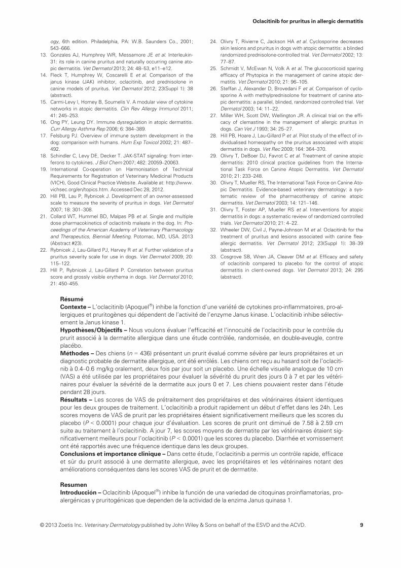
day 0) were collected on enrolment at day 0. An enhanced visual
analog scale (VAS) score was used by both dog owners and veteri-
narians. The VAS scale consisted of a 10 cm line with word descrip-
tors at 2 cm intervals. Owners were asked to assess the severity of
the ‘itch’, and veterinarians were asked to assess the severity of ‘der-
matitis’ (Figure 1a,b). The enhanced Owner Pruritus VAS had six de-
scriptors of pruritus evenly spaced at 2 cm intervals with ‘normal
dog’ at 0 cm and ‘extremely severe itching’ at 10 cm. The enhanced
Veterinarian Dermatitis VAS had six descriptors of dermatitis evenly
spaced at 2 cm intervals with ‘normal dog’ at 0 cm and ‘extremely
severe dermatitis’ at 10 cm.
Owners and veterinarians were instructed to place a mark on the
VAS line at the location that best represented the dog’s pruritus or
dermatitis, respectively. At study completion, the distance (in centi-
metres) from the bottom of the line (‘normal dog’) to the owner’s or
veterinarian’s mark on the line was measured and recorded. Owners
performed a VAS assessment on days 0, 1, 2, 3, 4, 5, 6 and 7. Veteri-
narians performed a VAS assessment on days 0 and 7 and (when
applicable) at all visits occurring during the continuation phase.
Assessments were required to be performed by the same owner or
veterinarian at all time points.
Blood samples (complete blood count and serum chemistry) were
collected on day 0 (prior to dosing), on day 7 and (when applicable) at
the conclusion of the continuation phase. Samples for urinalysis were
collected on day 0. All samples for haematology (complete blood
count), serum chemistry and urinalysis were sent to a central labora-
tory (Heska Corporation, Loveland, CO, USA).
Efficacy outcomemeasuresTo be included in the efficacy analyses, dogs had to have been on
study until day 5 and had to have received a minimum of 80% of their
intended doses (as recorded in a daily log) on days 0–7. The same
individuals (owner and veterinarian) had to perform all assessments
for the enrolled dogs. For the analysis of the owner (not veterinarian)
assessments, there was an additional requirement that dogs had
been properly dosed (two doses in the previous 24 h) and that there
were at least five of eight evaluable Owner Pruritus VAS scores
between days 0 and 7. Data were analysed using SAS version 9.2
(SAS Institute, Cary, NC, USA). The level of significance was set at
P < 0.05.
Owner Pruritus VAS scores were analysed with a linear mixed
model for repeated measures. The model included the fixed
effects of baseline, treatment and baseline by treatment interac-
tion, time and the interaction of treatment by time. Random
effects included clinic, clinic by treatment interaction, between-
animal error, clinic by treatment by time interaction and the resid-
ual variation. Baselines were centred for inclusion in the model by
subtracting the mean baseline value from an individual animal’s
value at day 0. Given that the treatment by time interaction was
significant (P < 0.05), treatment was compared at each time
point.
The effectiveness variables assessed were as follows: (i) Owner
Pruritus VAS scores at each assessment day; (ii) Veterinarian Derma-
titis VAS scores on day 7; (iii) dogs achieving a 2 cm reduction com-
pared with baseline in Owner Pruritus VAS scores at each
assessment day; (iv) dogs achieving a ≥50% reduction compared
with baseline in Owner Pruritus VAS scores on day 7; and (v) the pro-
portion of dogs that were treatment successes based on the Owner
Pruritus VAS assessment on days 1–7. Treatment success was
defined as achieving at least a 2 cm reduction from baseline score on
day X on the Owner Pruritus VAS assessment (day 0 score minus
day X score ≥ 2 cm) on at least five of the seven study days
assessed. If the criteria for treatment success were not met, the
Extremely severe itching. Dog is scratching,chewing, licking almost continuously. Itchingpractically never stops, regardless of what elseis happening around the dog.
Severe itching. Prolonged episodes of itchingwhen the dog is awake. Itching occurs at night and also when eating, playing, exercising, orwhen otherwise distracted.
Moderate itching. Regular episodes of itchingwhen the dog is awake. Itching might occur at night and wake the dog. No itching when eating,playing, exercising or when being distracted.
Mild itching. More frequent episodes of itching.May notice occasional episodes of itching atnight. No itching when sleeping, eating, playing,exercising or when being distracted.
Very mild itching. Occasional episodes ofitching. The dog is slightly more itchy than before the problem began.
Normal Dog. Itching is not a problem.
Extremely severe dermatitis. Extensiveevidence of chronic lesion and/or activeinfections/excoriations.
Severe dermatitis.
Moderately severe dermatitis.
Moderate dermatitis.
Mild dermatitis.
Normal Dog. Dermatitis is not a problem.
(a) (b)
Figure 1. (a) Enhanced Owner Pruritus visual analog scale (VAS). (b) Enhanced Veterinarian Dermatitis VAS.
© 2013 Zoetis Inc. Veterinary Dermatology published by John Wiley & Sons on behalf of the ESVD and the ACVD. 3
Oclacitinib for pruritus in allergic dermatitis

case was a treatment failure; this included dogs withdrawn prior to
day 7 for an adverse event or for worsening skin condition.
The Veterinarian VAS scores were analysed with a linear mixed
model including the fixed effects of (centred) baseline, treatment and
(centred) baseline by treatment interaction, and the random effects
of clinic, clinic by treatment interaction and residual variation.
A generalized linear mixed model for repeated measures using a
logit link function and binomial error distribution was used to analyse
a 2 cm and a 50% reduction from baseline in Owner Pruritus VAS
scores. The fixed effects were treatment, time and treatment by
time interaction, while the random effects were clinic, clinic by treat-
ment interaction, animal nested within clinic and treatment, and clinic
by treatment by time interaction. Treatments were compared at each
time.
Treatment success was analysed using a generalized linear mixed
model with a logit link and binomial error. The model included the
fixed effect of treatment and the random effects of clinic and treat-
ment by clinic interaction. The proportion of success with 95% confi-
dence interval for each treatment and the odds ratio with 95%
confidence interval comparing the treatments was reported.
To evaluate the effect of flea or sarcoptic mange treatment on the
effectiveness of oclacitinib for pruritus, the primary variable, treat-
ment success at day 7, was stratified by flea control/sarcoptic mange
control/none and the proportion of treatment success calculated for
each strata.
Safety outcome measuresAll enrolled dogs that were administered at least one dose of test arti-
cle were included in the safety analysis. For each continuous haema-
tology and serum chemistry measure, summary statistics (mean,
median, SDs, minimum and maximum) were calculated by treatment
and time point. Frequencies of dogs reported to experience at least
one abnormal health event were displayed by clinical sign for all
unique terms. Frequency tables summarizing the number of dogs
receiving each medication over the course of the study were pre-
pared.
Results
Demographics
A total of 436 dogs were enrolled (Table 1). Approxi-
mately 69% of the dogs were purebred. Retrievers and
terriers were the most common dog breed groups, com-
prising 15.8% (Labrador retrievers 10.3% and golden
retrievers 5.5%) and 13.9% of the study population,
respectively.
Presumptive diagnoses
The presumptive diagnoses for dogs enrolled in the study
are shown in Table 2. It was not always possible to stipu-
late a single presumptive diagnosis, and enrolled dogs
could have had more than one presumptive cause for the
reason for their pruritus associated with allergic dermati-
tis. The presumptive diagnoses were similar in each of
the two treatment groups. Over 80% of the dogs in each
group had a presumptive diagnosis of atopic dermatitis,
but only 41.5% had atopic dermatitis alone. Slightly more
than 30% of the dogs were presumed to have flea allergy
dermatitis, slightly more than 20% had food allergy der-
matitis, and approximately 10% had contact dermatitis.
Approximately 5% of the dogs had a presumptive diagno-
sis of sarcoptic mange, although mange mites were not
always observed. A variety of other reasons were noted
for the remaining 5% of the dogs enrolled. All of the
cases with presumptive diagnoses of ‘other’ also had ato-
pic dermatitis, except for one oclacitinib group dog with
the ‘other’ diagnosis of ‘unspecified allergic dermatitis’.
Assessment of effectiveness
The effectiveness data set for the Owner Pruritus VAS
comprised 407 (204 placebo- and 203 oclacitinib-treated)
dogs. The effectiveness data set for the Veterinarian Der-
matitis VAS comprised 413 (207 placebo- and 206 oclaciti-
nib-treated) dogs. Twenty-nine dogs (13 oclacitinib
treated and 16 placebo treated) were excluded from the
Owner Pruritus VAS analyses, and 23 dogs (10 oclacitinib
treated and 13 placebo treated) were excluded from the
Veterinarian Dermatitis VAS analyses for errors in compli-
ance with the trial and data collection protocols.
Owner Pruritus VAS scores by day of study
The mean day 0 Owner Pruritus VAS scores were very
similar between the treatment groups [7.39 and 7.58 cm
for the oclacitinib-treated dogs (range 4.7–10.0) and pla-
cebo-treated dogs (range 3.0–9.9), respectively; Figure 2]
corresponding to ‘severe itching’ on the enhanced Owner
Pruritus VAS score. After 1 day of treatment, a 2.20 cm
reduction of the least squares mean (mean) from the
average baseline including both treatment groups in
Owner Pruritus VAS scores was observed for dogs
receiving oclacitinib, while the dogs receiving placebo
treatment had a 0.95 cm reduction (Figure 2). For oclaciti-
nib-treated dogs, mean Owner Pruritus VAS scores con-
tinued to decrease over the remaining 6 days of the
study. Owner Pruritus VAS scores in the oclacitinib-trea-
ted dogs were significantly lower than those in the pla-
cebo-treated dogs (P < 0.0001) on each day of
assessment. At day 7, the Owner Pruritus VAS score had
Table 1. Baseline characteristics of enrolled dogs
Variable Placebo group Oclacitinib group
Breed distribution [n (%)]
Mixed breed 153 (69.5) 148 (68.5)
Purebred 67 (30.5) 68 (31.5)
Sex distribution [n (%)]
Female 114 (51.8) 105 (48.6)
Male 106 (48.2) 111 (51.4)
Mean age at study
onset [years (range)]
5.8 (1.0–16) 6.0 (0.5–18)
Mean weight at study
onset [kg (range)]
20.0 (3.0–61.7) 20.6 (3.0–56.0)
Owner Pruritus VAS
score at study onset
(arithmetic mean; cm)
7.58 7.39
Veterinarian Dermatitis
VAS score at study
onset (arithmetic
mean; cm)
6.18 6.20
Abbreviation: VAS, visual analog scale.
Table 2. Presumptive diagnoses at enrolment
Presumptive diagnosis
Oclacitinib group
[n (%)]
Placebo group
[n (%)]
Atopic dermatitis 175 (81.0) 179 (81.4)
Flea allergy dermatitis 72 (33.3) 70 (31.8)
Food allergy dermatitis 48 (22.2) 51 (23.2)
Contact dermatitis 24 (11.1) 23 (10.5)
Sarcoptic mange 2 (0.9) 8 (3.6)
Other 12 (5.6) 10 (4.5)
© 2013 Zoetis Inc. Veterinary Dermatology published by John Wiley & Sons on behalf of the ESVD and the ACVD.4
Cosgrove et al.

decreased for oclacitinib-treated dogs to 2.59 cm (a
4.89 cm reduction in VAS pruritus scores, which corre-
sponds to an approximate reduction from ‘severe itching’
to ‘very mild itching’) and for placebo-treated dogs to
5.54 cm (a 1.94 cm reduction in VAS pruritus scores,
which corresponds to an approximate reduction from
‘severe itching’ to ‘moderate itching’). The reduction in
the Owner Pruritus VAS scores (2.20 cm) for oclacitinib-
treated dogs after 1 day of treatment exceeded the
reduction in pruritus scores for placebo-treated dogs after
7 full days of therapy (1.94 cm).
Veterinarian Dermatitis VAS scores by day of study
The mean day 0 Veterinarian Dermatitis VAS scores were
similar for the two treatment groups [6.20 and 6.18 cm
for the oclacitinib-treated dogs (range 4.0–10.0) and pla-
cebo-treated dogs (range 0.0–9.9), respectively]. At
day 7, the mean Veterinarian Dermatitis VAS score for
the oclacitinib-treated dogs had decreased to 2.22 cm (a
3.98 cm reduction in VAS dermatitis scores, which corre-
sponds to an approximate reduction from ‘moderately
severe dermatitis’ to ‘mild dermatitis’) and for the pla-
cebo-treated dogs to 4.89 cm (a 1.29 cm reduction in
VAS dermatitis scores, which corresponds to an approxi-
mate reduction from ‘moderately severe dermatitis’ to
‘moderate dermatitis’; Figure 3). The Veterinarian
Dermatitis VAS scores in the oclacitinib-treated dogs
were significantly lower than those in the placebo-treated
dogs (P < 0.0001).
Veterinarian Dermatitis VAS scores were also assessed
at the end of the continuation phase (days 8–28) of thestudy; ~2.5 times more oclacitinib-treated dogs (n = 179)
than placebo-treated dogs (n = 73) were treated during
that phase. Owing to this imbalance, Veterinarian Derma-
titis VAS scores were not compared during the continua-
tion phase.
Dogs achieving a 2 cm Owner Pruritus VAS score
reduction each day of the study
On day 1, a 2 cm reduction in Owner Pruritus VAS was
observed in 44% of the oclacitinib-treated dogs compared
with 19% of the placebo-treated dogs. By day 7, 86.4%
of the oclacitinib-treated dogs compared with 42.5% of
the placebo-treated dogs achieved a 2 cm reduction in
Owner Pruritus VAS scores. The numbers and percent-
ages of dogs achieving a 2 cm reduction in the Owner
Pruritus VAS score for each day of the study are shown in
Figure 4.
Dogs achieving a ≥50% reduction from baseline in
Owner Pruritus VAS and Veterinarian Dermatitis VAS
scores on day 7
On day 7, 70.5% of the oclacitinib-treated dogs com-
pared with 23.2% of the placebo-treated dogs achieved a
≥50% reduction in Owner Pruritus VAS scores
(P < 0.0001). The numbers and percentages of dogs
achieving a ≥50% reduction in the Owner Pruritus VAS
score for each day of the study are shown in Figure 5.
Treatment success
Sixty-seven per cent of oclacitinib-treated dogs and 29%
of placebo-treated dogs were considered a treatment
success; the difference was significant (P < 0.0001). The
study also evaluated the effect of flea treatment on treat-
ment success. Flea treatment was initiated on day 0 for
19% (n = 41) and 13% (n = 29) of the dogs in the oclaciti-
nib and placebo treatment groups, respectively. Within
0
2
4
6
8
10
0 1 2 3 4 5 6 7
Mea
n VA
S sc
ore
(cm
)
Day of study
Placebo (n = 203) Oclaci nib (n = 204)
*
**
*
*
*
*
Extremely severe
Severe
Moderate
Mild
Very mild
Normal
Pruritus severity descriptuons (owner VA
S)
Figure 2. Owner Pruritus VAS score by day of study (day 0, arithme-
tic mean; days 1–7, least squares mean � 95% confidence interval).
6.18
4.89
6.20
2.22
0
1
2
3
4
5
6
7
8
9
10
Day 0 Day 7
Mea
n VA
S sc
ore
(cm
)
Placebo (n = 207) (n = 206)
*
Extremely severe
Severe
Moderate
Mild
Very mild
Normal
* = Oclaci nib is significantly different from Placebo (P < 0.0001)
Derm
as severity descriptors (veterinarian VA
S)
Figure 3. Veterinarian Dermatitis VAS score by day of study (day 0,
arithmetic mean; day 7, least squares mean � 95% confidence
interval).
0
20
40
60
80
100
0 1 2 3 4 5 6 7
Perc
ent
of d
ogs
Day of study
Placebo (P) Oclaci nib (O)
*
*
**
*
* *
Day 0 Day 1 Day 2 Day 3 Day 4 Day 5 Day 6 Day 7
Placebo
Oclaci nib
0
0
19
44*
32.5
61.3*
32.50
69.4*
39.7
80.1*
42.7
83*
42.3
84.2* 86.4*
42.5
Figure 4. Dogs achieving a 2 cm Owner Pruritus VAS score reduc-
tion each day of the study.
© 2013 Zoetis Inc. Veterinary Dermatology published by John Wiley & Sons on behalf of the ESVD and the ACVD. 5
Oclacitinib for pruritus in allergic dermatitis

the oclacitinib group, the percentage of dogs that were
treatment successes was similar regardless of whether
they received or did not receive flea treatment on day 0
(63.2% success with flea treatment and 67.7% success
without flea treatment). Within the placebo group, flea
treatment initiated on day 0 doubled the percentage of
dogs that were treatment successes, i.e. success rate
increased from 25.9 to 51.9%. The small number (n = 4)
of dogs receiving sarcoptic mange control treatment on
day 0 made it impossible to assess the effect of treat-
ment.
Safety assessment
All 436 of the enrolled dogs (i.e. 220 placebo- and 216
oclacitinib-treated dogs) were included in the safety
assessment, regardless of the number of doses adminis-
tered during the study; these ranged from 4 to 63 doses.
Safety data were also collected from 179 dogs that were
treated with oclacitinib for up to 32 days after the study.
Abnormal health events
There were no fatalities and no abnormal health events
that necessitated hospitalization in either the study phase
[day 0–7 (+3 days)] or the continuation phase [day 8–28(�2 days)] of the study. Given that the majority of dogs in
the placebo group withdrew after the completion of the
study phase, the incidence of abnormal clinical signs was
similar in both groups (Table 3). In most cases, the clinical
signs resolved spontaneously and did not require cessa-
tion of treatment. Treatment was stopped in one oclaciti-
nib-treated dog after 7 days because of darkening areas
of skin and fur.
The continuation phase (days 8–30) of the study was
three times longer than the study phase of the study and
contained approximately 2.5 times more oclacitinib male-
ate (179) than placebo group dogs (73). Six dogs (four
oclacitinib and two placebo group) were withdrawn from
the study during the continuation phase for abnormal
health events. Abnormal health events were reported in
11 of 179 oclacitinib-treated dogs post-study. These were
as follows: diarrhoea (four dogs; severe enough to war-
rant cessation of treatment in one dog); vomiting (four
dogs); fever, lethargy and cystitis (one dog); an inflamed
footpad and vomiting (one dog); and diarrhoea, vomiting
and lethargy (one dog).
Clinical pathology
Minor changes were seen in clinical pathology parame-
ters, but these remained within normal laboratory refer-
ence ranges. Mean lymphocyte counts for dogs in the
oclacitinib group were increased at day 7, but these
returned to pretreatment levels within 28 days without a
break in oclacitinib administration. These dogs also had a
slight decrease in mean white blood cell counts (neutro-
phil, eosinophil and monocyte counts), but these
remained within the normal reference ranges. Serum
cholesterol increased in 25% of oclacitinib-treated dogs,
but levels remained within the reference range. The inci-
dence of elevated liver enzyme activity for alkaline phos-
phatase, alanine aminotransferase and aspartate
aminotransferase was similar in dogs in the oclacitinib
and placebo groups.
Concomitant medications
A wide variety of concomitant medications were used in
conjunction with either placebo or oclacitinib treatment.
The concomitant medications administered most often
(in ≥2% of the oclacitinib-treated dogs) are summarized
by drug class and treatment group in Table 4. A variety of
*
*
*
*
*
**
0 8 8.5 13.0 17.1 19.6 24.9 23.2
Placebo (P) Oclaci nib (O)
0
20
40
60
80
100
0 1 2 3 4 5 6 7
Perc
ent
of d
ogs
Day of study
Day 0 Day 1 Day 2 Day 3 Day 4 Day 5 Day 6 Day 7
PlaceboOclaci nib 0 23* 37.7* 48.0* 57.7* 63.9* 69.9* 70.5*
Figure 5. Dogs achieving a ≥50% Owner Pruritus VAS score reduc-
tion each day of the study.
Table 3. Abnormal clinical signs during study phase (days 0–7)*
Abnormal
clinical sign
Oclacitinib-treated
dogs
[n = 216; n (%)]†
Placebo-treated
dogs
[n = 220; n (%)]
Diarrhoea 5 (2.3) 2 (0.9)
Vomiting 5 (2.3) 4 (1.8)
Lethargy 4 (1.8) 3 (1.4)
Anorexia 3 (1.4) 0 (0.0)
Polydipsia 3 (1.4) 0 (0.0)
*Seen in >1% of dogs.
†Number and percentage are given on a per animal basis.
Table 4. Concomitant medications*†
Drug class
Oclacitinib group
[n = 216; n (%)]‡Placebo group
[n = 220; n (%)]‡
Endectocides 145 (67.1) 150 (68.2)
Ectoparaciticides,
insecticides and
repellents
105 (48.6) 101 (45.9)
Canine vaccines 26 (12.0) 25 (11.4)
Glucosamine (with
and without
chondroitin) and
nonsteroidal anti-
inflammatory products
6 (2.8) 17 (7.7)
Systemic antibacterials§ 17 (7.9) 11 (5.0)
Omega-3 fatty
acid preparations
7 (3.2) 7 (3.2)
Otologicals 13 (6.0) 6 (2.7)
Ophthalmologicals 2 (0.9) 5 (2.3)
*Administered to ≥2% of the oclacitinib-treated dogs.
†Administered in decreasing order of frequency in the oclacitinib
treatment group.
‡Number and percentage are given on a per animal basis.
§Administered during the continuation phase.
© 2013 Zoetis Inc. Veterinary Dermatology published by John Wiley & Sons on behalf of the ESVD and the ACVD.6
Cosgrove et al.

other products were used less frequently (in ≤2% of the
oclacitinib-treated dogs) but were administered to a simi-
lar number and percentage of dogs in both treatment
groups, including thyroid medications, antibacterial prod-
ucts, systemic and topical antifungal products, while skin
emollients and skin protectives, as well as vitamins were
given to slightly more dogs in the oclacitinib-treated group
than in the placebo-treated group.
Discussion
This study provides evidence of the effectiveness of ocla-
citinib in the control of pruritus associated with allergic
dermatitis in dogs. There was a highly significant
improvement (P ≤ 0.0001) for all of the efficacy variables
in oclacitinib-treated dogs compared with placebo-treated
dogs. Following 7 days of oclacitinib treatment, there
was a 65% reduction in pruritus scores (from ‘severe itch-
ing’ to ‘very mild itching’) and a 64% reduction in clinical
severity scores (from ‘moderately severe dermatitis’ to
‘mild dermatitis’). Within the first 24 h of treatment, pruri-
tus scores were reduced by at least 2 cm in 44% of ocla-
citinib-treated dogs compared with 19% of the placebo-
treated dogs. By day 7, 86.4% of the oclacitinib-treated
dogs compared with <42.5% of the placebo-treated dogs
achieved a 2 cm reduction in Owner Pruritus VAS scores.
Additionally, by day 7, 70.5% of oclacitinib-treated dogs
showed a >50% reduction in Owner Pruritus VAS scores
compared with <23.2% of the placebo-treated dogs.
Based on the binary treatment success analysis, the
majority of oclacitinib-treated dogs (66.5%) were a treat-
ment success compared with only 29.4% of the placebo-
treated dogs, with owners and veterinarians noting sub-
stantial improvement in pruritus and dermatitis VAS
scores. Oclacitinib therefore appears to improve pruritus
and dermatitis substantially, affording the damaged skin
an opportunity to heal, while allowing the veterinarian
time correctly to diagnose and treat the underlying cause.
The rapid and effective reduction in pruritus could also
greatly improve the quality of life for the affected dogs
and their owners.
Immediate downregulation of the action of pruritogenic
cytokines, including IL-31, may in part explain the rapid
reduction in pruritus following oclacitinib treatment.13 The
placebo-treated group also had an immediate but lesser
reduction in pruritus score on the first treatment day,
which may be attributed in part to a placebo effect but
could also be explained by the flea control administered
at the start of the study in many of the dogs. The improve-
ment in the clinical severity scores was probably a conse-
quence of controlling the dogs’ pruritus, but may also
have reflected a direct anti-inflammatory action in the
skin. These findings are consistent with the pharmacolog-
ical properties of oclacitinib, which is a targeted therapy
that inhibits key pathways involved in the pathophysiol-
ogy of skin inflammation.14
Our study used enhanced VAS scales for the assess-
ment of both pruritus and dermatitis. Enhanced VAS
scales with severity descriptors at equally spaced inter-
vals along the line have been shown to be an easy and
repeatable method for users to assess the severity of pru-
ritus.20,22 Unique to this study was the use of a Veterinar-
ian Dermatitis VAS to assess changes in the severity of
the dog’s dermatitis at each clinic visit. In previous stud-
ies, veterinarians have used a VAS scale to assess pruri-
tus.23 However, dogs may not reliably demonstrate
pruritic behaviour in the veterinary clinic and therefore the
veterinarian’s pruritus VAS score may have to rely heavily
on what the owner describes rather than what is
observed. By comparison, the dermatitis VAS allowed the
veterinarian to assess changes in the dog’s skin lesions.
More objective and validated assessment tools, such as
Canine Atopic Dermatitis and Severity Index (CADESI),
are available, but these are limited to specific dermato-
ses, such as atopic dermatitis, and would have been
unsuitable to assess the severity of the variety of skin
conditions seen in this study. The enhanced dermatitis
VAS was simple and could be used successfully and reli-
ably by general practitioners without special training in
dermatology.
For the treatment success analysis, the continuous var-
iable of Owner Pruritus VAS scores collected repeatedly
over 7 days was converted to a single binary score for
each case, either treatment success or treatment failure.
To be classified as a treatment success, the following
two criteria had to be met: first, the pruritus score had to
improve by a full category (2 cm or more reduction from
baseline) on the enhanced Owner Pruritus VAS; and sec-
ond, the reduction in pruritus (≥2 cm) had to be achieved
on ≥5 of the first 7 days of Owner Pruritus VAS assess-
ments. Analysis of treatment success established that
the pruritus score improvement was not only statistically
different from the placebo group but was of a repeated
magnitude (≥2 cm) anticipated to be clinically relevant to
both the owner and veterinarian without extrapolating
data for cases withdrawn early or interpolating data for a
missing case at day 7. The downside to this analysis is
that cases with efficacy satisfactory to the owner (e.g.
1.9 cm pruritus reduction or ≥2 cm on four of
seven days) were counted as treatment failures. Not sur-
prisingly, the proportion of dogs that were a treatment
success (66.5%) was lower than the percentage of dogs
that achieved a 2 cm pruritus VAS score reduction from
baseline (85%) after 7 days of oclacitinib treatment.
Treatment success analysis adds methodological rigour
but may underestimate the oclacitinib effectiveness at
reducing pruritus observed by the owner and/or veterinar-
ian.
It is difficult to compare the efficacy of oclacitinib
directly with that of other antipruritic and anti-inflamma-
tory treatments. Previously reported clinical trials have
been conducted in different target populations, predomi-
nantly in dogs with atopic dermatitis, without the inclu-
sion of cases with other causes of allergic dermatitis, and
have used different measures to assess efficacy (pre-
dominantly CADESI).10,24–29 However, the proportion of
dogs that achieved a >50% reduction in pruritus in this
study is comparable to or better than the proportion of
atopic dogs that improved to this extent following treat-
ment with topical hydrocortisone aceponate, topical tri-
amcinolone, systemic glucocorticoids and systemic
ciclosporin.4,30,31
To the authors’ knowledge, there are no published
studies reporting the efficacy of glucocorticoids in dogs
© 2013 Zoetis Inc. Veterinary Dermatology published by John Wiley & Sons on behalf of the ESVD and the ACVD. 7
Oclacitinib for pruritus in allergic dermatitis

suffering from allergic dermatitis (i.e. not only atopic der-
matitis) or nonspecific pruritus despite the fact that this
class of drug is the most frequently used for the short-
term control of pruritus in dogs. The popularity of gluco-
corticoids seems to be based on the fast speed of onset
and reliable results in any of a number of conditions. This
study shows that oclacitinib shares these advantages in
dogs with pruritus associated with a number of underly-
ing causes of allergic dermatitis, including atopic dermati-
tis, flea allergy, food allergy, contact dermatitis and
sarcoptic mange. In particular, oclacitinib demonstrated
significantly better efficacy than placebo at 24 h, indicat-
ing a rapid speed of onset. The rapid onset of response to
oclacitinib administration has also been reported in a
model of IL-31-induced pruritus and a model of flea
allergy dermatitis.14,32 In both studies, oclacitinib adminis-
tered orally as a single dose at 0.4 mg/kg resulted in a sig-
nificant (P < 0.05) reduction in pruritus within 1 h after
administration compared with prednisolone administered
at doses of 0.25 and 0.5 mg/kg.
Oclacitinib was well tolerated in these dogs. The fre-
quency and type of abnormal health events were similar
between the oclacitinib- and placebo-treated dogs. The
most common adverse events were gastrointestinal
upsets, such as decreased appetite, vomiting and diar-
rhoea. These were mostly mild and only rarely required
cessation of treatment. The acute effects commonly
observed with systemic glucocorticoids (e.g. polyuria,
polydipsia, panting, polyphagia and changes in serum bio-
chemistry) were seen in <2% of the oclacitinib-treated
dogs. The mean values of all of the clinical pathology
parameters analysed fell within the normal reference
range for both treatment groups. The favourable safety
results reported here are supported by the results of a
field trial, in which oclacitinib was administered for
4 months to dogs with atopic dermatitis.33
This study was carried out to good clinical practice stan-
dards.19 Selection bias in breed, age, sex, weight and clin-
ical severity was not apparent. Randomized treatment
allocation was made according to a predetermined alloca-
tion code. Detection bias by the owners and investigators
was unlikely because they were blinded to treatment allo-
cation. Performance bias was possible, because antipara-
site treatment and dietary management changes for
some dogs on day 0 could have improved their clinical
signs. However, the impact of this on the comparison
between oclacitinib and placebo is likely to have been low
because the type and frequency of concomitant treat-
ments was comparable between the treatment groups.
Attrition bias was present; the analysis excluded dogs
that were considered to have had a protocol deviation
that affected the collection or integrity of their efficacy
data. It is possible that this biased towards a favourable
response to treatment, although the numbers never
exceeded 8% of the treated dogs and were comparable
between the treatment groups and analyses. Inclusion
and exclusion criteria established before the trial were
used to establish a working diagnosis of pruritus associ-
ated with allergic dermatitis. Rigorous criteria to establish
a firm diagnosis were not employed, because the aim and
design of this study were to assess the efficacy of oclacit-
inib in short-term management of allergic dermatitis. The
authors have also reported a study of longer term
(112 days) efficacy in the control of a more specific condi-
tion, atopic dermatitis.33
In the conditions of this study, oclacitinib, a selective
Janus kinase inhibitor, administered orally at a dose of
0.4–0.6 mg/kg twice daily, was safe and efficacious in
controlling the pruritus associated with allergic dermatitis.
Oclacitinib provided itch relief within 24 h that persisted
through the treatment period, with >70% of the treated
dogs achieving a >50% reduction in pruritus by day 7.
Acknowledgements
We would like to thank the following veterinarians who
enrolled dogs in this study and performed the clinical
investigations: Brett Berryhill, Glen Burkett, Jay Butan,
Randall R. Carpenter, Terry Clekis, Jeffrey N. Dizik, Sam
Geller, Mary Grabow, Robert Jackson, Mark Lelli, Marc
Leven, David Lukof, Patrick McSweeney, Kathleen Neu-
hoff, Karan Oberhansley, Gregory Paplawsky, Andrew
Pickering, Jeffrey Pinkston, Dean Rund, Roger Sifferman,
Jason St Romain, Kathy Tater, Bradford J. Theodoroff,
Philip VanVranken, Philip Waguespack and Melissa Wiest.
We would also like to thank the following Zoetis Inc. col-
leagues for their invaluable assistance: Candace A. Sousa,
Marcia J. Adams, Anne E. Daniels and Nancy L. Savicke.
References
1. VPI� Pet Insurance website. Top 10 pet medical conditions of
2010. Available at: http://press.petinsurance.com/pressroom/
02222011Pet_Conditions_2010.aspx. Accessed Dec 28, 2012.
2. Hill PB, Lo A, Eden CAN et al. Survey of the prevalence, diagno-
sis and treatment of dermatological conditions in small animals
in general practice. Vet Rec 2006; 158: 533–539.
3. Nuttall T, McKeever PJ, Harvey RG. Pruritic dermatitis. In: Bey-
non P, ed. A Colour Handbook of Skin Diseases of the Dog and
Cat. 2nd edn. London: Manson Publishing, 2009; 35–37.
4. Olivry T, Bisikova P. A systematic review of randomized con-
trolled trials for prevention or treatment of atopic dermatitis in
dogs: 2008–2011 update. Vet Dermatol 2013; 24: 97–117, e25–
e26.
5. Bloom P. Nonsteroidal, nonimmunosuppressive therapies for
pruritus. Vet Clin North Am: Small Anim Pract 2013; 43: 173–
187.
6. Scott DW, Miller WH, Griffin CE. Dermatologic therapy. In:
Muller and Kirk’s Small Animal Dermatology, 6th edition. Phila-
delphia: W.B. Saunders Co., 2001; 207–274.
7. Steinhoff M, Bienenstock J, Schmelz M et al. Neurophysiologi-
cal, neuroimmunological, and neuroendocrine basis of pruritus. J
Invest Dermatol 2006; 126: 1705–1718.
8. Metz M, St€ander S. Chronic pruritus – pathogenesis, clinical
aspects and treatment. J Eur Acad Dermatol Venereol 2010; 24:
1249–1260.
9. Plumb DC. Glucocorticoid agents, general information. Veteri-
nary drug handbook. 4th edn. Ames, IA: Blackwell Publishing;
2002; 387–389.
10. Nuttall T, Mueller R, Bensignor E et al. Efficacy of a 0.0584%
hydrocortisone aceponate spray in the management of canine
atopic dermatitis: a randomised, double blind, placebo-controlled
trial. Vet Dermatol 2009; 20: 191–198.
11. Olivry T, Mueller RS. Evidence-based veterinary dermatology: a
systematic review of the pharmacotherapy of canine atopic der-
matitis. Vet Dermatol 2003; 14: 121–146.
12. Scott DW, Miller WH, Griffin CE. Skin immune system and aller-
gic skin diseases. In: Muller and Kirk’s Small Animal Dermatol-
© 2013 Zoetis Inc. Veterinary Dermatology published by John Wiley & Sons on behalf of the ESVD and the ACVD.8
Cosgrove et al.
ogy, 6th edition. Philadelphia, PA: W.B. Saunders Co., 2001;
543–666.
13. Gonzales AJ, Humphrey WR, Messamore JE et al. Interleukin-
31: its role in canine pruritus and naturally occurring canine ato-
pic dermatitis. Vet Dermatol 2013; 24: 48–53, e11–e12.
14. Fleck T, Humphrey W, Coscarelli E et al. Comparison of the
janus kinase (JAK) inhibitor, oclacitinib, and prednisolone in
canine models of pruritus. Vet Dermatol 2012; 23(Suppl 1): 38
(abstract).
15. Carmi-Levy I, Homey B, Soumelis V. A modular view of cytokine
networks in atopic dermatitis. Clin Rev Allergy Immunol 2011;
41: 245–253.
16. Ong PY, Leung DY. Immune dysregulation in atopic dermatitis.
Curr Allergy Asthma Rep 2006; 6: 384–389.
17. Felsburg PJ. Overview of immune system development in the
dog: comparison with humans. Hum Exp Toxicol 2002; 21: 487–
492.
18. Schindler C, Levy DE, Decker T. JAK-STAT signaling: from inter-
ferons to cytokines. J Biol Chem 2007; 482: 20059–20063.
19. International Co-operation on Harmonisation of Technical
Requirements for Registration of Veterinary Medicinal Products
(VICH), Good Clinical Practice Website. Available at: http://www.
vichsec.org/en/topics.htm. Accessed Dec 28, 2012.
20. Hill PB, Lau P, Rybnicek J. Development of an owner-assessed
scale to measure the severity of pruritus in dogs. Vet Dermatol
2007; 18: 301–308.
21. Collard WT, Hummel BD, Malpas PB et al. Single and multiple
dose pharmacokinetics of oclacitinib maleate in the dog. In: Pro-
ceedings of the American Academy of Veterinary Pharmacology
and Therapeutics, Biennial Meeting. Potomac, MD, USA. 2013
(Abstract #23).
22. Rybnicek J, Lau-Gillard PJ, Harvey R et al. Further validation of a
pruritus severity scale for use in dogs. Vet Dermatol 2009; 20:
115–122.
23. Hill P, Rybnicek J, Lau-Gillard P. Correlation between pruritus
score and grossly visible erythema in dogs. Vet Dermatol 2010;
21: 450–455.
24. Olivry T, Rivierre C, Jackson HA et al. Cyclosporine decreases
skin lesions and pruritus in dogs with atopic dermatitis: a blinded
randomized prednisolone-controlled trial. Vet Dermatol 2002; 13:
77–87.
25. Schmidt V, McEwan N, Volk A et al. The glucocorticoid sparing
efficacy of Phytopica in the management of canine atopic der-
matitis. Vet Dermatol 2010; 21: 96–105.
26. Steffan J, Alexander D, Brovedani F et al. Comparison of cyclo-
sporine A with methylprednisolone for treatment of canine ato-
pic dermatitis: a parallel, blinded, randomized controlled trial. Vet
Dermatol 2003; 14: 11–22.
27. Miller WH, Scott DW, Wellington JR. A clinical trial on the effi-
cacy of clemastine in the management of allergic pruritus in
dogs. Can Vet J 1993; 34: 25–27.
28. Hill PB, Hoare J, Lau-Gillard P et al. Pilot study of the effect of in-
dividualised homeopathy on the pruritus associated with atopic
dermatitis in dogs. Vet Rec 2009; 164: 364–370.
29. Olivry T, DeBoer DJ, Favrot C et al. Treatment of canine atopic
dermatitis: 2010 clinical practice guidelines from the Interna-
tional Task Force on Canine Atopic Dermatitis. Vet Dermatol
2010; 21: 233–248.
30. Olivry T, Mueller RS, The International Task Force on Canine Ato-
pic Dermatitis. Evidence-based veterinary dermatology: a sys-
tematic review of the pharmacotherapy of canine atopic
dermatitis. Vet Dermatol 2003; 14: 121–146.
31. Olivry T, Foster AP, Mueller RS et al. Interventions for atopic
dermatitis in dogs: a systematic review of randomized controlled
trials. Vet Dermatol 2010; 21: 4–22.
32. Wheeler DW, Civil J, Payne-Johnson M et al. Oclacitinib for the
treatment of pruritus and lesions associated with canine flea-
allergic dermatitis. Vet Dermatol 2012; 23(Suppl 1): 38–39
(abstract).
33. Cosgrove SB, Wren JA, Cleaver DM et al. Efficacy and safety
of oclacitinib compared to placebo for the control of atopic
dermatitis in client-owned dogs. Vet Dermatol 2013; 24: 295
(abstract).
R�esum�e
Contexte – L’oclacitinib (Apoquel�) inhibe la fonction d’une vari�et�e de cytokines pro-inflammatoires, pro-al-
lergiques et pruritog�enes qui d�ependent de l’activit�e de l’enzyme Janus kinase. L’oclacitinib inhibe s�electiv-
ement la Janus kinase 1.
Hypoth�eses/Objectifs – Nous voulons �evaluer l’efficacit�e et l’innocuit�e de l’oclacitinib pour le controle du
prurit associ�e �a la dermatite allergique dans une �etude control�ee, randomis�ee, en double-aveugle, contre
plac�ebo.
M�ethodes – Des chiens (n = 436) pr�esentant un prurit �evalu�e comme s�ev�ere par leurs propri�etaires et un
diagnostic probable de dermatite allergique, ont �et�e enrol�es. Les chiens ont rec�u au hasard soit de l’oclaciti-
nib �a 0.4–0.6 mg/kg oralement, deux fois par jour soit un placebo. Une �echelle visuelle analogue de 10 cm
(VAS) a �et�e utilis�ee par les propri�etaires pour �evaluer la s�ev�erit�e du prurit des jours 0 �a 7 et par les v�et�eri-
naires pour �evaluer la s�ev�erit�e de la dermatite aux jours 0 et 7. Les chiens pouvaient rester dans l’�etude
pendant 28 jours.
R�esultats – Les scores de VAS de pr�etraitement des propri�etaires et des v�et�erinaires �etaient identiques
pour les deux groupes de traitement. L’oclacitinib a produit rapidement un d�ebut d’effet dans les 24h. Les
scores moyens de VAS de prurit par les propri�etaires �etaient significativement meilleurs que les scores du
placebo (P < 0.0001) pour chaque jour d’�evaluation. Les scores de prurit ont diminu�e de 7.58 �a 2.59 cm
suite au traitement �a l’oclacitinib. A jour 7, les scores moyens de dermatite par les v�et�erinaires �etaient sig-
nificativement meilleurs pour l’oclacitinib (P < 0.0001) que les scores du placebo. Diarrh�ee et vomissement
ont �et�e rapport�es avec une fr�equence identique dans les deux groupes.
Conclusions et importance clinique – Dans cette �etude, l’oclacitinib a permis un controle rapide, efficace
et sur du prurit associ�e �a une dermatite allergique, avec les propri�etaires et les v�et�erinaires notant des
am�eliorations cons�equentes dans les scores VAS de prurit et de dermatite.
Resumen
Introducci�on – Oclacitinib (Apoquel�) inhibe la funci�on de una variedad de citoquinas proinflamatorias, pro-
alerg�enicas y pruritog�enicas que dependen de la actividad de la enzima Janus quinasa 1.
© 2013 Zoetis Inc. Veterinary Dermatology published by John Wiley & Sons on behalf of the ESVD and the ACVD. 9
Oclacitinib for pruritus in allergic dermatitis

Hip�otesis/Objetivos – nuestro prop�osito fue evaluar la seguridad y eficacia de oclacinitib para el control
del prurito asociado con dermatitis al�ergica en un estudio al azar, doble ciego y controlado con placebo.
M�etodos – perros de propietarios particulares (n = 436) con prurito de moderado a severo en opini�on de
los propietarios y con un diagnostico presuntivo de dermatitis al�ergica se incluyeron en el estudio. Los per-
ros fueron distribuidos al azar para recibir oclacitinib a dosis de 0,4-0,6 mg/kg dos veces al d�ıa o un placebo
compuesto del mismo excipiente. Se utiliz�o una escala visual an�aloga aumentada de 10 cm (VAS) para que
los propietarios evaluaran la severidad del prurito desde el d�ıa 0 al 7 y por los veterinarios para evaluar la
severidad de la dermatitis en los d�ıas 0 y 7. Los perros pod�ıan permanecer en el estudio hasta 28 d�ıas.
Resultados – los valores de VAS pretratamiento de los propietarios y veterinarios fueron similares en los
dos grupos de tratamiento. Oclacitinib produjo un efecto eficaz y r�apido a las 24 h. Los valores medios de
VAS obtenidos por los propietarios en perros tratados con oclacitinib fueron significativamente mejores
que los valores de placebo (P < 0,0001) en cada uno de los d�ıas evaluado. Los valores de prurito decrecier-
on de 7,58 a 2,59 cm tras el tratamiento con oclacitinib. La media de los valores de VAS en el d�ıa 7 obteni-
dos por los veterinarios tambi�en fue significativamente mejor (P < 0,0001) que los valores de animales con
placebo. V�omitos y diarrea fueron descritos con igual frecuencia en ambos grupos.
Conclusiones e importancia cl�ınica – en este estudio oclacitinib caus�o un control r�apido, efectivo y seg-
uro del prurito asociado con la dermatitis al�ergica, y tanto propietarios como veterinarios notaron una mejo-
ra sustancial del prurito y de los valores VAS de dermatitis.
Zusammenfassung
Hintergrund – Oclacitinib (Apoquel�) inhibiert die Wirkung einer Vielzahl von proinflammatorischen, pro-al-
lergenen und juckreizausl€osenden Zytokinen, die von der Aktivit€at der Janus Kinase abh€angen. Oclacitinib
inhibiert selektiv die Janus Kinase 1.
Hypothese/Ziele – Unser Ziel war eine Evaluierung der Sicherheit und der Wirksamkeit von Oclacitinib zur
Juckreizkontrolle im Zusammenhang mit allergischer Dermatitis in einer randomisierten, doppelblinden,
Plazebo-kontrollierten Studie.
Methoden – Hunde im Privatbesitz (n=436) mit moderatem bis starkem von den BesitzerInnen beurteiltem
Juckreiz und der Verdachtsdiagnose einer allergischen Dermatitis wurden in die Studie aufgenommen. Die
Hunde wurden zuf€allig aufgeteilt und entweder mit Oclacitinib bei einer Dosierung von 0,4-0,6mg/kg per os
zweimal t€aglich oder mit dem Tr€agermedium angepasstem Plazebo behandelt. Eine verst€arkte 10cm visu-
elle Analogskala (VAS) wurde von den BesitzerInnen verwendet, um den Schweregrad des Juckreizes von
Tag 0 bis 7 zu beurteilen und von Tier€arztInnen wurde sie verwendet, um den Schweregrad der Dermatitis
an den Tagen 0 und 7 zu beurteilen. Die Hunde konnten 28 Tage lang an der Studie teilnehmen.
Ergebnisse – Die Beurteilungen der BesitzerInnen und der Tier€arztInnen waren in den beiden Be-
handlungsgruppen €ahnlich. Oclacitinib zeigte innerhalb von 24 h eine rasche Wirksamkeit. Die durchschnitt-
lichen Oclacitinib Pruritus VAS Bewertungen der BesitzerInnen waren an jedem Bewertungtag signifikant
besser als die Plazebobewertungen (P<0,0001). Die Pruritus Bewertungen nahmen nach Oclacitinib von
7,58 auf 2,59cm ab. Die Dermatitis VAS Durchschnittswerte der Tier€arztInnen waren am Tag 7 signifikant
besser (P<0,0001) nach Oclacitinib Behandlung im Vergleich zu den Bewertungen mit Plazebobehandlung.
Von Durchfall und Erbrechen wurde in beiden Gruppen mit €ahnlicher Frequenz berichtet.
Schlußfolgerungen und klinische Bedeutung – In dieser Studie bewirkte Oclacitinib eine rasche, wirk-
same und sichere Kontrolle des Pruritus, der im Zusammenhang mit einer allergischen Dermatitis auftrat,
wobei sowohl BesitzerInnen als auch Tier€arztInnen bedeutende Verbesserungen des Juckreizes sowie der
Dermatitis VAS Werte bemerkten.
© 2013 Zoetis Inc. Veterinary Dermatology published by John Wiley & Sons on behalf of the ESVD and the ACVD.10
Cosgrove et al.

© 2013 Zoetis Inc. Veterinary Dermatology published by John Wiley & Sons on behalf of the ESVD and the ACVD. 11
Oclacitinib for pruritus in allergic dermatitis