Embed Size (px)
Citation preview

www.koenigsee-implantate.de
VAR I A B EL VARIABLE
Titânio / Titanium
Variável e com estabilidade angular / Variable angle-stable
Variable angle-stable Olecranon plate
Placa de olécrano variávele com estabilidade angular
Titanio / Titanium
Variable de ángulo estable / Variable angle-stable
Variable angle-stable Olecranon plate
Placa para olécranonvariable de ángulo estable
Titan / Titanium
Variabel winkelstabil / Variable angle-stable
Variable angle-stable Olecranon plate
OlecranonplatteVariabel winkelstabile

2
Introduction Introdução
IndicaçãoIndication
As fraturas complexas da ulna proximal são muitas vezes um grande desafio para o cirurgião de vítimas de acidentes.Após a procura de eventuais lesões secundárias relevantes, deverá ser prevista a reposição da estrutura da articulação e uma fixação segura do aparelho extensor. Embora a fratura complexa da ulna pro-ximal seja bastante rara, a parte dos casos de esteoporose vai subindo. Se o paciente apresentar osso osteoporótico, a utilização de implantes convencionais pode provocar a falha da osteossíntese.
Complex fractures of the proximal ulna frequently pose a considerable challenge for the trauma surgeon.Following the evaluation of any secondary injuries, the aim should be the anatomical reconstruction of the joint structure, together with the secure anchorage of the extensor system. However, even though the complex proximal ulnar fracture is a relatively rare fracture, the proportion of osteoporotic cases is rising. When there is more osteoporotic bone, osteosynthesis with conven-tional implants is associated with a higher failure rate.The aim of the new, variable, angle-stable olecranon plate is to increase the primary stability that can be achieved and to make surgery easier thanks to the anatomically pre-shaped design of the plate.
Tipo I: fraturas sem deslocação (deslocação < 2mm):I A: um só fragmentoI B: vários fragmentosTyp II: fratura deslocada e estável (deslocação > 2mm ligamentos colaterais intatos):II A: um só fragmentoII B: vários fragmentosTyp III: fratura deslocada e instávelIII A: um só fragmentoIII B: vários fragmentos, possivelmente com fratura do caput radii
Type I: non-dislocated fractures (dislocation < 2mm):I A: single fragmentI B: several fragmentsType II: dislocated but stable fracture (dislocation > 2mm, but intact collateral ligaments):II A: single fragmentII B: several fragmentsType III: dislocated and non-stable fractureIII A: single fragmentIII B: several fragments with possible fracture of the radial head
Geralmente, a placa de olécrano, variável e com estabilidade angular, permite tratar fraturas extra- ou intra-articulares simples e complexas da ulna proximal (Morrey tipo I A até tipo III A; B). Mais indicações são pseudartroses ou osteotomias da ulna proximal.
As a general rule, the variable, angle-stable olecranon plate can be used to treat both simple and complex extra- or intraarticular fractures of the proximal ulna (Morrey Type I A to Type III A;B). Further indications are pseudarthroses or osteotomies of the proximal ulna.
Contra-indicaçãoContra-indication
Existem as mesmas contra-indicações encontradas na osteossíntese convencional de placas, por exemplo, risco de infeções agudas, tecido mole instável ou falta de cobertura pelo tecido mole em pacientes muito magros.
The contraindications are essentially the same as those for conventional plate osteosynthesis, such as acute infections, childhood fractures, non-stable soft tissue conditions or insufficient soft tissue coverage in very thin patients.
Medidas pré-operatórias
Pre-operative measures
Além da anamnese atual e do resultado do exame clínico, na maioria das vezes são suficientes, para o diagnóstico pré-opera-tório, duas radiografias em dois planos. Em caso de fraturas complexas, pode ser útil fazer uma tomografia computadorizada com reconstrução tridimensional.Eventuais lesões secundárias existentes, p. ex. uma fratura do caput radii ou fratura Monteggia, deverão ser reconhecidos e, se necessário, adicionalmente tratados.
As well as taking the current case history and the findings of a clinical examination, X-rays in two planes are generally sufficient for the pre-operative diagnosis. In the case of complex fractures, a CT with accompanying reconstruction may be helpful.Any secondary injuries, such as radial head fractures or Monteggia fractures must be identified and treated additionally as required.
Classificação de MorreyClassification according to Morrey
Introduction Introducción
IndicaciónIndication
La fracturas complejas del cúbito proximal suponen con frecuencia un desafío consi-derable para el traumatógolo.Una vez evaluadas las lesiones asociadas, se debe intentar la reconstrucción anató-mica de la estructura de la articulación y un anclaje seguro del aparato extensor. Aunque la compleja fractura del cúbito proximal se presenta raramente, la propor-ción de casos osteoporóticos aumenta. Si además existe tejido óseo osteoporótico, la osteosíntesis falla frecuentemente si se utilizan implantes convencionales.
Complex fractures of the proximal ulna frequently pose a considerable challenge for the trauma surgeon.Following the evaluation of any secondary injuries, the aim should be the anatomical reconstruction of the joint structure, together with the secure anchorage of the extensor system. However, even though the complex proximal ulnar fracture is a relatively rare fracture, the proportion of osteoporotic cases is rising. When there is more osteoporotic bone, osteosynthesis with conven-tional implants is associated with a higher failure rate.The aim of the new, variable, angle-stable olecranon plate is to increase the primary stability that can be achieved and to make surgery easier thanks to the anatomically pre-shaped design of the plate.
Tipo I: Fracturas no dislocadas (dislocación < 2mm).I A: Fragmento únicoI B: Varios fragmentosTipo II: Fractura dislocada pero estable (dislocación > 2mm, pero ligamen tos colaterales intactos).II A: Fragmento únicoII B: Varios fragmentosTipo III: Fractura dislocada e inestableIII A: Fragmento únicoIII B: Varios fragmentos, con posibilidad de fractura de la cabeza del radio
Type I: non-dislocated fractures (dislocation < 2mm):I A: single fragmentI B: several fragmentsType II: dislocated but stable fracture (dislocation > 2mm, but intact collateral ligaments):II A: single fragmentII B: several fragmentsType III: dislocated and non-stable fractureIII A: single fragmentIII B: several fragments with possible fracture of the radial head
De forma general, se pueden tratar tanto fracturas extra o intraarticulares sencillas como complejas del cúbito proximal con la placa para olécranon variable de ángulo estable (Morrey tipo I A hasta tipo III A;B). Otras indicaciones son pseudartrosis u osteotomías en el cúbito proximal.
As a general rule, the variable, angle-stable olecranon plate can be used to treat both simple and complex extra- or intraarticular fractures of the proximal ulna (Morrey Type I A to Type III A;B). Further indications are pseudarthroses or osteotomies of the proximal ulna.
ContraindicaciónContra-indication
De forma general existen las mismas contra-indicaciones que con las osteosíntesis con placas convencionales, como p. ej., infeccio-nes agudas, fracturas infantiles, condición inestable de los tejidos blandos o falta de cobertura del tejido blando en pacientes muy delgados.
The contraindications are essentially the same as those for conventional plate osteosynthesis, such as acute infections, childhood fractures, non-stable soft tissue conditions or insufficient soft tissue coverage in very thin patients.
Medidas preoperatorias
Pre-operative measures
Además de la anamnesis y del diagnós-tico clínico actuales, para el diagnóstico preoperatorio suelen bastar radiografías en 2 planos. En el caso de fracturas complejas, una TC con reconstrucción siguiente pue-de ser de ayuda.Las lesiones asociadas existentes, como p. ej., fracturas de la cabeza del radio o lesiones de Monteggia, se deben identificar y tratar adicionalmente de ser necesario.
As well as taking the current case history and the findings of a clinical examination, X-rays in two planes are generally sufficient for the pre-operative diagnosis. In the case of complex fractures, a CT with accompanying reconstruction may be helpful.Any secondary injuries, such as radial head fractures or Monteggia fractures must be identified and treated additionally as required.
Clasificación según MorreyClassification according to Morrey
Introduction Einleitung
IndikationIndication
Komplexe Frakturen der proximalen Ulna stellen oft eine nicht unerhebliche Heraus-forderung für den Unfallchirurgen dar.Nach Abklärung evtl. zu beachtender Begleitverletzungen, ist eine anatomische Rekonstruktion der Gelenkstruktur und eine sichere Verankerung des Streckappa-rates anzustreben. Wenn es sich bei der komplexen proximalen Ulnafraktur auch eher um eine seltenere Fraktur handelt, so steigt jedoch der Anteil der osteoporoti-schen Fälle. Bei zusätzlich vorliegendem osteoporotischen Knochen kommt es mit konventionellen Implantaten häufiger zum Versagen der Osteosynthese.
Ziel der neuen variabel winkelstabilen Olecranonplatte ist die Erhöhung der erzielbaren Primärstabilität und die Erleichterung der Operation durch ein anatomisch vorgeformtes Plattendesign.
Typ I: nicht dislozierte Frakturen (Dislokation < 2mm):I A: einziges FragmentI B: mehrere FragmenteTyp II: dislozierte aber stabile Fraktur (Dislokation > 2mm, aber intakte Kollateralbänder):II A: einziges FragmentII B: mehrere FragmenteTyp III: dislozierte und instabile FrakturIII A: einziges FragmentIII B: mehrere Fragmente, mit Radius- köpfchenfraktur möglich
Type I: non-dislocated fractures (dislocation < 2mm):I A: single fragmentI B: several fragmentsType II: dislocated but stable fracture (dislocation > 2mm, but intact collateral ligaments):II A: single fragmentII B: several fragmentsType III: dislocated and non-stable fractureIII A: single fragmentIII B: several fragments with possible fracture of the radial head
Grundsätzlich können sowohl einfache als auch komplexe extra- oder intraartikulä-re Frakturen der proximalen Ulna mit der variabel winkelstabilen Olecranonplatte versorgt werden (Morrey Typ I A bis Typ III A;B). Weitere Indikationen sind Pseudarth-rosen oder Osteotomien an der proxima-len Ulna.
As a general rule, the variable, angle-stable olecranon plate can be used to treat both simple and complex extra- or intraarticular fractures of the proximal ulna (Morrey Type I A to Type III A;B). Further indications are pseudarthroses or osteotomies of the proximal ulna.
Kontra-IndikationContra-indication
Grundsätzlich bestehen die gleichen Kon-traindikationen wie bei konventionellen Plattenosteosynthesen wie z.B. akute Infek-tionen, kindliche Frakturen, instabile Weich-teilverhältnisse oder mangelnde Weichteil-deckung bei sehr hageren Patienten.
The contraindications are essentially the same as those for conventional plate osteosynthesis, such as acute infections, childhood fractures, non-stable soft tissue conditions or insufficient soft tissue coverage in very thin patients.
PräoperativeMaßnahmen
Pre-operative
measures
Neben der Erhebung der aktuellen Anam-nese und des klinischen Untersuchungsbe-fundes sind zur präoperativen Diagnostik in der Regel Röntgenaufnahmen in 2 Ebe-nen ausreichend. Bei komplexen Frakturen kann ein CT mit begleitender Rekonstruk-tion hilfreich sein.Vorliegende Begleitverletzungen, wie z.B. Radiusköpfchenfrakturen oder Monteg-giaverletzungen, müssen erkannt und evtl. zusätzlich versorgt werden.
As well as taking the current case history and the findings of a clinical examination, X-rays in two planes are generally sufficient for the pre-operative diagnosis. In the case of complex fractures, a CT with accompanying reconstruction may be helpful.Any secondary injuries, such as radial head fractures or Monteggia fractures must be identified and treated additionally as required.
Klassifikation nach MorreyClassification according to Morrey
Complex fractures of the proximal ulna frequently pose a considerable challenge for
the trauma surgeon.Following the evaluation of any secondary injuries, the aim should be the anatomical
reconstruction of the joint structure, together with the secure anchorage of the extensor
system. However, even though the complex proximal ulnar fracture is a relatively rare frac-
ture, the proportion of osteoporotic cases is rising. When there is more osteoporotic bone, osteosynthesis with conventional implants is
associated with a higher failure rate.The aim of the new, variable, angle-stable olecranon plate is to increase the primary
stability that can be achieved and to make surgery easier thanks to the anatomically
pre-shaped design of the plate.

3
Vantagens Advantages
A zona da ulna proximal caracteriza-se por uma cobertura bastante baixa pelo tecido mole. Pela tração do tendão do trí-ceps, o olécrano tende à deslocação. Com frequência, as áreas trocleares da articulação estão afetadas pela fratura. Um implante para a ulna proximal deverá preencher esses requisitos especiais.
Na zona da haste da ulna, a nova placa variável, com estabilidade angular, corres-ponde a uma placa DC para fragmentos pequenos. Apresenta tanto furos variáveis, com estabilidade angular, como furos DC. Por meio dos dois perfis REKO, a placa pode ser adaptada à ulna que na parte do meio muitas vezes mostra uma pequena curva.A placa amplia-se em direção à área troclear, apresentando uma forma levemente curva e perdendo um pouco da espessura para que não se produzam irritações do tecido mole.
Os quatro furos variáveis, com estabilidade angular, têm uma disposição desencontrada.
Nesta parte foram posicionados mais dois furos de 1,5 mm que servem para a fixação transiente da placa por meio de fios metálicos K ou a introdução de material de sutura com vista à fixação transósseo de fragmentos pequenos.
A parte da placa voltada ao olécrano encontra-se no meio. A parte proximal da placa está dobrada e tem dois furos variáveis, com estabilidade angular. Esta disposição especial permite, quando necessário, a montagem de um parafuso comprido que chega à haste da ulna.
Há duas versões da parte dobrada da placa: A primeira apresenta na sua cabeça duas garras que melhoram a estabilidade. A segunda apresenta dois ilhós na cabeça. Assim é possível fixar seguramente, através do tendão do tríceps, duas suturas à placa, por meio de material não dessorvível, reforçado por fibras. Na consequência, os esforços de tração podem ser bem neutralizados.
Existem diferentes comprimentos da pla-ca. A possibilidade de adaptar a placa per-mite renunciar à aquisição de uma versão esquerda e outra direita o que reduz as despesas de estocagem.
The proximal ulnar region has just a small amount of soft tissue coverage. The olecranon has a tendency to-wards dislocation due to the pull exerted by the triceps tendon. The trochlear articular surfaces are often impli-cated in the fracture. An implant in the proximal ulnar region therefore needs to be able to meet these special requirements.
The new, variable, angle-stable plate corresponds to a small-fragment DC plate in the ulnar shaft region and has both variable angle-stable holes and DC holes. Since the middle section of the ulna often has a slightly arched shape, the plate can be adapted to the ulna by means of two reconstruction profiles.The plate becomes broader as it approaches the troch-lear region, is bent slightly and is thinner here, in order to prevent it from irritating the soft tissues in this area.
A total of four variable angle-stable holes have been provided in a slightly offset arrangement.
A further two 1.5 mm holes have been positioned in this section, either for the temporary fixation of the plate with Kirschner wires or for the mounting of suture material for the trans osseous fixation of smaller fragments.
The plate section for the olecranon is in the center. The proximal plate section is bent in a fixed position and contains two variable angle-stable holes. This special design means that, if desired, a long screw can be inserted that extends into the shaft.
Two design versions of the bent section are available: one version has two claws at the head for additional stability, while the other version has two eyelets at the head, meaning that two interlacing sutures through the triceps tendon using non-absorbable fiber-reinforced suture material can be securely anchored to the plate. This enables the tensile forces that occur to be safely neutralized.
The plate is available in various lengths. Since the plate can be adapted, there is no need for a separate left and right version, which means that storage costs are reduced.
Ventajas Advantages
La región del cúbito proximal presenta sólo una reducida cobertura del tejido blando. El olécranon tiende a dislocarse por la trac-ción del tendón del tríceps. Las superficies trocleares de la articulación se ven afec-tadas frecuentemente por la fractura. Un implante en la región del cúbito proximal debe satisfacer estos requisitos especiales.
La nueva placa variable de ángulo estable corresponde en la región diafisaria del cúbito a una placa DC para fragmento pequeño y está equipada tanto con ori-ficios variables de ángulo estable, como con orificios DC. Como la parte central del cúbito presenta con frecuencia una forma ligeramente arqueada, la placa se puede ajustar al cúbito mediante dos perfiles de reconstrucción. La placa se ensancha hacia la región troclear, está ligeramente arqueada y es algo más estrecha aquí para no irritar los tejidos blandos.
Cuatro tornillos variables de ángulo estable se han dispuesto levemente desplazados.
En esta parte se han colocado adicional-mente dos orificios de 1,5 mm que se pue-den utilizar para fijación transitoria con agujas de Kirschner o para la instalación de material de sutura para la fijación transósea de fragmentos pequeños.
La parte de la placa al olécranon está en el centro. La parte proximal de la placa está doblada en una posición fija y está equi-pada con dos orificios variables de ángulo estable. Esta disposición especial permite introducir, si se desea, un tornillo largo que se extienda hasta la diáfisis.
Existen dos versiones de la parte doblada: Una de ella incorpora dos garras en la cabeza para conseguir mayor estabilidad. La otra versión tiene dos ojales en la cabeza que permiten fijar de forma segura a la placa dos suturas entrelazadas, de material de sutura no absorbible con refuerzo de fibra, en el tendón del tríceps. De este modo se pueden neutralizar de forma segura las fuerzas de tracción.
La placa se puede adquirir de diversas longitudes. La posibilidad de adaptación permite prescindir de una versión izquierda y una derecha y ahorrar costes de alma-cenaje.
The proximal ulnar region has just a small amount of soft tissue coverage. The olecranon has a tendency to-wards dislocation due to the pull exerted by the triceps tendon. The trochlear articular surfaces are often impli-cated in the fracture. An implant in the proximal ulnar region therefore needs to be able to meet these special requirements.
The new, variable, angle-stable plate corresponds to a small-fragment DC plate in the ulnar shaft region and has both variable angle-stable holes and DC holes. Since the middle section of the ulna often has a slightly arched shape, the plate can be adapted to the ulna by means of two reconstruction profiles.The plate becomes broader as it approaches the troch-lear region, is bent slightly and is thinner here, in order to prevent it from irritating the soft tissues in this area.
A total of four variable angle-stable holes have been provided in a slightly offset arrangement.
A further two 1.5 mm holes have been positioned in this section, either for the temporary fixation of the plate with Kirschner wires or for the mounting of suture material for the trans osseous fixation of smaller fragments.
The plate section for the olecranon is in the center. The proximal plate section is bent in a fixed position and contains two variable angle-stable holes. This special design means that, if desired, a long screw can be inserted that extends into the shaft.
Two design versions of the bent section are available: one version has two claws at the head for additional stability, while the other version has two eyelets at the head, meaning that two interlacing sutures through the triceps tendon using non-absorbable fiber-reinforced suture material can be securely anchored to the plate. This enables the tensile forces that occur to be safely neutralized.
The plate is available in various lengths. Since the plate can be adapted, there is no need for a separate left and right version, which means that storage costs are reduced.
Vorteile Advantages
Der Bereich der proximalen Ulna zeichnet sich durch eine nur geringe Weichteilde-ckung aus. Der Ellenhaken neigt durch den Zug der Tricepsehne zur Dislokation. Die trochlearen Gelenkflächen sind oft in die Fraktur mit einbezogen. Ein Implantat im proximalen Ulnabereich muss diesen be-sonderen Anforderungen gerecht werden.
Die neue variabel winkelstabile Platte ent-spricht im Schaftbereich der Ulna einer Kleinfragment DC-Platte und ist sowohl mit variabel winkelstabilen Löchern als auch mit DC-Löchern ausgestattet. Da die Ulna im mittleren Anteil oft leicht bogen-förmig verläuft, kann die Platte durch die vorhandenen zwei Reko-Profile der Ulna angepasst werden. Zum Trochleabereich hin verbreitert sie sich, ist leicht gebogen und verliert etwas an Plattenstärke, um hier die Weichteile nicht zu irritieren.
Leicht versetzt wurden insgesamt vier vari-abel winkelstabile Löcher angeordnet.
In diesem Anteil wurden zusätzlich zwei 1.5 mm Löcher platziert, entweder zur pas-sageren Fixierung der Platte mit K-Drähten oder zur Aufnahme von Nahtmaterial, um kleinere Fragmente transossär zu fixieren.
Der Plattenanteil zum Olecranon ist mittig angeordnet. Der proximale Plattenanteil ist fest umgebogen und mit zwei varia-bel winkelstabilen Löchern ausgestattet. Durch diese besondere Anordnung kann, falls erwünscht, auch eine lange bis in den Schaft reichende Schraube eingebracht werden.
Es stehen zwei Versionen des umgebo-genen Anteils zur Verfügung: Bei einer Version wurden am Kopf zwei Krallen an-geordnet, um eine zusätzliche Stabilität zu erreichen. Bei der anderen Version wurden am Kopf zwei Ösen angebracht. So kön-nen zwei Durchflechtungsnähte durch die Trizepsehne mit nicht resorbierbarem faserverstärktem Nahtmaterial sicher an der Platte verankert werden. Hierdurch können die auftretenden Zugkräfte sicher neutralisiert werden.
Die Platte steht in verschiedenen Längen zur Verfügung. Durch die Möglichkeit der Anpassung kann, zur Reduktion der Lager-haltungskosten, auf eine linke und rechte Version verzichtet werden.
The proximal ulnar region has just a small amount of soft tissue coverage. The olecranon has a tendency to-wards dislocation due to the pull exerted by the triceps tendon. The trochlear articular surfaces are often impli-cated in the fracture. An implant in the proximal ulnar region therefore needs to be able to meet these special requirements.
The new, variable, angle-stable plate corresponds to a small-fragment DC plate in the ulnar shaft region and has both variable angle-stable holes and DC holes. Since the middle section of the ulna often has a slightly arched shape, the plate can be adapted to the ulna by means of two reconstruction profiles.The plate becomes broader as it approaches the troch-lear region, is bent slightly and is thinner here, in order to prevent it from irritating the soft tissues in this area.
A total of four variable angle-stable holes have been provided in a slightly offset arrangement.
A further two 1.5 mm holes have been positioned in this section, either for the temporary fixation of the plate with Kirschner wires or for the mounting of suture material for the trans osseous fixation of smaller fragments.
The plate section for the olecranon is in the center. The proximal plate section is bent in a fixed position and contains two variable angle-stable holes. This special design means that, if desired, a long screw can be inserted that extends into the shaft.
Two design versions of the bent section are available: one version has two claws at the head for additional stability, while the other version has two eyelets at the head, meaning that two interlacing sutures through the triceps tendon using non-absorbable fiber-reinforced suture material can be securely anchored to the plate. This enables the tensile forces that occur to be safely neutralized.
The plate is available in various lengths. Since the plate can be adapted, there is no need for a separate left and right version, which means that storage costs are reduced.

4
Situação pré-operatória: Paciente de 91 anos, queda com trauma direto no braço direito
Pre-operative: 91-year-old female patient, fall onto the right arm
Exemplo 1Case study 1
Tratamento operatório com uma placa de olécrano com estabilidade angular, versão com furos, para estabilizar o tendão do tríceps por meio de material de sutura.
Treated surgically with an angle-stable olecranon plate, design version with holes for reinforcement of the triceps tendon with suture material.
Preoperatorio: Paciente: mujer de 91 años, caída sobre el brazo derecho
Pre-operative: 91-year-old female patient, fall onto the right arm
Caso de estudio 1Case study 1
Tratamiento quirúrgico con placa para olécranon de ángulo estable, versión con orificios para fijar el tendón del tríceps mediante material de sutura.
Treated surgically with an angle-stable olecranon plate, design version with holes for reinforcement of the triceps tendon with suture material.
Präoperativ: 91 jährige Patientin, Sturz auf rechten Arm
Pre-operative: 91-year-old female patient, fall onto the right arm
Fallbeispiel 1Case study 1
Operative Versorgung mit winkelstabiler Olecranonplatte, Version mit Löchern zur Armierung der Trizepssehne mittels Fadenmaterial.
Treated surgically with an angle-stable olecranon plate, design version with holes for reinforcement of the triceps tendon with suture material.

5
Exemplo 2Case study 2
Paciente de 85 anos, queda com trauma direto no braço direito, fratura multifragmentada da cabeça do úmero.
85-year-old female patient, fall onto the right arm, additional multi-fragment fracture of the humeral head.
Tratamento operatório com uma placa de olécrano com estabilidade angular, modelo com garras.
Treated surgically with an angle-stable olecranon plate, design version with claws.
Caso de estudio 2Case study 2
Paciente: mujer de 85 años, caída sobre el brazo derecho con fractura de varios fragmentos de la cabeza del húmero.
85-year-old female patient, fall onto the right arm, additional multi-fragment fracture of the humeral head.
Tratamiento quirúrgico con placa para olécranon de ángulo estable, versión con garras
Treated surgically with an angle-stable olecranon plate, design version with claws.
Fallbeispiel 2Case study 2
85 jährige Patientin, Sturz auf rechten Arm, zusätzliche Mehrfragmentfraktur des Humeruskopfes.
85-year-old female patient, fall onto the right arm, additional multi-fragment fracture of the humeral head.
Operative Versorgung mit winkelstabiler Olecranonplatte, Krallenplattenversion
Treated surgically with an angle-stable olecranon plate, design version with claws.

6
OP-Instruction Instruções de operação
Colocação do paciente: decúbito dorsal, com o braço posicionado sobre a parte superior do corpo ou, como alternativa, decúbito ventral, com antebraço suspenso.Um acesso dorsal ao longo da borda radial da ulna, com dissecação radial da ponta do olécrano, permite representar a fratura. A dissecação pode ser continuada em direção ao ponto distal, conforme a dimensão da fratura. Primeiramente, disseca-se o musculus anconaeus do lado radial da ulna para representar, tanto quanto necessário, as partes radiais da articulação.
The patient is placed in a supine position with the arm positioned on the upper body or, alternatively, in a prone position with the forearm hanging down.The fracture is exposed by means of a dorsal approach along the radial ulnar edge with radial circumcision of the tip of the olecranon. The incision can be extended in a distal direction – as required by the extent of the fracture. The anconeus muscle is firstly stripped away on the radial ulnar side, together with the radial joint parts, if required.
AcessoApproach
Realização da operação
OP procedure
A seguir à reposição dos fragmentos, os mesmos são fixados, preferencialmente de forma transiente, por meio de fios de Kirschner. Eventuais fragmentos peque-nos, com partes aderentes do ligamento, podem ser estabilizados mediante material de sutura adequado o que proporciona uma reposição indireta.As impressões, nas superfícies da articula-ção, são levantadas e, quando necessário, sustentadas por substancia spongiosa. Os fios de Kirschner devem ser posicionados de modo que a posição da placa não seja interferida. Como a ulna apresenta uma forma levemente curva, a placa, na parte do perfil REKO, pode ser adaptada à forma da ulna.
Following the appropriate reduction of the individual fragments, these are then fixed with Kirschner wires as temporarily as possible. Smaller fragments with attached ligament parts can also be reinforced with appropriate suture material and indirect reduction can therefore be achieved.Impressions of the articular surface are taken and can-cellous bone lining can be performed if required. Wher-ever possible, the Kirschner wires should be positioned in such a way that they do not interfere with the plate installation. Since the ulna is generally slightly curved, the plate can be bent slightly in the region of the recon-struction profile if necessary.
OP-Instruction Instrucciones de operación
El paciente se puede colocar en posición decúbito supino con el brazo colocado sobre el tórax o, alternativamente, en po-sición decúbito prono con el antebrazo colgando.La exposición de la fractura se consigue mediante un abordaje dorsal a lo largo del borde radial del cúbito con sección circular radial de la punta del olécranon. El corte se puede prolongar hacia distal según lo exija la magnitud de la fractura. Primero se expone el músculo ancóneo en el lado radial del cúbito y, de ser necesario, la parte radial de la articulación.
The patient is placed in a supine position with the arm positioned on the upper body or, alternatively, in a prone position with the forearm hanging down.The fracture is exposed by means of a dorsal approach along the radial ulnar edge with radial circumcision of the tip of the olecranon. The incision can be extended in a distal direction – as required by the extent of the fracture. The anconeus muscle is firstly stripped away on the radial ulnar side, together with the radial joint parts, if required.
AbordajeApproach
Desarrollo operatorioOP procedure
Tras la reposición de cada uno de los fragmentos, éstos se fijan con agujas de Kirschner de la forma más transitoria posible. Los fragmentos más pequeños con partes de ligamento adheridas se pueden fijar también con el material de sutura correspondiente y conseguir así una repo-sición indirecta.Se toman impresiones de la superficie de la articulación y, de ser necesario, se rellena con material de esponjosa. Las agujas de Kirschner se deben colocar de modo que no interfieran en la colocación de la placa. Como el cúbito suele estar ligeramente arqueado, la placa se puede moldear ligeramente en la parte del perfil de reconstrucción.
Following the appropriate reduction of the individual fragments, these are then fixed with Kirschner wires as temporarily as possible. Smaller fragments with attached ligament parts can also be reinforced with appropriate suture material and indirect reduction can therefore be achieved.Impressions of the articular surface are taken and can-cellous bone lining can be performed if required. Wher-ever possible, the Kirschner wires should be positioned in such a way that they do not interfere with the plate installation. Since the ulna is generally slightly curved, the plate can be bent slightly in the region of the recon-struction profile if necessary.
OP-Instruction OP-Anleitung
Der Patient wird entweder in Rückenla-gerung mit Positionierung des Armes auf dem Oberkörper oder alternativ in Bauch-lagerung mit hängendem Unterarm gela-gert.Über einen dorsalen Zugang entlang der radialen Ulnakante mit radialer Umschnei-dung der Olecranonspitze erfolgt die Darstellung der Fraktur. Der Schnitt kann nach distal - soweit es das Frakturausmaß erfordert - verlängert werden. Zunächst wird auf der radialen Ulnaseite der M. an-conaeus abgelöst und die radialen Gelenk-anteile, soweit erforderlich, dargestellt.
The patient is placed in a supine position with the arm positioned on the upper body or, alternatively, in a prone position with the forearm hanging down.The fracture is exposed by means of a dorsal approach along the radial ulnar edge with radial circumcision of the tip of the olecranon. The incision can be extended in a distal direction – as required by the extent of the fracture. The anconeus muscle is firstly stripped away on the radial ulnar side, together with the radial joint parts, if required.
ZugangApproach
OP AblaufOP procedure
Nach entsprechender Reposition der ein-zelnen Fragmente werden diese möglichst passager über Kirschner Drähte fixiert. Klei-nere Fragmente mit anhaftenden Bandan-teilen können auch mit entsprechendem Fadenmaterial armiert werden und hier-durch eine indirekte Reposition erreicht werden.Impressionen der Gelenkfläche werden angehoben und eventuell, falls erforder-lich, mit Spongiosa unterfüttert. Die Kirsch-ner Drähte sollten möglichst so platziert werden, dass die Anlage der Platte nicht gestört wird. Da die Ulna in der Regel leicht geschwungen verläuft, kann die Platte im Bereich des Reko-Profiles erforderlichen-falls leicht nachgebogen werden.
Following the appropriate reduction of the individual fragments, these are then fixed with Kirschner wires as temporarily as possible. Smaller fragments with attached ligament parts can also be reinforced with appropriate suture material and indirect reduction can therefore be achieved.Impressions of the articular surface are taken and can-cellous bone lining can be performed if required. Wher-ever possible, the Kirschner wires should be positioned in such a way that they do not interfere with the plate installation. Since the ulna is generally slightly curved, the plate can be bent slightly in the region of the recon-struction profile if necessary.

7
Seguidamente, o tendão do tríceps deve ser entrelaçado, nessa área, mediante fios PDS 2 ou PDS tapes, que depois são passados pelos dois furos da parte ascendente da placa. A placa é aplicada ao osso e primeiramente fixada à ulna distal, apertando levemente o parafuso no furo oblongo. Após o controle por intensificador de imagens, a posição da placa pode ser corrigida. Finalmente, o parafuso, no furo oblongo da placa, pode ser definitivamente apertado.
The triceps tendon is now interlaced in this region with PDS II sutures or PDS tapes which are then pulled through the two suture holes in the ascendant part of the plate. The plate is then put in position and is first fixed to the distal ulna by the moderate tightening of the screw in the longitudinal hole. Following appropri-ate fluoroscopy, the position of the plate is corrected if required. The screw in the longitudinal hole of the plate is then tightened securely.
A fim de posicionar a parte da placa, a qual ascende à ponta do olécrano, de forma que se ajuste diretamente ao osso, é necessário que o tendão do tríceps seja dividido longitudinalmente e, na parte dorsal do olécrano, dissecado exatamente da ponta.
If the surgeon wants to position the part of the plate that rises towards the tip of the olecranon directly against the bone, the triceps tendon must firstly be cut open lengthwise and cut away from the tip of the olecranon in this dorsal part of the olecranon.
Uma distância muito reduzida da placa ao osso, na área proximal da ulna, é tolerável.As duas variantes - com ou sem garras - da placa transmitem diferentemente os esforços de tração do tendão do tríceps à placa, i. é, ou através da extremidade em forma de gancho da placa ou, na segunda variante, por meio dos fios, no tendão, a serem passados pelos furos de fixação de fios.
A small space between the plate and the bone in the proximal ulnar region is acceptable.With the two available versions of plate design - with and without claws - the tensile forces of the triceps tendon are transmitted to the plate in different ways: either via the hook-shaped ends of the plate or by means of anchor sutures in the triceps tendon which are pulled through the suture fixation holes in the alternative plate design.
Furos de fixação para o material de suturaWire-fixation-holes for sutures
Posicionamento da placa por meio de 1 parafuso, no furo oblongoPositioning of plate with 1st screw in the longhole
Ahora, el tendón del tríceps se entrelaza en esta zona con 2 suturas PDS o cintas PDS, que se pueden hacer pasar a través de los dos orificios para suturas en la parte ascen-dente de la placa. Ahora se coloca la placa y primero se fija en el orificio largo en el cúbito distal apretando el tornillo de forma moderada. Tras el correspondiente control mediante fluoroscopia se realiza una nueva corrección de la placa de ser necesario. Seguidamente, se aprieta este tornillo en el orificio largo de la placa.
The triceps tendon is now interlaced in this region with PDS II sutures or PDS tapes which are then pulled through the two suture holes in the ascendant part of the plate. The plate is then put in position and is first fixed to the distal ulna by the moderate tightening of the screw in the longitudinal hole. Following appropri-ate fluoroscopy, the position of the plate is corrected if required. The screw in the longitudinal hole of the plate is then tightened securely.
Si se desea colocar la parte de la tapa que sube a la punta del olécranon directamente sobre el hueso, primero se debe separar longitudinalmente el tendón del tríceps y, en esta parte dorsal del olécranon, extirpar de la punta del olécranon.
If the surgeon wants to position the part of the plate that rises towards the tip of the olecranon directly against the bone, the triceps tendon must firstly be cut open lengthwise and cut away from the tip of the olecranon in this dorsal part of the olecranon.
Una pequeña distancia de la placa al hueso en el lado del cúbito proximal es aceptable. En las dos variantes de placas presentes, con y sin garras, las fuerzas de tracción del tendón del tríceps se transmiten de distinto modo a la placa: o bien mediante los extremos en forma de gancho de la placa, o bien mediante suturas de anclaje en el tendón del tríceps, que se introducen por los orificios de fijación de suturas en el diseño alternativo de la placa.
A small space between the plate and the bone in the proximal ulnar region is acceptable.With the two available versions of plate design - with and without claws - the tensile forces of the triceps tendon are transmitted to the plate in different ways: either via the hook-shaped ends of the plate or by means of anchor sutures in the triceps tendon which are pulled through the suture fixation holes in the alternative plate design.
Orificios de fijación para suturasWire-fixation-holes for sutures
Posicionamiento de la placa con el primer tornillo en el orificio largo.Positioning of plate with 1st screw in the longhole
Die Trizepssehne wird in diesem Bereich nun mit 2-PDS Fäden oder PDS Tapes durchflochten, die dann durch die bei-den Fädenlöcher im aufsteigenden Plat-tenanteil hindurch gezogen werden. Nun wird die Platte angelegt und zunächst im Langloch an die distale Ulna durch mode-rates Anziehen der Schraube fixiert. Nach entsprechender BV-Kontrolle erfolgt die Durchführung einer nochmaligen Nach-korrektur der Platte, falls erforderlich. An-schließend wird diese Schraube im Lang-loch der Platte festgezogen.
The triceps tendon is now interlaced in this region with PDS II sutures or PDS tapes which are then pulled through the two suture holes in the ascendant part of the plate. The plate is then put in position and is first fixed to the distal ulna by the moderate tightening of the screw in the longitudinal hole. Following appropri-ate fluoroscopy, the position of the plate is corrected if required. The screw in the longitudinal hole of the plate is then tightened securely.
Will man den zur Olecranonspitze aufstei-genden Anteil der Platte unmittelbar am Knochen anliegend positionieren, so muss die Trizepssehne zunächst längs gespalten und in diesem dorsalen Anteil des Olecra-nons scharf von der Olecranonspitze ab-präpariert werden.
If the surgeon wants to position the part of the plate that rises towards the tip of the olecranon directly against the bone, the triceps tendon must firstly be cut open lengthwise and cut away from the tip of the olecranon in this dorsal part of the olecranon.
Ein geringfügiger Plattenabstand zum Knochen im proximalen Ulna-Bereich ist akzeptabel. Bei den zwei vorliegenden Plattenvarian-ten - mit und ohne Krallen - werden die Zugkräfte der Trizepssehne auf unter-schiedliche Art auf die Platte übertragen. Entweder über die hakenförmig ausge-stalteten Enden der Platte oder mittels Ankerfäden in der Trizepssehne, die durch die Fadenfixationslöcher im alternativen Plattendesign hindurchgezogen werden.
A small space between the plate and the bone in the proximal ulnar region is acceptable.With the two available versions of plate design - with and without claws - the tensile forces of the triceps tendon are transmitted to the plate in different ways: either via the hook-shaped ends of the plate or by means of anchor sutures in the triceps tendon which are pulled through the suture fixation holes in the alternative plate design.
Armierungslöcher für NahtmaterialWire-fixation-holes for sutures
Positionierung der Platte mit 1. Schraube im LanglochPositioning of plate with 1st screw in the longhole

8
Pós-tratamentoAfter-Treatment Ainda na sala de operações é aplicada uma
tala protetiva ao braço.O pós-tratamento depende da estabilidade osteossintética obtida, da qualidade óssea atual e da compliance registrada do paciente.Geralmente, a imobilização do braço dura até a ferida ter curada, com a prática de exercícios guiados sem que a tala seja retirada. É possível que a imobilização deva ser prolongada, conforme a estabilidade obtida.
During surgery, a protective upper arm splint is applied.After-care depends on the osteosynthesis stability achieved, the bone quality and patient compliance.Immobilization until the wound has healed is the usual procedure, with guided exercises while wearing the splint. However, a more prolonged period of immobi-lization may also be required depending on the level of stability achieved.
Os fios entrelaçados pelo tendão do tríceps funcionam como uma banda de tensão protetiva do tendão, sendo atados à placa. No final da osteossíntese deve ser feito o controle intra-operatório por meio do intensificador de imagens para excluir totalmente eventual material osteossin-tético intra-articular. É razoável controlar o funcionamento quanto à estabilidade durante exercícios físicos. A cápsula da articulação deve ser fechada com mate-rial de sutura dessorvível. Depois deve eventualmente ser colocado um dreno de Redon e fechada a ferida.
The sutures looped through the triceps tendon inser-tion point act like a protective tensioning band for the triceps tendon and these are now knotted at the plate. The final step of the osteosynthesis procedure is intra-operative fluoroscopy in order to ensure that there is no intraarticular osteosynthesis material left. A func-tional check for existing exercise stability should also be performed. The joint capsule is closed with absorbable suture material and this is followed by the possible insertion of a Redon drain and the appropriate closure of the wound.
No fragmento da fratura, o qual sustenta o tendão do tríceps, deve ser enroscado, com posição levemente ascendente, um parafuso até que atinja a camada cortical oposta do osso, na zona da base do proc. coronoideus. Na área distal da fratura principal devem ser posicionados pelo menos dois ou, ainda melhor, três parafu-sos corticais. Conforme a osteossíntese desejada, podem ser montados parafusos convencionais ou parafusos com estabili-dade angular. Os furos com estabilidade angular, situados na área da articulação, devem ser ocupados de forma divergente para impedir uma irritação dos parafusos.
A screw is then inserted from a dorsal position into the fracture fragment supporting the triceps tendon; the screw is inserted at a slight upwards angle and grips the opposite cortical layer in the region of the base of the coronoid process. A minimum of two - and ideally three - cortical screws should be placed distally to the main fracture. Either standard or angle-stable screws can be used here, depending on the osteosynthesis desired. The screws should be inserted into the angle-stable screw holes in the actual joint region in as divergent a manner as possible in order to prevent screw irritation.
OP-Instruction Instruções de operação
Tratamiento posterior
After-Treatment
Durante la operación se coloca un férula para húmero protectora.El tratamiento posterior se basa en la esta-bilidad conseguida de la osteosíntesis, de la calidad del hueso y del grado de coope-ración del paciente.Normalmente, la inmobilización se realiza hasta que la herida se cura con ejercicios orientados mientras se lleva la férula. En función de la estabilidad conseguida, pue-de ser necesaria una inmobilización más larga.
During surgery, a protective upper arm splint is applied.After-care depends on the osteosynthesis stability achieved, the bone quality and patient compliance.Immobilization until the wound has healed is the usual procedure, with guided exercises while wearing the splint. However, a more prolonged period of immobi-lization may also be required depending on the level of stability achieved.
Las suturas introducidas a través del punto de inserción del tendón del tríceps actúan como una banda de tensión protectora del tendón del tríceps y ahora se anudan a la placa. Concluida la osteosíntesis, tiene lugar un control mediante fluoroscopia intraoperatorio para excluir la posibilidad de que haya quedado material de osteo-síntesis. Además, se debe realizar un con-trol funcional de estabilidad al ejercicio. La cápsula de la articulación se cierra con material de sutura absorbible, seguida-mente se realiza un eventual drenaje al vacío y se cierra la herida.
The sutures looped through the triceps tendon inser-tion point act like a protective tensioning band for the triceps tendon and these are now knotted at the plate. The final step of the osteosynthesis procedure is intra-operative fluoroscopy in order to ensure that there is no intraarticular osteosynthesis material left. A func-tional check for existing exercise stability should also be performed. The joint capsule is closed with absorbable suture material and this is followed by the possible insertion of a Redon drain and the appropriate closure of the wound.
En el fragmento de la fractura que soporta el tendón del tríceps se inserta desde dorsal un tornillo, ligeramente ascendente, agarrando la contracortical en la zona de la base del proceso coronoides. Distal a la fractura principal se deben colocar dos, o mejor, tres tornillos corticales. Dependiendo de la osteosíntesis que se desee, aquí se pueden utilizar tornillos estándar o de ángulo estable.Los orificios para tornillos de ángulo estable que se encuentran en la zona de la articulación propiamente dicha se deben ocupar de la forma más divergente posible para evitar una irritación de los tornillos.
A screw is then inserted from a dorsal position into the fracture fragment supporting the triceps tendon; the screw is inserted at a slight upwards angle and grips the opposite cortical layer in the region of the base of the coronoid process. A minimum of two - and ideally three - cortical screws should be placed distally to the main fracture. Either standard or angle-stable screws can be used here, depending on the osteosynthesis desired. The screws should be inserted into the angle-stable screw holes in the actual joint region in as divergent a manner as possible in order to prevent screw irritation.
OP-Instruction Instrucciones de operación
NachbehandlungAfter-Treatment Im OP erfolgt die Anlage einer protektiven
Oberarmschiene.Die Nachbehandlung richtet sich nach der erzielten Osteosynthesestabilität, der vor-liegenden Knochenqualität und der beste-henden Compliance.Üblicherweise erfolgt die Ruhigstellung bis zur Wundheilung mit geführter Beübung aus der Schiene heraus. Je nach erzielter Stabilität ist jedoch auch längere Ruhig-stellung erforderlich.
During surgery, a protective upper arm splint is applied.After-care depends on the osteosynthesis stability achieved, the bone quality and patient compliance.Immobilization until the wound has healed is the usual procedure, with guided exercises while wearing the splint. However, a more prolonged period of immobi-lization may also be required depending on the level of stability achieved.
Die durch den Trizepssehnenansatz hin-durch geschlungenen Fäden wirken im Sinne einer protektiven Zuggurtung der Trizepssehne und werden nun an der Platte verknotet. Zum Abschluss der Osteosynthese erfolgt die intraoperative BV Kontrolle zum sicheren Ausschluss von intraartikulär liegendem Osteosynthese-material. Zusätzlich sollte eine Funktions-kontrolle auf bestehende Übungsstabilität durchgeführt werden. Die Gelenkkapsel wird mit resorbierbarem Nahtmaterial verschlossen, danach erfolgt eventuell die Einlage einer Redon-Drainage und ent-sprechender Wundverschluss.
The sutures looped through the triceps tendon inser-tion point act like a protective tensioning band for the triceps tendon and these are now knotted at the plate. The final step of the osteosynthesis procedure is intra-operative fluoroscopy in order to ensure that there is no intraarticular osteosynthesis material left. A func-tional check for existing exercise stability should also be performed. The joint capsule is closed with absorbable suture material and this is followed by the possible insertion of a Redon drain and the appropriate closure of the wound.
In das Trizepssehnentragende Frakturfrag-ment wird nun von dorsal eine Schraube, leicht aufsteigend, mit Fassen der Gegen-kortikalis im Bereich der Basis des Proc. co-ronoideus eingebracht. Distal der Haupt-fraktur sollten mindestens zwei, besser drei Kortikalisschrauben platziert werden. Je nach gewünschter Osteosynthese können hier entweder Standard- oder winkelstabi-le Schrauben gesetzt werden.Die im eigentlichen Gelenkbereich liegen-den winkelstabilen Schraubenlöcher sind möglichst divergierend zu besetzen, um eine Irritation der Schrauben zu vermei-den.
A screw is then inserted from a dorsal position into the fracture fragment supporting the triceps tendon; the screw is inserted at a slight upwards angle and grips the opposite cortical layer in the region of the base of the coronoid process. A minimum of two - and ideally three - cortical screws should be placed distally to the main fracture. Either standard or angle-stable screws can be used here, depending on the osteosynthesis desired. The screws should be inserted into the angle-stable screw holes in the actual joint region in as divergent a manner as possible in order to prevent screw irritation.
OP-Instruction OP-Anleitung

9VAR I A B EL VARIABLE
VAR I A B EL VARIABLEVAR I A B EL VARIABLE
Pedido de material Ordering information
Placa umeral distal radial para parafusos diâmetro 3,5 mm, com extensãoDistal radial humerus plate with conical thread 3.5 mm, with extension
5.8522.54 à direita / right 78 mm, lateral 5/4 15.8522.56 à direita / right 102 mm, lateral 5/6 15.8522.58 à direita / right 126 mm, lateral 5/8 -5.8522.510 à direita / right 150 mm, lateral 5/10 -5.8532.54 à esquerda / left 78 mm, lateral 5/4 15.8532.56 à esquerda / left 102 mm, lateral 5/6 15.8532.58 à esquerda / left 126 mm, lateral 5/8 -5.8532.510 à esquerda / left 150 mm, lateral 5/10 -
Placa distal ulnar de estabilidade angular variável para parafusos diâmetro 3,5 mmDistal ulnar humerus plate with conical thread 3.5 mm, variable angle-stable
5.8563.44 à direita / right 76 mm, medial 4/4 15.8563.46 à direita / right 100 mm, medial 4/6 15.8563.48 à direita / right 124 mm, medial 4/8 -5.8563.410 à direita / right 148 mm, medial 4/10 -5.8573.44 à esquerda / left 76 mm, medial 4/4 15.8573.46 à esquerda / left 100 mm, medial 4/6 15.8573.48 à esquerda / left 124 mm, medial 4/8 -5.8573.410 à esquerda / left 148 mm, medial 4/10 -
ImplantesImplants
Placa de olécrano variável e com estabilidade angular com garra e rosca cônica de 3,5 mmVariable angle-stable olecranon plate, with claw and conical thread 3.5 mm
5.08743.08 76 mm 8 -5.08743.10 99 mm 10 15.08743.14 151 mm 14 1
Placa de olécrano variável e com estabilidade angular sem garra, com rosca cônica de 3,5 mmVariable angle-stable olecranon plate, without claw, with conical thread 3.5 mm
5.08753.08 76 mm 8 -5.08753.10 99 mm 10 -5.08753.14 151 mm 14 -
N°de pedido titânio Comprimento Número de furos Unidades no conjuntoItem N° titanium Length No of hole Quantity in set
Pedido suplementario Ordering information
Placa de humero radial distal con cabeza cónica roscada 3,5 mm, con extensionesDistal radial humerus plate with conical thread 3.5 mm, with extension
5.8522.54 derecha / right 78 mm, lateral 5/4 15.8522.56 derecha / right 102 mm, lateral 5/6 15.8522.58 derecha / right 126 mm, lateral 5/8 -5.8522.510 derecha / right 150 mm, lateral 5/10 -5.8532.54 izquierda / left 78 mm, lateral 5/4 15.8532.56 izquierda / left 102 mm, lateral 5/6 15.8532.58 izquierda / left 126 mm, lateral 5/8 -5.8532.510 izquierda / left 150 mm, lateral 5/10 -
Placa de humero cubito distal con cabeza cónica roscada 3,5 mm de angulo variableDistal ulnar humerus plate with conical thread 3.5 mm, variable angle-stable
5.8563.44 derecha / right 76 mm, medial 4/4 15.8563.46 derecha / right 100 mm, medial 4/6 15.8563.48 derecha / right 124 mm, medial 4/8 -5.8563.410 derecha / right 148 mm, medial 4/10 -5.8573.44 izquierda / left 76 mm, medial 4/4 15.8573.46 izquierda / left 100 mm, medial 4/6 15.8573.48 izquierda / left 124 mm, medial 4/8 -5.8573.410 izquierda / left 148 mm, medial 4/10 -
ImplantesImplants
Placa para olécranon variable de ángulo estable con garra y rosca cónica de 3,5 mmVariable angle-stable olecranon plate, with claw and conical thread 3.5 mm
5.08743.08 76 mm 8 -5.08743.10 99 mm 10 15.08743.14 151 mm 14 1
Placa para olécranon variable de ángulo estable sin garra y con rosca cónica de 3,5 mmVariable angle-stable olecranon plate, without claw, with conical thread 3.5 mm
5.08753.08 76 mm 8 -5.08753.10 99 mm 10 -5.08753.14 151 mm 14 -
N° de pedido Titanio Longitud Número de orificios Cantidad en el setItem N° titanium Length No of hole Quantity in set
Nachbestellung Ordering information
Distale Radiale Humerusplatte mit konischem Gewinde 3,5 mm, mit AuslegerDistal radial humerus plate with conical thread 3.5 mm, with extension
5.8522.54 rechts / right 78 mm, lateral 5/4 15.8522.56 rechts / right 102 mm, lateral 5/6 15.8522.58 rechts / right 126 mm, lateral 5/8 -5.8522.510 rechts / right 150 mm, lateral 5/10 -5.8532.54 links / left 78 mm, lateral 5/4 15.8532.56 links / left 102 mm, lateral 5/6 15.8532.58 links / left 126 mm, lateral 5/8 -5.8532.510 links / left 150 mm, lateral 5/10 -
Distale Ulnare Humerusplatte mit konischem Gewinde 3,5 mm, variabel winkelstabilDistal ulnar humerus plate with conical thread 3.5 mm, variable angle-stable
5.8563.44 rechts / right 76 mm, medial 4/4 15.8563.46 rechts / right 100 mm, medial 4/6 15.8563.48 rechts / right 124 mm, medial 4/8 -5.8563.410 rechts / right 148 mm, medial 4/10 -5.8573.44 links / left 76 mm, medial 4/4 15.8573.46 links / left 100 mm, medial 4/6 15.8573.48 links / left 124 mm, medial 4/8 -5.8573.410 links / left 148 mm, medial 4/10 -
ImplantateImplants
Variabel winkelstabile Olecranonplatte mit Kralle und konischem Gewinde 3,5 mmVariable angle-stable olecranon plate, with claw and conical thread 3.5 mm
5.08743.08 76 mm 8 -5.08743.10 99 mm 10 15.08743.14 151 mm 14 1
Variabel winkelstabile Olecranonplatte ohne Kralle mit konischem Gewinde 3,5 mmVariable angle-stable olecranon plate, without claw, with conical thread 3.5 mm
5.08753.08 76 mm 8 -5.08753.10 99 mm 10 -5.08753.14 151 mm 14 -
Bestell-Nr. Titan Länge Lochanzahl Anzahl im SetItem N° titanium Length No of hole Quantity in set
10
Ordering information Pedido de material
Parafusos corticais, D 3,5 mm, rosca inteira, autorrosqueanteCortical screws, diameter 3,5 mm, fully threaded, self-tapping
N°de pedido titânio Comprimento Unidades no conjuntoItem N° titanium Length Quantity in set
3.132.10 10 mm 23.132.12 12 mm 23.132.14 14 mm 23.132.16 16 mm 23.132.18 18 mm 23.132.20 20 mm 43.132.22 22 mm 43.132.24 24 mm 43.132.26 26 mm 43.132.28 28 mm 43.132.30 30 mm 43.132.32 32 mm 43.132.34 34 mm 43.132.36 36 mm 43.132.38 38 mm 43.132.40 40 mm 43.132.45 45 mm 23.132.50 50 mm 23.132.55 55 mm 23.132.60 60 mm 2
Parafusos corticais, D 3,5 mm, com rosca cônica na cabeça, rosca inteira, autorrosqueanteCortical screws, diameter 3,5 mm, with conical head thread, fully threaded, self-tapping
N°de pedido titânio Comprimento Unidades no conjuntoItem N° titanium Length Quantity in set
3.133.10 10 mm 53.133.12 12 mm 53.133.14 14 mm 53.133.16 16 mm 53.133.18 18 mm 53.133.20 20 mm 53.133.22 22 mm 53.133.24 24 mm 53.133.26 26 mm 53.133.28 28 mm 53.133.30 30 mm 53.133.32 32 mm 53.133.34 34 mm 53.133.36 36 mm 53.133.38 38 mm 53.133.40 40 mm 53.133.45 45 mm 53.133.50 50 mm 53.133.55 55 mm 53.133.60 60 mm 5
Ordering information Pedido suplementario
Tornillos corticales, diámetro 3,5 mm, rosca maciza, autoroscantesCortical screws, diameter 3,5 mm, fully threaded, self-tapping
N° de pedido Titanio Longitud Cantidad en el setItem N° titanium Length Quantity in set
3.132.10 10 mm 23.132.12 12 mm 23.132.14 14 mm 23.132.16 16 mm 23.132.18 18 mm 23.132.20 20 mm 43.132.22 22 mm 43.132.24 24 mm 43.132.26 26 mm 43.132.28 28 mm 43.132.30 30 mm 43.132.32 32 mm 43.132.34 34 mm 43.132.36 36 mm 43.132.38 38 mm 43.132.40 40 mm 43.132.45 45 mm 23.132.50 50 mm 23.132.55 55 mm 23.132.60 60 mm 2
Tornillos corticales, diámetro 3,5 mm, con rosca de cabeza cónica, rosca maciza, autoroscantesCortical screws, diameter 3,5 mm, with conical head thread, fully threaded, self-tapping
Bestell-Nr. Titan Länge Anzahl im SetCode No titanium Length Quantity in set
3.133.10 10 mm 53.133.12 12 mm 53.133.14 14 mm 53.133.16 16 mm 53.133.18 18 mm 53.133.20 20 mm 53.133.22 22 mm 53.133.24 24 mm 53.133.26 26 mm 53.133.28 28 mm 53.133.30 30 mm 53.133.32 32 mm 53.133.34 34 mm 53.133.36 36 mm 53.133.38 38 mm 53.133.40 40 mm 53.133.45 45 mm 53.133.50 50 mm 53.133.55 55 mm 53.133.60 60 mm 5
Ordering information Nachbestellung
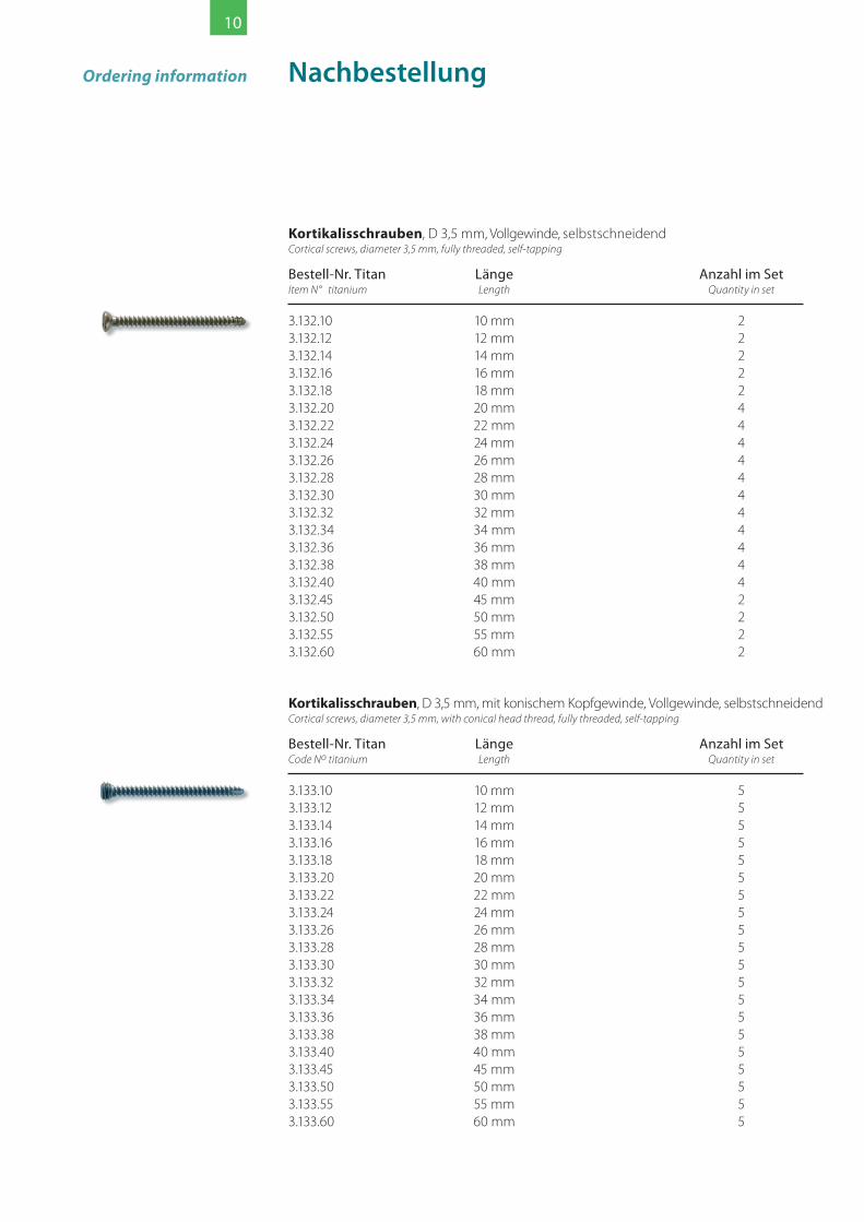
Kortikalisschrauben, D 3,5 mm, Vollgewinde, selbstschneidendCortical screws, diameter 3,5 mm, fully threaded, self-tapping
Bestell-Nr. Titan Länge Anzahl im SetItem N° titanium Length Quantity in set
3.132.10 10 mm 23.132.12 12 mm 23.132.14 14 mm 23.132.16 16 mm 23.132.18 18 mm 23.132.20 20 mm 43.132.22 22 mm 43.132.24 24 mm 43.132.26 26 mm 43.132.28 28 mm 43.132.30 30 mm 43.132.32 32 mm 43.132.34 34 mm 43.132.36 36 mm 43.132.38 38 mm 43.132.40 40 mm 43.132.45 45 mm 23.132.50 50 mm 23.132.55 55 mm 23.132.60 60 mm 2
Kortikalisschrauben, D 3,5 mm, mit konischem Kopfgewinde, Vollgewinde, selbstschneidendCortical screws, diameter 3,5 mm, with conical head thread, fully threaded, self-tapping
Bestell-Nr. Titan Länge Anzahl im SetCode No titanium Length Quantity in set
3.133.10 10 mm 53.133.12 12 mm 53.133.14 14 mm 53.133.16 16 mm 53.133.18 18 mm 53.133.20 20 mm 53.133.22 22 mm 53.133.24 24 mm 53.133.26 26 mm 53.133.28 28 mm 53.133.30 30 mm 53.133.32 32 mm 53.133.34 34 mm 53.133.36 36 mm 53.133.38 38 mm 53.133.40 40 mm 53.133.45 45 mm 53.133.50 50 mm 53.133.55 55 mm 53.133.60 60 mm 5

11
Conjunto completo para operaçõesOP-Set completeN°de pedido 19.551.00Item No
Broca para acoplamento rápidoD 2,5 mm, 125 mm de comprimento 2.904.06 1Twist drill for quick coupler diameter 2.5 mm, length 125 mm
Pinça para segurar parafusos, com efeito de auto retenção 2.954.01 1Screw forceps, self holding
Medidor com gancho 2.953.60 1para parafusos D 3,5 - 4,0 mm, MB 60 mmScrew gauge with clasp, for screws diameter 3.5 - 4.0 mm, MR 60 mm
Instrumentos no conjunto N°de pedido Unidades no conjuntoInstuments in set, Item N° Quantity in set
Chave para parafusos hexagonais, forma cônica com cabo,para parafusos de D 2,7 mm a 4,0 mm, SW 2,5 2.9406.25 1Hexagonal screwdriver, conical with handle, for screws diameter 2.7 mm - 4.0 mm, wrench width 2.5
Tabuleiro com base para instrumentos e implantes 19.550.00 1Perforated autoclavable container with inset for instruments and implants
Bucha de furar para fragmento pequenoDrill guide for small fragment
para a união roscada variável e com estabilidade angular, 2.977.08 1comprimento de 45 mmfor variable angle-stable screwing, length 45 mm
com pegadouro de 55 mm de comprimento 10.500.27A 1with handle, handle length 55 mm
para a união roscada com estabilidade angular, 2.977.01 2comprimento de 45 mmfor angle-stable screwing, length 45 mm
Fio de Kirschner „Trokar“, extremidade arredondada,D 1,6 mm, 150 mm de comprimento 6.031.16 5Kirschner wire with trocar point and round end, diameter 1.6 mm, length 150 mm
VAR I A B EL VARIABLE
Set quirúrgico completoOP-Set complete
N° de pedido 19.551.00Item No
Broca espiral para acoplamiento rápidodiámetro 2,5 mm, longitud 125 mm 2.904.06 1Twist drill for quick coupler diameter 2.5 mm, length 125 mm
Pinza de sujeción de tornillos autosujetante 2.954.01 1Screw forceps, self holding
Instrumento de medición con gancho 2.953.60 1para tornillos diámetro 3,5 - 4,0 mm, RM 60 mmScrew gauge with clasp, for screws diameter 3.5 - 4.0 mm, MR 60 mm
Instrumentos en el set N° de pedido Cantidad en el setInstuments in set Item N° Quantity in set
Destornillador hexagonal, cónico, con mango, para tornillos diámetro 2,7 mm - 4,0 mm, ancho de llave 2,5 2.9406.25 1Hexagonal screwdriver, conical with handle, for screws diameter 2.7 mm - 4.0 mm, wrench width 2.5
Tamiz de almacenamiento con insertos 19.550.00 1para instrumentos e implantesPerforated autoclavable container with inset for instruments and implants
Casquillo de taladrado para fragmentos pequeñosDrill guide for small fragment,
para atornilladura variable con ángulo estable, 2.977.08 1longitud 45 mmfor variable angle-stable screwing, length 45 mm
con mango, longitud del mango 55 mm 10.500.27A 1with handle, handle length 55 mm
para atornilladura con ángulo estable, longitud 45 mm 2.977.01 2for angle-stable screwing, length 45 mm
Aguja de Kirschner trocar, extremo redondo,diámetro 1,6 mm, longitud 150 mm 6.031.16 5Kirschner wire with trocar point and round end, diameter 1.6 mm, length 150 mm
VAR I A B EL VARIABLE
OP-Set komplettOP-Set completeBestell-Nr. 19.551.00Item No
Spiralbohrer für SchnellkupplungD 2,5 mm, Länge 125 mm 2.904.06 1Twist drill for quick coupler diameter 2.5 mm, length 125 mm
Schraubenhaltepinzette selbsthaltend 2.954.01 1Screw forceps, self holding
Messinstrument mit Haken 2.953.60 1für Schrauben D 3,5 - 4,0 mm, MB 60 mmScrew gauge with clasp, for screws diameter 3.5 - 4.0 mm, MR 60 mm
Instrumente im Set Bestell-Nr. Anzahl im SetInstuments in set, Item N° Quantity in set
Sechskantschraubendreher,konisch mit Griff für Schrauben D 2,7 mm - 4,0 mm, SW 2,5 2.9406.25 1Hexagonal screwdriver, conical with handle, for screws diameter 2.7 mm - 4.0 mm, wrench width 2.5
Lagerungssieb mit Einsatz für Instrumente und Implantate 19.550.00 1Perforated autoclavable container with inset for instruments and implants
Bohrbuchse für KleinfragmentDrill guide for small fragment,
für variabel winkelstabile Verschraubung, Länge 45 mm 2.977.08 1for variable angle-stable screwing, length 45 mm
mit Griff, Grifflänge 55 mm 10.500.27A 1with handle, handle length 55 mm
für winkelstabile Verschraubung, Länge 45 mm 2.977.01 2for angle-stable screwing, length 45 mm
Kirschnerdraht Trokar, Ende rund, D 1,6 mm, Länge 150 mm 6.031.16 5Kirschner wire with trocar point and round end, diameter 1.6 mm, length 150 mm
VAR I A B EL VARIABLE

www.koenigsee-implantate.de
2004 -
Certificação conformeDiretriz 93/42/CEE
e norma DIN EN ISO 13485:2012certified according to
EC directive 93/42/ECC DIN EN ISO 13485: 2012
OT Aschau • Am Sand 4 • 07426 Allendorf • República Federal da AlemanhaFon +49 (0) 36738 498-550 • Fax +49 (0) 36738 498-559e-mail: [email protected]© Copyright Königsee Implantate GmbH
O conteúdo, mesmo parcial, não pode ser reproduzido sem a autorização por escrito do editor.
No part of this publication may be reproduced, stored in a retrieval system or transmitted in any
form or by any means electronic, mechanical, photocopying, recording or other-wise without the prior written permission of the
publisher.
Apresentado por:Presented by:
Autor médico:Medical author:
Dr.med. Detlef Drüppel
Médico-chefe, Cirurgia do trauma / OrtopediaDiretor médicoHospital „St. Walburga Krankenhaus Meschede“
Desenhos:Drawings:
PD Dr. Diethard Wahl
Berlin
Edição: fevereiro de 2014 Nºde referência: 1.033.00 Item No
Certificado según Directiva CE 93/42/CEE DIN EN ISO 13485:2012
certified according to EC directive 93/42/ECC DIN EN ISO 13485: 2012
OT Aschau • Am Sand 4 • 07426 Allendorf • República Federal de AlemaniaFon +49 36738 498-550 • Fax +49 36738 498-559e-mail: [email protected]© Copyright Königsee Implantate GmbH
Reproducción parcial o total del contenido sólo con autorización
por escrito del editor.
No part of this publication may be reproduced, stored in a retrieval system or transmitted in any
form or by any means electronic, mechanical, photocopying, recording or other-wise without the prior written permission of the
publisher.
Facilitado por:Presented by:
Autor médico:Medical author:
Dr. méd. Detlef Drüppel
Médico Jefe de Traumatología / OrtopediaDirector MédicoSt. Walburga Krankenhaus Meschede
Ilustraciones:Drawings:
PD Dr. Diethard Wahl
Berlin
Fecha: Febrero 2014 N° de pedido: 1.033.00 Item No
zertifiziert nachEG Richtlinie 93/42/EWG
DIN EN ISO 13485: 2012certified according to
EC directive 93/42/ECC DIN EN ISO 13485: 2012
OT Aschau • Am Sand 4 • 07426 Allendorf • Bundesrepublik DeutschlandFon +49 (0) 36738 498-550 • Fax +49 (0) 36738 498-559e-mail: [email protected]© Copyright Königsee Implantate GmbH
Wiedergabe des Inhalts, auch auszugsweise, nur mit schriftlicher Genehmigung des Herausgebers.
No part of this publication may be reproduced, stored in a retrieval system or transmitted in any
form or by any means electronic, mechanical, photocopying, recording or other-wise without the prior written permission of the
publisher.
Überreicht durch:Presented by:
Ärztlicher Autor:Medical author:
Dr.med. Detlef Drüppel
Chefarzt Unfallchirurgie / OrthopädieÄrztlicher DirektorSt. Walburga Krankenhaus Meschede
Zeichnungen:Drawings:
PD Dr. Diethard Wahl
Berlin
Version: Februar 2014 Bestell-Nr.: 1.033.00 Item No


![Convex Push Plate Placa de Empuje Convexa Plaque de ... · para montar la placa de empuje en el activador. 7) Si el grosor de la pared es menor a 1-1/2” [38mm], rompa las barras](https://img.dokumen.tips/doc/110x75/60029c33743c2364fb2419f3/convex-push-plate-placa-de-empuje-convexa-plaque-de-para-montar-la-placa-de.jpg)