Embed Size (px)
Citation preview

UvA-DARE is a service provided by the library of the University of Amsterdam (http://dare.uva.nl)
UvA-DARE (Digital Academic Repository)
The extrinsic coagulation pathway in coronary artery disease and endotoxemiaMoons, A.H.M.
Link to publication
Citation for published version (APA):Moons, A. H. M. (2003). The extrinsic coagulation pathway in coronary artery disease and endotoxemia.
General rightsIt is not permitted to download or to forward/distribute the text or part of it without the consent of the author(s) and/or copyright holder(s),other than for strictly personal, individual use, unless the work is under an open content license (like Creative Commons).
Disclaimer/Complaints regulationsIf you believe that digital publication of certain material infringes any of your rights or (privacy) interests, please let the Library know, statingyour reasons. In case of a legitimate complaint, the Library will make the material inaccessible and/or remove it from the website. Please Askthe Library: http://uba.uva.nl/en/contact, or a letter to: Library of the University of Amsterdam, Secretariat, Singel 425, 1012 WP Amsterdam,The Netherlands. You will be contacted as soon as possible.
Download date: 08 Apr 2019

C h a p t e r r 3 3 Thee value of hemostatic markers in the triage of
patientss with chest pain presenting with a normal or non-diagnosticc electrocardiogram
Arnoo H.M. Moons', Petrus M. van der Zee', Radha Bholasingh1, Auguestee Sturk2, C. Erik Hack3, Joost CM. Meijers4, Otto Kamp5, Jan H.
Cornel6,, Ron J.G. Peters1, Robbert J. de Winter'
'Departmentt of Cardiology, -Clinical Chemistry, and 4Vascular Medicine, Academic Medicall Center, University of Amsterdam, department of Pathophysiology of Plasma
Proteins,, Central Laboratory of the Netherlands Red Cross Blood Transfusion, 'Departmentt of Cardiology, University Hospital VU, Amsterdam, ^Department of
Cardiology,, Medical Center Alkmaar, Alkmaar, The Netherlands.
Chapterr 3
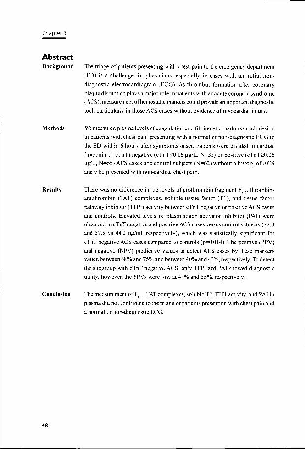
Abstrac t t Background d Thee triage of patients presenting with chest pain to the emergency department
(ED)) is a challenge for physicians, especially in cases with an initial non-
diagnosticc electrocardiogram (ECG). As thrombus formation after coronary
plaquee disruption plays a major role in patients with an acute coronary syndrome
(ACS),, measurement of hemostatic markers could provide an important diagnostic
tool,, particularly in those ACS cases without evidence of myocardial injury.
Methodss We measured plasma levels of coagulation and fibrinolytic markers on admission
inn patients with chest pain presenting with a normal or non-diagnostic ECG to
thee ED within 6 hours after symptoms onset. Patients were divided in cardiac
Troponinn T (cTnT) negative (cTnT<0.06 Mg/L, N-33) or positive (cTnT>0.06
ug/L,, N=65) ACS cases and control subjects (N=62) without a history of ACS
andd who presented with non-cardiac chest pain.
Resultss There was no difference in the levels of prothrombin fragment F]+,, thrombin-
antithrombinn (TAT) complexes, soluble tissue factor (TF), and tissue factor
pathwayy inhibitor (TFPI) activity between cTnT negative or positive ACS cases
andd controls. Elevated levels of plasminogen activator inhibitor (PAI) were
observedd in cTnT negative and positive ACS cases versus control subjects (72.3
andd 57.8 vs 44.2 ng/ml, respectively), which was statistically significant for
cTnTT negative ACS cases compared to controls (p=0.014). The positive (PPV)
andd negative (NPV) predictive values to detect ACS cases by these markers
variedd between 68% and 75% and between 40% and 43%, respectively. To detect
thee subgroup with cTnT negative ACS, only TFPI and PAI showed diagnostic
utility ,, however, the PPVs were low at 43% and 55%, respectively.
Conclusionn The measurement of F^,, TAT complexes, soluble TF, TFPI activity, and PAI in
plasmaa did not contribute to the triage of patients presenting with chest pain and
aa normal or non-diagnostic ECG.
48 8

Triagee of patients with chest pain and a non-diagnostic ECG using hemostatic markers
Introductio n n Off the patients presenting with chest pain at the emergency department (ED), the earlyy diagnosis of acute coronary syndromes (ACS) is a major challenge for physicians.. In addition to medical history and physical examination, the electrocardiogramm (ECG) is critical for the initial evaluation of these patients. However,, in 50%-80% of the cases, the ECG is normal or non-diagnostic at presentation',, which makes the early differentiation from non-cardiac causes of chest painn difficult. Currently, biochemical cardiac markers have become an important diagnosticc tool for ACS. Moreover, elevations in myocardial injury markers, such as cardiacc Troponin T (cTnT) and I, are associated with early and late recurrent adverse cardiacc events20. Serial assessment of cardiac markers, including troponins, creatine kinase-MBB isoenzyme and myoglobin, results in a high sensitivity to detect myocardial damage66 7, but the absence of evidence of myocardial damage does not exclude ACS*. Therefore,, novel early markers of ACS are needed.
Thee main cause of ACS is atherosclerotic plaque disruption with superimposed arterial thrombuss formation91', and tissue factor (TF) induced thrombin generation plays a pivotall role in this process12. Significantly higher levels of circulating TF13~16, that containss procoagulant activity17, free and total tissue factor pathway inhibitor-antigen (TFPI,, the physiologic inhibitor of TF)1516'8, and TFPI-activity'819, were found in patientss with ACS compared to patients with stable angina and healthy control subjects.. Conflicting results, however, have been reported about thrombin generation markerss as prothrombin fragment F]+, and thrombin-antithrombin (TAT) complexes, whichh showed to be significantly increased15202', while others observed comparable orr even reduced levels2224 in patients with unstable angina or myocardial infarction comparedd with stable angina patients or healthy individuals. In all of these studies, patientss with ACS presented with characteristic changes on the ECG, whereas the diagnosticc value of hemostatic markers may be useful in the triage of patients presentingg with a non-diagnostic ECG.
Thee purpose of this study was to evaluate the diagnostic value of coagulation markers (i.e.. markers of thrombin generation (F, and TAT complexes), soluble TF, and TFPI activity),, and a fibrinolytic marker (plasminogen activator inhibitor (PAI)) for the earlyy identification of ACS in patients presenting to the ED with chest pain and a normall or non-diagnostic ECG
49 9
Chapterr 3
Method s s Patients s Studyy patients were selected from a consecutive, observational cohort of patients whoo were admitted to the ED of one large teaching hospital and two academic hospitals.. All patients suffered from typical chest pain suggestive of myocardial ischemiaa that started within 6 hours of presentation and without electrocardiographic signs,, at presentation, that are typical of acute myocardial infarction25 or ischemia26. Exclusionn criteria were severe skeletal muscle damage or trauma, cardiac resuscitation, knownn or suspected thromboembolic disease, current use of oral anticoagulation, and infectiouss disease or signs of inflammation. All patients were observed at the ED for <244 hours before discharge or hospital admission. The study population of the present studyy consisted of cases with an ACS and control subjects. The cases were divided into twoo groups: group 1, which included cases with recurrent chest pain, a negative cTnT (definedd as a peak cTnT< 0.06 ug/L in two serial measurements), and dynamic ST-segmentt or T-wave changes on the ECG during observation; and group 2, which included casess with recurrent chest pain and a positive cTnT (defined as a peak cTnT > 0.06 ug/ LL in two serial measurements). The control subjects were patients without a history of cardiovascularr disease, who were considered to have chest pain of non-cardiac origin, andd who were event free during subsequent 6 months of follow-up. Thee study protocol was approved by the institutional review boards, and all patients gavee informed consent.
Bloodd sampling and assays Onn admission to the ED, venous blood samples were taken from each subject, and collectedd in citrate containing tubes for the measurements of hemostatic markers, andd in another tube without additional substrates for the measurement of cTnT. At 122 hours after the onset of symptoms, an additional blood sample was collected from alll study patients for the second measurement of cTnT. All blood samples were centrifugedd for 15 minutes at 1500g immediately after collection. Plasma was stored att -70 °C until assayed.
FF and cTnT levels were assessed using commercial enzyme-linked immunosorbent assayss (ELISAs, Dade Behring, Marburg, Gemany and Roche Diagnostics, Almere, thee Netherlands, respectively). Plasma levels of TAT complexes and PAI were also quantifiedd by ELISA as previously described27. Soluble TF concentrations were determinedd by ELISA using specific monoclonal antibodies raised against recombinant TFF {Central Laboratory of the Netherlands Red Cross Blood Transfusion, Amsterdam,
50 0

Triagee of patients with chest pain and a nondiagnostic ECG using hemostatic markers
Thee Netherlands). TFPI activity was measured on a Behring Coagulation System (Dade Behring,, Marburg, Germany) according to a method described by Sandset et al.28
Statisticall analysis Descriptivee statistics included mean values and standard deviation (SD), or median valuess and interquartile ranges UQR), for outcomes with or without a normal distribution,, respectively. Inter-group comparisons between the control group and eachh ACS case group were tested for all study measurements. Comparison of baseline characteristicss was by y} analysis for categorical variables and either the unpaired Student'ss t test or Mann-Whitney U test for continuous variables where appropriate. Thee comparisons of the hemostatic markers were performed by Kruskall-Wallis H testss and where there was a statistically significant result, subsequent comparisons betweenn the control and both case groups were made using a Mann-Whitney U test. Receiverr operator characteristic (ROC) curves were constructed for the sensitivities andd specificities of the hemostatic markers for the detection of ACS (i.e. cTnT negative orr positive ACS). Additional ROC curves were constructed to specifically detect cTnTT positive ACS or cTnT negative ACS. The positive (PPVs) and negative predictivee values (NPVs) were calculated from the section of the ROC curve with thee optimal balance between sensitivity and specificity. Statisticall significance was defined as p<0.05.
Result s s Baselinee characteristics AA total of 160 patients were included in this study: 62 controls and 98 cases divided inn two groups (group 1: 33 cases with cTnT negative ACS, and group 2: 65 cases withh cTnT positive ACS). Baseline characteristics are summarized in Table 1. Comparedd to controls who did not have a history of cardiovascular disease, both casee groups had more often a history of myocardial infarction, coronary angioplasty andd coronary bypass grafting, which was statistically significant for all three variables exceptt for the number of previous coronary bypass grafting in the cTnT positive ACSS case group. Both case groups also showed a trend to a higher number of patients withh risk factors for coronary atherosclerosis, compared to the control group. However, thiss was only statistically significant for a history of hypertension. The median duration off chest pain at presentation to the ED was approximately 2 hours for all three groups.
51 1

Chapterr 3
Tablee 1: Baseline characteristics
Characteristic c
Controls s
(N=62) )
Cases s
cTnTT negative ACS cTnT positive ACS (cTnT<0.06;; N=33) (cTnT>0.06; N=65)
Agee (y): mean SD Malee gender: N (%) Previouss Ml: N (%) Previouss PCI: N (%) Previouss CABG: N (%) Hypertension:: N (%) Hypercholesterolemia:: N (%) Diabetes:: N (%) Currentt smoking: N (%) Familyy history: N (%) Totall cholesterol (mmol/L): mean tt SD Durationn of chest pain (min): median (1QR)
600 10 45(73) ) 0(0) ) 0(0) ) 0(0) )
15(24) ) 14(23) ) 5(8) )
19(31) ) 21(34) )
5.66 1.1 123(88-210) )
600 9 25(76) )
"(21)+ + 10(30)f f 4(12)t t 155 (46)T
13(39) ) 7(21) ) 9(27) ) 16(49) )
5.22 8 120(74-180) )
633 12 45(69) ) 12(19)t t 6(9)t t 3(5) )
30(46)+ + 19(29) ) 11(17) ) 18(28) ) 33(51) )
5.77 1.1 1255 (75-240)
fpp value vs controls <().()5. cTnT=cardiac troponin T (jag/L). ACS=acute coronary syndrome. SD=standardd deviation. MI=myocardial infarction. PCl=percutaneous coronary intervention. CABG=coronaryy artery bypass grafting. IQR=interquartile range.
Plasmaa levels of hemostatic markers
FjFj,, 2 and TA T complexes Plasmaa levels of F, , and TAT complexes were slightly higher in cTnT positive ACS casess compared to controls (Figure 1, median (IQR) levels of F^,: 0.9 (0.7-1.3) vs 0.8 (0.6-1.1)) nmol/L; median (IQR) levels of TAT: 2.3 (1.0-7.6) vs 1.0 (<1.0-4.8) ng/ml). Plasmaa levels in the cTnT negative ACS case group were equal to those found in the controll group.
Fl+2 2 TAT T 1.44 -I
1.22
11
OX X
0.66
0.4 4
00 7
Controls s cTnTT negative
ACSS cases
(c c TnT<0.0 0 6) )
cTnTT positive
ACC S cases
(c c TnT T >0.0 0 6) )
B B
88 -I
7 7
66
55
44
33
2 2
00
TT -|
Controlss c l nT negative c'l
ACSS cases
(cTnl l '0 .0f t )) (c
nn 1 positive
VCSS cases
TnT T >0.0 0 6) )
Figuree 1. Median (+ 75th percentile) plasma levels of prothrombin fragment F (Panel A) and thrombin-antithrombinn (TAT) complexes (Panel B) of controls and cTnT negative and positive ACS casess at presentation to the emergency department. cTnT=cardiac Troponin T (ug/L). ACS=acute coronaryy syndrome.
52 2
Triagee of patients with chest pain and a non-diagnostic ECG using hemostatic markers
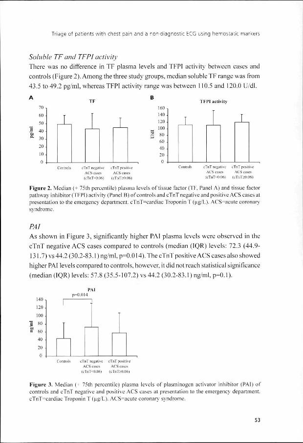
SolubleSoluble TF and TFPI activity Theree was no difference in TF plasma levels and TFPI activity between cases and controlss (Figure 2). Among the three study groups, median soluble TF range was from 43.55 to 49.2 pg/ml, whereas TFPI activity range was between 110.5 and 120.0 U/dl.
I F F 700 ,
600
40--
30--
20--
10--
Controls s
( (
nT T /\CS S cTn n
ega a
r<o. .
ivee c
36)) (
I I 1 1
l"nTT posi ACSS cas TnT>0. .
ive e s s
)6) )
160-, ,
140--
120--
100--
|| 80 -
600 -
400 -
200 -
T T
Controls s
TFPII activity
cTnTT negative ACSS cases (cTnT<0.06) )
1 1
cTnTT positive ACSS cases
<cTnT>0.06) )
Figuree 2. Median (+ 75th percentile) plasma levels of tissue factor (TF. Panel A) and tissue factor pathwayy inhibitor (TFPI) activity (Panel B) of controls and cTnT negative and positive ACS cases at presentationn to the emergency department. cTnT=cardiac Troponin T(ug/L). ACS=acute coronary syndrome. .
PAI PAI Ass shown in Figure 3, significantly higher PAI plasma levels were observed in the cTnTT negative ACS cases compared to controls (median (IQR) levels: 72.3 (44.9-131.7)) vs 44.2 (30.2-83.1) ng/ml, p=0.014). The cTnT positive ACS cases also showed higherr PAI levels compared to controls, however, it did not reach statistical significance (mediann (IQR) levels: 57.8 (35.5-107.2) vs 44.2 (30.2-83.1) ng/ml, p=0.1).
PAI I p=0.014 4
1400 ,
1200
100 0
800
60 0
40 0
20 0
Controlss cTnT negative ACSS cases
(c c TnT T <0.0 0 6) )
cTnTT positive ACSS cases
(c c TnT T >0.0 0 6) )
Figuree 3. Median (- 75th percentile) plasma levels of plasminogen activator inhibitor (PAI) of controlss and cTnT negative and positive ACS cases at presentation to the emergency department. cTnT=cardiacc Troponin T (ug/L). ACS^acute coronary syndrome.
53 3

Chapterr 3
Diagnosticc utility of hemostatic markers Thee diagnostic utility of the hemostatic markers to detect patients suffering form ACSS or to identify a subgroup of cTnT positive ACS cases is shown in Table 2. Solublee TF did not have any diagnostic value as the area under the ROC curves was beloww 0.5 (area under the curves: 0.435 and 0.452 for detecting ACS at large and the subgroupp with cTnT positive ACS, respectively). The cut-off values presented in tablee 2 correspond to the points of the ROC curves showing the best balance between sensitivityy and specificity. Except for PAI antigen, each marker had the same optimal cutpointt for detecting ACS at large or the subgroup with cTnT positive ACS. The proportionss of study patients with plasma levels above the optimal cutpoint varied betweenn 23% and 27% for the different markers.
Ass shown in Table 2, the PPVs and NPVs to identify cases with an ACS were comparablee for all 4 markers varying between 68% and 75%, and between 40% and 43%,, respectively. To detect cases with a cTnT positive ACS, F demonstrated a betterr accuracy compared to the other markers, however, its PPV and NPV were onlyy 62% and 66%, respectively.
Afterr exclusion of cTnT positive ACS cases, only TFPI and PAI demonstrated diagnosticc value to detect cTnT negative ACS (area under the ROC curves (95% confidencee interval): 0.57 (0.44-0.70) and 0.65 (0.54-0.77), respectively). At cut-off valuess of TFPI>140 U/dl and of PAI>110 ng/ml, the sensitivity, specificity, PPV,
Tablee 2: Utilit y of the hemostatic markers to identify cases with an ACS or cases specifically evolving aa cTnT positive ACS
Cutpointt hemostatic marker
Detectingg ACS at large F l+2>> 1.1 nmol/L TATT complexes > 6.0 ng/ml TFPII activity > 140 U/dl PA1>> 110 ng/ml
Detectingg cTnT positive ACS FI+2>> 1.1 nmol/L TATT complexes > 6.0 ng'ml TFPII activity > 140 U/dl PAII > 95 ng/ml
Areaa under the ROCC curve
0,533 (0.44-0.63) 0.52(0.42-0.61) ) 0.57(0.48-0.66) ) 0.611 (0.52-0.70)
0.62(0.53-0.71} } 0.59(0.50-0.68) ) 0.55(0.46-0.64) ) 0.54(0.45-0.63) )
Sens(%) )
311 (22-40) 27(18-36) ) 29(20-38) ) 28(19-37) )
40(28-52) ) 29(18-40) ) 28(17-39) ) 29(18-40) )
Specc (%)
80(70-90) ) 80(70-90) ) 799 (69-89) 855 (76-94)
83(75-91) ) 79(71-87) ) 76(67-85) ) 76(67-85) )
PPVV (%)
71(57-85) ) 68(53-83) ) 68(54-82) ) 75(61-89) )
622 (47-77) 50(34-66) ) 44(29-59) ) 455 (30-60)
NPVV (%)
42(33-51) ) 40(31-49) ) 42(33-51) ) 433 (34-52)
66(57-75) ) 61(52-70) ) 60(51-69) ) 611 (52-70)
Dataa presented as percentages (95% confidence interval). ACS-acute coronary syndrome. cTnT=cardiac Troponinn T. ROC=receiver operator characteristic. Sens=sensitivity. Spec=specificity. PPV=positive predictivee value. NPV=negative predictive value. F ^prothrombin fragment 1-2. TAT=thrombin-antithrombin.. TFPI=tissue factor pathway inhibitor. PA!=plasminogen activator inhibitor.
54 4

Triagee of patients with chest pain and a non-diagnostic ECG using hemostatic markers
andd NPV (with respective 95% confidence intervals) to identify cTnT negative ACS wass forTFPI: 31% (15%-47%), 79% (69%-89%), 43% (23%-63%), and 69% (58%-80%),, respectively, and for PAI; 34% (18%-50%), 85% (76%-94%), 55% (33%-77%),, and 72% (62%-82%), respectively. Combiningg the results of markers of thrombin generation, TFPI and PAI, did not improvee the diagnostic accuracies.
Discussion n Inn the present study, we measured coagulation markers (thrombin generation markers (FF and TAT complexes), soluble TF, and TFPI activity) and a fibrinolytic marker (PAI)) in patients admitted to the ED with chest pain and an initially normal or non-diagnosticc ECG, within 6 hours after symptom onset. Except for PAI antigen, there weree no significant differences in the plasma levels of these markers between cases withh a cTnT negative or positive ACS and control subjects. AA large study of Pope et al. showed that 5.4% of patients with a non-diagnostic ECG whoo were discharged from the ED were ultimately found to have an ACS29. Serial measurementt of specific markers of myocardial necrosis such as creatine kinase-MBB isoenzymes and troponins are used as a diagnostic tool. However, an ACS can nott be excluded when these markers are not increased8. Although TF mediated thrombuss formation after coronary plaque disruption plays a significant role in the initiationn of ACS12, systemic measurement of hemostatic markers did not demonstrate additionall diagnostic value to the early triage of our study patients with chest pain.
Ourr results do not confirm earlier studies showing significantly increased plasma concentrationss of TF, TFPI, Fl t , and TAT-complexes on admission, in patients with unstablee angina or acute myocardial infarction compared to patients with stable angina1-1'15-20-21.. However, in these studies, patients with ACS were included if the ECGG demonstrated characteristic changes of the ST-segments or T-waves and were likelyy at higher risk. It has been demonstrated that among patients with unstable angina,, those with reversible ST-segment changes have significantly higher levels off thrombin activity, as measured by fibrinopeptide A levels, compared to those withoutt these changes, and ST changes correlated significantly with the presence of angiographicc thrombus1011. However, this could not be confirmed by Becker et al, whoo reported no differences in thrombin activity and thrombin generation between
55 5
Chapterr 3
patientss with and without ECG changes32. In addition, others failed to show significant differencess in plasma levels of F,_, and TAT complexes between patients with acute myocardiall infarction, unstable angina and stable angina, despite the characteristic ECGG changes presented by patients with an ACS2™3. Thus, symptomatic myocardial ischemiaa may not primarily be related to acute thrombin generation, but is possibly thee result of a combination with platelet aggregation, transient increased vasomotor tonee and transient increased myocardial oxygen demand33. Alternatively, local thrombinn generation in the coronary arteries may be insufficiently reflected by systemicc plasma levels.
Wee observed elevated plasma levels of PAI antigen in both ACS case groups compared too controls, but this was only significant in cases with a cTnT negative ACS. An importantt source of PAI during atherosclerotic thrombus formation may be the secretionn by endothelial cells and activated platelets34. However, increased PAI plasma levelss are also associated with common risk factors for coronary artery disease such ass hypertension and particularly diabetes34. These two factors were more prevalent inn both case groups, in particular in the cTnT negative ACS case group, and this contributedd to the enhanced PAI levels in these groups compared to controls.
Patientss suffering from cTnT positive ACS previously demonstrated a significantly increasedd coagulation activation compared to patients with cTnT negative unstable angina3-3''.. However, we could not confirm this in our study population. We observed noo differences in plasma levels of the hemostatic markers between cases with cTnT positivee and negative ACS. Despite evidence of myocardial necrosis, the extent of coagulationn activation may be limited in cases with a cTnT positive ACS, and whose ECGG does not show characteristic changes of myocardial ischemia. This was reflected byy the poor diagnostic performance of the hemostatic markers to identify these cases. Prothrombinn fragment F, t, showed the best diagnostic utility, but the PPV and NPV weree only 62% and 66%, respectively. Since these patients can be identified after seriall measurement of cardiac serum markers, it remains a difficult task to detect patientss suffering from an ACS, without evidence of myocardial damage and presentingg with a non-diagnostic ECG. In the present study, after excluding the cTnT positivee ACS cases from the analyses, only TFPI and PAI demonstrated diagnostic valuee to detect cTnT negative ACS cases, but with low PPVs of 43% and 55%, respectively.. Based on these markers, as much as 66%-69% of the patients with cTnTT negative ACS would still not have been detected. Thus in our study population
56 6
Triagee of patients with chest pain and a non-diagnostic ECG using hemostatic markers
off low risk chest pain patients, the utility of hemostatic markers showed limited diagnosticc value to identify patients with either a cTnT positive or negative ACS.
Studyy limitations Ass shown in Figure 1 and 3, the assays measuring TAT complexes and PAI antigen demonstratedd marked variability in each group. We could not identify clinical characteristicss that appeared to be responsible to these wide ranges, including the samplingg times for PAI, since significantly higher PAI levels have been found in the morningg than in the evening both in control subjects and patients with ACS37. However,, we cannot exclude the possibility that other factors such as platelet activation duringg venipuncture have resulted in enhanced PAI levels. In addition, as patients withh unstable angina have been shown to develop frequent bursts of coagulation activation™,, the short plasma half-life of TAT and PAI of 5 and 8-10 minutes, respectively,, compared to that of F, ̂ (90 minutes), may also have contributed to the variabilityy in both ACS case groups.
Conclusions s Thee measurement in plasma of thrombin generation markers (Fj,, and TAT complexes),, soluble TF, TFPI activity, and PAI antigen, did not contribute to the triagee of patients presenting with chest pain and a normal or non-diagnostic ECG in thee ED. Although plaque disruption with thrombus formation is the most common underlyingg pathogenic mechanism during an ACS, in this patient population in whom theree is a need for additional markers of ACS, this does not lead to elevation of systemicc plasma levels that are clinically useful for the triage of low risk chest pain patients. .
Acknowledgements s Wee thank Ms M.C.L. Schaap and Mrs L.Y. Ko for their excellent technical assistance.
57 7

Chapterr 3
Reference s s 1.. Abbott BG, Jain D. Nuclear cardiology in the evaluation of acute chest pain in the emergency
department.. Echocardiography 2000; 17: 597-604. 2.. Lindahl B, Venge P, Wallentin L, for the FRISC Study Group. Relation between troponin T
andd the risk of subsequent cardiac events in unstable coronary artery disease. Circulation 1996; 93:: 1651-1657.
3.. Newby LK, Christenson RH, Ohman EM, et al. Value of serial troponin T measures for early andd late risk stratification in patients with acute coronary syndromes. Circulation 1998; 98: 1853-1859. .
4.. Antman EM, Tanasijevic MJ, Thompson B, et al. Cardiac-specific troponin 1 levels to predict thee risk of mortality in patients with acute coronary syndromes. N Engl J Med 1996; 335: 1342-1349. .
5.. Sonel A,Sasseen BM, FinebergN, etal. Prospective study correlating fibrinopeptide A, troponin 1, myoglobin,, and myosin light chain levels with early and late ischemic events in consecutive patients presentingg to the emergency department with chest pain. Circulation 2000; 102: 1107-1113.
6.. de Winter RJ, Koster RW, Sturk A, et al. Value of myoglobin, troponin T, and CK-MB mass inn ruling out an acute myocardial infarction in the emergency room. Circulation 1995; 92: 3401-3407. .
7.. Zimmerman J, Fromm R, Meyer D, et al. Diagnostic marker cooperative study for the diagnosis off myocardial infarction. Circulation 1999; 99: 1671-1677.
8.. de Winter RJ. Risk stratification with cardiac troponin I in acute coronary syndromes. J Am Colll Cardiol 2000; 36: 1824-1826.
9.. Fuster V, Badimon L, Badimon JJ, et al. The pathogenesis of coronary artery disease and the acutee coronary syndromes (1). N Engl J Med 1992; 326: 242-250.
10.. Fuster V, Badimon L, Badimon JJ, et al. The pathogenesis of coronary artery disease and the acutee coronary syndromes (2). N Engl J Med 1992; 326: 310-318.
11.. Falk E, Shah PK., Fuster V. Coronary plaque disruption. Circulation 1995; 92: 657-671. 12.. Moons AH, Levi M, Peters RJ. Tissue factor and coronary artery disease. Cardiovasc Res
2002;53:313-325. . 13.. Suefuji H, Ogawa H, Yasue H, et al. Increased plasma tissue factor levels in acute myocardial
infarction.. Am Heart J 1997; 134: 253-259. . 14.. Misumi K., Ogawa H, Yasue H, et al. Comparison of plasma tissue factor levels in unstable and
stablee angina pectoris. Am J Cardiol 1998; 81: 22-26. 15.. Soejima H, Ogawa H, Yasue H, et al. Heightened tissue factor associated with tissue factor pathway
inhibitorr and prognosis in patients with unstable angina. Circulation 1999; 99: 2908-2913. 16.. Falciani M, Gori AM, Fedi S, et al. Elevated tissue factor and tissue factor pathway inhibitor
circulatingg levels in ischaemic heart disease patients. Thromb Haemost 1998; 79: 495-499, 17.. Sambola A, Osende J, Hathcock J, et al. Role of risk factors in the modulation of tissue factor
activityy and blood thrombogenecity. Circulation 2003; 107: 973-977. 18.. He M, Wen Z, He X, et al. Observation on tissue factor pathway and some other coagulation
parameterss during the onset of acute cerebrocardiac thrombotic diseases. Thromb Res 2002; 107:223-228. .
19.. Sandset PM, Sirnes PA, Abildgaard U. Factor VII and extrinsic pathway inhibitor in acute coronaryy disease. BrJ Haematol 1989; 72: 391-396.
20.. Merlini P, Bauer K.A, Oltrona L, et al. Persistent activation of coagulation mechanism in unstable anginaa and myocardial infarction. Circulation 1994; 90: 61-68.
21.. Hoffmeister HM, Jur M, Wendel HP, et al. Alterations of coagulation and fibrinolytic and kallikrein-kininn systems in the acute and postacute phases in patients with unstable angina pectoris.. Circulation 1995; 91: 2520-2527.
58 8

Triagee of patients with chest pain and a non-diagnostic ECG using hemostatic markers
22.. Manten A, de Winter RJ, Minnema MC, et al. Procoagulant and proinflammatory activity in acutee coronary syndromes. Cardiovasc Res. 1998; 40: 389-395.
23.. Jude B, Agraou B, Mc Fadden EP, et al. Evidence for time-dependent activation of monocytes inn the systemic circulation in unstable angina but not in acute myocardial infarction or in stable angina.. Circulation 1994; 90: 1662-1668.
24.. McKenzie ME, Pothula A, Gurbel PA, et al. Failure of thrombin generation markers to triage patientss presenting with chest pain. Cardiology 1999; 92: 53-58.
25.. Alpert JS,Thygesen K, Antman E, etal. Myocardial infarction redefined: a consensus document off The Joint European Society of Cardiology/American College of Cardiology Committee for thee redefinition of myocardial infarction. J Am Coll Cardiol 2000; 36: 959-969.
26.. Braunwald E, Antman EM, Beasley JW, et al. ACC/AHA guidelines for the management of patientss with unstable angina and non-ST-segment elevation myocardial infarction: a report of thee American College of Cardiology/American Heart Association Task Force on Practice Guideliness (Committee on the Management of Patients With Unstable Angina). J Am Coll Cadioll 2000; 36: 970-1062.
27.. Boermeester MA, van Leeuwen PA, Coyle SM, et al. Interleukin-1 blockade attenuates mediator releasee and dysregulation of the hemostatic mechanism during human sepsis. Arch Surg 1995; 130:: 739-748.
28.. Sandset PM, Abildgaard U, Pettersen M. A sensitive assay of extrinsic coagulation pathway inhibitorr (EPI) in plasma and plasma fractions. Thromb Res 1987; 47: 389-400.
29.. Pope JH, Aufderheide TP, Ruthazer R, et al. Missed diagnoses of acute cardiac ischemia in the emergencyy department. N Engl J Med 2000; 342: 1163-1170.
30.. Eisenberg PR, Kenzora JL, Sobel BE, et al. Relation between ST segments shift during ischemia andd thrombin activity in patients with unstable angina. J Am Coll Cardiol 1991; 18: 898-903.
31.. Wilensky RL, Bourdillon PD, Vix VA, et al. Intracoronary artery thrombus formation in unstable angina:: a clinical, biochemical and angiographic correlation. J Am Coll Cardiol 1993; 21: 692-699.
32.. Becker RC, Tracy RP, Bovill EG, et al. Surface 12-lead electrocardiographic findings and plasmaa markers of thrombin activity and generation in patients with myocardial ischemia at rest.. J Thromb Thrombolysis 1994; 1: 101-107.
33.. Ambrose JA, Hjemdahl-Monsen CE. Arteriographic anatomy and mechanisms of myocardial ischemiaa in unstable angina. J Am Coll Cardiol 1987; 9: 1397-1402.
34.. Bastard J-P, Piéroni L, Hainque B. Relationship between plasma plasminogen activator inhibitor 11 and insulin resistance. Diabetes Metab Res Rev 2000; 16: 192-201.
35.. Terres W, Kümmel P, Sudrow A, et al. Enhanced coagulation activation in troponin T-posttive unstablee angina pectoris. Am Heart J 1998; 135:281-286.
36.. Giannitsis E, Bardorff MM, Schweikart S, et al. Relationship of cardiac troponin T and procoagulantt activity in unstable angina. Thromb Haemost 2000; 83: 224-228.
37.. Angleton P, Chandler WL, Schmer G. Diurnal variation of tissue-type plasminogen activator andd its rapid inhibitor (PA1-1). Circulation 1989; 79: 101-106.
38.. Biasucci LM, Liuzzo G, Caligiuri G, et al. temporal elation between ischemic episodes and activationn of the coagulation system in unstable angina. Circulation 1996; 93: 2121-2127.
59 9

1 1 * *