Embed Size (px)
DESCRIPTION
Utility of Dorsal Pits and Pubic Tubercle Height in Parity Assessment
Citation preview
Despite considerable research, accurate parity determinationfrom human skeletal remains continues to elude forensic anthropol-ogists. Skeletal changes on the dorsal aspect of the pubis and in theauricular surface region have been linked to pregnancy and parturi-tion through studies of human skeletal remains from archaeologicalsites. In particular, pubic bones have been the subject of intense re-search for indicators of childbirth. Pitting on its dorsal surface (1–3)and the height of the associated pubic tubercle (4,5) have been pro-posed as markers indicating parity. Unfortunately, the results ofthese studies are often conflicting (see review in Ref 6).
The reliability of dorsal pitting as an indicator of parity is uncer-tain, as studies with documented skeletal series have produced con-tradictory results (7–10). Even some males possess dorsal pits,which calls into question the causal factor involved in producingthe scars. Some studies have achieved positive results through theuse of remains with documented histories of parity. For instance,Suchey and colleagues (11) found a correlation, though weak, be-tween pitting and full-term pregnancies.
The height of the pubic tubercle has been proposed as an indica-tor of parity (4,5), but has yielded mixed results. The most promis-ing study to date used the Spitalfields skeletal series, dating from1729 to 1859, for which parturition information could be recon-
structed from historical records. In this study, Cox and Scott (5)found a correlation between pubic tubercle height and parity status.The study assessed the degree of extension of the pubic tubercle us-ing a four-stage classification system, after Bergfelder and Herr-mann (4). While most parous females (87%) had an extended pu-bic tubercle, some nulliparous females (33%) also had extendedtubercles.
Forensic anthropologists have a vested interest in discoveringmarkers indicative of parity, as they may be helpful in forming adescription of an individual. This, combined with the conflictingresults of previous studies, led us to examine the utility of dorsalpitting and pubic tubercle height in parity assessment using a largesample of pubic bone pairs with associated information on numberof births.
Methods
The sample consisted of 148 sets of pubic bones randomly se-lected from an extensive sample of 486 females previously de-scribed (11). This sample was collected at autopsy at the Los An-geles County Department of the Coroner in the late 1970s andincludes information on height, weight, age, and reported numberof births. Body mass index (BMI) was calculated as the weight inkilograms divided by the square of height in meters. The samplewas entirely female and the age range from 17 to 99 years, with amean age of 44.7 years.
Approximately one third of the women (n � 49) in the study hadno reported births, while one third had one or two children (n �50), and one third had three or more children (n � 48). Parity his-tory and age at death was missing for one individual. The numberof reported births ranged from zero to 17, with a mean of 2.1 births.
Copyright © 2003 by ASTM International, 100 Barr Harbor Drive, PO Box C700, West Conshohocken, PA 19428-2959.
TECHNICAL NOTE
J. Josh Snodgrass,1 M.A. and Alison Galloway,2 Ph.D.
Utility of Dorsal Pits and Pubic Tubercle Height inParity Assessment*
ABSTRACT: Parity indicators in human skeletal material are highly desirable yet elusive. In this study, the relationships of dorsal pits and pubictubercle elongation to parity are investigated in a sample of 148 modern female sets of pubic bones with associated birth information. The elonga-tion of the pubic tubercle shows no significant correlation with number of births, but instead is associated with the distance this feature is from thepubic symphysis (p � 0.01) and the size of the arcuate angle (p � 0.05). Dorsal pits show a strong association with increasing numbers of births (p � 0.01), especially in younger women. However, in women over 50 years old, dorsal pitting is correlated with BMI (p � 0.05) and is not sig-nificantly correlated with number of births. While this study lends support to the correlation of dorsal pitting and parity, it currently does not reachthe level of accuracy needed for forensic applications at the level of the individual.
KEYWORDS: forensic science, forensic anthropology, parity, dorsal pitting, pubic tubercle, body mass index (BMI)
J Forensic Sci, November 2003, Vol. 48, No. 6Paper ID JFS2003027_486
Available online at: www.astm.org
1
1 Department of Anthropology, Northwestern University, 1810 Hinman Av-enue, Evanston, IL.
2 Department of Anthropology, Social Sciences One Faculty Services, Uni-versity of California, Santa Cruz, 1156 High Street, Santa Cruz, CA.
* A version of this paper was presented at the 50th annual meeting of theAmerican Academy of Forensic Sciences in San Francisco, California, inFebruary 1998. Financial support for the project was provided by the Universityof California, Santa Cruz Division of Social Sciences, in a grant to A. Galloway.
Received 31 Jan. 2003; and in revised form 17 May 2003; accepted 10 July2003; published 25 Sept. 2003.
2 JOURNAL OF FORENSIC SCIENCES
Individuals were classified into parity groups according to numberof reported births: 0 births, 1–2 births, 3–4 births, and over 5 births.
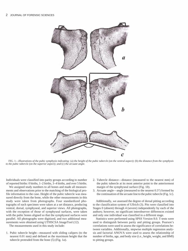
We assigned study numbers to all bones and made all measure-ments and observations prior to the matching of the biological pro-file information to the case. Height of the pubic tubercle was mea-sured directly from the bone, while the other measurements in thisstudy were taken from photographs. Four standardized pho-tographs of each specimen were taken at a set distance, producingventral, dorsal, symphyseal, and superior views. All photographs,with the exception of those of symphyseal surfaces, were takenwith the pubic bones aligned so that the symphyseal surfaces wereparallel. All photographs were digitized, and two additional mea-surements were obtained using UTHSCSA ImageTool (12).
The measurements used in this study include:
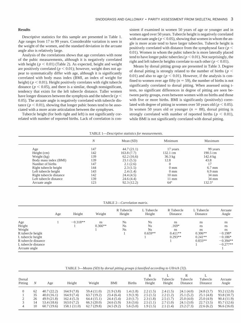
1. Pubic tubercle height—measured with sliding calipers (to thenearest 0.01 mm) and defined as the maximum height that thetubercle protruded from the bone (5) (Fig. 1a).
2. Tubercle distance—distance (measured to the nearest mm) ofthe pubic tubercle at its most anterior point to the anteriormostmargin of the symphyseal surface (Fig. 1b).
3. Arcuate angle—angle (measured to the nearest 0.5°) formed bythe continuation of the arcuate line to the pubic tubercle (Fig. 1c).
Additionally, we assessed the degree of dorsal pitting accordingto the classification system of Ullrich (3). Pits were classified intoStages 0 (absent) through 4 (severe) independently by each of theauthors; however, no significant interobserver differences existedand only one individual was classified in a different stage.
Statistics were performed using SPSS Version 8.0. T-tests wereused to distinguish between parity and pitting groups. Pearson’scorrelations were used to assess the significance of correlations be-tween variables. Additionally, stepwise multiple regression analy-sis and factorial ANOVA were used to assess the relationship ofnumber of births, age, and body size (i.e., height, weight, and BMI)to pitting groups.
FIG. 1—Illustrations of the pubic symphysis indicating: (a) the height of the pubic tubercle (on the ventral aspect); (b) the distance from the symphysisto the pubic tubercle (on the superior aspect); and (c) the arcuate angle.
Results
Descriptive statistics for this sample are presented in Table 1.Age ranges from 17 to 99 years. Considerable variation is seen inthe weight of the women, and the standard deviation in the arcuateangle also is relatively large.
Analysis of the correlations shows that age correlates with noneof the pubic measurements, although it is negatively correlatedwith height (p � 0.01) (Table 2). As expected, height and weightare positively correlated (p � 0.01); however, weight does not ap-pear to systematically differ with age, although it is significantlycorrelated with body mass index (BMI, an index of weight forheight) (p � 0.01). Height positively correlates with right tubercledistance (p � 0.05), and there is a similar, though nonsignificant,tendency that exists for the left tubercle distance. Taller womenhave longer distances between the symphysis and the tubercle (p �0.05). The arcuate angle is negatively correlated with tubercle dis-tance (p � 0.01), showing that longer pubic bones tend to be asso-ciated with a more acute articulation between the symphyses.
Tubercle height (for both right and left) is not significantly cor-related with number of reported births. Lack of correlation is con-
sistent if examined in women 50 years of age or younger and inwomen aged over 50 years. Tubercle height is negatively correlatedwith arcuate angle (p � 0.05), showing that women in whom the an-gle is more acute tend to have larger tubercles. Tubercle height ispositively correlated with distance from the symphyseal face (p �0.01). Women in whom the pubic tubercle is more laterally placedtend to have longer pubic tubercles (p � 0.01). Not surprisingly, theright and left tubercle heights correlate to each other (p � 0.01).
Means by dorsal pitting group are presented in Table 3. Degreeof dorsal pitting is strongly related to the number of births (p �0.01) and also to age (p � 0.01). However, if the analysis is con-fined to women over age fifty (n � 59), the number of births is notsignificantly correlated to dorsal pitting. When assessed using t-tests, no significant differences in degree of pitting are seen be-tween parity groups, even between women with no births and thosewith five or more births. BMI is significantly (positively) corre-lated with degree of pitting in women over 50 years old (p � 0.05).In women 50 years old or younger (n � 88), dorsal pitting isstrongly correlated with number of reported births (p � 0.01),while BMI is not significantly correlated with dorsal pitting.
SNODGRASS AND GALLOWAY • PARITY ASSESSMENT FROM SKELETAL REMAINS 3
TABLE 1—Descriptive statistics for measurements.
N Mean (SD) Minimum Maximum
Age 147 44.7 (21.1) 17 years 99 yearsHeight (cm) 142 163.8 (7.7) 132.1 cm 190.5 cmWeight (kg) 139 62.2 (16.6) 36.3 kg 142.4 kgBody mass index (BMI) 139 23.1 (5.5) 12.8 43.8Number of births 147 2.1 (2.6) 0 17Right tubercle height 144 2.3 (1.5) 0 mm 6.7 mmLeft tubercle height 143 2.4 (1.4) 0 mm 6.9 mmRight tubercle distance 142 24.4 (4.5) 10 mm 34 mmLeft tubercle distance 139 24.6 (4.4) 11 mm 34 mmArcuate angle 123 92.3 (12.2) 64° 132.5°
TABLE 2—Correlation matrix.
R Tubercle L Tubercle R Tubercle L Tubercle ArcuateAge Height Weight Height Height Distance Distance Angle
Age 1 �0.318** ns Ns Ns ns ns nsHeight 1 0.366** Ns Ns .169* ns nsWeight 1 Ns Ns ns ns nsR tubercle height 1 0.659** 0.411** 0.306** �0.198*L tubercle height 1 0.293** 0.241** �0.186*R tubercle distance 1 0.833** �0.394**L tubercle distance 1 �0.277**Arcuate angle 1
TABLE 3—Means (SD) by dorsal pitting groups (classified according to Ullrich [3]).
R L R LDorsal Tubercle Tubercle Tubercle Tubercle ArcuatePitting N Age Height Weight BMI Births Height Height Distance Distance Angle
0 62 40.7 (22.2) 164.9 (7.8) 59.4 (11.0) 21.9 (3.8) 1.4 (1.8) 2.2 (1.5) 2.4 (1.5) 24.1 (4.0) 24.8 (3.7) 93.2 (12.0)1 35 40.0 (16.1) 164.9 (7.4) 63.7 (19.2) 23.4 (6.4) 1.9 (1.9) 2.5 (1.5) 2.2 (1.2) 25.1 (5.2) 25.1 (4.8) 93.8 (10.7)2 26 49.9 (21.8) 162.4 (5.3) 64.4 (15.1) 24.4 (5.4) 2.0 (1.7) 2.3 (1.8) 2.5 (1.7) 25.0 (4.0) 25.0 (4.9) 90.4 (11.9)3 14 53.4 (18.6) 163.0 (7.2) 66.3 (20.0) 24.6 (5.9) 3.6 (3.6) 2.5 (1.1) 2.7 (1.0) 24.1 (3.0) 22.7 (3.5) 85.7 (12.6)4 10 60.7 (19.6) 158.1 (11.0) 62.7 (29.8) 24.5 (9.2) 5.6 (5.0) 1.9 (1.5) 2.1 (1.4) 23.2 (7.3) 22.6 (6.2) 96.6 (16.0)

4 JOURNAL OF FORENSIC SCIENCES
In stepwise multiple regression analysis of number of births, age,and BMI to pitting group (dependent variable), the combination ofnumber of births ( p � 0.001; � � 0.337) and age ( p � 0.05; � �0.192) is the best predictor of pitting stage (r2 � 0.184). When therelationship of parity group (fixed factor), age (covariate), and BMI(covariate) to pitting group (dependent variable) is assessed usingfactorial ANOVA, parity group ( p � 0.01; F � 5.352), age ( p �0.01; F � 6.753), and BMI (p � 0.05; F � 4.477) are significantin the dorsal pitting stage (r2 � 0.207). Parity group is a significantpredictor of dorsal pitting stage even when controlled for the ef-fects of age and BMI.
In the subset of women age 50 or younger, when the relationshipof the parity group (fixed factor), age (covariate), and BMI (co-variate) to pitting group (dependent variable) is assessed using fac-torial ANOVA, only the parity group is significant (p � 0.001; F � 10.483; r2 � 0.351). In stepwise multiple regression analysisof number of births, age, and BMI to pitting group (dependent vari-able) for the subset of women age 50 or younger, only the numberof births is significant in predicting the degree of dorsal pitting(p � 0.001; � � 0.532; r2 � 0.283).
In the subset of women over 50 years of age, when the relationshipof number of births, age, and BMI to pitting group (dependent vari-able) is assessed using stepwise multiple regression analysis, onlyBMI is significant (and the direction of influence is positive) in pre-dicting degree of dorsal pitting (p � 0.05; � � 0.339; r2 � 0.115).
Pitting is negatively correlated with height ( p � 0.05); shorterwomen have more pronounced pitting. However, in multivariatemodels, height does not add significant predictive power. Whenanalyses are confined to women age 50 or younger (n � 88), the re-lationship of pitting and height is not statistically significant. Pit-ting also may be affected by pubic shape; those with the most se-vere pitting tend to have somewhat shorter pubic bones and obtusearcuate angles, although in multivariate models these variables donot explain more of the variation.
When the sample is examined by the number of births, thosewomen with more reported births tend to be older and slightlyheavier (Table 4). Tubercle height, tubercle distance, and arcuateangle do not seem to affect or reflect birth rates.
Discussion
This study fails to support the relationship between pubic tuber-cle height and number of births, as previously suggested by Coxand Scott (5). Instead, pubic tubercle height appears to reflect otherfactors, including distance of the tubercle from the symphysealface and the acuteness of the arcuate angle. This suggests thatwomen in whom the arcuate angle is obtuse but who have long pu-bic bones are more prone to enhancement of the tubercle. In theparity ranges observed in most contemporary forensic populations,tubercle height does not provide a reliable assessment of parity.
Consequently, tubercle height should not be used to assess parityinformation.
As reported previously (11), dorsal pits were found to be associ-ated with a greater number of reported births. Age also appears tobe an important factor in the development of pitting. However,when age and number of births are considered together, number ofbirths is the strongest predictor of dorsal pitting stage. This ap-proach, however, masks considerable variation within the sample.Dorsal pitting in individuals over the age of 50 is most closely re-lated to BMI, while number of reported births is not significantlycorrelated with dorsal pitting. In this group of older individuals, tenindividuals have dorsal pitting, yet have no reported births. In con-trast, in women under 50 years old, dorsal pitting is strongly corre-lated to the number of reported births. However, 13 individuals inthis group have no reported births, yet show dorsal pitting (thoughmost of the pitting is concentrated in the lower stages). Changes tothe pubic region are likely the result of the interplay of multiple fac-tors and are not solely the result of parity. This study suggests thatage and body size play an important role in the development of dor-sal pits, with older individuals (�50 years old) with higher BMIstending to develop more pitting. Even within the reproductiveevent, there are probably a number of factors that affect pit forma-tion, including levels of relaxin production, interval since last preg-nancy, infant sizes, obstetric practices, body shape, weight gain,and activity levels during pregnancy. Andersen (10) suggests thatso-called parity indicators are the result of pelvic instability, whichis more common in females than males. Since females have widerhips than males, but smaller articular surfaces at both the pubicsymphysis and auricular surfaces, their pelves should be moreprone to movement throughout their adult lives. Age-relatedchanges in the development of pitting may be the result of changesin hormonal levels that occur at the end of childbearing years,which may affect pelvic stability directly or through bone loss inthe immediate structural environment of the pubic joint.
Since the present study was confined to the extracted pubic sym-physis itself, greater pelvic dimensions could not be assessed. Pre-vious studies have indicated that in females certain measures ofpelvic size, such as the diameter of the pelvic inlet, are positivelycorrelated with stature, although the overall strength of the rela-tionships is low (13,14). However, although some measures of fe-male pelvic size do increase with stature, other measures of pelvicsize are not significantly correlated with stature (15,16). Otherstudies indicate that female pelvic dimensions are significantly cor-related with several other measures of body size, namely bodyweight, femoral head diameter, and biacromial diameter (16–18).A recent study by Tague (19) concludes that clavicular length (anindicator of torsal breadth) and femoral head diameter (a proxy forbody weight) are more broadly linked to pelvic size than femorallength (an indicator of stature). These results combined with the re-sults of the present study suggest that differences in shape may oc-
TABLE 4—Means (SD) by parity groups.
R L R LBirth Tubercle Tubercle Tubercle Tubercle ArcuateGroup N Age Height Weight BMI Height Height Distance Distance Angle
0 births 49 38.2 (21.1) 165.3 (6.3) 61.1 (10.3) 22.4 (3.8) 1.9 (1.5) 2.3 (1.6) 23.6 (4.2) 24.0 (4.4) 91.3 (12.2)1–2 births 50 43.2 (19.7) 164.2 (8.7) 59.6 (13.6) 22.1 (4.6) 2.7 (1.5) 2.5 (1.5) 24.9 (3.7) 25.2 (3.5) 93.5 (11.1)3–4 births 28 52.0 (22.7) 161.3 (8.0) 69.7 (25.3) 25.7 (7.6) 2.1 (1.6) 2.1 (1.3) 25.9 (3.9) 25.5 (3.9) 92.5 (9.6)5� births 20 54.4 (16.1) 163.0 (7.1) 62.7 (18.9) 23.5 (6.5) 2.5 (1.4) 2.5 (1.1) 23.3 (6.8) 23.3 (6.9) 91.8 (17.0)
cur, but that increases in linear growth of the limbs and overallstature may not directly translate into increased pelvic diameter.
In the matter of dorsal pits, the placement of the soft tissue asso-ciated with the pits and pressure from surrounding structures maybe a critical factor. Future studies are needed to understand the in-terplay of variables involved in the formation of dorsal pits. Whilethis study lends support to the correlation of dorsal pitting and par-ity, especially in younger women, it currently does not reach thelevel of accuracy needed for forensic applications at the level of theindividual.
Acknowledgments
We thank William Leonard and Lauren Rockhold Zephro forcomments on the paper. In addition, we thank the Los AngelesCounty Coroner’s Office for providing access to the pubic bonecollection.
References1. Stewart TD. Distortion of the pubic symphyseal surface in females and
its effect on age determination. Am J Phys Anthropol 1957;15(1):9–18.2. Stewart TD. Identification of the scars of parturition in the skeletal re-
mains of females. In: Stewart TD, editor. Personal identification in massdisasters. Washington, DC: Smithsonian Press, 1970;127–35.
3. Ullrich H. Estimation of fertility by means of pregnancy and childbirthalterations at the pubis, the ilium, and the sacrum. Ossa 1975;2:23–39.
4. Bergfelder T, Herrmann B. Estimating fertility on the basis of birth trau-matic changes in the pubic bone. J Hum Evol 1980;9:611–3.
5. Cox M, Scott A. Evaluation of the obstetric significance of some pelviccharacters in an 18th century British sample of known parity status. AmJ Phys Anthropol 1992;89:431–40.
6. Galloway A. Determination of parity from the maternal skeleton: an ap-praisal. Riv di Anthropol (Roma) 1995;73:83–98.
7. Gilbert BM, McKern TW. A method for aging the female os pubis. AmJ Phys Anthropol 1973;38:31–8.
8. Holt CA. A re-examination of parturition scars on the human femalepelvis. Am J Phys Anthropol 1978;49:91–4.
9. Kelley MA. Parturition and pelvic changes. Am J Phys Anthropol1979;51:541–6.
10. Andersen BC. Parturition scarring as a consequence of flexible pelvic ar-chitecture [dissertation]. Burnaby: Simon Fraser University, 1986.
11. Suchey JM, Wiseley D, Green RF, Noguchi TT. Analysis of dorsal pit-ting in the os pubis in an extensive sample of modern American females.Am J Phys Anthropol 1979;51:517–40.
12. Wilcox D, Dove B, McDavid D, Greer D. UTHSCSA ImageTool forWindows, Version 1.27. San Antonio: University of Texas Health Sci-ence Center in San Antonio, 1997.
13. Holland EL, Cran GW, Elwood JH, Pinkerton JHM, Thompson W. As-sociations between pelvic anatomy, height and year of birth of men andwomen in Belfast. Ann Hum Biol 1982;9:113–20.
14. Adadevoh SWK, Hobbs C, Elkins TE. The relation of the true conjugateto maternal height and obstetric performance in Ghanaians. Int J Gy-naecol Obstet 1989;28:243–51.
15. Moerman ML. A longitudinal study of growth in relation to body sizeand sexual dimorphism in the human pelvis [dissertation]. Ann Arbor:University of Michigan, 1981.
16. Walrath DE, Glantz MM. Sexual dimorphism in the pelvic midplane andits relationship to Neandertal reproductive patterns. Am J Phys Anthro-pol 1996;100:89–100.
17. Ince JGH, Young M. The bony pelvis and its influence on labour: a ra-diological and clinical study of 500 women. J Obstet Gynaecol Br Emp1940;47:130–90.
18. Rosenberg KR. The functional significance of Neanderthal pubic length.Curr Anthropol 1988;29:595–617.
19. Tague R. Do big females have big pelves? Am J Phys Anthropol2000;112:377–93.
Additional information and reprint requests:J. Josh SnodgrassDepartment of Anthropology1810 Hinman AvenueNorthwestern UniversityEvanston, IL 60208E-mail: [email protected]
SNODGRASS AND GALLOWAY • PARITY ASSESSMENT FROM SKELETAL REMAINS 5
![Femme de 75 ans Douleurs abdominales diffuses et ...onclepaul.fr/wp-content/uploads/2011/07/hepe1.pdf · « [Value of the pubic tubercle as a CT reference point in groin hernias]](https://img.dokumen.tips/doc/110x75/5ec3b3136c58b6555e567b71/femme-de-75-ans-douleurs-abdominales-diffuses-et-value-of-the-pubic-tubercle.jpg)