Embed Size (px)
Citation preview

Chapter 10Chapter 10The Knee JointThe Knee Joint

FemurFemurHead
Patellar Surface
Lateral condyleMedial
condyle
Adductor Tubercle
Linea Aspera
Gluteal Tuberosity
Quadrate Tubercle
Intertrochanteric Crest
Intertrochanteric Line Lesser
Trochanter
Greater Trochanter
Neck
Fovea Capitus
Popliteal Surface
Intercondyloid Fossa
Right Femur (Anterior) Label
Right Femur (Posterior)
Tibia & FibulaTibia & Fibula
Anterior Border
Talus Articulation
Medial Malleolus
Inferior Fibular Articulation
Anterior Border
Interosseous Border
Superior Fibular Articulation
Soleal Line
Tibial Tuberosity
Lateral Condyle Medial
Condyle
Intercondyloid Eminence
Talus Articulation
Lateral Malleolus
Styloid Process
Head
Interosseus Border
Tibia Label
Fibula Label

PatellaPatella
Base
Apex
Lateral Border
Medial Border
Proximal Border
LabelRight Patella

a.a. Tibial (medial) Collateral Tibial (medial) Collateral LigamentLigament
c. Anterior Cruciate Ligamentc. Anterior Cruciate Ligament
d. Posterior Cruciate Ligamentd. Posterior Cruciate Ligament
b. Fibular (lateral) Collateral b. Fibular (lateral) Collateral LigamentLigament
e. Medial Meniscus (semicircular “C” shape) & Lateral Meniscus (more circular shaped)
JointsJoints
• Tibiofemoral (ginglymus)Tibiofemoral (ginglymus)– Trochoginglymus (pivot & ginglymus)Trochoginglymus (pivot & ginglymus)
• Attaches medial condyle of femur to medial condyle of tibia (medial meniscus attachment)
•Maintains stability by resisting valgus forces
Tibial (medial) Collateral Ligament
• Patellofemoral (arthrodial)
Fibular (lateral) Collateral Ligament
•Attaches lateral femoral condyle to styloid process of fibula
•Maintains lateral stability by resisting valgus forces
Anterior Cruciate Ligament
•Connects the anterior aspect of the intercondylar eminence of the tibia with the medial aspect of lateral femoral condyle•Prevents anterior displacement of the tibia on femur•Mode of Injury
Posterior Cruciate Ligament
•Connects the posterior aspect of intercondyloid fossa of the tibia to the anterolateral aspect of the medial condyle of femur•Prevents posterior displacement of the tibia on femur•Mode of Injury
Medial Meniscus (semicircular “C” shape) & Lateral Meniscus (more circular shaped)
•Sit atop tibial condyles•Serve to deepen the surface of the articular fossa of tibial condyles for reception of femoral condyles•Mode of Injury

Range of Motion (knee joint)Range of Motion (knee joint)
• Flexion: 135Flexion: 135°°
• Hyperextension: 0Hyperextension: 0°°-10-10°°– ““Screw Home” = Screw Home” =
approx. 10approx. 10°° external external rotation to align rotation to align condylescondyles
• With knee flexed 30With knee flexed 30°° or more: or more: – 3030°° internal rotation & internal rotation &
4545°° external rotation external rotation

MovementsMovements
• FlexionFlexion
• ExtensionExtension
• Internal RotationInternal Rotation
• External RotationExternal Rotation

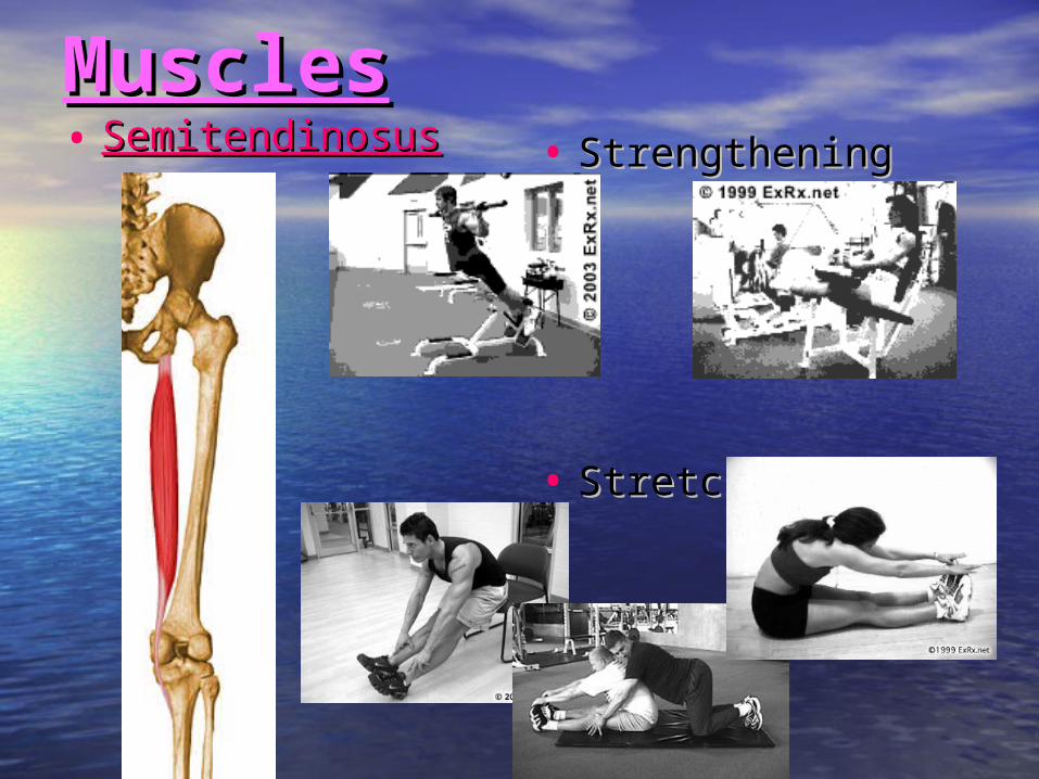
MusclesMuscles• Rectus Rectus FemorisFemoris • StrengtheningStrengthening
• StretchingStretching

MusclesMuscles• VastusVastus LateralisLateralis • StrengtheningStrengthening
• StretchingStretching

MusclesMuscles• VastusVastus IntermediusIntermedius • StrengtheningStrengthening
• StretchingStretching

MusclesMuscles• VastusVastus MedialisMedialis • StrengtheningStrengthening
• StretchingStretching

MusclesMuscles• StrengtheningStrengthening
• StretchingStretching
• Biceps Biceps FemorisFemoris– Long headLong head– Short headShort head
MusclesMuscles• StrengtheningStrengthening
• StretchingStretching
• SemitendinosusSemitendinosus

MusclesMuscles• StrengtheningStrengthening
• StretchingStretching
• SemimembranosusSemimembranosus

MusclesMuscles• GracilisGracilis • StrengtheningStrengthening
• StretchingStretching

MusclesMuscles• SartoriousSartorious • StrengtheningStrengthening
• StretchingStretching

MusclesMuscles• PopliteusPopliteus • StrengtheningStrengthening
• StretchingStretching
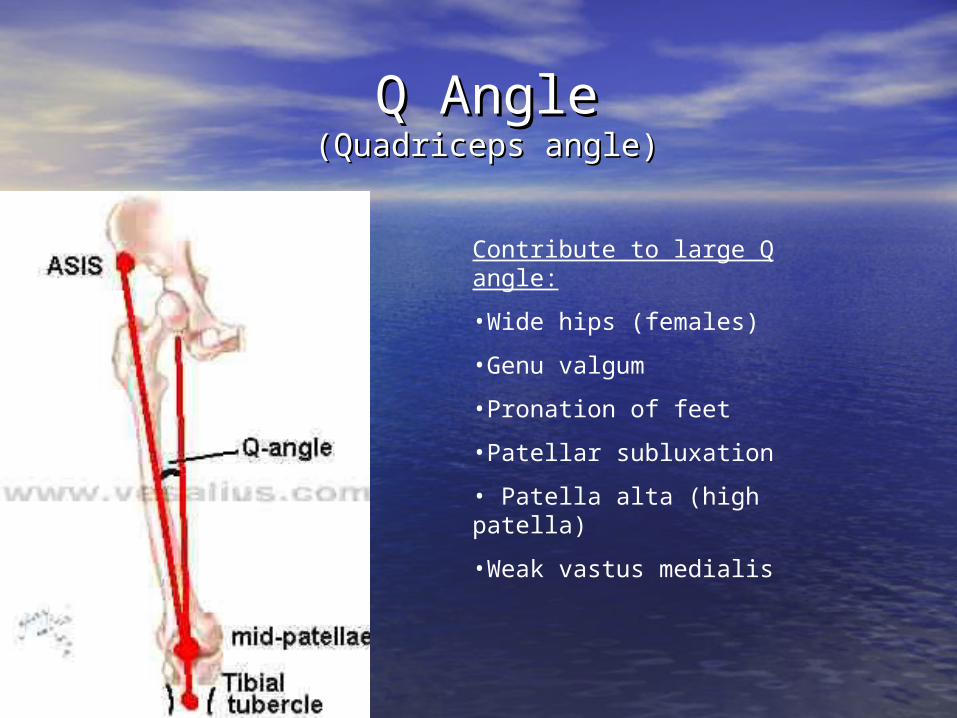
Q AngleQ Angle(Quadriceps angle)(Quadriceps angle)
Contribute to large Q angle:
•Wide hips (females)
•Genu valgum
•Pronation of feet
•Patellar subluxation
• Patella alta (high patella)
•Weak vastus medialis

Knee DeviationsKnee Deviations
• Genu Genu VarumVarum
a.a. Condyle to Condyle to malleoli malleoli relationshiprelationship
b.b. Relationship to Relationship to pronationpronation
c.c. Strain on soft Strain on soft tissuestissues
• Genu Genu ValgumValgum
a.a. Condyle to Condyle to malleoli malleoli relationshiprelationship
b.b. Relationship to Relationship to pronationpronation
c.c. Strain on soft Strain on soft tissuestissues
• Genu Genu RecurvatumRecurvatum
– May be caused by:May be caused by:
a.a. EquinusEquinus
b.b. Hamstring Hamstring weaknessweakness
c.c. Quadriceps Quadriceps weaknessweakness

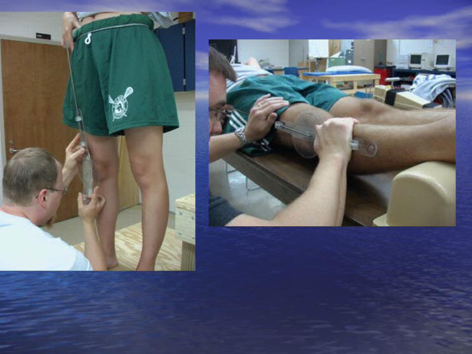
TorsionTorsion• an internal twist of a bone on itself. an internal twist of a bone on itself.
Requires a force - counterforce. Requires a force - counterforce.– Types of torsion. Types of torsion.
(Distal part is the reference)(Distal part is the reference)• Internal tibialInternal tibial• External tibialExternal tibial• Internal femoralInternal femoral• External femoralExternal femoral
– Testing procedureTesting procedure• Step #1: Determine that a torsion is present while subject Step #1: Determine that a torsion is present while subject
is standing (patella and feet do not line up)is standing (patella and feet do not line up)• Step #2: Have subject sit on table with feet dangling free.Step #2: Have subject sit on table with feet dangling free.
– Femoral torsion: Femoral torsion: Feet point straight aheadFeet point straight ahead
– Tibial torsion:Tibial torsion: Feet point out = external tibial torsionFeet point out = external tibial torsion Feet point in = internal tibial torsionFeet point in = internal tibial torsion