Embed Size (px)
Citation preview

Uterus Didelphys
Maria Georgiades, MDJune 24, 2010
Morbidity and Morbidity
downstatesurgery.org

Case Presentation
• 13 year old female presented with dull, intermittent abdominal pain x 2 days in the lower abdomen.
• The pain initially relieved with Motrin
• Similar pains in 12/2009.
downstatesurgery.org
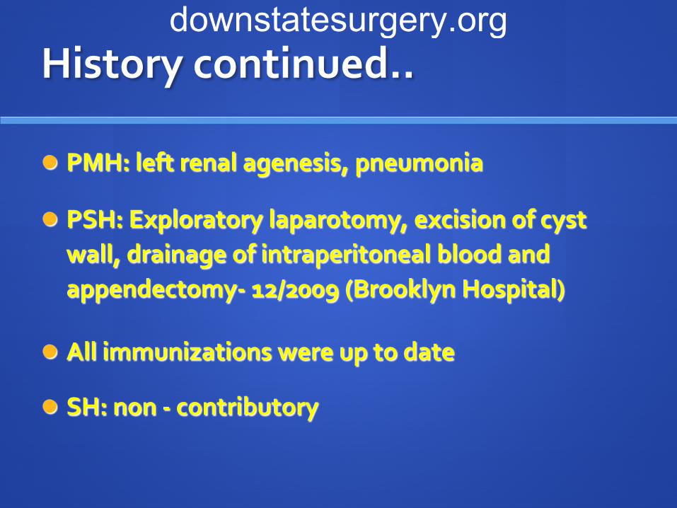
History continued..
PMH: left renal agenesis, pneumonia
PSH: Exploratory laparotomy, excision of cyst wall, drainage of intraperitoneal blood and appendectomy- 12/2009 (Brooklyn Hospital)
All immunizations were up to date
SH: non - contributory
downstatesurgery.org
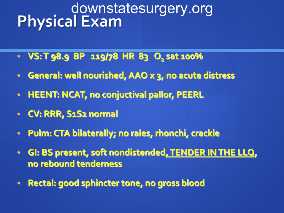
Physical Exam
• VS: T 98.9 BP 119/78 HR 83 O2 sat 100%
• General: well nourished, AAO x 3, no acute distress
• HEENT: NCAT, no conjuctival pallor, PEERL
• CV: RRR, S1S2 normal
• Pulm: CTA bilaterally; no rales, rhonchi, crackle
• GI: BS present, soft nondistended, TENDER IN THE LLQ, no rebound tenderness
• Rectal: good sphincter tone, no gross blood
downstatesurgery.org

Labs
CBC : 9.2/13/41/325
BMP: 139/5 101/26 12/0.8 <80
LFTs: 9.7/7.1/29/11/180/0.1
downstatesurgery.org
Differential Diagnosis
Ruptured ovarian cyst
Hematosalpinx
Ureterovaginal malformation
Uterus didelphys
downstatesurgery.org
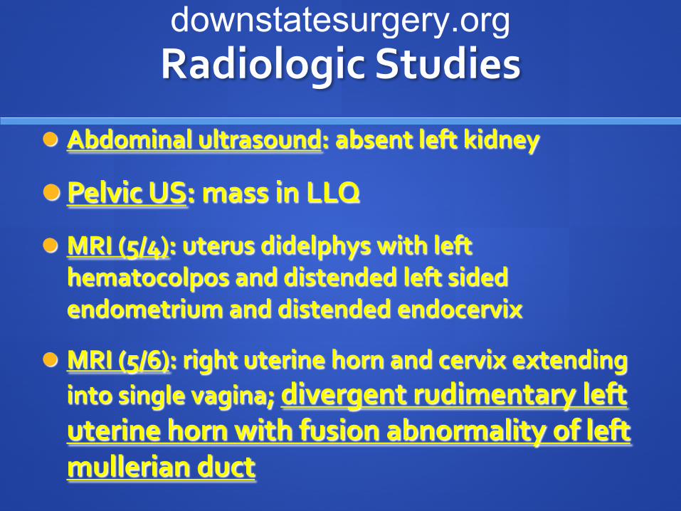
Radiologic Studies
Abdominal ultrasound: absent left kidney
Pelvic US: mass in LLQ
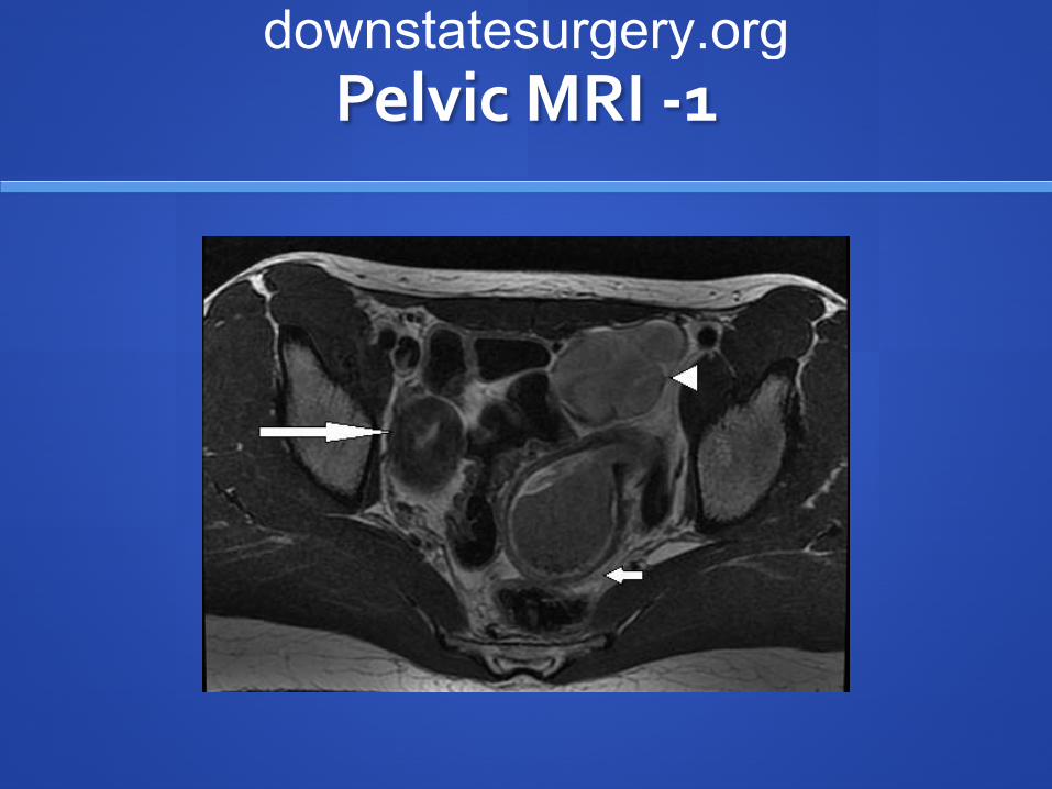
MRI (5/4): uterus didelphys with left hematocolpos and distended left sided endometrium and distended endocervix
MRI (5/6): right uterine horn and cervix extending into single vagina; divergent rudimentary left uterine horn with fusion abnormality of left mullerian duct
downstatesurgery.org

Pelvic Ultrasounddownstatesurgery.org
Pelvic MRI -1downstatesurgery.org

Pelvic MRI-2downstatesurgery.org

Hospital Course
• Taken for EUA, vaginoscopy and possible vaginal aspiration on HD # 2
• Patient was and taken for an exploratory laparotomy on HD #3
• Intra-operative findings: didelphys uterus– Right: normal uterine system with a right ovary and a
cervix opening into the vagina– Left: hematosalpinx with a distended uterus filled with
dark blood and normal ovary.
downstatesurgery.org

Intra-Operative Imagingdownstatesurgery.org
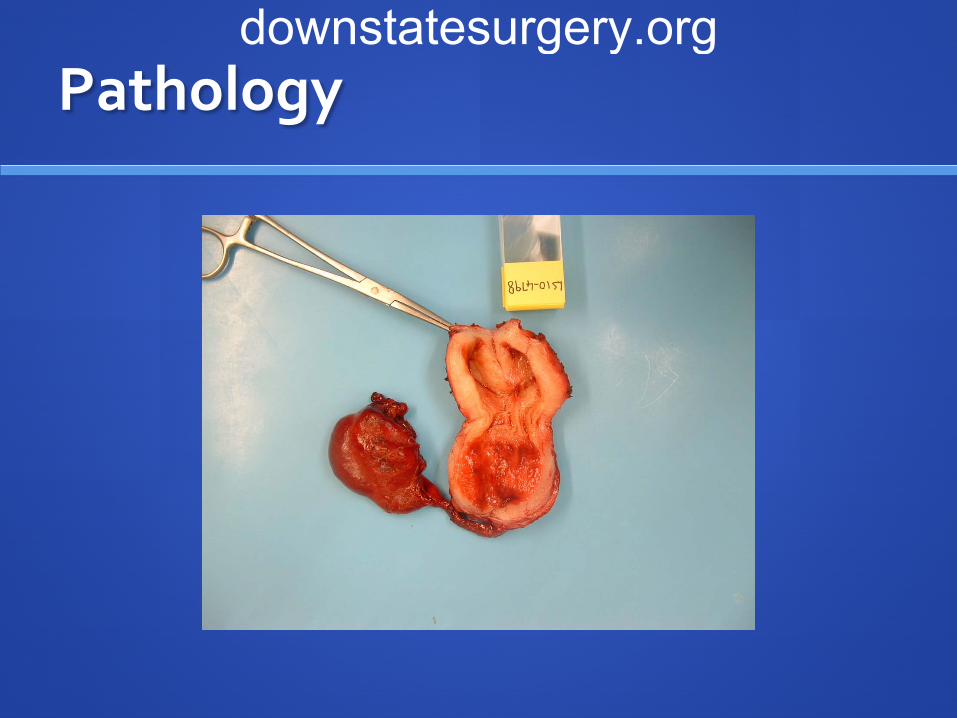
Pathologydownstatesurgery.org
Postoperative Course
Pathology: weak proliferation of endometrium; endocervix with hematosalpinx with paratubal
adenomatoid tumor Foci of endometriosis
Diet was advanced on POD # 1
Discharged on POD#2
downstatesurgery.org

Normal Uterusdownstatesurgery.org
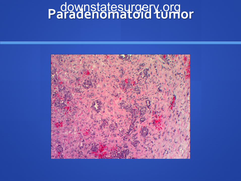
Paradenomatoid tumordownstatesurgery.org

Calretinin immunostaindownstatesurgery.org

Outline
Uterovaginal development
Mullerian malformations
Congenital fusion abnormalities
Herlyn-Werner-Wunderlich Syndrome
downstatesurgery.org

Figure 106-23 Development of uterus and vagina. During the 10th week, the paramesonephric ducts fuse at their caudal ends to establish a common channel and come into contact with a thickened portion of the posterior urogenital sinus called the sinovaginal bulb. This is followed by development of the vaginal plate, which elongates between the 3rd and 5th months and becomes canalized to form the inferior vaginal lumen. (Modified from Sadler TW: Langman's Medical Embryology. Baltimore, Williams & Wilkins, 1985.)
DEVELOPMENT downstatesurgery.org

Müllerian malformations
True duplication of the uterus is rare
Failed resorption of the common medial wall of
the paired mullerian ducts
More than 50 cases of uterus didelphys with a
unilateral imperforate vagina
Renal and axial skeletal abnormalies
downstatesurgery.org
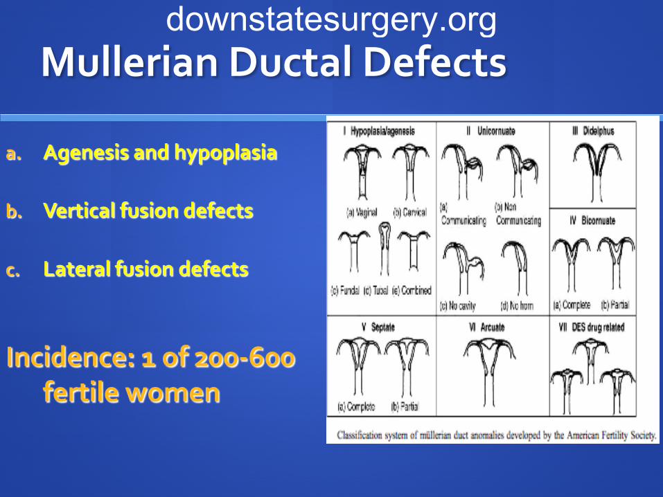
Mullerian Ductal Defects
a. Agenesis and hypoplasia
b. Vertical fusion defects
c. Lateral fusion defects
Incidence: 1 of 200-600 fertile women
downstatesurgery.org
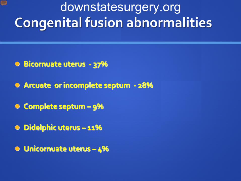
Congenital fusion abnormalities
Bicornuate uterus - 37%
Arcuate or incomplete septum - 28%
Complete septum – 9%
Didelphic uterus – 11%
Unicornuate uterus – 4%
downstatesurgery.org
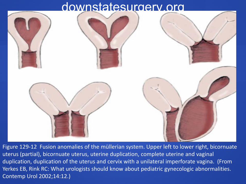
Figure 129-12 Fusion anomalies of the müllerian system. Upper left to lower right, bicornuate uterus (partial), bicornuate uterus, uterine duplication, complete uterine and vaginal duplication, duplication of the uterus and cervix with a unilateral imperforate vagina. (From Yerkes EB, Rink RC: What urologists should know about pediatric gynecologic abnormalities. Contemp Urol 2002;14:12.)
downstatesurgery.org

Herlyn-Werner-Wunderlich Syndrome
Didelphic uterus, obstructed hemivagina and ipsilateral renal and ureteral agenesis
Failure of both lateral fusion and vertical fusion
Theory: Damage to the caudal portion of the wolffian duct
downstatesurgery.org

Anatomical defect is a failure of lateral fusion of mullerian ducts combined with failure of vertical fusion between mullerian duct and urogenital sinus (hemivaginal septum ipsilateral to the side of renal ageneis. (Journal of Ped Surgery 2006. 41, 987-992)
downstatesurgery.org
Associated anomalies
Ipsilateral renal agensis is commonly seen
o Inferred that an early teratogenic process active in 4th
week of gestation resulted in arrested growth of one
mesonephric ductAgenesis of the ureteric bud
IVC duplication, intestinal malformation, ovarian
malposition
downstatesurgery.org
On history and physical…
Sx: asymptomatic until menarche cyclic or chronic pelvic pain Does not result in primary amenorrhea
PE: Unilateral abdomino-pelvic mass that
terminates in a bluish bulge in lateral vaginal mass
downstatesurgery.org

Diagnosis
Imaging: MRI: two uterine horns usually widely
separated, with preservation of the endometrial and myometrial widths
US: renal anomalies on the side ipsilateral to the obstructed system
downstatesurgery.org

Complications
• If left untreated:
– Endometriosis
– Pelvic adhesions
– Pyosalpinx or pyocolpos
• Spontaneous abortion – 40%
• Fertility is comparatively good
downstatesurgery.org
Management
Diagnostic Laparoscopy
Treatment: Wide incision of vertical vaginal septum to
release the entrapped menstrual blood Resection of the vaginal septum Marsupialization of vaginal margins Hemihysterectomy and salpingo-oophorectomy
downstatesurgery.org
Management and outcome of patients with combined vaginal septum, bifid uterus, and ipsilateral renal agenesis
(Herlyn-Werner-Wunderlich syndrome
80 patients with uterine and vaginal abnormalities 12 patients with HWWS Preferred surgical approach: full excision and
marsupialization of the vaginal septum
Hemihysterectomy with or without salpingooophoretomy is rarely indicated
J Pediatr Surg. 2006 May;41(5):987-92.
downstatesurgery.org

References
Management and outcome of patients with combined vaginal septum, bifid uterus, and ipsilateral renal agenesis (Herlyn-Werner-Wunderlich syndrome). Gholoum S, Puligandla PS, Hui T, Su W, Quiros E, Laberge JM. J Pediatr Surg. 2006 May;41(5):987-92.
Uterus didelphys with unilateral obstructed hemivagina with hematometrocolpos and hematosalpinx with ipsilateral renal agenesis Jindal G, Kachhawa S, Meena GL, Dhakar GJ Hum Reprod Sci. 2009 Jul;2(2):87-9
Wein: Campbell-Walsh Urology, 9th ed.; Chapter 129 - Surgical Management of Intersexuality, Cloacal Malformation, and other Abnormalities of the Genitalia in Girls > ... > Obstructive Genital Anomalies
Adam: Grainger & Allison's Diagnostic Radiology, 5th ed.
Uterus didelphys and longitudinal vaginal septum coincident with an obstructive transverse vaginal septum. Moawad NS - J Pediatr Adolesc Gynecol - 01-OCT-2009; 22(5): e163-5
downstatesurgery.org

Thank you
Special Thanks To Dr. Velcek.
downstatesurgery.org