Embed Size (px)
Citation preview

Printed in Japan F0386E2-062015
More than a capsule. A comprehensive system for capsule endoscopy.

Usabilityfor Streamlined Workflows
Efficiencyfor Stable Operations
Visualizationfor Detailed Observations
Trusted
Trusted
Trusted
Trusted. Revolutionary. And decidedly OLYMPUS.A comprehensive system for capsule endoscopy.
ENDOCAPSULE 10 SYSTEM reflects our vast experience in opto-digital technology for endoscopes.
This small-intestine endoscope system produces extremely high-quality images for fast, efficient and
precise examinations that you can trust — the ideal solution for medical institutions looking to expand
diagnostic capabilities in this critical field. OLYMPUS endoscopic imaging technology makes diagnosis easier than ever thanks to high-quality
images along with excellent usability and efficiency, all of which are hallmarks of our continually evolving
advancements in the field of endoscopy.
ENDOCAPSULE 10 SYSTEM. More than a capsule. A comprehensive system you can trust.
OLYMPUS’ trusted opto-digital technology
results in improved high-quality images
and a wide angle of view for accurate
observations and diagnosis.
Feature-rich and highly intuitive,
OLYMPUS software is the advanced
solution for fast, efficient analysis of
small-intestine examinations.
Trust OLYMPUS to fully support you and
your patients thanks to a new all-in-one
recorder, more convenient antenna and
functional reporting features.
2 3

As the undisputed leader in the field of endoscopy, OLYMPUS is renowned for exceptionally high-quality images.
This translates into easier analysis for more reliable and consistent diagnosis than before. You’ll also appreciate
the expanded angle of view, which makes it less likely to miss abnormalities. Another advancement made possible by renowned OLYMPUS optical technology is the expanded angle of view: 160° as opposed to 145° on the previous model. This wider coverage offers a significantly enhanced field of observation for refined examinations.
Battery life has been extended from eight hours to twelve hours to considerably increase the ratio of completed small-intestine observations. The long observation time maximizes the detection rate of lesions for more reliable diagnosis.
Eight user-selectable sharpness settings let you optimize image enhancement in order to observe tiny mucosal architecture clearly. You can also adjust color tone (red/blue) and brightness levels for more comfortable viewing in the color of your choice.
A difference you can immediately see.Crisp, clear images support accurate diagnosis.
Trusted Visualization for Detailed Observations
Advanced OLYMPUS optical technology delivers high-quality images that reveal individual villi with superb clarity. Noise has been markedly reduced along with halation, optimizing brightness levels for detailed observation of small-intestine mucosa and identification of abnormalities. The clear visual information facilitates highly accurate diagnosis.
Wide angle of view Longer observation time
Image adjustment function
High-quality images
Previousmodel
EC-S10
8H
12H
50% longer observationEC-S10
145°
160°
Previousmodel
Previous model EC-S10 Previous model EC-S10
Previous model
EC-S10
Level 3Level 1 Level 8
Angioma (no bleeding)
Observable symptomsNormal
Angioma(bleeding)
Multiple inflammations with Crohn disease stenosis
Less noise Less halation
EC-S10
Red 0 / Blue 0 Red -3 / Blue +3
Structure enhancement Color tone
The white lines indicate the angle of
view of the previous model.
4 5

Normal mode
Omni-selectedmode
Playback all images.
Playback only selected images.
Adjust modePlayback all images
at optimal speed.
High speed
Normal speed
Previous model
ENDOCAPSULE 10 SYSTEM with new algorithm
Adjacent image display function
Enlarging image functionBriefly place mouse over an image.
Click selected image.
Overview tab
Playback view tab
Red coloroverview tab
“Adjust” button
“Normal” button
“Omni” button
3D Track area
Change playback speed depending on differences in images. In Adjust mode, images showing no change are superimposed on each other, and review speed is optimized to move quickly past images indicating no characteristic differences compared to preceding images. This mode vastly reduces playback time to increase reading effectiveness.
Bubbles and debris can sometimes adhere to the capsule and degrade image quality. ENDOCAPSULE 10 SYSTEM automatically detects poor-quality images and displays only those that can be accurately read. This algorithm also enhances the performance of Adjust mode, and the Overview function.
This function displays a library of characteristic images. The new Adjacent image display and Enlarging image functions provide a quick way for further observation without having to switch to Playback view mode. In addition, the new Red color overview function gives you a quick overview only of images showing an excessive amount of red.
Images that overlap with previous ones are skipped, and new images are selected even when only minute changes are present. This algorithm can recognize that an image is similar, even when the capsule is displaying the same section of small intestine from a different angle. This intelligent approach helps to speed diagnosis by analyzing a wider number of attributes
than ever before.**Compared to ENDOCAPSULE 10 SYSTEM Express-selected mode
Track the capsule as it moves through the small intestine with the 3D Track function. A high-precision antenna recognizes the detailed signals from the capsule, allowing the system to display the capsule track in 3D. The track progress bar is useful for estimating capsule location in the small intestine. It also indicates on the 3D tracking screen where each thumbnail image was captured in order to assess the locations of abnormalities. The 3D Track function operates intuitively, showing capsule location to help you decide what approach should be taken for subsequent procedures.
ENDOCAPSULE 10 SYSTEM software facilitates reading with a variety of unique functions to detect images
requiring closer inspection, providing the means for quick review of results to ultimately speed diagnosis.
An absolute time-saver.Intelligent reading functions simplify analysis.
Trusted Efficiency for Stable Operations
Overview function Bubble and debris image detection algorithm
3D Track function
Adjust mode Omni-selected mode
6 7
3D Track area
Track progress bar

8 9
Preparation times are markedly reduced thanks to the slim, lightweight antenna unit, which is incorporated in the belt harness. The unit can be worn over light clothing, and offers more sensitive detection capability compared to the previous model while enhancing patient comfort.
Personalized instructions for each patient can be displayed by registering data. Instructions are delivered as text messages preceded by beep and vibration alerts. The messages direct patient activity, such as eating, drinking water and returning to the hospital. Making it easy for patients to follow correct procedures helps you conduct safer, more accurate examinations.
Confirm capsule location during the entire procedure from images displayed in real-time. Monitoring the capsule’s progress in real-time lets you uncover any anomalies, such as bleeding, and take immediate action if needed.
Considering the needs of medical personnel and patients, ENDOCAPSULE 10 SYSTEM is designed for optimal
clinical performance as well as outstanding ease-of-use and mobility. The all-in-one recorder and belt-style
antenna simplify procedures, making for a smooth and relaxed examination environment.
Check images of the small intestine as the capsule passes through it. Images of interest can be captured then downloaded to a workstation for further review.
Up to 15 captured images can be displayed as thumbnails, making it easy to quickly find suspected anomalies and further speeding observation procedures.
Ingest capsule
“Please come back to Hospital.”
“You can take a light meal from now.”
8:00
0:000:30
2:00
4:00
“You can drink water from now.”
“Please come back to procedure room.”
Guidance example
Designed for medical staff and patients.Examinations have never been this comfortable.
Trusted Usability for Streamlined Workflows
The recorder combines a receiver and viewer ina compact and easy-to-handle unit, allowing you to playback and capture images any time during the procedure. The recorder is rechargeable, and comes with a charging cradle. Just place the unit in the cradle to recharge.
Patient guidance function
Real-time view/Capture Playback/Capture Captured images screen
Smart recorder
Belt-style antenna unit
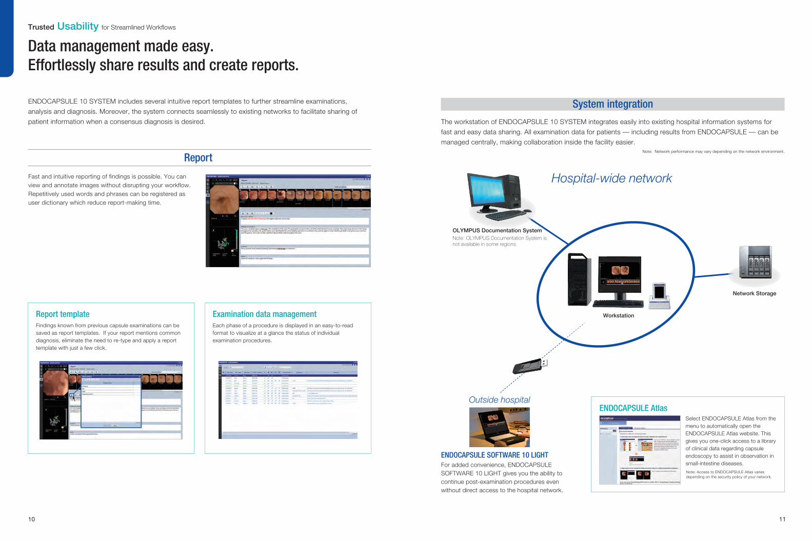
ENDOCAPSULE 10 SYSTEM includes several intuitive report templates to further streamline examinations,
analysis and diagnosis. Moreover, the system connects seamlessly to existing networks to facilitate sharing of
patient information when a consensus diagnosis is desired.
Fast and intuitive reporting of findings is possible. You can view and annotate images without disrupting your workflow. Repetitively used words and phrases can be registered as user dictionary which reduce report-making time.
The workstation of ENDOCAPSULE 10 SYSTEM integrates easily into existing hospital information systems for
fast and easy data sharing. All examination data for patients — including results from ENDOCAPSULE — can be
managed centrally, making collaboration inside the facility easier.
Hospital-wide network
ENDOCAPSULE AtlasSelect ENDOCAPSULE Atlas from the menu to automatically open the ENDOCAPSULE Atlas website. This gives you one-click access to a library of clinical data regarding capsule endoscopy to assist in observation in small-intestine diseases.
Note: Network performance may vary depending on the network environment.
Note: Access to ENDOCAPSULE Atlas varies depending on the security policy of your network.
Data management made easy.Effortlessly share results and create reports.
Trusted Usability for Streamlined Workflows
Note: OLYMPUS Documentation System is not available in some regions.
OLYMPUS Documentation System
Network Storage
Workstation
For added convenience, ENDOCAPSULE SOFTWARE 10 LIGHT gives you the ability to continue post-examination procedures even without direct access to the hospital network.
ENDOCAPSULE SOFTWARE 10 LIGHT
Outside hospital
Report
System integration
Report templateFindings known from previous capsule examinations can be saved as report templates. If your report mentions common diagnosis, eliminate the need to re-type and apply a report template with just a few click.
Examination data managementEach phase of a procedure is displayed in an easy-to-read format to visualize at a glance the status of individual examination procedures.
10 11

Printed in Japan F0386E2-062015
Specifications
ENDOCAPSULE RECORDER SET: MAJ-2029
Components 1. ENDOCAPSULE RECORDER: OLYMPUS RE-10
2. BATTERY PACK: MAJ-2030
3. ANTENNA UNIT: MAJ-2031
4. CRADLE: MAJ-2032
5. RECORDER HOLDER: MAJ-2033
6. ANTENNA UNIT HOLDER: MAJ-2034
7. CAPSULE ACTIVATOR: MAJ-1478
1 piece
1 piece
1 piece
1 piece
1 piece
1 set
2 pieces
ENDOCAPSULE SMALL INTESTINAL CAPSULE ENDOSCOPE: OLYMPUS EC-S10Optics
Sampling Rate
Battery Life
Size
Note: EC-S10 is not sold as single product but as MAJ-2027
Field of view
Depth of field
Weight
Dimensions
160 degrees
0-20 mm
2 fps
12 hours
3.3 g
ø11 mm (diameter) × 26 mm (length)
ENDOCAPSULE SOFTWARE 10: MAJ-2188
Components ENDOCAPSULE SOFTWARE 10 (DVD-R) 1 piece
ENDOCAPSULE SOFTWARE 10 LIGHT: MAJ-2189
Components ENDOCAPSULE SOFTWARE 10 LIGHT (DVD-R) 1 piece
ENDOCAPSULE SMALL INTESTINAL CAPSULE ENDOSCOPE SET: MAJ-2027
Components ENDOCAPSULE SMALL INTESTINAL CAPSULE
ENDOSCOPE: OLYMPUS EC-S10 5 pieces
RECORDER HOLDER: MAJ-2033
Size Weight
Dimensions
110 g (incl. strap)
Pouch: 100 mm (W) × 175 mm (H) × 45 mm (D)
ENDOCAPSULE RECORDER: OLYMPUS RE-10
Battery Life
Size
LCD display size
Weight
Dimensions
Typ. 12 hours
320 g
87 mm (W) × 154 mm (H) × 33 mm (D)
3.5 inch
BATTERY PACK: MAJ-2030
Type
Capacity
Voltage
Recharging Time
Size Weight
Dimensions
Lithium-ion storage cell
2860 mAh
3.7 V
Approx. 2 hours
70 g
70 mm (W) × 10 mm (H) × 55 mm (D) (without projection parts)
ANTENNA UNIT: MAJ-2031
Size Weight
Dimensions
150 g
87mm(W) x 51mm(H) x 15mm(D) (without projection parts)
CRADLE: MAJ-2032
Power supply
Size
Components
Weight
Dimensions
Cradle, AC Adapter, AC cable, USB cable
DC 6 V / 2 A
Main body: 315 g
142 mm (W) × 79 mm (H) × 85 mm (D)
ANTENNA UNIT HOLDER: MAJ-2034
Size Weight
Dimensions
260 g
Pouch: 340 mm (W) × 160 mm (H) × 15 mm (D)
Long belt: 50 mm (W) × 1000 mm (L)
Short belt: 50 mm (W) × 700 mm (L)
Plate: 290 mm (W) x 149 mm (D)
Cable band: 38 mm (W) x 200 mm (L)
1.
2.
4.
4.
4.
3.
5.
6.
6. 6.
6.
6.
7.