Embed Size (px)
Citation preview
URINARY SYSTEM
General Introduction
The urinary system is one of the most important excretory pathways in the body. The other excretory
pathways are (1) the alimentary tract; (2) the biliary system, which eliminates waste into the digestive tract via the
common bile duct; (3) the lungs, from which gases and chemicals are exhaled; and (4) the exocrine glands of the skin.
Metabolism of nutrients produces waste products, such as carbon dioxide, nitrogenous wastes, ammonia, and
detoxified organic and non-organic compounds, which must be eliminated from the body if normal function is to
continue. The urinary system is primarily concerned with the removal of nitrogenous wastes from the body.
Functions of the Urinary System
• Filters Waste Products from Blood - The urinary system eliminates in the urine different waste products
such as ammonia and urea (both formed when amino acids are broken down), Creatinine – by product of
muscle metabolism and uric acid (formed when nucleic acids are broken down).
• Regulates Ion levels in the Plasma - The urinary system also regulates ion levels in the plasma by regulating
the amount of sodium, potassium, chloride and other ions lost in the urine.
• Regulates Blood pH - The urinary system regulates blood pH by regulating the number of H+ and bicarbonate
ions (HCO3-) lost in the urine.
• Conserves Valuable Nutrients - At the same time, the urinary system makes sure that glucose, amino acids
and other valuable nutrients are not lost from the urine. The blood is filtered by the kidney through 3
processes called filtration, reabsorption, and secretion. The wastes leave the body as urine.
• Regulates Blood Volume - The urinary system regulates blood volume by releasing renin, a
chemical/hormone (do not confuse with rennin, a digestive product) restricts salt and water loss at the kidneys.
• Regulates RBC Production - If oxygen levels in the blood are low, the kidneys release erythropoietin, a
hormone that stimulates the hemocytoblasts (stem cells in the bone marrow) to increase red blood cell
formation. Having more RBCs allows the blood to transport more oxygen.
• Stores Urine - The bladder stores the urine until it is convenient to excrete it.
• Excretes Urine - The urethra transports urine from the urinary bladder to the outside of the body.
Kidneys:
A. Gross Anatomy: See textbook and L2S1 Lecture sheet
B. Microscopic Structure: See Texbook and L1S2 Lecture sheet
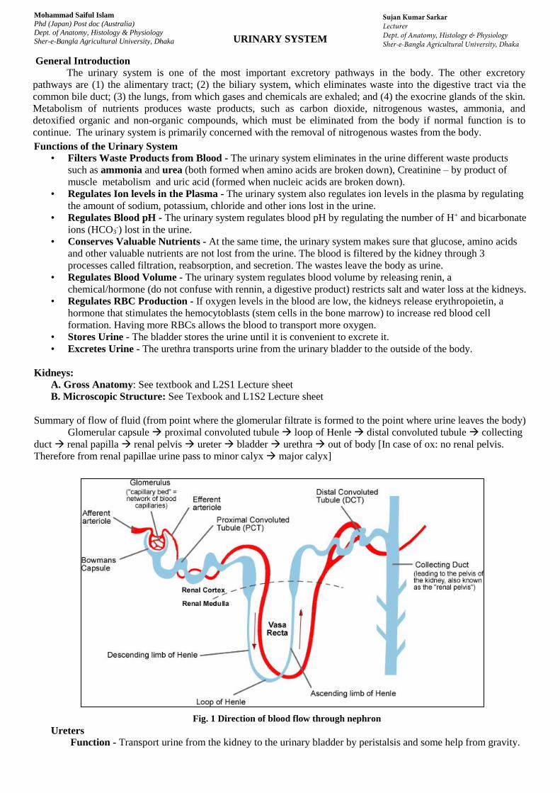
Summary of flow of fluid (from point where the glomerular filtrate is formed to the point where urine leaves the body)
Glomerular capsule proximal convoluted tubule loop of Henle distal convoluted tubule collecting
duct renal papilla renal pelvis ureter bladder urethra out of body [In case of ox: no renal pelvis.
Therefore from renal papillae urine pass to minor calyx major calyx]
Ureters
Function - Transport urine from the kidney to the urinary bladder by peristalsis and some help from gravity.
Fig. 1 Direction of blood flow through nephron
Mohammad Saiful Islam
Phd (Japan) Post doc (Australia)
Dept. of Anatomy, Histology & Physiology
Sher-e-Bangla Agricultural University, Dhaka
Sujan Kumar SarkarLecturerDept. of Anatomy, Histology & PhysiologySher-e-Bangla Agricultural University, Dhaka

Urinary Bladder
Gross/microscopic Structure: See Textbook and previous lecture sheet Function - Stores and expels urine. The bladder can hold about 1 pint ( about 500 mL.) of urine. When about 300
ml of urine collect, stretch receptors initiates a reflex called micturition. It causes the internal urethral sphincter to
contract. This sphincter is a thickening of the detrusor smooth muscle at the junction of the bladder and urethra.
This is involuntary smooth muscle. It keeps urine from dribbling between voidings. There is also another
sphincter, the external urethral sphincter which surrounds the urethra. This is voluntary skeletal muscle.
Urethra
• Function - This is a tube that transports urine from the bladder to the outside of the body.
• Gross structure/Histology: See Textbook and previous lecture
Blood Flow through the Kidney
Arterial flow:
The two renal arteries receive about one-fourth of the total cardiac output.
Each renal artery enters the hilus of the kidney and divides into a number of relatively large branches, the
interlobar arteries. These pass peripherally between pyramids almost to the cortex, where they bend abruptly
and become arcuate arteries (which derive their name from the arched manner) and they pass along the
junction between cortex and medulla. Each arcuate artery gives off a number of interlobular arteries that
extend into the cortex and in turn give rise to the afferent arterioles. Then afferent artery gives off tuft of
capillaries (glomerulus). Glomeruli produce filtrate that moves through the uriniferous tubule and becomes
urine. Then efferent arteriole - receives blood from the glomerulus. Peritubular capillaries and vasa recta.
The peritubular capillaries arise from the efferent arterioles draining the cortical glomeruli. They thread
around the rental tubule of the nephron. They absorb solutes and water from the tubule cells after these
substances are reabsorbed from the filtrate. The vasa recta arise from the efferent arterioles draining the
juxtamedullary glomeruli. (vas=vessel; recta=straight). The vasa recta are part of the kidney's urine-
concentrating mechanism.
Venous flow: The veins drain the blood in reverse of the arteries.
Interlobar veins>Arcuate veins> Renal vein> Caudal vena cava

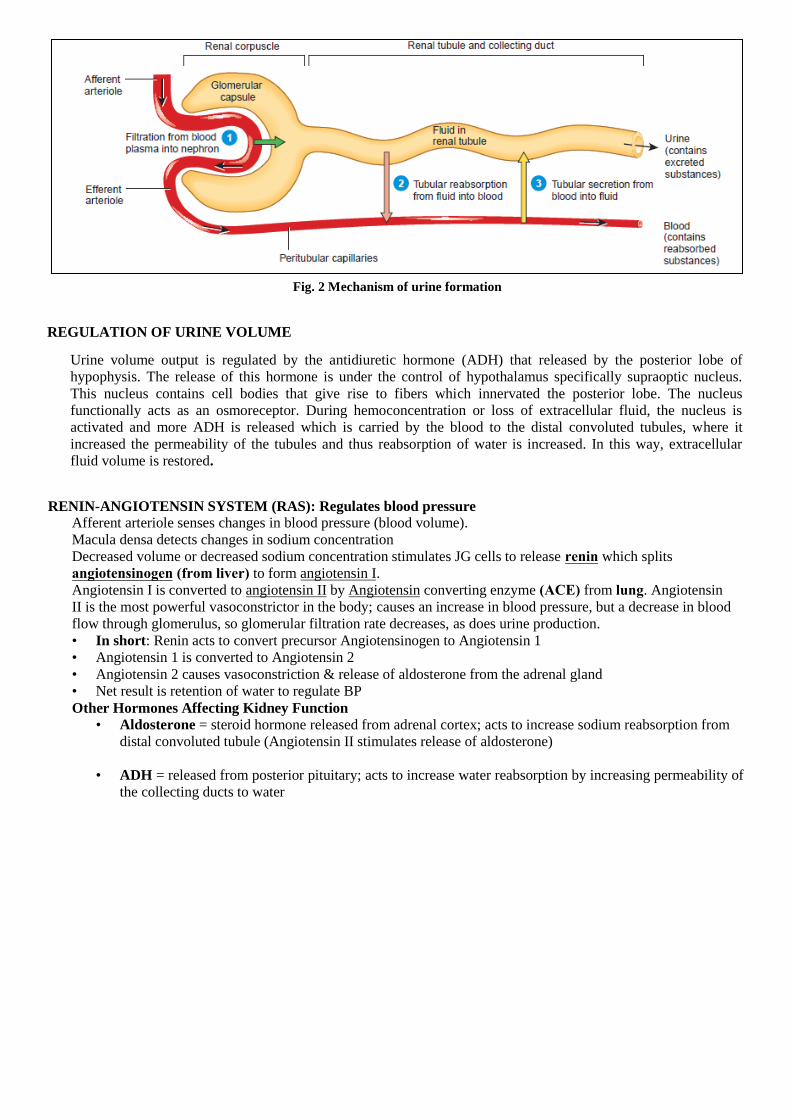
Formation of urine
The kidneys filter blood and produce urine. They filter out wastes and excess ions, which leave the body in the
urine, and return any needed substances back to the blood. In one day, the kidneys filter over 140 liters of blood (the
body’s entire volume of blood is filtered every 40 minutes).
Process: The kidneys produce urine through 3 processes:
A. Glomerular filtration – It is the first step of urine formation. Here, water, solutes smaller than proteins, and most
wastes passively pass out of the glomerulus capillaries into urinary pole of the Bowman’s capsule. This fluid is called
filtrate. Proteins and blood cells cannot pass through and so they are not part of the filtrate. In a glomerulus, there is an
ultrafiltration occur due to difference in hydrostatic pressure (HP). The HP in the glomerular capillary is estimated to
be about 70mmHg which is considerably higher than in the other capillaries. The blood osmotic pressure is
approximately 28mmHg. There is a back pressure, that is HP in capsule and tubule is about 10mmHg. Thus effective
glomerular filtration pressure is equal to about [70-(28+10)] = [70-38] = 32mmHg. So, the glomerular filtration takes
place due to the pressure difference of 32mmHg.
B.Tubular reabsorption - The filtrate moves into tubules that recover most of the nutrients, water, and essential ions
from the filtrate and return it to the blood of capillaries in surrounding connective tissue. The term reabsorption refers
to the return of substances to the bloodstream. The term absorption, by contrast, means entry of new substances into
the body, as occurs in the gastrointestinal tract. What is left becomes “urine” and is eventually excreted out.
C. Tubular secretion - As fluid flows along the renal tubule and through the collecting duct, the tubule and duct cells
secrete other materials, such as wastes, drugs, and excess ions, into the fluid. Tubular secretion removes a substance
from the blood.
Ureterovesicular junction: oblique entrance of ureter into the urinary bladder.
(A) Urine is conveyed to the urinary bladder from therenal pelvis by peristalsis and enters at the ureterovesicular junction.
(B)During micturition (emptying of the urinary bladder), urine is directed through the neck of the bladder to the urethra.
Urine does not reenter the ureter because the ureterovesicular junction is closed by the hydrostaticpressure of urine associated with contraction of the detrusor muscle of the bladder wall.
Figure: Ureterovesicular junction
REGULATION OF URINE VOLUME
Urine volume output is regulated by the antidiuretic hormone (ADH) that released by the posterior lobe of
hypophysis. The release of this hormone is under the control of hypothalamus specifically supraoptic nucleus.
This nucleus contains cell bodies that give rise to fibers which innervated the posterior lobe. The nucleus
functionally acts as an osmoreceptor. During hemoconcentration or loss of extracellular fluid, the nucleus is
activated and more ADH is released which is carried by the blood to the distal convoluted tubules, where it
increased the permeability of the tubules and thus reabsorption of water is increased. In this way, extracellular
fluid volume is restored.
RENIN-ANGIOTENSIN SYSTEM (RAS): Regulates blood pressure
Afferent arteriole senses changes in blood pressure (blood volume).
Macula densa detects changes in sodium concentration
Decreased volume or decreased sodium concentration stimulates JG cells to release renin which splits
angiotensinogen (from liver) to form angiotensin I.
Angiotensin I is converted to angiotensin II by Angiotensin converting enzyme (ACE) from lung. Angiotensin
II is the most powerful vasoconstrictor in the body; causes an increase in blood pressure, but a decrease in blood
flow through glomerulus, so glomerular filtration rate decreases, as does urine production.
• In short: Renin acts to convert precursor Angiotensinogen to Angiotensin 1
• Angiotensin 1 is converted to Angiotensin 2
• Angiotensin 2 causes vasoconstriction & release of aldosterone from the adrenal gland
• Net result is retention of water to regulate BP
Other Hormones Affecting Kidney Function • Aldosterone = steroid hormone released from adrenal cortex; acts to increase sodium reabsorption from
distal convoluted tubule (Angiotensin II stimulates release of aldosterone)
• ADH = released from posterior pituitary; acts to increase water reabsorption by increasing permeability of
the collecting ducts to water
Fig. 2 Mechanism of urine formation

ROLE OF COLLECTING DUCT
• Urine passes into COLLECTING DUCT for final run down through the gradient
• Walls of CD are permeable: water is drawn out and urine is concentrated
• COUNTERCURRENT FLOW assures maximum concentration
• Urine is low in volume and high in osmolarity; depends on steepness of gradient & length of loop
DIURESIS: excretion of copious amount of urine
• If CT wall is NOT permeable to water, copious dilute urine is released
• Permeability of CT wall is under hormonal control
– ADH from pituitary causes increased permeability and hence decreased volume
Fig. 3 Regulation of blood Pressure
Diuretics: Diuretics or diuretic agents are the substances, which enhance the output of urine.
Type of diuretics-Diuretics are classified into seven types:
1. Osmotic diuretics
2. Diuretics which inhibit active reabsorption of electrolytes
3. Diuretics which inhibit action of aldosterone
4. Diuretics which inhibit the activity of carbonic anhydrase
5. Diuretics which increase glomerular filtration rate
6. Diuretics which inhibit secretion of ADH
7. Diuretics which inhibit ADH receptors
MICTURITION
Micturition is the act of expelling urine from the bladder. It is normally a reflex activity but can be overridden
by voluntary control from the brain. The steps involved are:
• Bladder distends with urine formed by the kidneys
• Stretch receptors in the smooth muscle of the bladder wall are stimulated and send nerve impulses to centres
in the appropriate segment of the spinal cord
• Nerve impulses are transmitted via parasympathetic nerves back to the smooth muscle, and initiate contraction
• Nerve impulses also stimulate relaxation in the internal bladder sphincter and urine is expelled.
• The neck of the bladder ends in the bladder sphincter, whose function is to control the flow of urine out of the
bladder and down the urethra. It consists of two concentric parts:
Internal sphincter – made of smooth muscle; under involuntary control
External sphincter – ring of striated muscle; under voluntary control.
Creatinine is a nitrogenous byproduct of muscle metabolism. The major reaction that produces creatinine is
the spontaneous loss of phosphoric acid from creatine phosphate in muscle. Creatinine production is independent of
protein metabolism. The amount produced depends on the mass of muscle in the body and is very constant from day
to day. Because it is constantly produced, it is constantly excreted, and normal plasma creatinine concentrations are 0.5 to 2.0 mg/dL.
URINALYSIS: ANALYSIS OF URINE
Urine is derived from the ultrafiltrate of plasma so it reflects the health status of the whole animal. The analysis of
urine, or urinalysis, is a useful diagnostic tool. Normal urine contains only water, salts and urea.
Normal constituents of urine
• Water – 95%
• Solutes – 5%
– Urea – from metabolism of amino acids
– Creatinine – by product of muscle
metabolism
– Uric acid – from catabolism of nucleic acids
– Urobilinogen – breakdown of hemoglobin
– Hippuric acid, indican, and ketone bodies
– Ions
Abnormal Constituents of Urine
• Albuminuria
• Glucosuria
• Hematuria
• Pyuria
• Ketonuria (Ketosis)
• Bilirubinuria
• Casts
• Renal calculi
• Microbes
Urine is formed to keep the composition of the ECF constant. Color. Urine is usually yellow in color. The yellow color is derived from bilirubin that was excreted into the intestine
and reabsorbed into the portal circulation as urobilinogen. Much of the urobilinogen is reexcreted by the liver into the
intestine, but urobilinogen that bypasses the liver can be excreted by the kidneys into the urine. The various bilinogens
are colorless but are spontaneously oxidized on exposure to oxygen. Thus urobilinogen, when partially oxidized, is
known as urobilin, and it is largely responsible for the yellow color of urine.

Odor. The odor of urine is characteristic for a species and is probably influenced by diet. For example, the
characteristic odor
imparted to human urine after the ingestion of asparagus is caused by the formation of asparagine (the amide form of
the amino
acid, aspartic acid).
Consistency. Urine has a watery consistency in most species. Horse urine is somewhat thick and syrupy, however,
because of the secretion of mucus from glands in the pelvis of the kidneys and the upper part of the ureters. The urine
of the horse has high concentrations of carbonates and phosphates, which seem to precipitate on standing. The
secretion of mucus provides a carrier for the precipitated carbonates and phosphates and prevents their collection in
the renal pelvis.
Nitrogenous component. The principal nitrogenous constituent of mammalian urine is urea. Urea is formed by the
liver from ammonia, which is produced during amino acid metabolism. The body expends considerable energy in
producing urea so that the toxicity of ammonia can be avoided. As compared with ammonia, urea is relatively
nontoxic at normal concentrations.
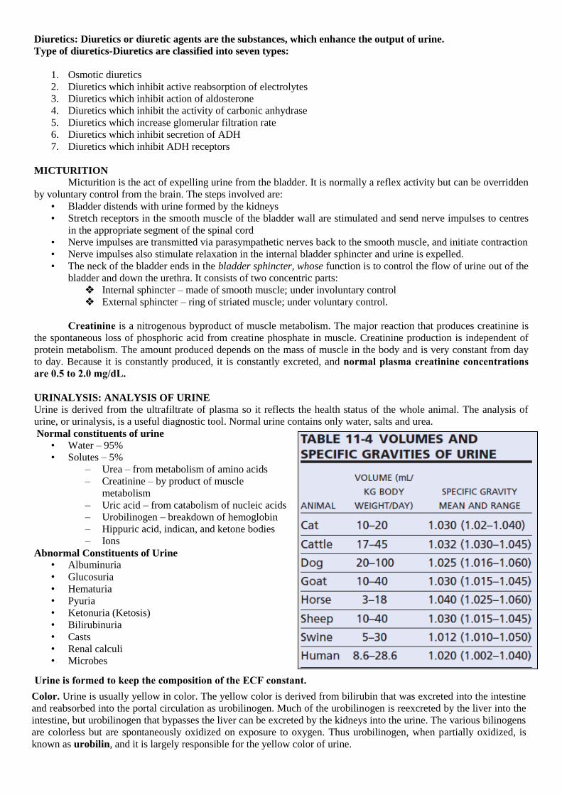
Amount and specific gravity. The amount of urine excreted daily varies with diet, work, external temperature, water
consumption, season, and other factors. Marked pathologic variations may occur. The specific gravity of urine varies
with the relative proportion of dissolved matter and water. In general, the greater the volume, the lower the specific
gravity. Volume and specific gravities for several domestic animals are shown in Table 11-4.
Descriptive Terms Urinary continence is the normal condition of storing urine in the bladder while it fills. Continence is maintained by
continuous
tone of the external sphincter muscle and by closure of the neck of the bladder
Polyuria refers to increased urine output, oliguria means decreased output, and anuria describes the condition of no
output. Dysuria is a term used to describe diffi cult or painful micturition. Stranguria is slow, dropwise, painful
discharge of the urine caused by spasm of the urethra and bladder.
Renal blood flow (RBF): refers to the rate at which blood flows to the kidneys. In as much as plasma is the fluid part of the blood, from which the glomerular filtrate is formed, renal plasma flow (RPF) refers to that part of the RBF that is plasma. As long as there continues to be an RBF, a glomerular filtrate will be formed from the plasma at the glomerulus. The rate at which it is formed is known as the glomerular filtration rate (GFR) and is measured in milliliters per minute.RBF and RPF are also measured in milliliters per minute, and the ratio of GFR to RPF is referred to as the filtration fraction (FF).
Renal Clearance
Volume of blood or plasma that is completely cleared or emptied, of any substance in one minute is called renal clearance.is a measurement of the kidney’s ability to remove substances from the plasma.
UVC = ----------
PWhere, C = plasma clearance in ml/min U = concentration of the substances in urine V = rate of urine flow/ formation in ml/min
P = concentration of the substances in the plasma

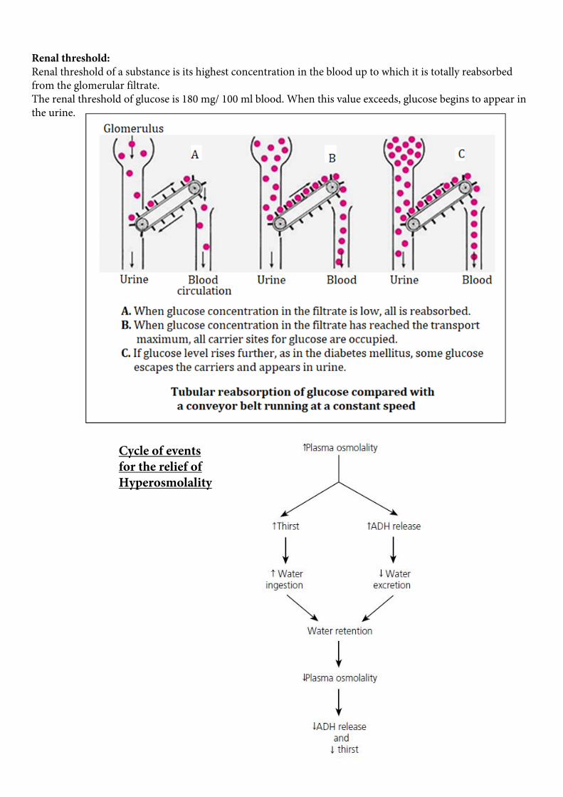
Cycle of events for the relief of Hyperosmolality
Renal threshold:Renal threshold of a substance is its highest concentration in the blood up to which it is totally reabsorbed from the glomerular filtrate. The renal threshold of glucose is 180 mg/ 100 ml blood. When this value exceeds, glucose begins to appear in the urine.
Dialysis:Dialysis is the procedure to remove the waste materials and toxic substances and to restore normal volume and composition of body fluid in severe renal failure . It is also called hemodialysis.
Artificial kidney:Artificial kidney is the machine that is used to carry out dialysis during renal failure.
Condition when Artificial kidney required:Artificial kidney is used to treat patients suffering from - # Acute renal failure that occur due to circulatory shock or mercury poisoning # Chronic / permanent renal failure
Frequency and duration of dialysis:Frequency and duration of dialysis depends upon the severity of renal function. Dialysis is done usually thrice a week in severe uremia. Each time artificial kidney is used for about 6 hours.
Complication of dialysis:Sleep disorders, Anxiety , Depression
RENAL DISORDERS
1. Renal failureThe term renal failure refers the excretory function which is characterized by decrease of GFR. Types of renal failureA. Acute renal failure- It is the abrupt stoppage of renal functions.Causes:Acute nephritis, Renal ischemia, Acute tubular necrosis, Severe transfusion reactions, Sudden fall in blood pressure during hemorrhage, diarrhea, severe burns and cholera, Blockage of ureter due to formation of calculi, Damage of renal tissues by poisons like lead (Pb), mercury (Hg),Carbon tetrachloride(CCll4)Features: Oliguria -Decreased urinary output, Anuria -Cessation of urine formation , Proteinuria -Appearance of proteins in urine, Hematuria -Presence of blood in urine
B. Chronic renal failure- It is the progressive, long standing and irreversible impairement of renal functions.Causes:Chronic nephritis, Polycystic kidney disease, Renal calculi, Hypertension, Atherosclerosis, Tuberculosis, Slow poisoning by drugs or metal.Features: Uremia- Presence of urinary constituents in the blood, Acidosis, Edema, Anemia
2. Polyuria- A large increase in urine volume output than normal.Physiological causes of polyuria: Increase water conjumption, Diuretics, Intake of salting food, Fear, emotional stages.Pathological causes of polyuria: Diabetes mellitus, Diabetes insipidus, Pyometra, Generalized liver disease.
3. Glycosuria- When glucose come out with urine called glycosuria.

AVIAN URINARY SYSTEM
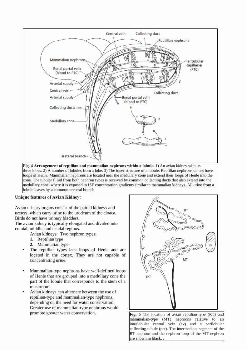
Unique features of Avian Kidney:
Avian urinary organs consist of the paired kidneys and
ureters, which carry urine to the urodeum of the cloaca.
Birds do not have urinary bladders.
The avian kidney is typically elongated and divided into
cranial, middle, and caudal regions.
Avian kidneys: Two nephron types:
1. Reptilian type
2. Mammalian type
• The reptilian types lack loops of Henle and are
located in the cortex. They are not capable of
concentrating urine.
• Mammalian-type nephrons have well-defined loops
of Henle that are grouped into a medullary cone the
part of the lobule that corresponds to the stem of a
mushroom.
• Avian kidneys can alternate between the use of
reptilian-type and mammalian-type nephrons,
depending on the need for water conservation.
Greater use of mammalian-type nephrons would
promote greater water conservation. Fig. 5 The location of avian reptilian-type (RT) and
mammalian-type (MT) nephrons relative to an
intralobular central vein (cv) and a perilobular
collecting tubule (pct). The intermediate segment of the
RT nephron and the nephron loop of the MT nephron
are shown in black. .
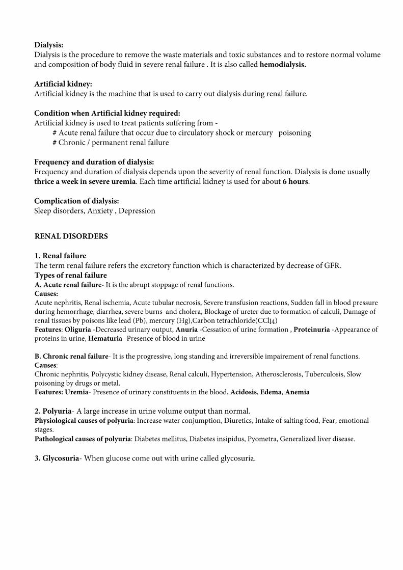
Fig. 4 Arrangement of reptilian and mammalian nephrons within a lobule. 1) An avian kidney with its
three lobes. 2) A number of lobules from a lobe. 3) The inner structure of a lobule. Reptilian nephrons do not have
loops of Henle. Mammalian nephrons are located near the medullary cone and extend their loops of Henle into the
cone. The tubular fl uid from both nephron types is received by common collecting ducts that also extend into the
medullary cone, where it is exposed to ISF concentration gradients similar to mammalian kidneys. All urine from a
lobule leaves by a common ureteral branch
• 25% of the filtrate comes from mammalian-type nephrons and 75% from reptilian-type nephrons.
Renal Portal System: A unique feature of the avian kidney is its renal portal system. The renal portal blood is venous blood that comes to
the kidney from the hindlimbs via the external iliac and isciatic veins. Blood enters the kidney from its periphery,
supplying afferent blood to the peritubular capillaries. Within the peritubular capillaries, it is mixed with efferent
arteriolar blood coming from the glomeruli. The mixture perfuses the tubules and proceeds to the central vein. Cortical
nephrons are arranged radially around central (intralobular) veins of the cortex of the lobule. The renal portal system
supplies 1/2 to 2/3 of the blood to the kidneys. There is a valve, known as a renal portal valve, located at the juncture
of the right and left renal veins.
Formation of Uric Acid:In reptiles and birds, uric acid is formed instead of urea because these animals develop in eggshells that are
impervious to water. The excretion of urea obligates water excretion. Because there is only limited water in eggs, it
must be conserved. Uric acid reaches a certain concentration, and it precipitates. As a precipitate (no effective osmotic
pressure), there is no water obligated in its excretion. If urea were excreted it would be necessary to eliminate the
liquid urine formed, and this is not possible within eggs.
Just as urea is formed in the liver of mammals from ammonia, so is uric acid formed in the liver of birds from
ammonia. The kidneys of birds are also a site for the formation of uric acid. Uric acid precipitates in the tubules
because the extra blood from the renal portal system that perfuses the tubules leads to greater tubular secretion and
consequently greater tubular concentration. The greater amounts in the tubules exceed uric acid solubility, and it
precipitates. Uric acid continues in the tubules in its precipitated form and appears in the urine as a white coagulum.
Because uric acid is no longer in solution, it does not contribute to the effective osmotic pressure of the tubular fluid,
and obligatory water loss is avoided.
Modification of Ureteral Urine
After presentation of ureteral urine to the cloaca, there is backflow (retrograde flow) of urine into the cecum and colon
by antiperistalsis. In the cecum and colon, Na+ is reabsorbed and water is reabsorbed by osmosis. There is no water
reabsorbed from the cloaca even though there may be some Na + reabsorption.
Urine Characteristics and Flow
Bird urine is cream-colored and contains thick mucus. The precipitated uric acid is mixed with the mucus, whereby
the mucus secretion facilitates transport of the precipitate, similar to the mucus in equine urine that facilitates
the transport of the carbonates and phosphates that precipitate. Urine flow for hydrated chickens is reported to be about 18 mL/kg/hr, and for hydrated turkeys it is about 30 mL/kg/hr.
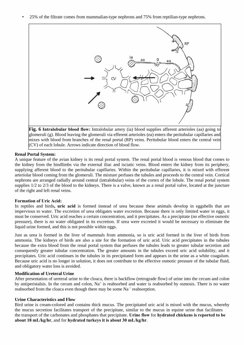
Fig. 6 Intralobular blood flow: Intralobular artery (ia) blood supplies afferent arterioles (aa) going to
glomeruli (g). Blood leaving the glomeruli via efferent arterioles (ea) enters the peritubular capillaries and
mixes with blood from branches of the renal portal (RP) veins. Peritubular blood enters the central vein
(CV) of each lobule. Arrows indicate direction of blood flow.

The Avian Salt Gland/ Nasal GlandAll birds have nasal or salt glands located above the eye that are distinct from Harderian and lacrimal glands which in many species produces a nonserous, nonmucoidal secretion having uncertain function. These glands are well developed in marine species and are capable of producing copious secretions containing high concentrations of NaCl. Because of their osmoregulatory function in these species, they have been called salt glands. The ostrich, cormorant, duck, goose, falcon, gull, and penguin all have functioning salt glands.
Structure: Avian salt glands are derived embryologically from invaginations in the nasal epithelium that persist as the main ducts of the gland. They are paired and are composed of tubular lobes (Figure A,B) that are parallel and run the length of the gland. Each lobe has a central canal that is continuous with a duct of gland. The secretion is formed in secretory tubules, arranged radially around the central canal of each lobe and are continuous with it (Figure C,D). The epithelial cells comprising the tubules are responsible for the secretion process.
Blood Flow: The blood flow to the gland forms a network of capillaries that course along the tubules to the periphery of the lobes where veins collect the blood near the surface (Figure C,D). The salt glands have a structure entirely different from that of the kidney and can excrete a salt solution of up to twice the concentration of sea water.
Secretion/ Function: These glands secrete the excess salt when food with high salt content is ingested or when sea water is drunk. The salt secretion flows through the salt gland ducts into the nasal cavity, runs out through the nares, and drips from the tip of the beak to be sneezed out of the nostrils. The salt glands secrete only NaCl and none of the other substances excreted by the kidneys. They function only when there is a salt load; otherwise they are at rest.