Embed Size (px)
Citation preview

C26
Urinary System(Slides Matched to Science Department Lecture Objectives)

Review of Anatomy


Female Urethra• 3 to 4 cm long
• bound to anterior wall of vagina
• external urethral orifice
– between vaginal orifice and clitoris
• internal urethral sphincter
– detrusor muscle thickening– smooth muscle under
involuntary control
• external urethral sphincter
– where the urethra passes through the pelvic floor
– skeletal muscle under voluntary control
Ureter
Urethra
(a) Female
DetrusormuscleUreteralopenings
UrogenitaldiaphragmExternal urethralorifice
Trigone
External urethralsphincter
Internal urethralsphincter

Male Urethra • 18 cm long
• 3 regions of male urethra
– prostatic urethra (2.5 cm) // passes through prostate gland
– membranous urethra (.5 cm) // passes through muscular floor of pelvic cavity
– spongy (penile) urethra (15 cm) // passes through penis in corpus spongiosum
• internal urethral sphincter // detrusor muscle thickening
• external urethral sphincter // part of skeletal muscle of pelvic floor
Ureter
Prostatic urethraProstate gland
Penis
External urethral orifice
Rugae
(b) Male
Ureteralopenings
Trigone
MembranousurethraBulbourethralgland
Spongy (penile)urethra
Detrusormuscle
Internal urethralsphincter
UrogenitaldiaphragmExternal urethralsphincter

(1) Describe the function of the urinary system:

Functions of the Kidney (1 of 2)
• filters blood plasma, separates waste from useful chemicals, returns useful substances to blood, eliminates wastes
• regulate blood volume and pressure by eliminating or conserving water
• regulate osmolarity of the body fluids by controlling the relative amounts of water and solutes eliminated
• secretes enzyme, renin, which activates hormonal mechanisms that control blood pressure and electrolyte balance

Functions of the Kidney (2 of 2)
• secretes the hormone, erythropoietin, which stimulates the production of red blood cells
• collaborate with the lungs to regulate the PCO2 and acid-base balance of body fluids
• final step in synthesizing hormone, calcitriolconverted to calcitriol in kidney // active form of Vit D / which contributes to calcium homeostasis
• gluconeogenesis from amino acids in extreme starvation
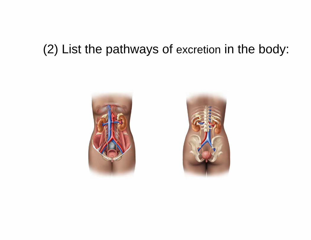
(2) List the pathways of excretion in the body:

Waste Products & Kidney Function
• ‘To live is to metabolize’ // metabolism creates a variety of toxic waste products (e.g. nitrogen and acids)
• These waste products of metabolism must be removed from the body using these systems
– Respiratory
– Digestive
– Sweat glands
– Urinary system

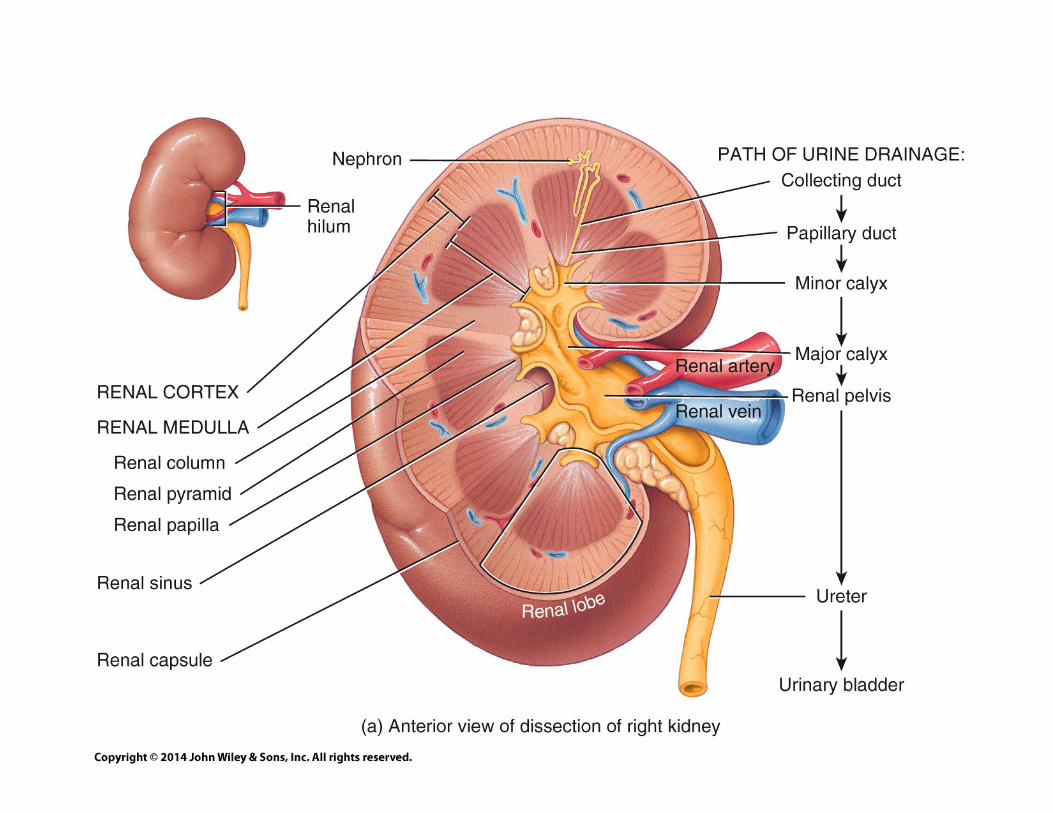
(3) List the major organs of the urinary system and give the generalized functions of each:

urinary system consists of 6 organs:
2 kidneys, 2 ureters, urinary bladder, and urethra
Urinary System
Ureter
Diaphragm
(b) Posterior view
11th and 12th ribs
Urinary bladder
Urethra
Inferior vena cavaAorta
Renal arteryRenal vein
Adrenal gland
KidneyVertebra L2
(a) Anterior view

Kidney Location
Small intestine
Ureter
KidneySpleen
Fibrous capsule
Renal fascia Lumbar muscles
L1
Inferior vena cavaAorta
Hilum
Pancreas
Stomach
Peritoneum
Colon
Anterior
Posterior
Renal arteryand vein
Perirenalfat capsule

The Ureter
• retroperitoneal, muscular tube that extends from the kidney to the urinary bladder
– about 25 cm long
– passes posterior to bladder and enters it from below
– flap of mucosa acts as a valve into bladder
• keeps urine from backing up in the ureter when bladder contracts

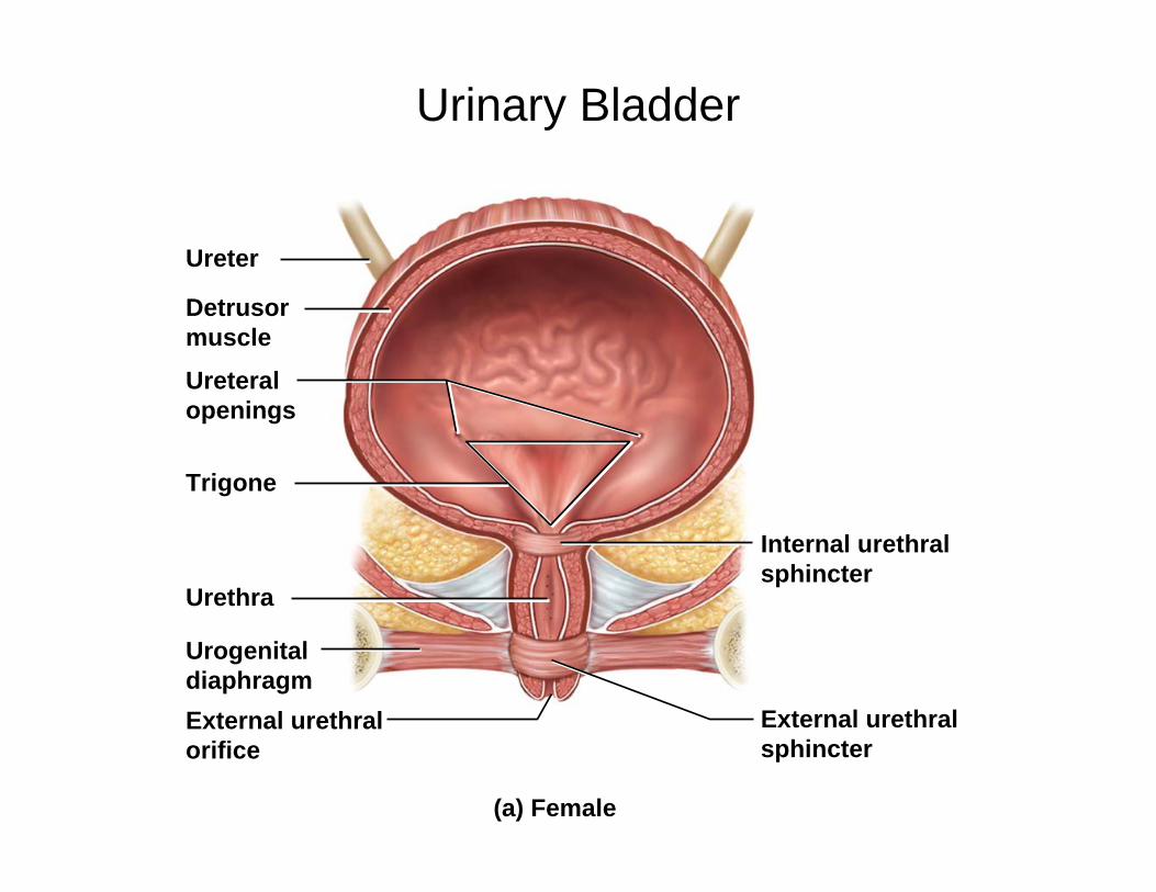
Urinary Bladder (1 of 2)
• urinary bladder - muscular sac located on floor of pelvic cavity
– inferior to peritoneum and posterior to pubic symphysis
• 3 layers
– parietal peritoneum, superiorly, fibrous adventitia other areas
– muscularis /// detrusor muscle /// 3 layers of smooth muscle
– mucosa - transitional epithelium
• rugae - conspicuous wrinkles in relaxed bladder

Urinary Bladder (1 of 2)
• trigone – smooth-surfaced triangular area marked with openings of ureters and urethra
• capacity - full is 500 ml, max. is 700 - 800 ml
– highly distensible
– as it fills, it expands superiorly
– rugae flatten
– epithelium thins from five or six layers to two or three
Urinary Bladder
Ureter
Urethra
(a) Female
Detrusormuscle
Ureteralopenings
UrogenitaldiaphragmExternal urethralorifice
Trigone
External urethralsphincter
Internal urethralsphincter

Female Urethra• 3 to 4 cm long
• bound to anterior wall of vagina
• external urethral orifice
– between vaginal orifice and clitoris
• internal urethral sphincter
– detrusor muscle thickening– smooth muscle under
involuntary control
• external urethral sphincter
– where the urethra passes through the pelvic floor
– skeletal muscle under voluntary control
Ureter
Urethra
(a) Female
DetrusormuscleUreteralopenings
UrogenitaldiaphragmExternal urethralorifice
Trigone
External urethralsphincter
Internal urethralsphincter

Male Urethra• 18 cm long
• 3 regions of male urethra
– prostatic urethra (2.5 cm) // passes through prostate gland
– membranous urethra (.5 cm) // passes through muscular floor of pelvic cavity
– spongy (penile) urethra (15 cm) // passes through penis in corpus spongiosum
• internal urethral sphincter // detrusor muscle thickening
• external urethral sphincter // part of skeletal muscle of pelvic floor
Ureter
Prostatic urethraProstate gland
Penis
External urethral orifice
Rugae
(b) Male
Ureteralopenings
Trigone
MembranousurethraBulbourethralgland
Spongy (penile)urethra
Detrusormuscle
Internal urethralsphincter
UrogenitaldiaphragmExternal urethralsphincter

(4) Name the parts of a nephron and describe the role of each component in the formation of urine:

Renal capsule
Collecting ductNephron
(a)
(c)
Cortical nephron
CortexMedulla
GlomerulusGlomerular capsule
Renal corpuscle:
Nephron loop:Descending limbAscending limb
Thick segmentThin segment
Flow of tubular fluidFlow of blood
Key
(b)
Renalcortex
Renalmedulla
Renalpapilla
Minorcalyx
Efferentarteriole
Afferentarteriole
Proximalconvolutedtubule (PCT)
Distalconvolutedtubule (DCT)
Collectingduct (CD)
Papillaryduct
Collectingduct
Nephronloops
Juxtamedullarynephron
Convoluted tubules(PCT and DCT)
The NephronThe nephron is the “functional unit” of a kidney
each kidney has about 1.2 million nephrons
The nephron is composed of two principal parts:
renal corpuscle – filters the blood plasma
renal tubules – long coiled tube that converts the filtrate into urine

Renal Corpuscle
• glomerular filtrate collects in capsular space, flows into proximal convoluted tubule. Note the vascular and urinary poles. Note the afferent arteriole is larger than the efferent arteriole.
Flow of filtrateFlow of blood
Key
Afferentarteriole
Bloodflow
Efferentarteriole
Blood flow
Glomerularcapillaries(podocytesand capillarywallremoved)
Proximalconvolutedtubule
Glomerulus
Podocytes ofvisceral layer
Capsularspace
Parietal layer
Glomerular capsule:

Renal Tubules
• A duct system that leads away from the glomerular capsule and ends at the tip of the medullarypyramid
– divided into four regions
• proximal convoluted tubule
• nephron loop
• distal convoluted tubule
• collecting duct
Copyright © The McGraw-Hill Companies, Inc. Permission required for reproduction or display.
Arcuate vein
Arcuate artery
Vasa recta
Nephron loop
Collecting duct
Cortical nephron
Juxtamedullary nephron Glomerulus
Efferent arteriole
Afferent arteriole
Interlobular artery
Interlobular veinPeritubularcapillaries
Corticomedullaryjunction
PCT
DCT
Medulla
Cortex

Proximal convoluted tubule(PCT)
– arises from glomerularcapsule
– longest and most coiled region
– simple cuboidal epithelium with prominent microvilli for majority of absorption
Copyright © The McGraw-Hill Companies, Inc. Permission required for reproduction or display.
Arcuate vein
Arcuate artery
Vasa recta
Nephron loop
Collecting duct
Cortical nephron
Juxtamedullary nephron Glomerulus
Efferent arteriole
Afferent arteriole
Interlobular artery
Interlobular veinPeritubularcapillaries
Corticomedullaryjunction
PCT
DCT
Medulla
Cortex

Nephron loop (loop of Henle)
– long U-shaped portion of renal tubule
– descending limb and ascending limb
– thick segments have simple cuboidal epithelium // initial part of descending limb and part or all of the ascending limb // heavily engaged in the active transport of salts and have many mitochondria
– thin segment has simple squamous epithelium // forms lower part of descending limb // cells very permeable to water
Copyright © The McGraw-Hill Companies, Inc. Permission required for reproduction or display.
Arcuate vein
Arcuate artery
Vasa recta
Nephron loop
Collecting duct
Cortical nephron
Juxtamedullary nephron Glomerulus
Efferent arteriole
Afferent arteriole
Interlobular artery
Interlobular veinPeritubularcapillaries
Corticomedullaryjunction
PCT
DCT
Medulla
Cortex

Overview of Urine Formation
• kidneys convert blood plasma to urine in four stages
– glomerular filtration– tubular reabsorption– tubular secretion– water conservation
• glomerular filtrate // fluid in capsular space // blood plasma without protein
• tubular fluid // fluid in renal tubule // similar to above except tubular cells have removed and added substances
• urine // once it enters the collecting duct // only remaining change is water content
Glomerular filtrationCreates a plasmalikefiltrate of the blood
Tubular reabsorption Removes useful solutes from the filtrate, returns them to the blood
Tubular secretionRemoves additionalwastes from the blood,adds them to the filtrate
Water conservation Removes water from theurine and returns it toblood; concentrates wastes
Renal corpuscle
Flow of filtrate
Peritubularcapillaries
Renal tubule
H2O
H2O
H2O
Urine
Blood flow
1
2
3
and

(5) Describe the renal blood supply and trace blood flow through the specialized vessels of the kidney:

Blood Supply Diagram
kidneys receive 21% of cardiac output
Inferior vena cava
Arcuate v.
Peritubular capillaries Vasa recta
Efferent arterioleGlomerulus
Afferent arteriole
Interlobular a.
Arcuate a.
Interlobar a.
Segmental a.
Renal a.
(b)
Aorta
Renalmedulla
Renalcortex
Interlobularartery and vein
Interlobarartery and vein
Segmentalartery
Renalarteryandvein
Arcuatearteryand vein
Interlobular v.
Interlobar v.
Renal v.

Microcirculation of the Kidney
• in the cortex, peritubularcapillaries branch off of the efferent arterioles supplying the tissue near the glomerulus, the proximal and distal convoluted tubules
• in medulla, the efferent arterioles give rise to the vasa recta, supplying the nephron loop portion of the nephron.
Copyright © The McGraw-Hill Companies, Inc. Permission required for reproduction or display.
Arcuate vein
Arcuate artery
Vasa recta
Nephron loop
Collecting duct
Cortical nephron
Juxtamedullary nephron Glomerulus
Efferent arteriole
Afferent arteriole
Interlobular artery
Interlobular veinPeritubularcapillaries
Corticomedullaryjunction
PCT
DCT
Medulla
Cortex
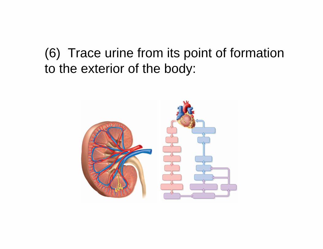
(6) Trace urine from its point of formation to the exterior of the body:

Renal TubulesThis is the path for the flow of fluid from the point wherethe glomerular filtrate is formed to the point where urine leaves the body:
glomerular capsule →proximal convoluted tubule →nephron loop →distal convoluted tubule →collecting duct →papillary duct →minor calyx →major calyx →renal pelvis →ureter →urinary bladder →urethra → (pass urine from body)

(7) Explain the importance of filtration, tubular re-absorption, and tubular secretion in urine formation:

Overview of Urine Formation
• kidneys convert blood plasma to urine in four stages
– glomerular filtration– tubular reabsorption– tubular secretion– water conservation
• glomerular filtrate // fluid in capsular space // blood plasma without protein
• tubular fluid // fluid in renal tubule // similar to above except tubular cells have removed and added substances
• urine // once it enters the collecting duct // only remaining change is water content
Glomerular filtrationCreates a plasmalikefiltrate of the blood
Tubular reabsorption Removes useful solutes from the filtrate, returns them to the blood
Tubular secretionRemoves additionalwastes from the blood,adds them to the filtrate
Water conservation Removes water from theurine and returns it toblood; concentrates wastes
Renal corpuscle
Flow of filtrate
Peritubularcapillaries
Renal tubule
H2O
H2O
H2O
Urine
Blood flow
1
2
3
and

(8) Describe the fate of most of the water that leaves the glomerulus:

Urine Formation III:Water Conservation
• The kidney eliminates metabolic wastes from the body
• The Kidney also must prevents excessive water lossas well
• Kidney needs to returns water from the tubules back into the tissue fluid and bloodstream
• Any fluid remaining in the renal tubules will pass from body as urine
• As more water is conserved then the more the tubular fluid (i.e. the urine) is concentrated!

Collecting Duct Concentrates Urine
• collecting duct (CD) begins in the cortex where it receives tubular fluid from several nephrons
• as CD passes through the medulla, water is reabsorbed and concentrates urine in CD up to four times
• medullary portion of CD is more permeable to water than to NaCl
• as urine passes through the increasingly salty medulla, water leaves by osmosis which concentrates urine
MedullaCortex
Osm
olar
ityof
tiss
ue fl
uid
(mO
sm/L
)
300
600
900
1,200
H2O
Tubular fluid(300 mOsm/L)
Collectingduct
Nephronloop
Urine(up to 1,200 mOsm/L)
H2O
H2O
H2O
H2O

Control of Water Loss (1 of 2)
• How concentrated the urine becomes depends on body’s state of hydration:
• water diuresis – drinking large volumes of water will produce a large volume of hypotonic urine

Control of Water Loss (2 of 2)
– When urine is hypertonic:
• dehydration causes the urine to be low volume and more concentrated
• Dehydration also causes high blood osmolarity which stimulates posterior pituitary to release ADH /// result in an increase in synthesis of aquaporin channels by renal tubule cells
• more water is reabsorbed by collecting duct
• urine is more concentrated
– If BP is low in a dehydrated person, GFR will also be low
• filtrate moves more slowly and more time for reabsorption
• more salt removed, more water reabsorbed and less urine produced

Proximal Convoluted Tubule
• Reabsorbs about 65% of glomerular filtrate from the PCT segment
– removes some substances from the blood, and secretes them into the tubular fluid for disposal in urine
– prominent microvilli and great length
– abundant mitochondria provide ATP for active transport
– PCTs alone account for about 6% of one’s resting ATP and calorie consumption

The Role of Sodium Chloride in Reabsorption
• sodium reabsorption is the key to the reabsorption of all the solutes as well as the reabsorption of water
– Transporting sodium creates an osmotic and electrical gradient that drives the reabsorption of water and other solutes
– most abundant cation in filtrate // creates steep concentration gradient that favors its diffusion into the epithelial cells
• two types of transport proteins in the apical cell surface are responsible for sodium uptake
– symports that simultaneously bind Na+ and another solute such as glucose, amino acids or lactate
– a Na+ - H+ antiport that pulls Na+ into the cell while pumping out H+ into tubular fluid

Reabsorption in the PCT // Other Electrolytes
• potassium, magnesium, and phosphate ions diffuse through the paracellular route with water
• phosphate is also cotransported into the epithelial cells with Na+
• some calcium is reabsorbed through the paracellular route in the PCT, but most Ca+2 occurs later in the nephron
• glucose is cotransported with Na+ by sodium-glucose transport (SGLT) proteins.
• urea diffuses through the tubule epithelium with water – reabsorbs 40 – 60% in tubular fluid– kidneys remove about half of the urea from the blood - creatinine is not reabsorbed at all
Copyright © The McGraw-Hill Companies, Inc. Permission required for reproduction or display.
Aquaporin
Solvent drag
Glucose
Paracellular route
Cl–H+
Na+K+Na+
Na+
Glucose
Cl–
H2O
Anions
Peritubularcapillary
Tissuefluid Tubule epithelial cells Tubular fluid
Sodium–glucosetransport protein(SGLT) (Symport)
Na+–H+ antiport
Cl––anion antiport
Brushborder
Transcellular route
Tight junction
H2O, urea, uric acid,Na+, K+, Cl–, Mg2+, Ca 2+, Pi
K+–Cl–symport
ADP + Pi
ATP
Na+–K+ pump
K+

Water Reabsorption
• kidneys reduce 180 L of glomerular filtrate to 1 or 2 liters of urine each day
• two-thirds of water in filtrate is reabsorbed by the PCT
• reabsorption of all the salt and organic solutes makes the tubule cells and tissue fluid hypertonic
– water follows solutes by osmosis through both paracellular and transcellular routes through water channels called aquaporins
– in PCT, water is reabsorbed at constant rate called obligatory water reabsorption

(9) Describe the fate of glucose in the glomerular filtrate:

Transport Maximum of Glucose
• there is a limit to the amount of solute that the renal tubules can reabsorb
• limited by the number of transport proteins in the plasma membrane
• if all transporters are occupied as solute molecules pass
– excess solutes appear in urine
• transport maximum is reached when transporters are saturated
• each solute has its own transport maximum
– any blood glucose level above 220 mg/dL results in glycosuria
– When glucose exceeds Tm then glucose becomes an osmotic diuretic
Normoglycemia
(a) (b)
Glucose reabsorption
Hyperglycemia
Glomerularfiltration
Glucosetransportprotein
Normalurine volume,glucose-free
Increasedurine volume,with glycosuria

(10) Describe the control mechanisms affecting the volume of urine production:

Regulation of Glomerular Filtration
• If GFR too high
– fluid flows through the renal tubules too rapidly for them to reabsorb the usual amount of water and solutes
– urine output rises – Greater chance of dehydration and electrolyte depletion
• If GFR too low
– wastes not filtered – Wastes stay in plasma– azotemia may occur

Regulation of Glomerular Filtration
• GFR controlled by adjusting glomerular blood pressure from moment to moment
• GFR regulated by three homeostatic mechanisms
– renal autoregulation
– sympathetic control
– hormonal control

Renal Autoregulation of GFR (1 of 7)
• renal autoregulation
– the ability of the nephrons to adjust their own blood flow and GFR without external (nervous or hormonal) control
– enables them to maintain a relatively stable GFR in spite of changes in systemic arterial blood pressure
– two methods of autoregulation
• myogenic mechanism
• tubuloglomerular feedback

Renal Autoregulation of GFR (2 of 7)
• myogenic mechanism // based on the tendency of smooth muscle to contract when stretched
• increased arterial blood pressure stretches the afferent arteriole
• arteriole constricts and prevents blood flow into the glomerulus from changing much
• when blood pressure falls /// the afferent arteriole relaxes
• allows blood to flow more easily into glomerulus
• filtration remains stable

Renal Autoregulation of GFR (3 of 7)
• tubuloglomerular feedback
– mechanism by which glomerulus receives feedback on the status of the downstream tubular fluid and adjust filtration to regulate the composition of the fluid, stabilize its own performance, and compensate for fluctuation in systemic blood pressure
– juxtaglomerular apparatus – complex structure found at the very end of the nephron loop where it has just reentered the renal cortex
– loop comes into contact with the afferent and efferent arterioles at the vascular pole of the renal corpuscle
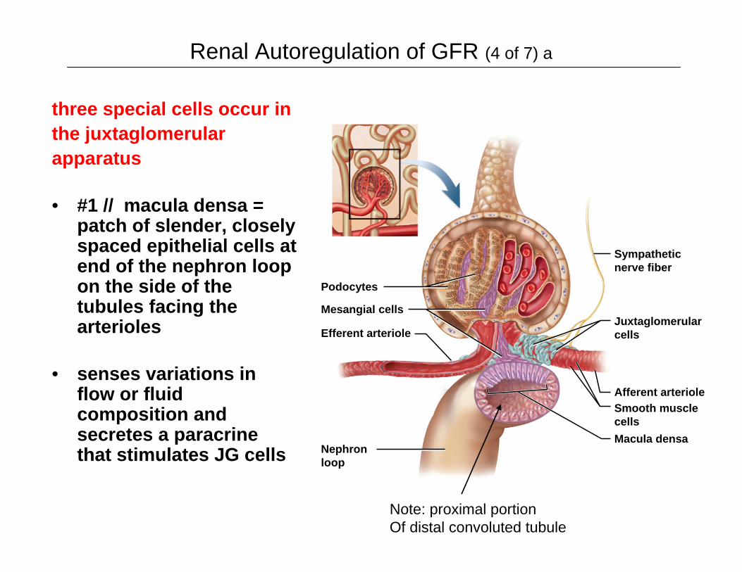
Renal Autoregulation of GFR (4 of 7) a
three special cells occur inthe juxtaglomerularapparatus
• #1 // macula densa = patch of slender, closely spaced epithelial cells at end of the nephron loop on the side of the tubules facing the arterioles
• senses variations in flow or fluid composition and secretes a paracrinethat stimulates JG cells
Podocytes
Afferent arteriole
Efferent arteriole
Macula densa
Mesangial cells
Nephronloop
Smooth musclecells
Juxtaglomerularcells
Sympatheticnerve fiber
Note: proximal portionOf distal convoluted tubule

Renal Autoregulation of GFR (4 of 7) b
• #2 // juxtaglomerular (JG) cells – enlarged smooth muscle cells in the afferent arteriole directly across from macula densa
– when stimulated by the macula
– they dilate or constrict the arterioles
– they also contain granules of renin, which they secrete in response to drop in blood pressure
Podocytes
Afferent arteriole
Efferent arteriole
Macula densa
Mesangial cells
Nephronloop
Smooth musclecells
Juxtaglomerularcells
Sympatheticnerve fiber
Note: proximal portionOf distal convoluted tubule

Renal Autoregulation of GFR (4 of 7) c
#3 // mesangial cells – in thecleft between the afferent andefferent arterioles and amongthe capillaries of theglomerulus
– connected to macula densa and JG cells by gap junctions and communicate by means of paracrines
– build supportive matrix for glomerulus, constrict or relax capillaries to regulate flow
Podocytes
Afferent arteriole
Efferent arteriole
Macula densa
Mesangial cells
Nephronloop
Smooth musclecells
Juxtaglomerularcells
Sympatheticnerve fiber
Note: proximal portionOf distal convoluted tubule
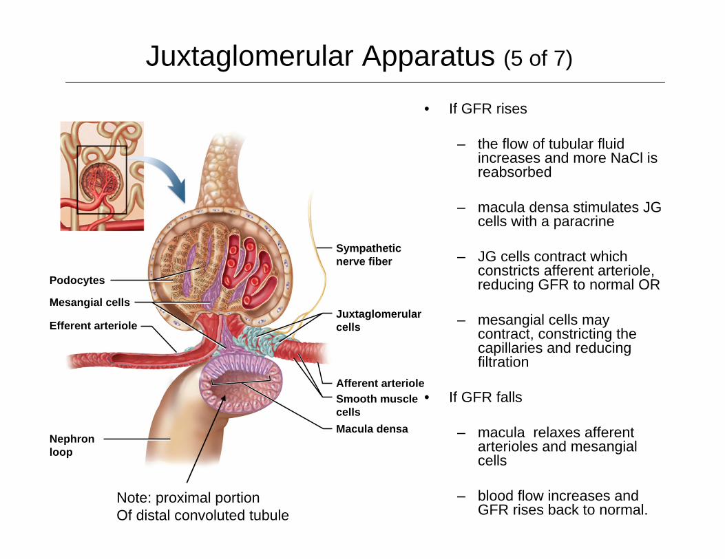
Juxtaglomerular Apparatus (5 of 7)
• If GFR rises
– the flow of tubular fluid increases and more NaCl is reabsorbed
– macula densa stimulates JG cells with a paracrine
– JG cells contract which constricts afferent arteriole, reducing GFR to normal OR
– mesangial cells may contract, constricting the capillaries and reducing filtration
• If GFR falls
– macula relaxes afferent arterioles and mesangialcells
– blood flow increases and GFR rises back to normal.
Podocytes
Afferent arteriole
Efferent arteriole
Macula densa
Mesangial cells
Nephronloop
Smooth musclecells
Juxtaglomerularcells
Sympatheticnerve fiber
Note: proximal portionOf distal convoluted tubule

Effectiveness of Autoregulation (6 of 7)
• maintains a dynamic equilibrium
– GFR fluctuates within narrow limits only
– blood pressure changes do affect GFR and urine output somewhat
• renal autoregulation can not compensate for extreme blood pressure variation
– over a MAP range of 90 – 180 mm Hg, the GFR remains quite stable
– below 70 mm Hg, glomerular filtration and urine output cease /// Likely to occurs in hypovolemic shock

Negative Feedback Control of GFR (7 of 7)
Copyright © The McGraw-Hill Companies, Inc. Permission required for reproduction or display.
High GFR Reduced GFR
Rapid flow offiltrate in renal tubules
Sensed bymacula densa
Constriction ofafferent arteriole
Paracrinesecretion

Sympathetic Control of GFR
• sympathetic nerve fibers richly innervate the renal blood vessels
• sympathetic nervous system and adrenal epinephrine constrict the afferent arterioles in strenuous exercise or acute conditions like circulatory shock
– reduces GFR and urine output
– redirects blood from the kidneys to the heart, brain, and skeletal muscles
– GFR may be as low as a few milliliters per minute

Renin-Angiotensin-Aldosterone Mechanism
• renin secreted by juxtaglomerular cells if BP drops dramatically
• renin converts angiotensinogen, a blood protein, into angiotensinI
• in the lungs and kidneys
– angiotensin-converting enzyme (ACE) converts angiotensin I to angiotensinII = active hormone
– works in several ways to restore fluid volume and BP
Liver
Kidney
Kidney
Lungs
Hypothalamus
Renin
Aldosterone
Drop in bloodpressure
Angiotensinogen(453 amino acids long)
Angiotensin I(10 amino acids long)
Angiotensin-convertingenzyme (ACE)
Angiotensin II(8 amino acids long)
Cardiovascularsystem
Vasoconstriction
Thirst anddrinking
Elevated bloodpressure
Sodium andwater retention
Adrenalcortex

Falling BP & Angiotensin II
• potent vasoconstrictor raising BP throughout body
• constricts efferent arteriole raising GFR despite low BP
• lowers BP in peritubularcapillaries enhancing reabsorption of NaCl & H2O
• angiotensin II stimulates adrenal cortex to secrete aldosteronepromoting Na+ and H2O reabsorption in DCT and collecting duct
• angiotensin II stimulates posterior pituitary to secrete ADH which promotes water reabsorption by collecting duct
• angiotensin II stimulates thirst & H2O intake
Normoglycemia
(a) (b)
Glucose reabsorption
Hyperglycemia
Glomerularfiltration
Glucosetransportprotein
Normalurine volume,glucose-free
Increasedurine volume,with glycosuria

(11) Differentiate between the normal composition of plasma and the glomerular filtrate.

Filtration Pores and Slits
Capsular space
Filtration slit
Filtration pore
Basement membrane
Passed through filter:
ElectrolytesGlucoseAmino acidsFatty acidsVitaminsUreaUric acidCreatinine
Turned back:Blood cellsPlasma proteinsLarge anionsProtein-bound
Most molecules> 8 nm indiameter
Bloodstream
Endothelial cell ofglomerular capillary
Foot process ofpodocyte
minerals andhormones
Water
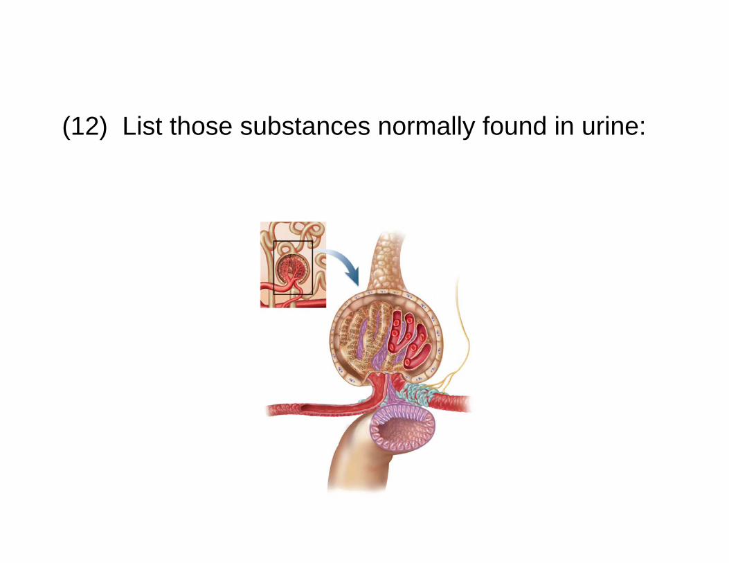
(12) List those substances normally found in urine:

Composition and Properties of Urine (1 of 2)
• urinalysis – the examination of the physical and chemical properties of urine
– appearance - clear, almost colorless to deep amber -yellow color due to urochrome pigment from breakdown of hemoglobin (RBCs) – other colors from foods, drugs or diseases
• cloudiness or blood could suggest urinary tract infection, trauma or stones
• pyuria – pus in the urine
• hematuria – blood in urine due to urinary tract infection, trauma, or kidney stones

Composition and Properties of Urine (1 of 2)
– odor - bacteria degrade urea to ammonia, some foods impart aroma
– specific gravity - compared to distilled water /// density of urine ranges from 1.001 -1.028

Composition and Properties of Urine (1 of 2)
– osmolarity - (blood = 300 mOsm/L) /// ranges from 50 mOsm/L to 1,200 mOsm/L in dehydrated person
– pH - range: 4.5 to 8.2, usually 6.0 (mildly acidic)
– chemical composition: 95% water, 5% solutes
• Normal to find /// urea, NaCl, KCl, creatinine, uric acid, phosphates, sulfates, traces of calcium, magnesium, and sometimes bicarbonate, urochrome and a trace of bilirubin
• Abnormal to find /// glucose, free hemoglobin, albumin, ketones, bile pigments
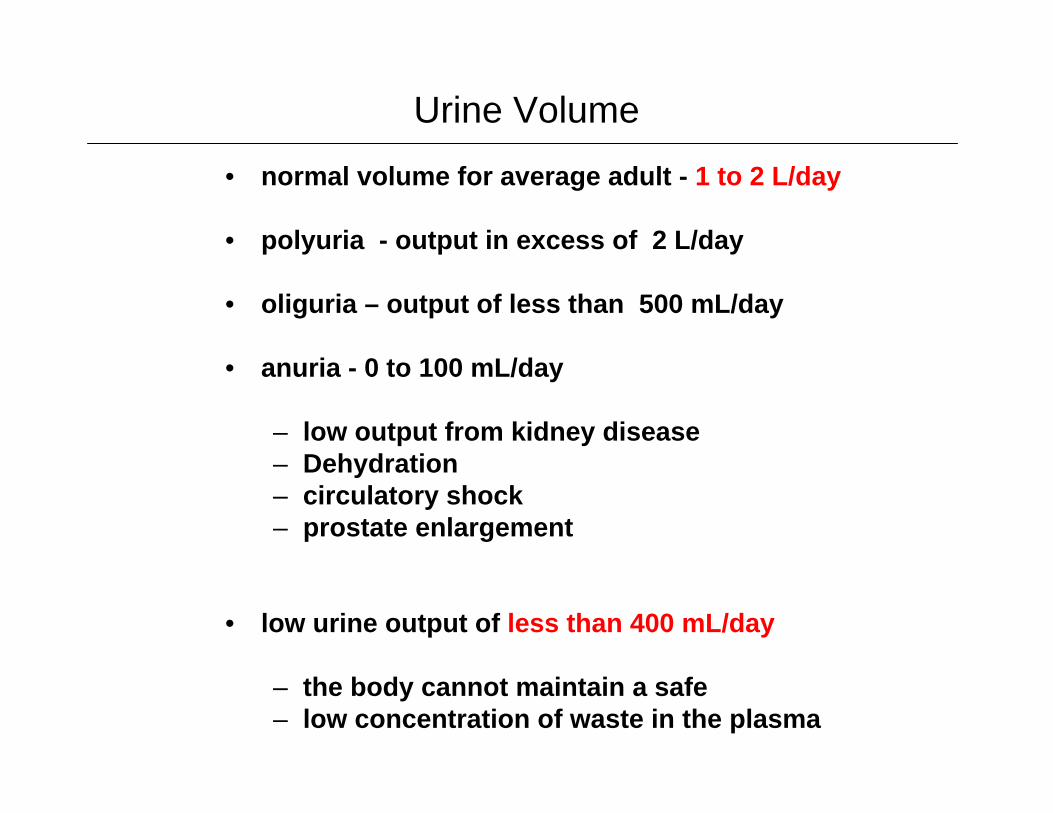
Urine Volume
• normal volume for average adult - 1 to 2 L/day
• polyuria - output in excess of 2 L/day
• oliguria – output of less than 500 mL/day
• anuria - 0 to 100 mL/day
– low output from kidney disease– Dehydration– circulatory shock– prostate enlargement
• low urine output of less than 400 mL/day
– the body cannot maintain a safe– low concentration of waste in the plasma

(13) Identify the hormones that influence urine output and blood volume and explain their modes of action:

Renin-Angiotensin-Aldosterone Mechanism
• renin secreted by juxtaglomerular cells if BP drops dramatically
• renin converts angiotensinogen, a blood protein, into angiotensinI
• in the lungs and kidneys
– angiotensin-converting enzyme (ACE) converts angiotensin I to angiotensinII = active hormone
– works in several ways to restore fluid volume and BP
Liver
Kidney
Kidney
Lungs
Hypothalamus
Renin
Aldosterone
Drop in bloodpressure
Angiotensinogen(453 amino acids long)
Angiotensin I(10 amino acids long)
Angiotensin-convertingenzyme (ACE)
Angiotensin II(8 amino acids long)
Cardiovascularsystem
Vasoconstriction
Thirst anddrinking
Elevated bloodpressure
Sodium andwater retention
Adrenalcortex

Falling BP & Angiotensin II
• potent vasoconstrictor raising BP throughout body
• constricts efferent arteriole raising GFR despite low BP
• lowers BP in peritubularcapillaries enhancing reabsorption of NaCl & H2O
• angiotensin II stimulates adrenal cortex to secrete aldosteronepromoting Na+ and H2O reabsorption in DCT and collecting duct
• angiotensin II stimulates posterior pituitary to secrete ADH which promotes water reabsorption by collecting duct
• angiotensin II stimulates thirst & H2O intake
Normoglycemia
(a) (b)
Glucose reabsorption
Hyperglycemia
Glomerularfiltration
Glucosetransportprotein
Normalurine volume,glucose-free
Increasedurine volume,with glycosuria

DCT and Collecting Duct
• fluid arriving in the DCT still contains about 20% of the water and 7% of the salts from glomerular filtrate /// if this were all to pass from kidneys as urine, it would amount to 36 L/day
• DCT and collecting duct reabsorb variable amounts of water and salt which are regulated by several hormones
– aldosterone, atrial natriuretic peptide, ADH, and parathyroid hormone
• two kinds of cells in the DCT and collecting duct
– principal cells /// most numerous with receptors for hormones /// involved in salt and water balance
– intercalated cells /// involved in acid/base balance by secreting H+ into tubule lumen and reabsorbing K+

DCT and Collecting Duct
• aldosterone - the “salt-retaining” hormone
– steroid secreted by the adrenal cortex
• when blood Na+ concentration falls
• when K+ concentration rises
• drop in blood pressure → renin release →angiotensin II formation → stimulates adrenal cortex to secrete aldosterone

DCT and Collecting Duct
• functions of aldosterone
– acts on thick segment of nephron loop, DCT, and cortical portion of collecting duct
• stimulates the reabsorption of more Na+ and secretion of K+
• water and Cl- follow the Na+
• net effect is that the body retains NaCl and water // helps maintain blood volume and pressure
• the urine volume is reduced
• the urine has an elevated K+ concentration

DCT and Collecting Duct
• atrial natriuretic peptide (ANP) /// secreted by atrialmyocardium of the heart in response to high blood pressure
• has four actions that result in the excretion of more salt and water in the urine, thus reducing blood volume and pressure
– dilates afferent arteriole, constricts efferent arteriole - ↑GFR
– inhibits renin and aldosterone secretion
– inhibits secretion of ADH
– inhibits NaCl reabsorption by collecting duct
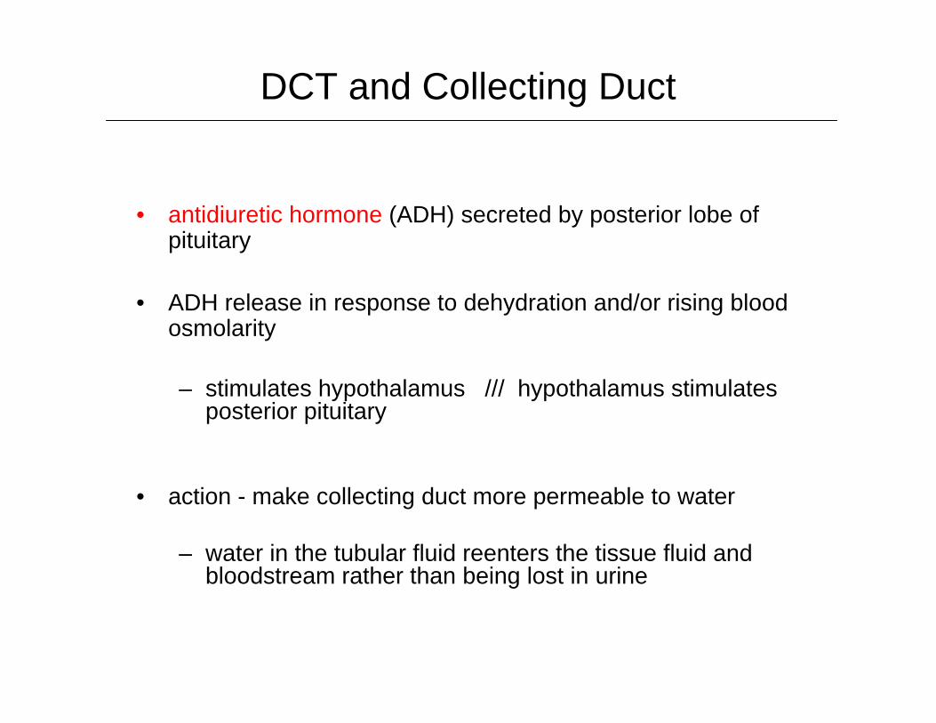
DCT and Collecting Duct
• antidiuretic hormone (ADH) secreted by posterior lobe of pituitary
• ADH release in response to dehydration and/or rising blood osmolarity
– stimulates hypothalamus /// hypothalamus stimulates posterior pituitary
• action - make collecting duct more permeable to water
– water in the tubular fluid reenters the tissue fluid and bloodstream rather than being lost in urine
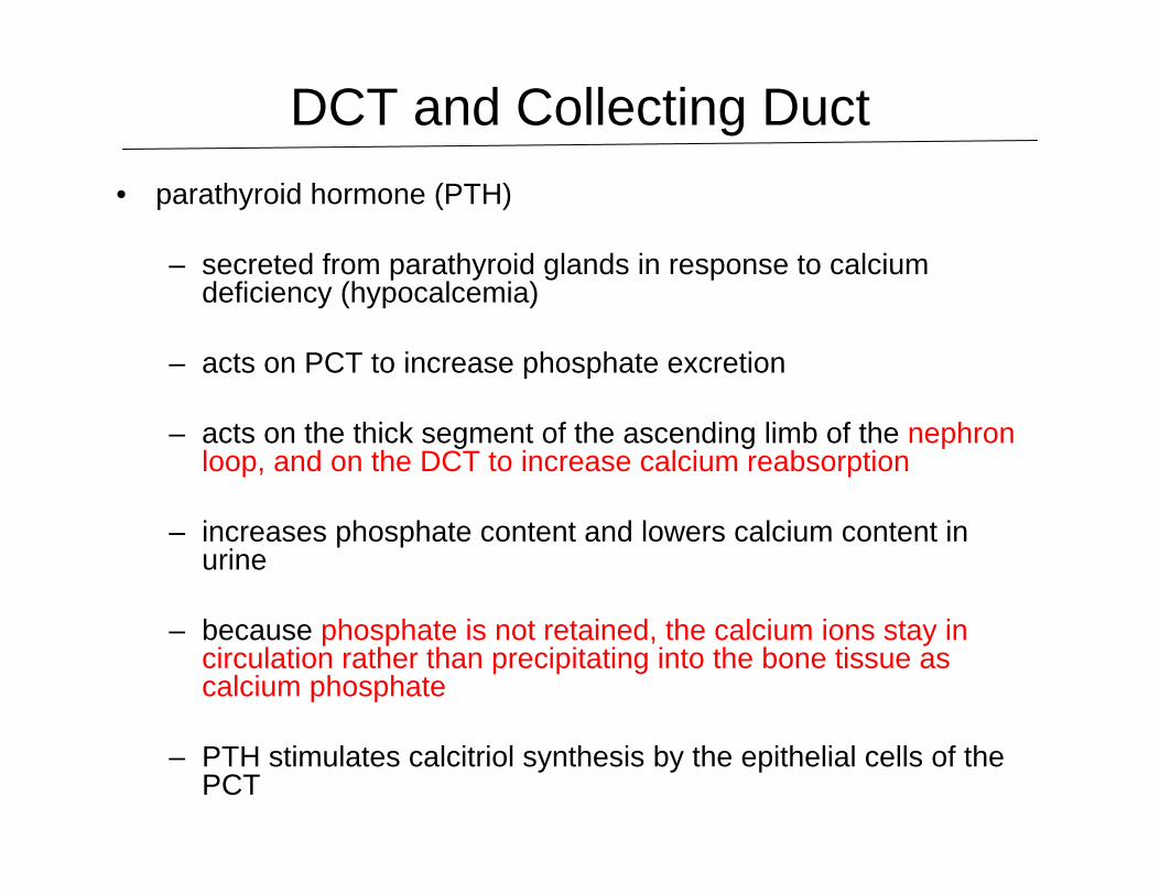
DCT and Collecting Duct• parathyroid hormone (PTH)
– secreted from parathyroid glands in response to calcium deficiency (hypocalcemia)
– acts on PCT to increase phosphate excretion
– acts on the thick segment of the ascending limb of the nephronloop, and on the DCT to increase calcium reabsorption
– increases phosphate content and lowers calcium content in urine
– because phosphate is not retained, the calcium ions stay in circulation rather than precipitating into the bone tissue as calcium phosphate
– PTH stimulates calcitriol synthesis by the epithelial cells of the PCT
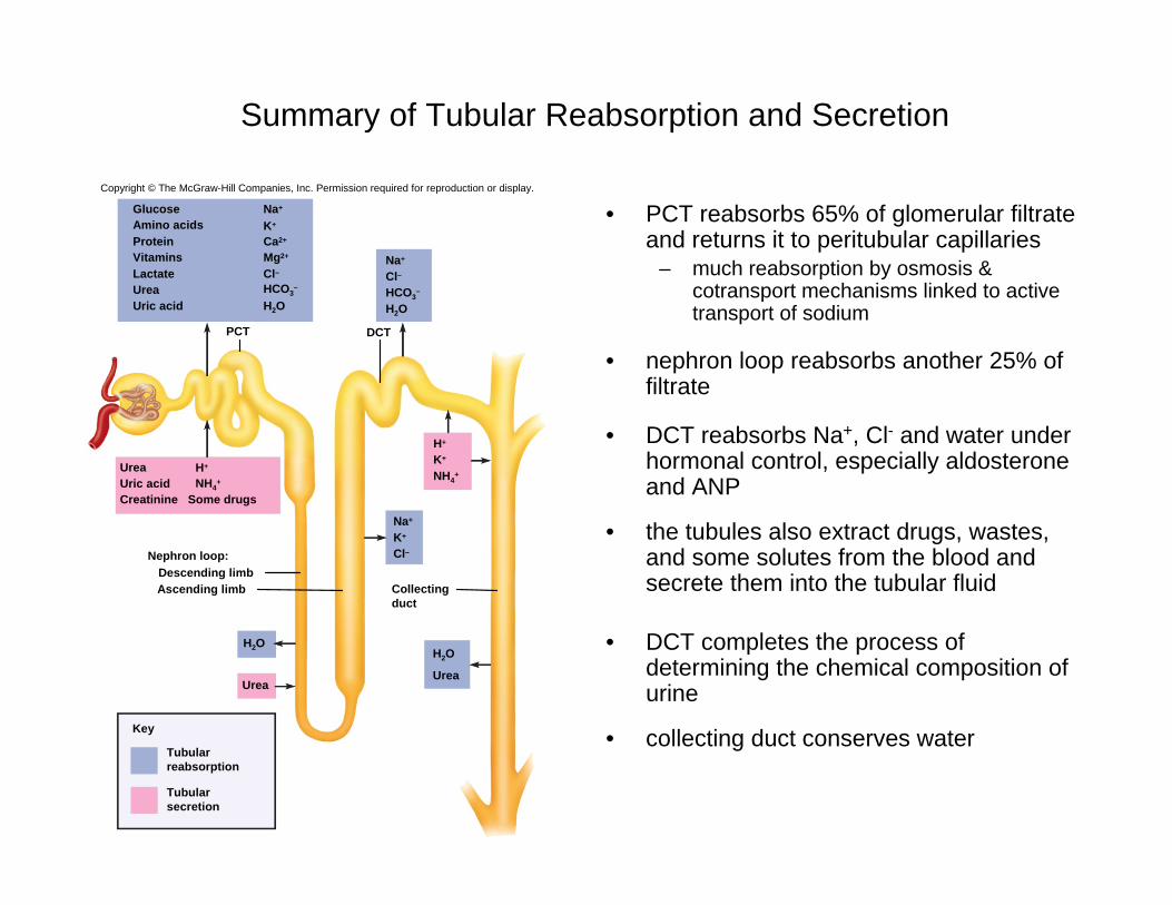
Summary of Tubular Reabsorption and Secretion
• PCT reabsorbs 65% of glomerular filtrate and returns it to peritubular capillaries
– much reabsorption by osmosis & cotransport mechanisms linked to active transport of sodium
• nephron loop reabsorbs another 25% of filtrate
• DCT reabsorbs Na+, Cl- and water under hormonal control, especially aldosteroneand ANP
• the tubules also extract drugs, wastes, and some solutes from the blood and secrete them into the tubular fluid
• DCT completes the process of determining the chemical composition of urine
• collecting duct conserves water
Copyright © The McGraw-Hill Companies, Inc. Permission required for reproduction or display.
H2O
UreaUrea
H+
K+
NH4+
Na+
Cl–
H2O
Urea H+
Uric acid NH4+
Creatinine Some drugs
Glucose Na+
Amino acids K+
Protein Ca2+
Vitamins Mg2+
Lactate Cl–Urea HCO3
–
Uric acid H2O
Na+
K+
Cl–
H2O
Nephron loop:Descending limbAscending limb
PCT DCT
Key
Collectingduct
Tubularreabsorption
Tubularsecretion
HCO3–

Water ConservationCountercurrent Multiplication & Countercurrent Exchange

Urine Formation &Water Conservation
• The kidney eliminates metabolic wastes from the body
• The Kidney also must prevents excessive water lossas well
• Kidney needs to returns water from the tubules back into the tissue fluid and bloodstream
• Any fluid remaining in the renal tubules will pass from body as urine
• As more water is conserved then the more the tubular fluid (i.e. the urine) is concentrated!

Collecting Duct Concentrates Urine
• collecting duct (CD) begins in the cortex where it receives tubular fluid from several nephrons
• as CD passes through the medulla, water is reabsorbed and concentrates urine in CD up to four times
• medullary portion of CD is more permeable to water than to NaCl
• as urine passes through the increasingly salty medulla, water leaves by osmosis which concentrates urine
MedullaCortex
Osm
olar
ityof
tiss
ue fl
uid
(mO
sm/L
)
300
600
900
1,200
H2O
Tubular fluid(300 mOsm/L)
Collectingduct
Nephronloop
Urine(up to 1,200 mOsm/L)
H2O
H2O
H2O
H2O

Control of Water Loss (1 of 2)
• How concentrated the urine becomes depends on body’s state of hydration:
– water diuresis – drinking large volumes of water will produce a large volume of hypotonic urine
• cortical portion of CD reabsorbs NaCl, but it is impermeable to water
• salt removed from the urine stays in the CD
• urine concentration may be as low as 50 mOsm/L

Control of Water Loss (2 of 2)
– producing hypertonic urine
• dehydration causes the urine to become low volume and more concentrated
• high blood osmolarity stimulates posterior pituitary to release ADH and then an increase in synthesis of aquaporinchannels by renal tubule cells
• more water is reabsorbed by collecting duct
• urine is more concentrated
– If BP is low in a dehydrated person, GFR will also be low
• filtrate moves more slowly and more time for reabsorption
• more salt removed, more water reabsorbed and less urine produced

Countercurrent Multiplier = Loop of Henle
• the ability of kidney to concentrate urine depends on creating a salinity gradient in renal medulla
– four times as salty in the renal medulla than the cortex
• nephron loop acts as countercurrent multiplier
– multiplier - continually recaptures salt and returns it to extracellular fluid of medulla which multiplies the salinity in adrenal medulla
– countercurrent - because of fluid flowing in opposite directions in adjacent tubules of nephron loop

Countercurrent Multiplier = Loop of Henle
• fluid flowing downward in descending limb– passes through environment of increasing osmolarity– most of descending limb very permeable to water but not to NaCl– water passes from tubule into the ECF leaving salt behind– concentrates tubular fluid to 1,200 mOsm/L at lower end of loop
• fluid flowing upward in ascending limb – impermeable to water– reabsorbs Na+, K+, and Cl- by active transport pumps into ECF– maintains high osmolarity of renal medulla– tubular fluid becomes hypotonic – 100 mOsm/L at top of loop
• recycling of urea: lower end of CD permeable to urea– urea contributes to the osmolarity of deep medullary tissue– continually cycled from collecting duct to the nephron loop and
back– urea remains concentrated in the collecting duct and some of it
always diffuses out into the medulla adding to osmolarity

Countercurrent Multiplier of Nephron Loop
300
400 200
100
1,200
700900
400600
Na+
K+
Cl–
H2O
1
2
3
5
4
The more salt thatis pumped out of theascending limb, thesaltier the ECF is inthe renal medulla.
Na+
K+
Cl–
Na+
K+
Cl–
Na+
K+
Cl–
Na+
K+
Cl–
Na+
K+
Cl–
H2O
The saltier the fluid in theascending limb, the moresalt the tubule pumps intothe ECF.
The more water that leavesthe descending limb, thesaltier the fluid is thatremains in the tubule.
H2O
H2O
H2O
The higher the osmolarityof the ECF, the more waterleaves the descending limbby osmosis.
More salt is continuallyadded by the PCT.

Countercurrent Exchange System = Vasa Recta
• vasa recta – capillary branching off efferent arteriole in medulla
– provides blood supply to medulla and does not remove NaCl and urea from medullary ECF
• countercurrent system - formed by blood flowing in opposite directions in adjacent parallel capillaries

Countercurrent Exchange System = Vasa Recta
• descending capillaries /// exchanges water for salt– water diffuses out of capillaries and salt diffuses in
• ascending capillaries // / exchanges salt for water
– water diffuses into and NaCl diffuses out of blood– the vasa recta gives the salt back and does not
subtract from the osmolarity of the medulla
• absorb more water on way out than the way in, and thus they carry away water reabsorbed from the urine by collecting duct and nephron loop

Maintenance of Osmolarity in Renal Medulla
Medulla
Cortex
Nephron loop
Key
Collecting duct Vasa recta
300
400
600
900
1,200
300
300
400
900
600
700
400
400
200
200
100
100
300
500
700
1,200
1,200
Urea
Urea
Urea
UreaUrea
Urea
UreaNaCl
NaCl NaCl
NaCl
Na+
K+
Cl–
Na+
K+
Cl–
Active transport
300 300
400
600
900
400
600
1,200
900
H2O
H2O
H2O
Key
Osmolarity ofECF(mOsm/L)
Na+
K+
Cl–
Na+
K+
Cl–
Na+
K+
Cl–H2O
H2O
H2O
H2O
Diffusion througha membrane channel
Countercurrent MultiplierLoop of Henle
Countercurrent ExchangeVasa Recta

Summary of Tubular Reabsorption and Secretion
H2O
UreaUrea
H+
K+
NH4+
Na+
Cl–
H2O
Urea H+
Uric acid NH4+
Creatinine Some drugs
Glucose Na+
Amino acids K+
Protein Ca2+
Vitamins Mg2+
Lactate Cl–Urea HCO3
–
Uric acid H2O
Na+
K+
Cl–
H2O
Nephron loop:Descending limbAscending limb
PCT DCT
Key
Collectingduct
Tubularreabsorption
Tubularsecretion
HCO3–