Embed Size (px)
Citation preview

INVITED REVIEW ABSTRACT: Neuropathy is a common complication of end-stage kidneydisease (ESKD), typically presenting as a distal symmetrical process withgreater lower-limb than upper-limb involvement. The condition is of insidiousonset, progressing over months. and has been estimated to be present in60%–100% of patients on dialysis. Neuropathy generally only develops atglomerular filtration rates of less than 12 ml/min. The most frequent clinicalfeatures reflect large-fiber involvement, with paresthesias, reduction in deeptendon reflexes, impaired vibration sense, muscle wasting, and weakness.Nerve conduction studies demonstrate findings consistent with a general-ized neuropathy of the axonal type. Patients may also develop autonomicfeatures, with postural hypotension, impaired sweating, diarrhea, constipa-tion, or impotence. The development of uremic neuropathy has been relatedpreviously to the retention of neurotoxic molecules in the middle molecularrange, although this hypothesis lacked formal proof. Studies utilizing novelaxonal excitability techniques have recently shed further light on the patho-physiology of this condition. Nerves of uremic patients have been shown toexist in a chronically depolarized state prior to dialysis, with subsequentimprovement and normalization of resting membrane potential after dialysis.The degree of depolarization correlates with serum K�, suggesting thatchronic hyperkalemic depolarization plays an important role in the develop-ment of nerve dysfunction in ESKD. These recent findings suggest thatmaintenance of serum K� within normal limits between periods of dialysis,rather than simple avoidance of hyperkalemia, is likely to reduce the inci-dence and severity of uremic neuropathy.
Muscle Nerve 35: 273–290, 2007
UREMIC NEUROPATHY: CLINICAL FEATURESAND NEW PATHOPHYSIOLOGICAL INSIGHTS
ARUN V. KRISHNAN, MB, BS, PhD, and MATTHEW C. KIERNAN, MB, BS, PhD
Prince of Wales Medical Research Institute and Prince of Wales Clinical School, Universityof New South Wales, Barker Street, Randwick, Sydney, NSW 2031, Australia
Accepted 24 October 2006
End-stage kidney disease (ESKD) occurs whennephrons are irretrievably impaired to the extentthat the retention of metabolic waste products, salt,and water becomes potentially fatal.128 ESKD mayoccur due to either a primary renal disorder or as acomplication of a multisystem disorder. The com-mon causes of ESKD remain diabetes, glomerulone-phritis, and hypertension.136 When renal functionreaches critically low levels, renal replacement ther-apy either in the form of dialysis or transplantation is
required in order to remove waste products andexcess fluid.
Uremic neuropathy has for many decades beenrecognized as a common complication of ESKD.7,72,145
Although the advent of dialysis and transplant pro-grams has led to reductions in the rate of severe neu-ropathy, the prevalence of this condition remainshigh.113 This review covers the clinical and electrophys-iological features of uremic neuropathy, with a focuson advances in understanding the pathophysiology ofthis condition.
HISTORICAL ASPECTS
The possibility of peripheral neuropathy in patientstreated with hemodialysis was first raised shortly afterthe introduction of the first formal hemodialysis pro-gram.172 The first clinical documentation of neuropa-thy was provided in 1961 in two young male patientswith hereditary interstitial nephritis and deafness.209
The development of neuropathy in these cases, how-
Available for Category 1 CME credit through the AANEM at www.aanem.org.
Abbreviations: ADH, activity-dependent hyperpolarization; CSF, cerebro-spinal fluid; CMAP, compound muscle action potential; CTS, carpal tunnelsyndrome; EPO, erythropoietin; ESKD, end-stage kidney disease; NSS, neu-ropathy symptom score; PTH, parathyroid hormoneKey words: end-stage kidney disease; middle molecules; peripheral neurop-athy; potassium; uremic neuropathyCorrespondence to: M.C. Kiernan; e-mail: [email protected]
© 2006 Wiley Periodicals, Inc.Published online 28 December 2006 in Wiley InterScience (www.interscience.wiley.com). DOI 10.1002/mus.20713
Uremic Neuropathy MUSCLE & NERVE March 2007 273

ever, was attributed to the underlying hereditary disor-der, rather than viewed as a complication of ESKD.
Following this report, Asbury et al.6 providedextensive clinical and pathological findings in fourmen who developed neuropathy as a consequence ofESKD of varying etiologies. All four patients hadclinical features of renal disease for many years be-fore the development of neuropathy, which mani-fested as a symmetrical length-dependent sensorimo-tor neuropathy. Nerve biopsies established axonaldegeneration, maximal distally, with sparing of prox-imal nerve segments and nerve roots. Moreover,there was no evidence to suggest nerve compression,inflammation, or the superimposition of a systemicdisease process, such as diabetes or amyloid, leadingto the conclusion that the development of neurop-athy was a consequence of the underlying renal dis-order.
Early clinical neurophysiological investigations inESKD patients demonstrated reductions in motornerve conduction velocity in symptomatic andasymptomatic patients.158,185 Jebsen et al.,82 studyingthe natural history of uremic neuropathy, comparedclinical and nerve conduction findings in patientstreated conservatively to those receiving dialysis ther-apy. Whereas the development of neuropathy in theconservatively treated group was related to deterio-rating renal function, those patients treated withlong-term dialysis manifested improvement in bothclinical and neurophysiological parameters. Follow-ing these early reports and in light of the increasinguse of dialysis and renal transplantation therapies,greater attention has been focused on uremic neu-ropathy, with numerous studies reporting high ratesof neuropathy in ESKD patients, generally relatingthe development of neuropathy to the severity ofrenal failure.5,20,82,107,144,146,149,158,185,187 Of particularnote, studies by Nielsen144,149–152 and Bolton etal.20,21 in the 1970s demonstrated nerve conductionslowing in clinically unaffected nerve segments, withcorrelation between the extent of renal impairmentand degree of conduction slowing, as well as im-provement in neurophysiological parameters follow-ing renal transplantation. These studies providedclinical evidence to suggest that a uremic toxin wasresponsible for the development of neuropathy inESKD patients, a hypothesis that was to become amajor focus of future neurophysiological research inthis condition.
INCIDENCE AND CLINICAL FEATURES
Peripheral neuropathy in ESKD generally presentsas a distal symmetrical polyneuropathy with greater
lower-limb than upper-limb involvement. The condi-tion is of insidious onset, progressing over months,and has been noted to have a male predominance. Itgenerally only develops at glomerular filtration ratesof less than 12 ml/min.39 The most frequent clinicalfeatures are those of large-fiber involvement, withparesthesias, reduction in deep tendon reflexes,impaired vibration sense, weakness, and muscle wast-ing (Fig. 1). In the 1970s, Nielsen144,145 demon-strated the presence of neuropathic symptoms inover 50% of patients with ESKD. Other studies havedemonstrated prevalence rates varying from 60% to100%, depending on the diagnostic criteria ap-plied.1,23,122,191
Laaksonen et al.122 staged the clinical severity ofuremic neuropathy in 21 ESKD patients, using amodified version of the neuropathy symptom score(NSS) developed by Dyck et al.,57,58 and combined
FIGURE 1. Prominent wasting of intrinsic hand muscles in apatient with established uremic neuropathy. In addition to theprominent wasting and resultant clinical weakness, the patientcomplained of numbness and had impaired proprioception.
274 Uremic Neuropathy MUSCLE & NERVE March 2007

this assessment with results of nerve conductionstudies. The NSS quantified symptoms that weregrouped into three categories to reflect alteration inmotor, sensory, and autonomic systems. Within eachgroup, further subsets were used to group symptomsaccording to the region affected and the presence ofpositive or negative symptoms. Using the NSS andthe staging procedure previously used in studies ofdiabetic patients,57,58 81% of ESKD patients receiveda diagnosis of neuropathy. Stage 1 neuropathy(asymptomatic neuropathy) was diagnosed in 19%,stage 2 neuropathy (symptoms nondisabling) waspresent in 48%, and stage 3 neuropathy (disablingsymptoms) was noted in 14%. In a more recentstudy,113 93% of ESKD patients had neuropathicsymptoms on NSS testing, with 72% diagnosed withstage 2 neuropathy and 21% with stage 3 neuropa-thy, despite all patients meeting currently acceptedguidelines of dialysis adequacy.143
CLINICAL AND NEUROPHYSIOLOGICALFINDINGS IN GENERALIZEDUREMIC NEUROPATHY
Early studies of uremic neuropathy utilizing nervebiopsy techniques revealed prominent axonal de-generation, most severe in the distal parts of nervetrunks. Although initial studies suggested that demy-elination was a significant feature of uremic neurop-athy,5,54 subsequent reviews demonstrated that de-myelination was secondary to axonal loss and thatproximal segments of the nerves were relativelyspared.3,55,67,187 These findings supported the con-cept that uremic neuropathy was a dying-back neu-ropathy, with metabolic failure of the neuron caus-ing distal axonal degeneration.55
Numerous neurophysiological series have beenundertaken in patients with uremic neurop-athy and have demonstrated findings consistentwith a generalized neuropathy of the axonaltype.1,4,13,20,41,46,49,68,80,107,113,121,130,148,155,156,179,183,185,191
Early studies focused on motor nerve conduction pa-rameters and demonstrated slowing of conduction ve-locity in patients prior to the development of clinicalneuropathy.45,158 Subsequent studies demonstrated ab-normalities of nerve conduction148 with generalizedslowing in both sensory and motor nerves, accompa-nied by reduction in sensory response amplitudes. Mo-tor response amplitudes tend to remain relatively pre-served, although abnormalities in lower-limb motornerves were noted in some patients, accompanied byneurogenic changes in distal lower-limb muscles onelectromyography.148,149
In a recent study, amplitude of the sural sensorynerve action potential was found to be the most sensi-tive indicator of uremic neuropathy, being reduced in50% of ESKD patients.113 Other groups have con-firmed similar findings, demonstrating reductions insensory and motor response amplitudes in addition toabnormalities of late responses.1,4,49,122,130,137,157,191 Re-duction in peroneal nerve motor conduction veloc-ity46,137,148 and prolongation of tibial F-wave minimumlatencies122 have been established as sensitive indica-tors of neuropathy in ESKD patients. Prolongation ofsoleus H reflexes has also been demonstrated in pa-tients without clinical evidence of neuropathy, suggest-ing that this parameter may be more sensitive in de-tecting early neuropathy.68,191
Studies of quantitative sensory testing in ESKDpatients have demonstrated increased vibratory per-ception thresholds, most marked in the lowerlimbs,147 whereas somatosensory-evoked potentialsin ESKD patients demonstrate abnormalities of con-duction along both the distal and proximal segmentsof peripheral somesthetic pathways, but less com-monly along intracranial sensory pathways.25,153,166
A study of single-fiber electromyography demon-strated normal fiber densities in motor units of ESKDpatients.186 This finding suggested that reinnervation,characterized by increased fiber density, had failed tooccur. However, this was accompanied by increasedjitter, possibly reflecting peripheral demyelination inthe setting of axonal degeneration. A further single-fiber EMG study established that jitter abnormalitiesimproved following a year of dialysis.106
Early studies of nerve excitability, utilizing a lim-ited range of excitability parameters, demonstratedan elevated threshold for excitation even when nerveconduction values were normal, in addition to dem-onstrating prolongation of absolute and relative re-fractory periods.25,125,182,208 As a consequence, it wasconcluded that the safety factor for neural transmis-sion at the nodes of Ranvier would be lowered. Un-expectedly, uremic nerves retained vibratory percep-tion and their sensory response amplitudes for alonger period than control nerves when renderedischemic.43 Uremic nerves also behaved differentlywhen temperature was lowered, with a less rapid risein response amplitude compared to controls.24,25
In addition to the slowly progressive sensorimo-tor axonal neuropathy, a more rapidly progressivemotor neuropathy has been described. A small num-ber of ESKD patients with diabetes have also beenshown to develop a subacute neuropathy progress-ing over a few months, with severe muscle weakness.In this group of patients, nerve conduction studiesmay demonstrate features of either a demyelinating
Uremic Neuropathy MUSCLE & NERVE March 2007 275

or axonal neuropathy.26,27,165 Although the presenceof diabetes complicates assessment of nerve conduc-tion data, the absence of preexisting neuropathicsymptoms and the clinical improvement noted fol-lowing dialysis or renal transplantation suggest ametabolic basis for the neuropathy, related to theunderlying ESKD. Analysis of cerebrospinal fluid(CSF) is rarely helpful, as CSF protein concentrationis frequently elevated in ESKD patients and maysimulate the albuminocytologic dissociation that ischaracteristic of Guillain–Barre syndrome.23
Small-fiber neuropathy may develop as a clinicalentity in ESKD patients. Lindblom and Tegner124
demonstrated abnormalities of thermal sensation in30% of ESKD patients and concluded that small-fiber neuropathy may exist as a distinct entity inthese patients. These results, however, differed fromthose of other groups who demonstrated minimalimpairment of thermal sensation in ESKD.56,188 In astudy of 20 ESKD patients, abnormalities in standardnerve conduction studies were demonstrated in 16patients,4 whereas abnormal thermal thresholdswere found in only 6 patients and, when present, didnot correlate with clinical evidence of polyneurop-athy.4 Such findings are consistent with those ofpathological studies that demonstrated greater vul-nerability of larger-diameter fibers in ESKD pa-tients.55
MONONEUROPATHIES IN ESKD
Mononeuropathies are a frequent clinical complica-tion in ESKD patients and most typically occur in themedian, ulnar, and femoral nerves.39
Carpal tunnel syndrome (CTS) is the most com-mon mononeuropathy in ESKD, with prevalencerates varying from 6% to 31%.15,17,52,66,67,76,81,170 �2-Microglobulin amyloidosis is a major factor underly-ing the development of CTS in ESKD patients,63 acomplication noted in patients on long-term hemo-dialysis. Amyloid deposits have been identified insynovial specimens from dialysis patients with CTS63
and an increase in the rate of CTS has been demon-strated with increasing hemodialysis duration.63
Strategies geared at reducing the levels of �2-micro-globulin, such as the use of high-flux biocompatiblemembranes and �2-microglobulin adsorption col-umns, have resulted in reduced rates of CTS devel-opment and ultimately improvement in symp-toms.63,118,192
Other factors that may contribute to the in-creased incidence of CTS in ESKD patients includeuremic tumoral calcinosis47,203 and the placement ofarteriovenous fistulas (Fig. 2), inducing a “steal” of
blood from the distal limb.66,70,105,119,131,141,201 Theplacement of Brescia–Cimino arteriovenous fistulasbetween the radial artery and cephalic vein has beenassociated with the development of both the clinicalfeatures of CTS and subclinical neurophysiologicalabnormalities in median or ulnar nerve territo-ries.70,105 A recent study demonstrated CTS in 30.5%of limbs with fistulas compared to 12.2% on thecontralateral side.66 Although the site of the fistulahad no effect on the development of CTS, a signifi-cant correlation was noted between the age of thefistula and the presence of CTS.
With regard to treatment of CTS in ESKD, theoutcome of median nerve decompression appearsinferior in this group compared to patients withidiopathic CTS.76,102,173 In addition, recurrence ratesare higher. In ESKD patients with recurrent CTS, anendoscopic approach may prove to be effective inrelieving persistent symptoms.211
Ulnar neuropathy is also a common occurrencein ESKD, affecting up to 51% of patients undergoinghemodialysis.142 Causes include external compres-
FIGURE 2. Forearm arteriovenous fistula (basilic vein transposi-tion) in a patient referred for neurophysiological assessment ofright hand pain and numbness. Subsequent nerve conductionstudies established a generalized axonal neuropathy, with su-peradded features consistent with carpal tunnel syndrome.
276 Uremic Neuropathy MUSCLE & NERVE March 2007

sion at the elbow during prolonged dialysis thera-py142 and other risk factors as outlined for CTS,particularly arteriovenous fistulas and uremic tu-moral calcinosis.62,69
In addition to their possible contributions to thedevelopment of CTS and ulnar neuropathy, upper-limb arteriovenous fistulas may be associated with anacute-onset neuropathy, first described by Bolton etal.22 and later termed ischemic monomelic neurop-athy.206 In this condition, acute limb ischemia devel-ops due to shunting of arterial blood away from thedistal parts of the limb.135 The severity of ischemiatypically affects nerves, without causing changes inother tissues such as muscle, that possess a higherthreshold for ischemic injury. Diabetic patients areparticularly vulnerable, especially those with preex-isting peripheral vascular disease or neuropathy.162
The symptoms are those of multiple upper-limbmononeuropathies, with distal sensory loss andweakness in the muscles of the forearm and hand.Electromyography demonstrates neurogenic abnor-malities in distal limb muscles with sparing of prox-imal musculature.206 There may be predilection formedian nerve involvement and the presence of con-duction block in this nerve has been described as asign of reversible injury.87 Early ligation or revisionof the fistula frequently leads to significant clinicaland electrophysiological improvement.87,161
Femoral neuropathy in ESKD patients is a well-recognized complication of renal transplantation,with an incidence of �2%.83,134,175 Prolonged use ofself-retaining retractors in the region of the femoralnerve during renal transplantation may cause nervecompression and neuropraxic injury.175 Rapid recov-ery is expected in cases of neurapraxia but pro-longed ischemia may lead to axonal loss.210 Theprognosis is generally favorable, with most patientsachieving complete recovery,175 although residualdeficits may be present when significant axonal losshas occurred.134
AUTONOMIC NEUROPATHY IN ESKD
Autonomic neuropathy may develop in ESKD pa-tients, manifesting as postural hypotension, im-paired sweating, diarrhea, constipation, or impo-tence.14,197 In a study of 36 ESKD patients,gastrointestinal autonomic symptoms were evidentin 42% and impotence in 45%.200 Although posturalhypotension was an uncommon clinical finding,36% complained of episodes of postural dizziness,which was most prominent in elderly ESKD patients.Some studies have suggested that autonomic neu-ropathy occurs as a manifestation of generalized
polyneuropathy,89,200 but others have shown no cor-relation between autonomic dysfunction and periph-eral nervous system abnormalities.44,180,195 Themechanisms underlying the development of uremicautonomic neuropathy remain unknown, althoughan association with hyperparathyroidism has beensuggested.178,194
Studies utilizing objective measures of auto-nomic function, including R–R interval variationas a measure of parasympathetic function and sus-tained handgrip and sympathetic skin response asmeasures of sympathetic function, have estab-lished abnormalities in up to 62% of ESKD pa-tients on dialysis treatment.44,121,178,200 However,these abnormalities frequently occur in the ab-sence of clinical symptoms of autonomic dysfunc-tion.59,164 Parasympathetic dysfunction has beenshown to occur with greater frequency than sym-pathetic dysfunction, which is generally more com-mon in diabetic ESKD patients.73,164,168,195,196,200,212
The contribution of autonomic dysfunction tothe development of intradialytic hypotension re-mains a matter of ongoing debate, with some studiessuggesting a possible association89,168 and others sug-gesting no significant relationship.123,195 A recentreview of the literature on the use of the oral alpha-1-adrenoceptor agonist midodrine in the treatmentof intradialytic hypotension suggested a beneficialeffect, although the authors drew attention to thefact that most studies were not randomized and hadsmall sample sizes.137
EFFECTS OF DIALYSIS AND TRANSPLANTATIONON UREMIC NEUROPATHY
Early reports investigating the effects of hemodialysison uremic neuropathy suggested that some patientswith mild neuropathy recovered completely with ad-equate dialysis.72,82,107,185 In fact, failure to improvewas considered to be an indictor of insufficient dial-ysis. These reports, however, did emphasize that theextent of improvement was likely to be related to theseverity of neuropathy and that patients with severeneuropathy were unlikely to experience any signifi-cant recovery.82,107,185
More recent studies, however, have demon-strated that improvement in neuropathy with dialysisis an uncommon event.13,41,151,155 Although thesestudies suggest that dialysis retards the progressionof neuropathy in most patients, in some cases agradual deterioration of neuropathy may oc-cur.13,41,151,155 A comparison of hemodialysis andperitoneal dialysis with regard to neuropathy pro-
Uremic Neuropathy MUSCLE & NERVE March 2007 277

gression has demonstrated no significant differencebetween the two dialysis forms.184
Renal transplantation remains the only knowncure for uremic neuropathy,20,21 with clinical im-provement in sensory and, to a lesser extent, motorfunction occurring within a few days of transplanta-tion.80 Serial nerve conduction studies followingtransplantation demonstrated a correlation betweenthe improvement in nerve conduction and biochem-ical parameters, suggesting that metabolic phenom-ena may underlie the rapid improvement.156 Evenwith severe neuropathy, improvement in symptomsand signs may occur within 1 month of transplanta-tion, although in some patients the recovery is pro-longed or remains incomplete.21,152,183
Dialysis and transplantation are less beneficial forpatients with autonomic neuropathy compared tolarge-fiber neuropathy. An early study suggested thatautonomic function may be improved with dialysis,74
but a more recent report failed to show any signifi-cant benefit.198 Although renal transplantation maylead to improvement or normalization of autonomicfunction,127,164 the time course of such improvementis often slow and may be incomplete, with significantchanges often occurring after 4–8 years.177,199
Recent evidence suggests that treatment witherythropoietin (EPO) may prove beneficial in ESKDpatients with neuropathy71,174 as well as for patientswith neuropathy due to other etiologies.91,189 Treat-ment with EPO improved motor nerve conductionvelocity in ESKD patients, but had no effect on sen-sory indices. In vitro studies have shown that EPOreceptors are present on Schwann cells and in dorsalroot ganglion neurons.79,90 Upregulation of EPO re-ceptors occurs after axonal injury, mediated by re-lease of nitric oxide, and administration of exoge-nous EPO is associated with reduction in limbweakness and neuropathic pain behavior.90
DIALYZABLE TOXINS AND THEMIDDLE MOLECULE HYPOTHESIS
Hegstrom et al.72 postulated that uremic neuropathyoccurred due to accumulation of a dialyzable sub-stance on the basis of their observational studies thatdemonstrated improvement in neuropathy in twosubjects with long-standing ESKD following com-mencement of dialysis therapy. Later studies demon-strated that patients treated with peritoneal dialysishad lower rates of uremic neuropathy despite thefact that these patients frequently had higher bloodurea and creatinine concentrations.10 The lowerneuropathy rate in the peritoneal dialysis group wasthought to indicate that the substance responsible
for neuropathy was better dialyzed by the perito-neum than by the cellophane membranes used inhemodialysis. On this basis, the most likely group ofsubstances was thought to be the “middle mole-cules,” substances with a molecular weight of 300–12,000 Da,193 given that such substances were knownto be poorly cleared by hemodialysis membranes.
Marked elevations in the concentrations of mid-dle molecules have been demonstrated in ESKD pa-tients, a finding not observed in healthy controls.60,61
Examples of such molecules include parathyroidhormone (PTH) and �-2-microglobulin (�-2M), thelevels of which are elevated in patients with ESKD.193
Further studies demonstrated that the use of thinnerdialysis membranes and longer dialysis times, strate-gies that would have greater benefits for the clear-ance of middle molecules compared to small mole-cules, led to significant reductions in the rates ofsevere neuropathy.9,64 A study using a hemodialysismembrane highly permeable to middle moleculesalso demonstrated a dramatic reduction in the de-velopment of neuropathy.129
These early studies, however, were hampered bya number of difficulties, not least of which was theinability to measure middle molecule levels.10 An-other major shortcoming of the hypothesis has beenthe lack of conclusive evidence that any single mol-ecule in the middle molecular range is actually neu-rotoxic.104,193 In a study of nerve conduction velocityfollowing renal transplantation, correlation wasnoted between the postoperative concentration ofmyoinositol, a middle molecule, and median nervesensory conduction velocity.156 Although myoinosi-tol levels are elevated in ESKD patients, there is littleconvincing evidence for a neurotoxic effect.193,207
The only middle molecule for which some evi-dence of neurotoxicity exists is PTH, with some studiessuggesting a link between PTH and the neurologicalcomplications of ESKD.132,176 PTH has been shown toprolong motor nerve conduction velocities in animalstudies,65 although human studies of the effect of PTHon peripheral nerves have yielded conflicting results,with variable changes in motor nerve conduction ve-locity in patients with ESKD.8,53,169
Despite the shortcomings of the middle moleculehypothesis, the hypothesis that a dialyzable toxinmay be involved in the pathophysiology of this con-dition remains prevalent. More recently, it has beensuggested that the following criteria should be metin order for a substance to be truly regarded as auremic neurotoxin: (1) it must be an identifiablechemical; (2) it should be elevated in the blood ofuremic patients; (3) there should be a direct positiverelationship between blood level and neurological
278 Uremic Neuropathy MUSCLE & NERVE March 2007

dysfunction; (4) it should cause neurological dys-function in animals at appropriate blood levels; and(5) its removal from the blood should abolish thedysfunction.38 The middle molecule hypothesis failsto satisfy a number of these criteria, most impor-tantly criterion 3, as there is very little evidence tosuggest that such molecules are actually neurotoxic.
Despite the evidence that a dialyzable toxin mayunderlie the development of uremic neuropathy,the mechanism of this neurotoxicity remained un-clear. The possibility that the neurotoxic effect maybe due to alteration in membrane excitability wasfirst proposed by Nielsen who, drawing on evidencefrom in vitro studies of muscle and red blood cells inESKD patients,18,205 proposed that one or more ofthese toxins may cause neuropathy by inhibiting ac-tivity of the axonal Na�/K� pump. This energy-dependent pump is electrogenic, with three Na�
ions being pumped out for every two K� ionspumped into the axon,159 leading to a net deficit ofpositive charge on the inner aspect of the axonalmembrane. Paralysis of the Na�/K� pump abolishesthe direct contribution of the hyperpolarizing pumpcurrent to the membrane potential and leads to anaccumulation of extracellular K� that causes furtherdepolarization.84,163 The Na�/K� pump is thereforeof critical importance in maintaining normal ionicgradients, which are essential for axonal survival.Disruption of these gradients may cause reverse op-eration of the Na�/Ca2�exchanger, leading to in-creased levels of intracellular Ca2� and axonal loss.48
Although it is not possible to measure membranepotential directly in human axons in vivo, indirect
information regarding membrane potential and ax-onal ion function may be gained from nerve excit-ability studies.37,40 Axonal excitability can be investi-gated using threshold tracking, where “threshold”indicates the stimulus current required to produce atarget potential that can be adjusted online by com-puter (i.e., tracked) to assess excitability. The recentdevelopment of automated protocols has facilitatedthe use of excitability techniques in the clinical set-ting.93,95,108 Rather than relying on a single parame-ter, excitability techniques provide information re-garding alterations in membrane potential andaxonal ion channel function based on coherentchanges in a number of different indices. Nerveexcitability measures have been used to study periph-eral nerves in patients with neuropathy and haveprovided information about disease pathophysio-logy.42,85,88,96,110,111,120 Prior to discussing thechanges in axonal excitability that develop in pa-tients with ESKD, a general understanding of nervephysiology is critical for the further discussion re-lated to the pathophysiological mechanisms involvedin the development of neuropathy.
MOLECULAR STRUCTURE OF THE AXONAND SALTATORY CONDUCTION
Transmission of impulses in myelinated axons oc-curs by means of saltatory conduction (Fig. 3), withaction potentials advancing between successivenodes of Ranvier:
“Like a kangaroo travelling at speed, the actionpotential advances at near-uniform velocity, but it
FIGURE 3. (A) Saltatory conduction, with the action potential jumping from one node of Ranvier to the next. (B) Different channels aredistributed unevenly along the axonal membrane: Na� channels are found in high concentrations at the node, as are slow K� channels.Fast potassium channels (K�
f) are almost exclusively paranodal. Inward rectifier channels (Ih), permeable to both K� and Na� ions actto limit axonal hyperpolarization, whereas the Na�/K� pump serves to reverse ionic fluxes that may be generated through activity.
Uremic Neuropathy MUSCLE & NERVE March 2007 279
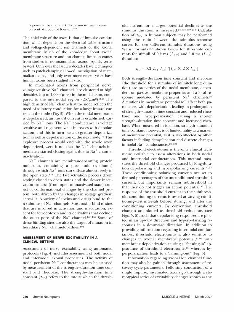
is powered by discrete kicks of inward membranecurrent at nodes of Ranvier.”33
The chief role of the axon is that of impulse conduc-tion, which depends on the electrical cable structureand voltage-dependent ion channels of the axonalmembrane. Much of the knowledge about axonalmembrane structure and ion channel function comesfrom studies in nonmammalian axons (squids, verte-brates). Only over the last few decades have techniquessuch as patch-clamping allowed investigation of mam-malian axons, and only over more recent years havehuman axons been studied in vitro.
In myelinated axons from peripheral nerve,voltage-sensitive Na� channels are clustered at highdensities (up to 1,000/�m2) in the nodal axon, com-pared to the internodal region (25/�m2).202 Thehigh density of Na� channels at the node reflects theneed of saltatory conduction for a large inward cur-rent at the node (Fig. 3). When the nodal membraneis depolarized, an inward current is established, car-ried by Na� ions. The Na� conductance is voltagesensitive and regenerative: it increases with depolar-ization, and this in turn leads to greater depolariza-tion as well as depolarization of the next node.75 Thisexplosive process would end with the whole axondepolarized, were it not that the Na� channels im-mediately started closing again, due to Na� channelinactivation.
Na� channels are membrane-spanning proteinmolecules, containing a pore unit (�-subunit)through which Na� ions can diffuse almost freely inthe open state.171 The fast activation process (fromresting closed to open state) and the slower inacti-vation process (from open to inactivated state) con-sist of conformational changes by the channel pro-tein, both driven by the changes in voltage gradientacross it. A variety of toxins and drugs bind to the�-subunits of Na� channels. Most toxins bind to sitesthat are involved in activation and inactivation, ex-cept for tetrodotoxin and its derivatives that occludethe outer pore of the Na� channel.100,154 Some ofthese binding sites are also the target of mutation inhereditary Na� channelopathies.101
ASSESSMENT OF NERVE EXCITABILITY IN ACLINICAL SETTING
Assessment of nerve excitability using automatedprotocols (Fig. 4) includes assessment of both nodaland internodal axonal properties. The activity ofnodal persistent Na� conductances may be assessedby measurement of the strength–duration time con-stant and rheobase. The strength–duration timeconstant (�SD) refers to the rate at which the thresh-
old current for a target potential declines as thestimulus duration is increased.28,138,139,204 Calcula-tion of �SD in human subjects may be performedusing the ratio between the stimulus–responsecurves for two different stimulus durations usingWeiss’ formula,204 shown below for threshold cur-rents for stimuli of 0.2 ms (I 0.2) and 1.0 ms (I 1.0)duration:
�SD � 0.2�I0.2–I1.0�/�I1.0–�0.2 � I0.2��
Both strength–duration time constant and rheobase(the threshold for a stimulus of infinitely long dura-tion) are properties of the nodal membrane, depen-dent on passive membrane properties and a local re-sponse mediated by persistent Na� channels.36
Alterations in membrane potential will affect both pa-rameters, with depolarization leading to prolongationof strength–duration time constant and reduced rheo-base; and hyperpolarization causing a shorterstrength–duration time constant and increased rheo-base. When measured in isolation, strength–durationtime constant, however, is of limited utility as a markerof membrane potential, as it is also affected by otherfactors including demyelination and discrete changesin nodal Na� conductances.28,100
Threshold electrotonus is the only clinical tech-nique available to assess alterations in both nodaland internodal conductances. This method mea-sures the threshold changes produced by long-dura-tion depolarizing and hyperpolarizing currents.30,37
These conditioning polarizing currents are set todefined percentages of the unconditioned thresholdcurrent, but importantly remain subthreshold inthat they do not trigger an action potential.37 Theresponse of the threshold current to the subthresh-old conditioning currents is tested at varying condi-tioning–test intervals before, during, and after theconditioning currents. By convention, thresholdchanges are plotted as threshold reductions (seeFigs. 5, 6), such that depolarizing responses are plot-ted in an upward direction and hyperpolarizing re-sponses in a downward direction. In addition toproviding information regarding internodal conduc-tances, threshold electrotonus is also sensitive tochanges in axonal membrane potential,11,92 withmembrane depolarization causing a “fanning-in” ap-pearance of threshold electrotonus,86 whereas hy-perpolarization leads to a “fanning-out” (Fig. 5).
Information regarding axonal ion channel func-tion may also be gained through assessment of re-covery cycle parameters. Following conduction of asingle impulse, myelinated axons go through a ste-reotypical series of excitability changes known as the
280 Uremic Neuropathy MUSCLE & NERVE March 2007

recovery cycle (Figs. 5, 6). For a period of 0.5–1.0 msafter an impulse, axons are completely inexcitableand cannot generate another impulse regardless ofthe strength of the depolarizing stimulus. This pe-riod is known as the absolute refractory period. Theaxon then enters a period of relative refractorinessthat can be measured either as the increase in cur-rent required to produce a potential of a specifiedsize, known as refractoriness, or as the duration ofthe relative refractory period until threshold hasreturned to baseline, usually 3–4 ms.
This period of refractoriness is followed by aperiod of superexcitability (or supernormality), dur-ing which there is a reduction in threshold occurringover a 10–15-ms interval. Finally, there is a late phaseof raised threshold known as late subexcitability,ending around 100 ms. These changes in threshold
are associated with changes in latency, which is in-creased during the refractory period, decreased dur-ing superexcitability, and increased during late sub-excitability.16,181
The relative refractory period results from in-activation of nodal transient Na� channels. It isprolonged by membrane depolarization and re-duced by hyperpolarization. Refractoriness maytherefore be used a measure of membrane poten-tial, although it is essential to take into accountthe effect of temperature, given that cooling leadsto an increase in refractoriness.94 Furthermore,measures of refractoriness may be unreliable insituations of impaired distal transmission, as mayoccur with axonal demyelination, neuromuscularjunction abnormalities, muscle disease, or anyother factor that reduces the security of impulse
FIGURE 4. (A) Arrangement for nerve excitability studies in ESKD patients undergoing hemodialysis. The current required to produce thedesired compound motor action potential (CMAP) amplitude is determined using a computerized threshold-tracking program that is runby a personal computer. Recordings are amplified and digitized using an analog-to-digital board. Stimulus waveforms are converted tocurrent with a purpose-built isolated linear bipolar constant current stimulator. The threshold tracking software consists of an automatedtracking system in which the amplitude of the test stimulus is automatically increased or decreased by a percentage step after eachresponse, depending on the difference between the recorded response and the target response, generally set to 40% of maximal CMAPamplitude for motor axons, as shown in (B). The protocol incorporates a proportional tracking system, in which the change in the stimulusis proportional to the difference between the actual and target responses.
Uremic Neuropathy MUSCLE & NERVE March 2007 281

transmission.37,114,120,190 Alteration in refractori-ness may also occur secondary to changes in nodalNa� conductances. Reductions in refractorinesshave been demonstrated in diabetic and toxic neu-ropathies, consistent with a reduction in the nodal
Na� conductances.100,103,126 In a recent study, anincrease in refractoriness was demonstrated in pa-tients treated with the chemotherapeutic agentoxaliplatin, in the absence of significant changesin other excitability parameters, suggesting that
FIGURE 5. Six plots of excitability parameters recorded from tibialis anterior for a single representative patient (continuous lines withcircles) prior to dialysis with 95% confidence intervals (broken lines) for a single subject.95 (A) Absolute stimulus–response relationshipfor stimuli of 0.2 ms duration (line without filled circle) and 1-ms duration (line with filled circle). The filled circle on the 1-ms response curvecorresponds to the threshold for a CMAP 50% of maximum and the broken ellipse corresponds to the 95% confidence limits for a singlesubject. (B) Normalized stimulus–response relationship. (C) Current–threshold relationship, reflecting rectifying properties of the axonfollowing polarizing currents. Threshold changes to hyperpolarizing current are represented on the left and to depolarizing current on theright. (D) Distribution of strength–duration time constants of nine populations of axons, from 5% to 95% of maximal CMAP in groups of10%. (E) Threshold electrotonus. Changes are plotted as threshold reductions, with depolarization represented in an upward direction andhyperpolarization in a downward direction. (F) Recovery cycle, showing the relative refractory period, superexcitability, and latesubexcitability. There is “fanning-in” of threshold electrotonus,76,82 an increase in refractoriness, and reductions in superexcitability andlate subexcitability (reproduced with permission from Oxford University Press; Krishnan et al.,113 fig. 1).
282 Uremic Neuropathy MUSCLE & NERVE March 2007

the neurotoxicity of this agent may be mediated byblockage of nodal transient Na� channels.110
Superexcitability is due to a depolarizing afterpo-tential that results from the capacitative charging ofthe internode by the action potential12 and subse-quently discharges through high resistance pathwaysunder or through the myelin sheath.40,99 Recentstudies have also suggested that activation of nodalNa� conductances may be a further contributingfactor.133 As for refractoriness, superexcitability alsovaries with membrane potential, with depolarizationleading to a reduction in superexcitability and hy-perpolarization causing an increase. Late subexcit-ability is determined by activation of nodal slow K�
channels and the difference between membrane po-tential (Er) and the K� equilibrium potential (Ek)and increases with depolarization if extracellular K�
is unchanged. It may therefore be used to differen-tiate between pure depolarization and depolariza-tion secondary to nerve ischemia in which there is nosignificant change in late subexcitability due to com-pensatory changes in extracellular K� ions.92
NERVE EXCITABILITY STUDIES IN ESKD
Nerve excitability studies in ESKD patients (Fig. 4)have recently demonstrated significant alterations inmembrane potential prior to hemodialysis, with re-covery in the postdialysis period.97,113,115–117 Prior todialysis, measures of nerve excitability were signifi-cantly abnormal in ESKD patients compared to con-trol data.93,95,108 Stimulus–response curves wereshifted to the right, indicating that axons were ofhigh threshold. This was accompanied by a fan-
FIGURE 6. Comparison of threshold electrotonus (A,C) and recovery cycle (B,D) in ESKD patients (continuous lines with circles) pre- andpostdialysis, with 95% confidence intervals for normal controls (broken lines). Predialysis traces demonstrate “fanning-in” of thresholdelectrotonus, increased refractoriness, and reduced superexcitability and late subexcitability. Abnormalities have largely resolved 1 hpostdialysis (reproduced with permission from Oxford University Press; Krishnan et al.,113 fig. 2).
Uremic Neuropathy MUSCLE & NERVE March 2007 283

ning-in of threshold electrotonus curves,86,92 whichassess alterations in both nodal and internodal con-ductances and are sensitive indicators of change inmembrane potential (Figs. 5, 6). Measurement ofrecovery cycles of excitability demonstrated that re-fractoriness, due to inactivation of voltage-gatedtransient Na� channels, was increased and superex-citability, due to the depolarizing afterpotential,12
was reduced. The findings were consistent with pre-dialysis axonal depolarization in ESKD patients.92
Measures of motor and sensory nerve excitabilityhave been assessed in relation to changes in serumlevels of potential neurotoxins, including K�, Ca2�
urea, uric acid, and middle molecules such as PTHand �-2M. Predialysis excitability abnormalities werenoted to be strongly correlated with serum K� in allstudies, suggesting that hyperkalemic depolarizationmay underlie the development of uremic neuropa-thy (Fig. 7). Furthermore, abnormalities of excitabil-ity became apparent with serum K� concentrationsin the high normal range, well below the levels re-quired to produce cardiac toxicity.38,115 The excit-ability abnormalities in ESKD patients were also dif-ferent from those noted in patients with diabeticneuropathy,111 another common metabolic neurop-athy, suggesting that the abnormalities noted inESKD patients were not purely a consequence ofstructural changes.
Predialysis excitability recordings also demon-strated an apparently paradoxical reduction in latesubexcitability, determined in part by nodal slow K�
channels. Since late subexcitability depends on thedifference between the resting potential and the K�
equilibrium potential and actually increases with de-polarization if extracellular K� is unchanged, thereduction in this parameter was thought to providefurther evidence that predialysis axonal membranedepolarization in patients with ESKD was due toeffects mediated by serum K�.97,113
Potassium satisfies criteria that have been sug-gested for a substance to be accepted as a uremicneurotoxin.38 It is an identifiable chemical that iselevated in the serum of ESKD patients and causesneurological dysfunction in both humans and ani-mals. It is also a critical determinant of axonal rest-ing membrane potential.97 Moreover, a direct rela-tionship exists between serum levels of K� andneurophysiological parameters, and its removalleads to considerable improvement in these indi-ces.97,113,115,117 The resting potentials of both nerveand muscle membranes are largely determined byK�, with relative changes in K� capable of depolar-izing membranes.
It may be argued that the abnormalities of serumK� noted in excitability studies are the consequenceof a transient homeostatic disturbance, rapidly cor-rected by dialysis, and therefore unlikely to play amajor role in the development of chronic irrevers-ible neuropathy. Against such an argument, pro-longed exposure to hyperkalemia in ESKD patientsseems likely, given that postdialysis rebound of K� isa well-recognized phenomenon,2,51 with hyperkale-mia typically recurring within 6 h of a dialysis sessiondue to reequilibration between intracellular and ex-tracellular fluid compartments.19 Such prolongedhyperkalemia may cause disruption of normal ionicgradients and activate damaging Ca��-mediatedprocesses, leading to axonal loss.48 Furthermore,given the importance of K� in mediating these ab-normalities, current measures of dialysis adequacy,50
which are based solely on blood urea concentrations,may be inappropriate for determining the adequacyof a dialysis regimen to prevent neurotoxicity. Abetter indication of adequate dialysis might be themaintenance of serum K� within normal limits be-tween periods of dialysis, which may require moreattention to dietary restriction of K� intake in somepatients.113
AXONAL NA�/K� PUMP FUNCTIONIN ESKD PATIENTS
Inhibition of the Na�/K� pump by uremic neurotox-ins, previously proposed as the mechanism underlyingthe development of uremic neuropathy,148 may inducemembrane depolarization.16,31,32,35,92 Of further rele-vance, previous studies have demonstrated that alter-ations in membrane potential and intra- and extracel-lular K� concentration have a direct effect on Na�/K�
pump function.78,77,109,140,159,160
Nerve excitability measurements provide a meansof detecting changes in membrane potential causedby activation of the Na�/K� pump.29,34,98 Vagg et
FIGURE 7. Relationship between predialysis depolarizing thresh-old electrotonus at the 90–100 ms interval (TEd 90–100ms) andK� for pooled data from median sensory studies (open square)and lower-limb motor studies (TA: filled diamond; EDB, opendiamond).113,117
284 Uremic Neuropathy MUSCLE & NERVE March 2007

al.190 showed that the activity-dependent hyperpolar-ization (ADH) of motor axons induced by voluntaryactivity causes threshold changes that can be used toassess Na�/K� pump function. Assessment of activ-ity-dependent excitability changes prior to hemodi-alysis in 10 ESKD patients demonstrated quantita-tively similar changes in ESKD patients and controls,arguing against any significant reduction in the ax-onal Na�/K� pump in ESKD (Fig. 8).115
A further study also assessed Na�/K� pump func-tion in six ESKD patients prior to dialysis by moni-toring the excitability changes that occurred before,during, and after 13 min of nerve ischemia.116 Withthe onset of ischemia, a short-lived threshold reduc-tion was followed by a rapid increase in threshold(Fig. 9). Whereas normal controls manifested a post-ischemic threshold increase due to increased activityof the Na�/K� pump and consequent membranehyperpolarization,112 a paradoxical reduction inthreshold was noted in ESKD patients. This patternsuggested that the axonal Na�/K� pump had a sta-bilizing effect on the axon and was attempting toreturn membrane potential toward baseline from
the highly depolarized levels of the ischemic period.Importantly, however, the rapid return of thresholdto baseline levels in the postischemic period con-firmed that the Na�/K� pump was functioning wellin ESKD.
CONCLUSION
Neuropathy is a common complication of ESKD,occurring in the majority of patients undergoingdialysis. At present, renal transplantation remainsthe only known cure for uremic neuropathy.20,21
Recent nerve excitability studies have suggested thathyperkalemia may underlie the development of neu-ropathy and have argued against any dysfunction ofthe axonal Na�/K� pump in the development ofthis condition. Excess K� fits the profile of the neu-rotoxin responsible for uremic neuropathy betterthan middle molecules, parathyroid hormone, orany other organic substance that has been previouslylinked to the development of uremic neuropathy.Recent findings from nerve excitability studies inESKD patients suggest that maintenance of serum
FIGURE 8. Comparison of excitability changes following maximalvoluntary contraction in ESKD patients predialysis (filled dia-mond) and normal controls (open diamond). The filled arrowindicates the time of contraction. (A) Changes in normalizedthreshold for a 1-ms stimulus duration. (B) Strength–durationtime constant, �SD. (C) Superexcitability. (D) Submaximal CMAPamplitude. Threshold and CMAP amplitude changes are normal-ized to precontraction values and are expressed as mean data �SEM. Although baseline superexcitability was significantly less inESKD patients, there were no significant differences betweenpatients and controls in the magnitude of excitability changesinduced by activity,99 arguing against Na�/K� pump dysfunctionin this condition (reprinted with permission from InternationalFederation of Clinical Neurophysiology; Krishnan et al.,115 fig. 3).
FIGURE 9. Excitability parameters recorded from tibialis anteriorfor ESKD patients and controls before, during, and after theischemic period (indicated by filled horizontal bar). Mean data �SEM. During ischemia refractoriness increased and superexcit-ability decreased in both groups, consistent with axonal depolar-ization. The reversal of these changes in the postischemic periodis consistent with axonal hyperpolarization. There is a smallerthreshold reduction during ischemia in ESKD patients and aprominent ischemic threshold increase beyond baseline values, afeature not observed in control subjects (reproduced with permis-sion from Oxford University Press; Krishnan et al.,116 fig. 3).
Uremic Neuropathy MUSCLE & NERVE March 2007 285

K� within normal limits between periods of dialysis,rather than simple avoidance of hyperkalemia, islikely to reduce the incidence and severity of uremicneuropathy.
Grant support was received from the Australian Brain Founda-tion, Sylvia and Charles Viertel Charitable Foundation, NationalHealth and Medical Research Council of Australia and the Aus-tralian Association of Neurologists. We thank Hugh Bostock andDavid Burke for help in the development of the excitability stud-ies, and Bruce Pussell and John Charlesworth for assistance instudies in uremic patients.
REFERENCES
1. Ackil AA, Shahani BT, Young RR, Rubin NE. Late responseand sural conduction studies. Usefulness in patients withchronic renal failure. Arch Neurol 1981;38:482–485.
2. Ahmed J, Weisberg LS. Hyperkalemia in dialysis patients.Semin Dial 2001;14:348–356.
3. Ahonen RE. Peripheral neuropathy in uremic patients andin renal transplant recipients. Acta Neuropathol 1981;54:43–53.
4. Angus-Leppan H, Burke D. The function of large and smallnerve fibers in renal failure. Muscle Nerve 1992;15:288–294.
5. Appenzeller O, Kornfeld M, MacGee J. Neuropathy inchronic renal disease. Arch Neurol 1971;24:449–461.
6. Asbury AK, Victor M, Adams RD. Uremic polyneuropathy.Arch Neurol Psychiatr 1963;8:413–428.
7. Asbury AK. Recovery from uremic neuropathy. N Engl J Med1971;284:1211–1212.
8. Avram MM, Feinfeld DA, Huatuco AH. Search for the ure-mic toxin. Decreased motor-nerve conduction velocity andelevated parathyroid hormone in uremia. N Engl J Med1978;298:1000–1003.
9. Babb AL, Popovich RP, Christopher TG, Scribner BH. Thegenesis of the square meter-hour hypothesis. Trans Am SocArtif Intern Org 1971;17:81–91.
10. Babb AL, Ahmad S, Bergstrom J, Scribner BH. The middlemolecule hypothesis in perspective. Am J Kidney Dis 1981;1:46–50.
11. Baker M, Bostock H. Depolarization changes the mechanismof accommodation in rat and human motor axons. J Physiol(Lond) 1989;411:545–561.
12. Barrett EF, Barrett JN. Intracellular recording from verte-brate myelinated axons: mechanism of the depolarizing af-terpotential. J Physiol (Lond) 1982;323:117–144.
13. Bazzi C, Pagani C, Sorgato G, Albonico G, Fellin G, D’AmicoG. Uremic polyneuropathy: a clinical and electrophysiolog-ical study in 135 short- and long-term hemodialyzed patients.Clin Nephrol 1991;35:176–181.
14. Bellinghieri G, Santoro D, Lo Forti B, Mallamace A, DeSanto RM, Savica V. Erectile dysfunction in uremic dialysispatients: diagnostic evaluation in the sildenafil era. Am JKidney Dis 2001;38:S115–117.
15. Benz RL, Siegfried JW, Teehan BP. Carpal tunnel syndromein dialysis patients: comparison between continuous ambu-latory peritoneal dialysis and hemodialysis populations. Am JKidney Dis 1988;11:473–476.
16. Bergmans J. The physiology of single human nerve fibres.Vander, Belgium: University of Louvain; 1970.
17. Bicknell JM, Lim AC, Raroque HG Jr, Tzamaloukas AH.Carpal tunnel syndrome, subclinical median mononeuropa-thy, and peripheral polyneuropathy: common early compli-cations of chronic peritoneal dialysis and hemodialysis. ArchPhys Med Rehabil 1991;72:378–381.
18. Bittar EE. Maia muscle fibre as a model for the study ofuraemic toxicity. Nature 1967;214:310–312.
19. Blumberg A, Roser HW, Zehnder C, Muller-Brand J. Plasmapotassium in patients with terminal renal failure during andafter haemodialysis; relationship with dialytic potassium re-moval and total body potassium. Nephrol Dial Transplant1997;12:1629–1634.
20. Bolton CF, Baltzan MA, Baltzan RB. Effects of renal trans-plantation on uremic neuropathy. A clinical and electro-physiologic study. N Engl J Med 1971;284:1170–1175.
21. Bolton CF. Electrophysiologic changes in uremic neuropa-thy after successful renal transplantation. Neurology 1976;26:152–161.
22. Bolton CF, Driedger AA, Lindsay RM. Ischaemic neuropathyin uraemic patients caused by bovine arteriovenous shunt.J Neurol Neurosurg Psychiatry 1979;42:810–814.
23. Bolton CF. Peripheral neuropathies associated with chronicrenal failure. Can J Neurol Sci 1980;7:89–96.
24. Bolton CF, Carter K, Koval JJ. Temperature effects on con-duction studies of normal and abnormal nerve. MuscleNerve 1982;5:S145–147.
25. Bolton CF, Young GB. Neurological complications of renaldisease. Stoneham, UK: Butterworth-Heinemann; 1990.
26. Bolton CF, McKeown MJ, Chen R, Toth B, Remtulla H.Subacute uremic and diabetic polyneuropathy. MuscleNerve 1997;20:59–64.
27. Bolton CF, Remtulla H, Toth B, Bernardi L, Lindsay RM,Maryniak O, et al. Distinctive electrophysiological features ofdenervated muscle in uremic patients. J Clin Neurophysiol1997;14:539–5542.
28. Bostock H. The strength–duration relationship for excita-tion of myelinated nerve: computed dependence on mem-brane parameters. J Physiol (Lond) 1983;341:59–74.
29. Bostock H, Grafe P. Activity-dependent excitability changesin normal and demyelinated rat spinal root axons. J Physiol(Lond) 1985;365:239–257.
30. Bostock H, Baker M. Evidence for two types of potassiumchannel in human motor axons in vivo. Brain Res 1988;462:354–358.
31. Bostock H, Baker M, Grafe P, Reid G. Changes in excitabilityand accommodation of human motor axons following briefperiods of ischaemia. J Physiol (Lond) 1991;441:513–535.
32. Bostock H, Baker M, Reid G. Changes in excitability ofhuman motor axons underlying post-ischaemic fascicula-tions: evidence for two stable states. J Physiol (Lond) 1991;441:537–557.
33. Bostock H. Impulse propagation in experimental neuropa-thy. In: Dyck PJ, Thomas PK, Griffin JW, Low PA, Poduslo JF,editors. Peripheral neuropathy. Philadelphia: Saunders;1993. p 109–120.
34. Bostock H, Bergmans J. Post-tetanic excitability changes andectopic discharges in a human motor axon. Brain 1994;117:913–928.
35. Bostock H, Burke D, Hales JP. Differences in behaviour ofsensory and motor axons following release of ischaemia.Brain 1994;117:225–234.
36. Bostock H, Rothwell JC. Latent addition in motor and sen-sory fibres of human peripheral nerve. J Physiol (Lond)1997;498:277–294.
37. Bostock H, Cikurel K, Burke D. Threshold tracking tech-niques in the study of human peripheral nerve. MuscleNerve 1998;21:137–158.
38. Bostock H, Walters RJ, Andersen KV, Murray NM, Taube D,Kiernan MC. Has potassium been prematurely discarded asa contributing factor to the development of uraemic neu-ropathy? Nephrol Dial Transplant 2004;19:1054–1057.
39. Brouns R, De Deyn PP. Neurological complications in renalfailure: a review. Clin Neurol Neurosurg 2004;107:1–16.
40. Burke D, Kiernan MC, Bostock H. Excitability of humanaxons. Clin Neurophysiol 2001;112:1575–1585.
41. Caccia MR, Mangili A, Mecca G, Ubiali E, Zanoni P. Effectsof hemodialytic treatment on uremic polyneuropathy. Aclinical and electrophysiological follow-up study. J Neurol1977;217:123–131.
286 Uremic Neuropathy MUSCLE & NERVE March 2007

42. Cappelen-Smith C, Lin CS, Kuwabara S, Burke D. Conduc-tion block during and after ischaemia in chronic inflamma-tory demyelinating polyneuropathy. Brain 2002;125:1850–1858.
43. Castaigne P, Cathala HP, Beaussart-Boulenge L, Petrover M.Effect of ischaemia on peripheral nerve function in patientswith chronic renal failure undergoing dialysis treatment.J Neurol Neurosurg Psychiatry 1972;35:631–637.
44. Chang MH, Chou KJ. The role of autonomic neuropathy inthe genesis of intradialytic hypotension. Am J Nephrol 2001;21:357–361.
45. Chaumont P, Lefebvre J, Lerique JL. Explorations elec-trologiques au cours des insufficicances renales graves. RevNeurol 1963;108:199.
46. Codish SD, Cress RH, Bolton CF. Uremic neuropathy.N Engl J Med 1971;285:752–753.
47. Cofan F, Garcia S, Combalia A, Segur JM, Oppenheimer F.Carpal tunnel syndrome secondary to uraemic tumoral cal-cinosis. Rheumatology (Oxford) 2002;41:701–703.
48. Craner MJ, Hains BC, Lo AC, Black JA, Waxman SG. Co-localization of sodium channel Nav1.6 and the sodium-cal-cium exchanger at sites of axonal injury in the spinal cord inEAE. Brain 2004;127:294–303.
49. D’Amour ML, Dufresne LR, Morin C, Slaughter D. Sensorynerve conduction in chronic uremic patients during the firstsix months of hemodialysis. Can J Neurol Sci 1984;11:269–271.
50. Daugirdas JT. Dialysis adequacy and kinetics. Curr OpinNephrol Hypertens 2000;9:599–605.
51. De Nicola L, Bellizzi V, Minutolo R, Cioffi M, Giannattasio P,Terracciano V, et al. Effect of dialysate sodium concentra-tion on interdialytic increase of potassium. J Am Soc Neph-rol 2000;11:2337–2343.
52. Delmez JA, Holtmann B, Sicard GA, Goldberg AP, HarterHR. Peripheral nerve entrapment syndromes in chronic he-modialysis patients. Nephron 1982;30:118–123.
53. Di Giulio S, Chkoff N, Lhoste F, Zingraff J, Drueke T.Parathormone as a nerve poison in uremia. N Engl J Med1978;299:1134–1135.
54. Dinn JJ, Crane DL. Schwann cell dysfunction in uraemia.J Neurol Neurosurg Psychiatry 1970;33:605–608.
55. Dyck PJ, Johnson WJ, Lambert EH, O’Brien PC. Segmentaldemyelination secondary to axonal degeneration in uremicneuropathy. Mayo Clin Proc 1971;46:400–431.
56. Dyck PJ, Johnson WJ, Nelson RA, Lambert EH, O’Brien PC.Uremic neuropathy. III. Controlled study of restricted pro-tein and fluid diet and infrequent hemodialysis versus con-ventional hemodialysis treatment. Mayo Clin Proc 1975;50:641–649.
57. Dyck PJ, Sherman WR, Hallcher LM, Service FJ, O’Brien PC,Grina LA, et al. Human diabetic endoneurial sorbitol, fruc-tose, and myo-inositol related to sural nerve morphometry.Ann Neurol 1980;8:590–596.
58. Dyck PJ. Detection, characterization, and staging of polyneu-ropathy: assessed in diabetics. Muscle Nerve 1988;11:21–32.
59. Ewing DJ, Winney R. Autonomic function in patients withchronic renal failure on intermittent haemodialysis.Nephron 1975;15:424–429.
60. Furst P, Asaba M, Gordon A, Zimmerman L, Bergstrom J.Middle molecules in uraemia. Proc Eur Dial TransplantAssoc 1975;11:417–426.
61. Furst P, Zimmerman L, Bergstrom J. Determination of en-dogenous middle molecules in normal and uremic bodyfluids. Clin Nephrol 1976;3:178–188.
62. Garcia S, Cofan F, Combalia A, Campistol JM, OppenheimerF, Ramon R. Compression of the ulnar nerve in Guyon’scanal by uremic tumoral calcinosis. Arch Orthop TraumaSurg 2000;120:228–230.
63. Gejyo F, Narita I. Current clinical and pathogenetic under-standing of beta2-m amyloidosis in long-term haemodialysispatients. Nephrology 2003;8(Supp):S45–49.
64. Ginn HE, Bugel HJ, James L, Hopkins P. Clinical experiencewith small surface area dialyzers (SSAD). Proc Clin DialTransplant Forum 1971;1:53–60.
65. Goldstein DA, Chui LA, Massry SG. Effect of parathyroidhormone and uremia on peripheral nerve calcium and mo-tor nerve conduction velocity. J Clin Invest 1978;62:88–93.
66. Gousheh J, Iranpour A. Association between carpel tunnelsyndrome and arteriovenous fistula in hemodialysis patients.Plast Reconstr Surg 2005;116:508–513.
67. Graf RJ, Halter JB, Pfeifer MA, Halar E, Brozovich F, Porte DJr. Glycemic control and nerve conduction abnormalities innon-insulin-dependent diabetic subjects. Ann Intern Med1981;94:307–311.
68. Halar EM, Brozovich FV, Milutinovic J, Inouye VL, BeckerVM. H-reflex latency in uremic neuropathy: correlation withNCV and clinical findings. Arch Phys Med Rehabil 1979;60:174–177.
69. Hamilton DV, Evans DB, Henderson RG. Ulnar nerve lesionas complication of Cimino–Brescia arteriovenous fistula.Lancet 1980;2:1137–1138.
70. Harding AE, Le Fanu J. Carpal tunnel syndrome related toantebrachial Cimino–Brescia fistula. J Neurol Neurosurg Psy-chiatry 1977;40:511–513.
71. Hassan K, Simri W, Rubenchik I, Manelis J, Gross B, ShashaSM, et al. Effect of erythropoietin therapy on polyneurop-athy in predialytic patients. J Nephrol 2003;16:121–125.
72. Hegstrom RM, Murray JS, Pendras JP, Burnell JM, ScribnerBH. Two year’s experience with periodic hemodialysis in thetreatment of chronic uremia. Trans Am Soc Artif InternOrgans 1962;8:266–280.
73. Heidbreder E, Schafferhans K, Heidland A. Autonomic neu-ropathy in chronic renal insufficiency. Comparative analysisof diabetic and nondiabetic patients. Nephron 1985;41:50–56.
74. Heidbreder E, Schafferhans K, Heidland A. Disturbances ofperipheral and autonomic nervous system in chronic renalfailure: effects of hemodialysis and transplantation. ClinNephrol 1985;23:222–228.
75. Hille B. Ionic channels of excitable membranes. Sunder-land, MA: Sinauer Associates; 1992.
76. Hirasawa Y, Ogura T. Carpal tunnel syndrome in patients onlong-term haemodialysis. Scand J Plast Reconstr Surg HandSurg 2000;34:373–381.
77. Hodgkin AL, Keynes RD. Movement of cations during recov-ery in nerve. Symp Soc Exp Biol 1954;8:423–437.
78. Hodgkin AL, Keynes RD. Active transport of cations in giantaxons from Sepia and Logilo. J Physiol (Lond) 1955;128:28–60.
79. Hoke A, Keswani SC. Neuroprotection in the PNS: erythro-poietin and immunophilin ligands. Ann N Y Acad Sci 2005;1053:491–501.
80. Ibrahim MM, Barnes AD, Crosland JM, Dawson-Edwards P,Honigsberger L, Newman CE, et al. Effect of renal transplan-tation of uraemic neuropathy. Lancet 1974;2:739–742.
81. Jain VK, Cestero RV, Baum J. Carpal tunnel syndrome inpatients undergoing maintenance hemodialysis. JAMA 1979;242:2868–2869.
82. Jebsen RH, Tenckhoff H, Honet JC. Natural history of ure-mic polyneuropathy and effects of dialysis. N Engl J Med1967;277:327–333.
83. Jog MS, Turley JE, Berry H. Femoral neuropathy in renaltransplantation. Can J Neurol Sci 1994;21:38–42.
84. Kaji R, Sumner AJ. Ouabain reverses conduction distur-bances in single demyelinated nerve fibers. Neurology 1989;39:1364–1368.
85. Kaji R, Bostock H, Kohara N, Murase N, Kimura J, ShibasakiH. Activity-dependent conduction block in multifocal motorneuropathy. Brain 2000;123:1602–1611.
86. Kaji R. Physiology of conduction block in multifocal motorneuropathy and other demyelinating neuropathies. MuscleNerve 2003;27:285–296.
Uremic Neuropathy MUSCLE & NERVE March 2007 287

87. Kaku DA, Malamut RI, Frey DJ, Parry GJ. Conduction blockas an early sign of reversible injury in ischemic monomelicneuropathy. Neurology 1993;43:1126–1130.
88. Kanai K, Kuwabara S, Arai K, Sung JY, Ogawara K, Hattori T.Muscle cramp in Machado-Joseph disease: altered motoraxonal excitability properties and mexiletine treatment.Brain 2003;126:965–973.
89. Kersh ES, Kronfield SJ, Unger A, Popper RW, Cantor S,Cohn K. Autonomic insufficiency in uremia as a cause ofhemodialysis-induced hypotension. N Engl J Med 1974;290:650–653.
90. Keswani SC, Buldanlioglu U, Fischer A, Reed N, Polley M,Liang H, et al. A novel endogenous erythropoietin mediatedpathway prevents axonal degeneration. Ann Neurol 2004;56:815–826.
91. Keswani SC, Leitz GJ, Hoke A. Erythropoietin is neuropro-tective in models of HIV sensory neuropathy. Neurosci Lett2004;371:102–105.
92. Kiernan MC, Bostock H. Effects of membrane polarizationand ischaemia on the excitability properties of human motoraxons. Brain 2000;123:2542–2551.
93. Kiernan MC, Burke D, Andersen KV, Bostock H. Multiplemeasures of axonal excitability: a new approach in clinicaltesting. Muscle Nerve 2000;23:399–409.
94. Kiernan MC, Cikurel K, Bostock H. Effects of temperatureon the excitability properties of human motor axons. Brain2001;124:816–825.
95. Kiernan MC, Lin CS, Andersen KV, Murray NM, Bostock H.Clinical evaluation of excitability measures in sensory nerve.Muscle Nerve 2001;24:883–892.
96. Kiernan MC, Guglielmi JM, Kaji R, Murray NM, Bostock H.Evidence for axonal membrane hyperpolarization in multi-focal motor neuropathy with conduction block. Brain 2002;125:664–675.
97. Kiernan MC, Walters RJ, Andersen KV, Taube D, MurrayNM, Bostock H. Nerve excitability changes in chronic renalfailure indicate membrane depolarization due to hyperkal-aemia. Brain 2002;125:1366–1378.
98. Kiernan MC, Lin CS, Burke D. Differences in activity-depen-dent hyperpolarization in human sensory and motor axons.J Physiol (Lond) 2004;558:341–349.
99. Kiernan MC, Burke D, Bostock H. Nerve excitability mea-sures: biophysical basis and use in the investigation of pe-ripheral nerve disease. In: Dyck PJ, Thomas PK, editors.Peripheral neuropathy. Philadelphia: Elsevier Saunders;2005. p 113–130.
100. Kiernan MC, Isbister GK, Lin CS, Burke D, Bostock H. Acutetetrodotoxin-induced neurotoxicity after ingestion of pufferfish. Ann Neurol 2005;57:339–348.
101. Kiernan MC, Krishnan AV, Lin CS, Burke D, Berkovic SF.Mutation in the Na� channel subunit SCN1B produces par-adoxical changes in peripheral nerve excitability. Brain2005;128:1841–1846.
102. Kim SJ, Shin SJ, Kang ES. Endoscopic carpal tunnel releasein patients receiving long-term hemodialysis. Clin Orthop2000;:141–148.
103. Kitano Y, Kuwabara S, Misawa S, Ogawara K, Kanai K,Kikkawa Y, et al. The acute effects of glycemic control onaxonal excitability in human diabetics. Ann Neurol 2004;56:462–467.
104. Kjellstrand CM. Do middle molecules cause uremic intoxi-cation? Am J Kidney Dis 1981;1:51–56.
105. Knezevic W, Mastaglia FL. Neuropathy associated with Bres-cia-Cimino arteriovenous fistulas. Arch Neurol 1984;41:1184–1186.
106. Konishi T, Nishitani H, Motomura S. Single fiber electro-myography in chronic renal failure. Muscle Nerve 1982;5:458–461.
107. Konotey-Ahulu FI, Baillod RA, Comty CM, Heron JR, Shal-don S, Thomas PK. Effect of periodic dialysis on the periph-eral neuropathy of end-stage renal failure. Br Med J 1965;2:1212–1215.
108. Krishnan AV, Lin CS-Y, Kiernan MC. Nerve excitability prop-erties in lower limb motor axons:evidence for a length-dependent gradient. Muscle Nerve 2004;29:645–655.
109. Krishnan AV, Colebatch JG, Kiernan MC. Hypokalemia in-duces activity-dependent conduction block. Neurology 2005;65:1309–1312.
110. Krishnan AV, Goldstein D, Friedlander M, Kiernan MC.Oxaliplatin-induced neurotoxicity and the development ofneuropathy. Muscle Nerve 2005;32:51–60.
111. Krishnan AV, Kiernan MC. Altered nerve excitability prop-erties in established diabetic neuropathy. Brain 2005;128:1178–1187.
112. Krishnan AV, Lin CS, Kiernan MC. Excitability differences inlower-limb motor axons during and after ischemia. MuscleNerve 2005;31:205–213.
113. Krishnan AV, Phoon RK, Pussell BA, Charlesworth JA, Bos-tock H, Kiernan MC. Altered motor nerve excitability inend-stage kidney disease. Brain 2005;128:2164–2174.
114. Krishnan AV, Kiernan MC. Axonal function and activity-dependent excitability changes in myotonic dystrophy. Mus-cle Nerve 2006;33:627–636.
115. Krishnan AV, Phoon RK, Pussell BA, Charlesworth JA, Bos-tock H, Kiernan MC. Neuropathy, axonal Na�/K� pumpfunction and activity-dependent excitability changes in end-stage kidney disease. Clin Neurophysiol 2006;117:992–997.
116. Krishnan AV, Phoon RK, Pussell BA, Charlesworth JA, Bos-tock H, Kiernan MC. Ischaemia induces paradoxical changesin axonal excitability in end-stage kidney disease. Brain2006;129:1585–1592.
117. Krishnan AV, Phoon RK, Pussell BA, Charlesworth JA, Kier-nan MC. Sensory nerve excitability and neuropathy in end-stage kidney disease. J Neurol Neurosurg Psychiatry 2006;77:548–551.
118. Kuchle C, Fricke H, Held E, Schiffl H. High-flux hemodial-ysis postpones clinical manifestation of dialysis-related amy-loidosis. Am J Nephrol 1996;16:484–488.
119. Kumar S, Trivedi HL, Smith EK. Carpal tunnel syndrome: acomplication of arteriovenous fistula in hemodialysis pa-tients. Can Med Assoc J 1975;113:1070–1072.
120. Kuwabara S, Ogawara K, Sung JY, Mori M, Kanai K, HattoriT, et al. Differences in membrane properties of axonal anddemyelinating Guillain–Barre syndromes. Ann Neurol 2002;52:180–187.
121. Laaksonen S, Voipio-Pulkki L, Erkinjuntti M, Asola M, FalckB. Does dialysis therapy improve autonomic and peripheralnervous system abnormalities in chronic uraemia? J InternMed 2000;248:21–26.
122. Laaksonen S, Metsarinne K, Voipio-Pulkki LM, Falck B. Neu-rophysiologic parameters and symptoms in chronic renalfailure. Muscle Nerve 2002;25:884–890.
123. Ligtenberg G, Blankestijn PJ, Boomsma F, Koomans HA. Nochange in automatic function tests during uncomplicatedhaemodialysis. Nephrol Dial Transplant 1996;11:651–656.
124. Lindblom U, Tegner R. Thermal sensitivity in uremic neu-ropathy. Acta Neurol Scand 1985;71:290–294.
125. Lowitzsch K, Gohring U, Hecking E, Kohler H. Refractoryperiod, sensory conduction velocity and visual evoked poten-tials before and after haemodialysis. J Neurology NeurosurgPsychiatry 1981;44:121–128.
126. Mackel R, Brink E. Conduction of neural impulses in dia-betic neuropathy. Clin Neurophysiol 2003;114:248–255.
127. Mallamaci F, Zoccali C, Ciccarelli M, Briggs JD. Autonomicfunction in uremic patients treated by hemodialysis or CAPDand in transplant patients. Clin Nephrol 1986;25:175–180.
128. Mallick NP, Gokal R. Haemodialysis. Lancet 1999;353:737–742.
129. Man NK, Granger A, Rondon-Nucete M, Zingraff J, JungersP, Sausse A, et al. One year follow-up of short dialysis with amembrane highly permeable to middle molecules. Proc EurDial Transplant Assoc 1973;10:236–246.
130. Mansouri B, Adybeig B, Rayegani M, Yasami S, Behshad V.Uremic neuropathy and the analysis of electrophysiological
288 Uremic Neuropathy MUSCLE & NERVE March 2007

changes. Electromyography Clin Neurophysiol 2001;41:107–115.
131. Martinelli P, Baruzzi A, Montagna P, Ravasio A, Poppi M.Carpal tunnel syndrome in a patient with a Cimino–Bresciafistula. Eur Neurol 1981;20:478–480.
132. Massry SG. Parathyroid hormone: a uremic toxin. Adv ExpMed Biol 1987;223:1–17.
133. McIntyre CC, Richardson AG, Grill WM. Modeling the ex-citability of mammalian nerve fibers: influence of afterpo-tentials on the recovery cycle. J Neurophysiol 2002;87:995–1006.
134. Meech PR. Femoral neuropathy following renal transplanta-tion. Aust N Z J Surg 1990;60:117–119.
135. Miles AM. Vascular steal syndrome and ischaemicmonomelic neuropathy: two variants of upper limb isch-aemia after haemodialysis vascular access surgery. NephrolDial Transplant 1999;14:297–300.
136. Meguid El Nahas A, Bello AK. Chronic kidney disease: theglobal challenge. Lancet 2005;365:331–340.
137. Mitz M, Prakash AS, Melvin J, Piering W. Motor nerve con-duction indicators in uremic neuropathy. Arch Phys MedRehabil 1980;61:45–48.
138. Mogyoros I, Kiernan MC, Burke D. Strength-duration prop-erties of human peripheral nerve. Brain 1996;119:439–447.
139. Mogyoros I, Lin C, Dowla S, Grosskreutz J, Burke D.Strength–duration properties and their voltage dependenceat different sites along the median nerve. Clin Neurophysiol1999;110:1618–1624.
140. Morita K, David G, Barrett JN, Barrett EF. Posttetanic hyper-polarization produced by electrogenic Na�-K� pump in liz-ard axons impaled near their motor terminals. J Neuro-physiol 1993;70:1874–1884.
141. Naito M, Ogata K, Goya T. Carpal tunnel syndrome inchronic renal dialysis patients: clinical evaluation of 62hands and results of operative treatment. J Hand Surg (Br)1987;12:366–374.
142. Nardin R, Chapman KM, Raynor EM. Prevalence of ulnarneuropathy in patients receiving hemodialysis. Arch Neurol2005;62:271–275.
143. National Kidney Foundation. NKF-DOQI clinical practiceguidelines for hemodialysis adequacy. Am J Kidney Dis 1997;30(Suppl 2):S15–66.
144. Nielsen VK. The peripheral nerve function in chronic renalfailure. II. Intercorrelation of clinical symptoms and signsand clinical grading of neuropathy. Acta Med Scand 1971;190:113–117.
145. Nielsen VK. The peripheral nerve function in chronic renalfailure. I. Clinical symptoms and signs. Acta Med Scand1971;190:105–111.
146. Nielsen VK, Winkel P. The peripheral nerve function inchronic renal failure. 3. A multivariate statistical analysis offactors presumed to affect the development of clinical neu-ropathy. Acta Med Scand 1971;190:119–125.
147. Nielsen VK. The peripheral nerve function in chronic renalfailure. IV. An analysis of the vibratory perception threshold.Acta Med Scand 1972;191:287–296.
148. Nielsen VK. The peripheral nerve function in chronic renalfailure. V. Sensory and motor conduction velocity. Acta MedScand 1973;194:445–454.
149. Nielsen VK. The peripheral nerve function in chronic renalfailure. VI. The relationship between sensory and motornerve conduction and kidney function, azotemia, age, sex,and clinical neuropathy. Acta Med Scand 1973;194:455–462.
150. Nielsen VK. The peripheral nerve function in chronic renalfailure. IX. Recovery after renal transplantation. Electro-physiological aspects (sensory and motor nerve conduction).Acta Med Scand 1974;195:171–180.
151. Nielsen VK. The peripheral nerve function in chronic renalfailure. VII. Longitudinal course during terminal renal fail-ure and regular hemodialysis. Acta Med Scand 1974;195:155–162.
152. Nielsen VK. The peripheral nerve function in chronic renalfailure. 8. Recovery after renal transplantation. Clinical as-pects. Acta Med Scand 1974;195:163–170.
153. Obeso JA, Marti-Masso JF, Asin JL, Remirez MV, Irigoyen V,Iragui M, et al. Conduction velocity through the somestheticpathway in chronic renal failure. J Neurol Sci 1979;43:439–445.
154. Ogata N, Ohishi Y. Molecular diversity of structure andfunction of the voltage-gated Na� channels. Jpn J Pharmacol2002;88:365–377.
155. Ogura T, Makinodan A, Kubo T, Hayashida T, Hirasawa Y.Electrophysiological course of uraemic neuropathy in hae-modialysis patients. Postgrad Med J 2001;77:451–454.
156. Oh SJ, Clements RS Jr, Lee YW, Diethelm AG. Rapid im-provement in nerve conduction velocity following renaltransplantation. Ann Neurol 1978;4:369–373.
157. Panayiotopoulos CP, Lagos G. Tibial nerve H-reflex andF-wave studies in patients with uremic neuropathy. MuscleNerve 1980;3:423–426.
158. Preswick G, Jeremy D. Subclinical polyneuropathy in renalinsufficiency. Lancet 1964:731.
159. Rakowski RF, Gadsby DC, De Weer P. Stoichiometry andvoltage dependence of the sodium pump in voltage-clamped, internally dialyzed squid giant axon. J Gen Physiol1989;93:903–941.
160. Rang HP, Ritchie JM. On the electrogenic sodium pump inmammalian non-myelinated nerve fibres and its activation byvarious external cations. J Physiol (Lond) 1968;196:183–221.
161. Redfern AB, Zimmerman NB. Neurologic and ischemiccomplications of upper extremity vascular access for dialysis.J Hand Surg (Am) 1995;20:199–204.
162. Riggs JE, Moss AH, Labosky DA, Liput JH, Morgan JJ, Gut-mann L. Upper extremity ischemic monomelic neuropathy:a complication of vascular access procedures in uremic dia-betic patients. Neurology 1989;39:997–998.
163. Ritchie J, Straub R. The hyperpolarization which followsactivity in mammalian non-medullated fibres. J Physiol(Lond) 1957;136:80–97.
164. Rockel A, Hennemann H, Sternagel-Haase A, Heidland A.Uraemic sympathetic neuropathy after haemodialysis andtransplantation. Eur J Clin Invest 1979;9:23–27.
165. Ropper AH. Accelerated neuropathy of renal failure. ArchNeurol 1993;50:536–539.
166. Rossini PM, Treviso M, Di Stefano E, Di Paolo B. Nervousimpulse propagation along peripheral and central fibres inpatients with chronic renal failure. Electroencephalogr ClinNeurophysiol 1983;56:293–303.
167. Said G, Boudier L, Selva J, Zingraff J, Drueke T. Differentpatterns of uremic polyneuropathy: clinicopathologic study.Neurology 1983;33:567–574.
168. Sato M, Horigome I, Chiba S, Furuta T, Miyazaki M, HottaO, et al. Autonomic insufficiency as a factor contributing todialysis-induced hypotension. Nephrol Dial Transplant 2001;16:1657–1662.
169. Schaefer K, Offermann G, von Herrath D, Schroter R, Stol-zel R, Arntz HR. Failure to show a correlation betweenserum parathyroid hormone, nerve conduction velocity andserum lipids in hemodialysis patients. Clin Nephrol 1980;14:81–88.
170. Schwarz A, Keller F, Seyfert S, Poll W, Molzahn M, Distler A.Carpal tunnel syndrome: a major complication in long-termhemodialysis patients. Clin Nephrol 1984;22:133–137.
171. Schwarz JR, Eikhof G. Na currents and action potentials inrat myelinated nerve fibres at 20 and 37 degrees C. PflugersArch 1987;409:569–577.
172. Scribner BH, Buri R, Caner JE, Hegstrom R, Burnell JM. Thetreatment of chronic uremia by means of intermittent he-modialysis: a preliminary report. Trans Am Soc Artif InternOrgans 1960;6:114–122.
173. Sekiya H, Sugimoto N, Kariya Y, Hoshino Y. Carpal tunnelpressure in patients with carpal tunnel syndrome due tolong-term hemodialysis. Int Orthop 2002;26:274–277.
Uremic Neuropathy MUSCLE & NERVE March 2007 289

174. Shaheen FA, Mansuri NA, al-Shaikh AM, Sheikh IA, HuraibSO, al-Khader AA, et al. Reversible uremic deafness: is itcorrelated with the degree of anemia? Ann Otol RhinolLaryngol 1997;106:391–393.
175. Sharma KR, Cross J, Santiago F, Ayyar DR, Burke G 3rd.Incidence of acute femoral neuropathy following renaltransplantation. Arch Neurol 2002;59:541–545.
176. Slatopolsky E, Martin K, Hruska K. Parathyroid hormonemetabolism and its potential as a uremic toxin. Am J Physiol1980;239:F1–12.
177. Solders G, Tyden G, Persson A, Groth CG. Improvement ofnerve conduction in diabetic neuropathy. A follow-up study4 yr after combined pancreatic and renal transplantation.Diabetes 1992;41:946–951.
178. Spallone V, Mazzaferro S, Tomei E, Maiello MR, LungaroniG, Sardella D, et al. Autonomic neuropathy and secondaryhyperparathyroidism in uraemia. Nephrol Dial Transplant1990;5(Suppl 1):128–130.
179. Stanley E, Brown JC, Pryor JS. Altered peripheral nervefunction resulting from haemodialysis. J Neurol NeurosurgPsychiatry 1977;40:39–43.
180. Stojcheva-Taneva OO, Polenakovic MH. Autonomic neurop-athy in hemodialysis patients treated with recombinant hu-man erythropoietin. Int J Artif Organs 1996;19:574–577.
181. Stys PK, Ashby P. An automated technique for measuring therecovery cycle of human nerves. Muscle Nerve 1990;13:750–758.
182. Tackmann W, Ullerich D, Cremer W, Lehmann HJ. Nerveconduction studies during the relative refractory period insural nerves of patients with uremia. Eur Neurol 1974;12:331–339.
183. Taylor N, Halar EM, Tenckhoff H, Marchioro TL, MasockAJ. Effects of renal transplantation on motor nerve conduc-tion velocity. Arch Phys Med Rehabil 1972;53:227–231.
184. Tegner R, Lindholm B. Uremic polyneuropathy: differenteffects of hemodialysis and continuous ambulatory perito-neal dialysis. Acta Med Scand 1985;218:409–416.
185. Tenckhoff HA, Boen ST, Jebsen RH, Spiegler JH. Polyneu-ropathy in chronic renal failure. JAMA 1965;192:1121–1124.
186. Thiele B, Stalberg E. Single fibre EMG findings in polyneu-ropathies of different aetiology. J Neurol Neurosurg Psychi-atry 1975;38:881–887.
187. Thomas PK, Hollinrake K, Lascelles RG, O’Sullivan DJ, Bail-lod RA, Moorhead JF, et al. The polyneuropathy of chronicrenal failure. Brain 1971;94:761–780.
188. Thomas PK. Screening for peripheral neuropathy in patientstreated by chronic hemodialysis. Muscle Nerve 1978;1:396–399.
189. Umapathi T, Chaudhry V. Toxic neuropathy. Curr OpinNeurol 2005;18:574–580.
190. Vagg R, Mogyoros I, Kiernan MC, Burke D. Activity-depen-dent hyperpolarization of human motor axons produced bynatural activity. J Physiol (Lond) 1998;507:919–925.
191. Van den Neucker K, Vanderstraeten G, Vanholder R. Pe-ripheral motor and sensory nerve conduction studies inhaemodialysis patients. A study of 54 patients. ElectromyogrClin Neurophysiol 1998;38:467–474.
192. van Ypersele de Strihou C, Jadoul M, Malghem J, MaldagueB, Jamart J. Effect of dialysis membrane and patient’s age onsigns of dialysis-related amyloidosis. The Working Party onDialysis Amyloidosis. Kidney Int 1991;39:1012–1019.
193. Vanholder R, De Smet R, Hsu C, Vogeleere P, Ringoir S.Uremic toxicity: the middle molecule hypothesis revisited.Semin Nephrol 1994;14:205–218.
194. Vita G, Savica V, Calabro R, Padovano I, Manna L, ToscanoA, et al. Uremic vagal neuropathy: has parathyroid hormonea pathogenetic role? Funct Neurology 1986;1:253–259.
195. Vita G, Dattola R, Calabro R, Manna L, Venuto C, ToscanoA, et al. Comparative analysis of autonomic and somaticdysfunction in chronic uraemia. Eur Neurol 1988;28:335–340.
196. Vita G, Dattola R, Puglisi RM, Marabello L, Venuto C, To-scano A, et al. Autonomic nervous system dysfunction inchronic uraemics on haemodialysis. Funct Neurol 1989;4:195–197.
197. Vita G, Messina C, Savica V, Bellinghieri G. Uraemic auto-nomic neuropathy. J Auton Nerv Syst 1990;30:S179–184.
198. Vita G, Savica V, Puglisi RM, Marabello L, Bellinghieri G,Messina C. The course of autonomic neural function inchronic uraemic patients during haemodialysis treatment.Nephrol Dial Transplant 1992;7:1022–1025.
199. Vita G, Savica V, Milone S, Trusso A, Bellinghieri G, MessinaC. Uremic autonomic neuropathy: recovery following bicar-bonate hemodialysis. Clin Nephrol 1996;45:56–60.
200. Wang SJ, Liao KK, Liou HH, Lee SS, Tsai CP, Lin KP, et al.Sympathetic skin response and R-R interval variation inchronic uremic patients. Muscle Nerve 1994;17:411–418.
201. Warren DJ, Otieno LS. Carpal tunnel syndrome in patientson intermittent haemodialysis. Postgrad Med J 1975;51:450–452.
202. Waxman SG, Ritchie JM. Molecular dissection of the myelin-ated axon. Ann Neurol 1993;33:121–136.
203. Weiber H, Linell F. Tumoral calcinosis causing acute carpaltunnel syndrome. Case report. Scand J Plast Reconstr SurgHand Surg 1987;21:229–230.
204. Weiss G. Sur la possibilite de rendre comparables entre euxles appareils servant l’excitation electrique. Arch Ital Biol1901;35:413–446.
205. Welt LG, Sachs JR, McManus TJ. An ion transport defect inerythrocytes from uraemic subjects. Trans Assoc Am Phys1964;77:169–181.
206. Wilbourn AJ, Furlan AJ, Hulley W, Ruschhaupt W. Ischemicmonomelic neuropathy. Neurology 1983;33:447–451.
207. Wills MR, Savory J. Biochemistry of renal failure. Ann ClinLab Sci 1981;11:292–299.
208. Wright EA, McQuillen MP. Hypoexcitability of ulnar nervein patients with normal motor nerve conduction velocities.Neurology 1973;23:78–83.
209. Wu FF, Ryan A, Devaney J, Warnstedt M, Korade-Mirnics Z,Poser B, et al. Novel CLCN1 mutations with unique clinicaland electrophysiological consequences. Brain 2002;125:2392–2407.
210. Yazbeck S, Larbrisseau A, O’Regan S. Femoral neuropathyafter renal transplantation. J Urol 1985;134:720–721.
211. Yoshida A, Okutsu I, Hamanaka I, Motomura T. Results ofendoscopic management of primary versus recurrent carpaltunnel syndrome in long-term haemodialysis patients. HandSurg 2004;9:165–170.
212. Zoccali C, Ciccarelli M, Mallamaci F, Maggiore Q. Parasym-pathetic function in haemodialysis patients. Nephron 1986;44:351–354.
290 Uremic Neuropathy MUSCLE & NERVE March 2007