Embed Size (px)
Citation preview
INTRODUCTIONThomas D. Brock defined extreme environments, considering that there are environ-
ments with high species diversity and others with low species diversity. Those
environments with low species diversity, in which whole taxonomic groups are missing,
are called ‘extreme’ (Brock, 1979). It is not easy to find a definition that is completely
acceptable for all environments that are considered as extreme, but we observe that in
some habitats environmental conditions such as pH, temperature, pressure, nutrients or
saline concentrations are extremely high or low and that only limited numbers of
species (that may grow at high cell densities) are well adapted to those conditions.
Hypersaline environments are typical extreme habitats, in which the high salt con-
centration is not the only environmental factor that may limit their biodiversity; they
have low oxygen concentrations, depending on the geographical area, high or low
temperatures, and are sometimes very alkaline. Other factors that may influence their
biodiversity are the pressure, low nutrient availability, solar radiation or the presence
of heavy metals and other toxic compounds (Rodriguez-Valera, 1988). With a few
exceptions, most inhabitants of these environments are micro-organisms that are called
‘halophiles’. However, different groups can be distinguished on the basis of their
physiological responses to salt. Several classifications have been proposed; one that is
very well accepted considers the optimum growth of the micro-organisms at different
salt concentrations. Thus, Kushner & Kamekura (1988) defined several categories of
micro-organisms on the basis of their optimal growth: non-halophiles are those that
grow best in media containing less than 0.2 M NaCl (some of which, the halotolerant,
SGM symposium 66: Prokaryotic diversity – mechanisms and significance.Editors N. A. Logan, H. M. Lappin-Scott & P. C. F. Oyston. Cambridge University Press. ISBN 0 000 00000 0 ©SGM 2006
Unusual micro-organisms from unusual habitats: hypersaline environmentsAntonio VentosaDepartment of Microbiology and Parasitology, Faculty of Pharmacy, University of Seville, 41012 Sevilla, Spain
224 A. Ventosa
SGM symposium 66
can tolerate high salt concentrations), slight halophiles (marine bacteria) grow best in
media with 0.2 to 0.5 M NaCl, moderate halophiles grow best with 0.5 to 2.5 M NaCl
and extreme halophiles show optimal growth in media containing 2.5 to 5.2 M (sat-
urated) NaCl.
In hypersaline habitats, especially in those in which salinities exceed 1.5 M (about
10 %), the two main groups of micro-organisms that predominate are the moderately
halophilic bacteria and the extremely halophilic micro-organisms (archaea and
bacteria). Archaea have been associated with extreme environments and, although
today they are also recognized as normal inhabitants of other non-extreme environ-
ments, they constitute a large proportion of the microbial biota of hypersaline
environments. Most are haloarchaea, but some methanogenic species have also been
described from these environments.
Halophiles are found in many saline environments; the most important are hypersaline
waters and soils, but the latter are much less studied. They can also be isolated from salt
or salt deposits and from a variety of salted products, from salted fish or meats to
fermented foods, as well as other materials such as salted animal hides. Hypersaline
waters, those with higher concentrations of salt than sea water, can be divided into
thalassohaline, which have a marine origin, if they have a composition similar to that of
sea water, or athalassohaline, if their composition reflects the composition of the
surrounding geology, topography and climatic conditions, often particularly influenced
by the dissolution of mineral deposits; thus the composition of such waters varies
widely (Rodriguez-Valera, 1988; Grant, 1990). Typical examples of thalassohaline
water systems are solar salterns, used for the natural evaporation of sea water for the
production of salt. They are excellent models for the study of halophiles, providing a
series of ponds with different salinities, from sea water to salt saturation (Rodriguez-
Valera, 1988; Grant, 1990). Typical examples of athalassohaline waters that have been
studied in more detail are the Dead Sea, Great Salt Lake, some cold hypersaline lakes in
Antarctica or alkaline lakes, particularly East African lakes, like Lake Magadi or the
lakes of Wadi Natrun (Rodriguez-Valera, 1988; Javor, 1989; Grant, 1990).
In this chapter, I will review the micro-organisms that inhabit hypersaline environments
and the features that make them unique. Some have very special characteristics that will
be emphasized in this review. I will devote special attention to some recent studies on
the square archaea and the extremely halophilic bacterium Salinibacter.
EXTREMELY HALOPHILIC ARCHAEAThe haloarchaea (also designated halobacteria) constitute a large group of extremely
halophilic, aerobic archaea that are placed in the order Halobacteriales, family Halo-
Micro-organisms from hypersaline habitats 225
SGM symposium 66
bacteriaceae (Grant et al., 2001). Classically, and for a long period of time, they were
easily differentiated microscopically as rods or cocci that were respectively included
within the genus Halobacterium or Halococcus, with a very limited number of species
(Gibbons, 1974), probably due to the homogeneity of the isolation techniques and
culture media in use as well as the limited number of hypersaline environments studied.
When different approaches were used and many new hypersaline habitats were stud-
ied, the diversity found within this microbial group increased considerably. In fact, they
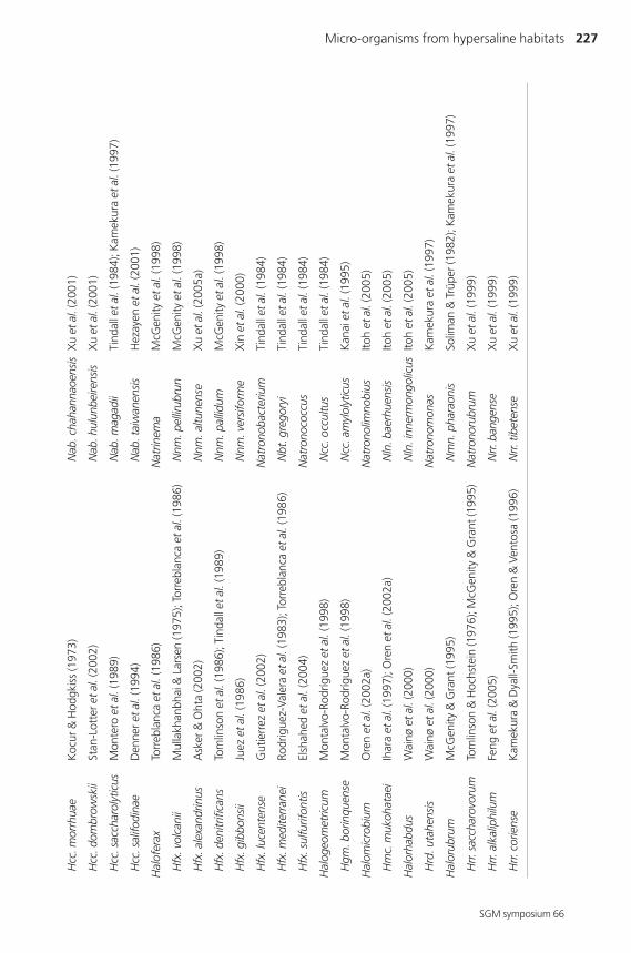
are currently represented by 20 different genera and a large number of species,
summarized in Table 1. Several other genera have recently been reported: Halovivax
asiaticus (Castillo et al., 2006) and ‘Halostagnum larsenii’ (A. M. Castillo, M. C.
Gutierrez, M. Kamekura, Y. Xue, Y. Ma, D. A. Cowan, B. E. Jones, W. D. Grant and
A. Ventosa, manuscript in preparation) were isolated from a saline lake in Inner
Mongolia, China. In addition to morphological and phenotypic features and compari-
son of 16S rRNA gene sequences, as is current practice in the systematics of other
prokaryotic micro-organisms, the taxonomy of haloarchaea is also largely based on
chemotaxonomic features: the polar lipid composition has proven to be an important
marker for differentiation at the genus level (Grant et al., 2001).
Haloarchaea require at least 1.5 M NaCl for growth and most species grow optimally
in media with 3.5–4.5 M NaCl; many are able to grow in saturated NaCl (5.2 M) (Grant
et al., 2001). However, haloarchaea isolated from coastal salt-marsh sediments able
to grow at lower salinities (around that of sea water) have been reported (Purdy et al.,
2004). They produce red- to pink-pigmented colonies due to the presence of bacterio-
ruberins, C50 carotenoids that are partially responsible for the typical colouration of
many natural environments in which they may develop in large numbers. However,
there are a few exceptions that are not pink- to red-pigmented, including species of the
genus Natrialba. Another interesting aspect of the haloarchaea is the presence in some
of them of retinal-based pigments, such as bacteriorhodopsin, that act as a proton
pump driven by light energy (Lanyi, 1995). They are found in many hypersaline
environments, such as salt lakes, soda lakes, salterns, solar salt and subterranean salt
deposits and salted foods (Grant et al., 2001).
Haloarchaea have typical archaeal characteristics such as the presence of ether-linked
phosphoglycerides that can be easily detected by thin-layer chromatography. All halo-
archaea contain phytanyl ether analogues of phosphatidylglycerol and phosphatidyl-
glycerol phosphate methyl ester. Many species also have phosphatidylglycerol sulfate
and one or more glycolipids and sulfated glycolipids (Grant et al., 2001; Kates, 1993).
All haloarchaea have diphytanyl (C20C20) glycerol ether core lipids and some may have
additional phytanyl-sesterterpanyl (C20C25) glycerol core lipids; furthermore, some
haloalkaliphiles have disesterterpanyl (C25C25) glycerol ether lipids (Grant et al., 2001).

226 A. Ventosa
SGM symposium 66
Tab
le 1
.Val
idly
pub
lishe
d ge
nus
and
spec
ies
nam
es w
ithin
the
fam
ily H
alob
acte
riace
ae
The
first
spe
cies
nam
e lis
ted
corr
espo
nds
to th
e ty
pe s
peci
es o
f eac
h ge
nus
(upd
ated
31
Dec
embe
r 200
5). T
he th
ree-
lett
er g
enus
abb
revi
atio
ns re
com
men
ded
byth
e IC
SP S
ubco
mm
ittee
on
the
taxo
nom
y of
the
fam
ily H
alob
acte
riace
aeha
ve b
een
used
. Bas
onym
s/sy
nony
ms
of o
rgan
ism
s th
at h
ave
been
tran
sfer
red
to o
ther
gene
ra a
re n
ot in
clud
ed.
Gen
us
Ref
eren
ce(s
)G
enu
sR
efer
ence
(s)
Hal
obac
teriu
mEl
azar
i-Vol
cani
(195
7); G
rant
(200
1a)
Hal
orub
rum
(con
t.)
Hbt
. sal
inar
umVe
ntos
a &
Ore
n (1
996)
; Gra
nt (2
001a
)H
rr. d
istr
ibut
umZv
yagi
ntse
va &
Tar
asov
(198
7); O
ren
& V
ento
sa (1
996)
Hbt
. nor
icen
seG
rube
r et a
l.(2
004)
Hrr.
lacu
spro
fund
iFr
anzm
ann
et a
l.(1
988)
; McG
enity
& G
rant
(199
5)
Hal
alka
licoc
cus
Xue
et a
l.(2
005)
Hrr.
sod
omen
seO
ren
(198
3); M
cGen
ity &
Gra
nt (1
995)
Hac
. tib
eten
sis
Xue
et a
l.(2
005)
Hrr.
tebe
nqui
chen
seLi
zam
a et
al.
(200
2)
Hal
oarc
ula
Torr
ebla
nca
et a
l.(1
986)
Hrr.
terr
estr
eVe
ntos
a et
al.
(200
4)
Har
. val
lism
ortis
Gon
zále
z et
al.
(197
9); T
orre
blan
ca e
t al.
(198
6)H
rr. ti
bete
nse
Fan
et a
l.(2
004)
Har
. arg
entin
ensi
sIh
ara
et a
l.(1
997)
Hrr.
trap
anic
umPe
tter
(193
1); M
cGen
ity &
Gra
nt (1
995)
Har
. his
pani
caJu
ez e
t al.
(198
6)H
rr. v
acuo
latu
mM
wat
ha &
Gra
nt (1
993)
; Kam
ekur
a et
al.
(199
7)
Har
. jap
onic
aTa
kash
ina
et a
l.(1
990)
Hrr.
xin
jiang
ense
Feng
et a
l.(2
004)
Har
. mar
ism
ortu
iO
ren
et a
l.(1
990)
Hal
osim
plex
Vree
land
et a
l.(2
002)
Har
. qua
drat
aO
ren
et a
l.(1
999)
Hsx
. car
lsba
dens
eVr
eela
nd e
t al.
(200
2)
Hal
obac
ulum
Ore
n et
al.
(199
5)H
alot
errig
ena
Vent
osa
et a
l.(1
999)
Hbl
. gom
orre
nse
Ore
n et
al.
(199
5)H
tg. t
urkm
enic
aZv
yagi
ntse
va &
Tar
asov
(198
7); V
ento
sa e
t al.
(199
9)
Hal
obifo
rma
Hez
ayen
et a
l.(2
002)
Htg
. sac
char
evita
nsX
u et
al.
(200
5c)
Hbf
. hal
oter
rest
risH
ezay
en e
t al.
(200
2)H
tg. t
herm
otol
eran
sM
onta
lvo-
Rodr
ígue
z et
al.
(200
0)
Hbf
. lac
isal
siX
u et
al.
(200
5b)
Nat
rialb
aK
amek
ura
& D
yall-
Smith
(199
5)
Hbf
. nitr
atire
duce
nsX
in e
t al.
(200
1); H
ezay
en e
t al.
(200
2)N
ab. a
siat
ica
Kam
ekur
a &
Dya
ll-Sm
ith (1
995)
Hal
ococ
cus
Scho
op (1
935)
; Gra
nt (2
001b
)N
ab. a
egyp
tiaH
ezay
en e
t al.
(200
1)
Micro-organisms from hypersaline habitats 227
Hcc
. mor
rhua
eK
ocur
& H
odgk
iss
(197
3)N
ab. c
haha
nnao
ensi
sX
u et
al.
(200
1)
Hcc
. dom
brow
skii
Stan
-Lot
ter e
t al.
(200
2)N
ab. h
ulun
beire
nsis
Xu
et a
l.(2
001)
Hcc
. sac
char
olyt
icus
Mon
tero
et a
l. (1
989)
Nab
. mag
adii
Tind
all e
t al.
(198
4); K
amek
ura
et a
l.(1
997)
Hcc
. sal
ifodi
nae
Den
ner e
t al.
(199
4)N
ab. t
aiw
anen
sis
Hez
ayen
et a
l.(2
001)
Hal
ofer
axTo
rreb
lanc
a et
al.
(198
6)N
atrin
ema
McG
enity
et a
l.(1
998)
Hfx
. vol
cani
iM
ulla
khan
bhai
& L
arse
n (1
975)
; Tor
rebl
anca
et a
l.(1
986)
Nnm
. pel
lirub
run
McG
enity
et a
l.(1
998)
Hfx
. ale
xand
rinus
Ask
er &
Oht
a (2
002)
Nnm
. altu
nens
eX
u et
al.
(200
5a)
Hfx
. den
itrifi
cans
Tom
linso
n et
al.
(198
6); T
inda
ll et
al.
(198
9)N
nm. p
allid
umM
cGen
ity e
t al.
(199
8)
Hfx
. gib
bons
iiJu
ez e
t al.
(198
6)N
nm. v
ersi
form
eX
in e
t al.
(200
0)
Hfx
. luc
ente
nse
Gut
ierr
ez e
t al.
(200
2)N
atro
noba
cter
ium
Tind
all e
t al.
(198
4)
Hfx
. med
iterr
anei
Rodr
igue
z-Va
lera
et a
l.(1
983)
; Tor
rebl
anca
et a
l.(1
986)
Nbt
. gre
gory
iTi
ndal
l et a
l.(1
984)
Hfx
. sul
furif
ontis
Elsh
ahed
et a
l.(2
004)
Nat
rono
cocc
usTi
ndal
l et a
l.(1
984)
Hal
ogeo
met
ricum
Mon
talv
o-Ro
dríg
uez
et a
l.(1
998)
Ncc
. occ
ultu
sTi
ndal
l et a
l.(1
984)
Hgm
. bor
inqu
ense
Mon
talv
o-Ro
dríg
uez
et a
l.(1
998)
Ncc
. am
ylol
ytic
usK
anai
et a
l.(1
995)
Hal
omic
robi
umO
ren
et a
l.(2
002a
)N
atro
nolim
nobi
usIto
h et
al.
(200
5)
Hm
c. m
ukoh
atae
iIh
ara
et a
l.(1
997)
; Ore
n et
al.
(200
2a)
Nln
. bae
rhue
nsis
Itoh
et a
l.(2
005)
Hal
orha
bdus
Wai
nøet
al.
(200
0)N
ln. i
nner
mon
golic
usIto
h et
al.
(200
5)
Hrd
. uta
hens
isW
ainø
et a
l.(2
000)
Nat
rono
mon
asK
amek
ura
et a
l. (1
997)
Hal
orub
rum
McG
enity
& G
rant
(199
5)N
mn.
pha
raon
isSo
liman
& T
rüpe
r (19
82);
Kam
ekur
a et
al.
(199
7)
Hrr.
sac
char
ovor
umTo
mlin
son
& H
ochs
tein
(197
6); M
cGen
ity &
Gra
nt (1
995)
Nat
rono
rubr
umX
u et
al.
(199
9)
Hrr.
alk
alip
hilu
mFe
ng e
t al.
(200
5)N
rr. b
ange
nse
Xu
et a
l.(1
999)
Hrr.
cor
iens
eK
amek
ura
& D
yall-
Smith
(199
5); O
ren
& V
ento
sa (1
996)
Nrr.
tibe
tens
eX
u et
al.
(199
9)
SGM symposium 66
228 A. Ventosa
SGM symposium 66
Their absolute requirement for NaCl and their optimal growth at high NaCl
concentrations are the most typical features of haloarchaea. The question of their
mechanisms of haloadaptation has attracted much attention and, in contrast to most
other prokaryotes, which accumulate intracellular organic compounds called com-
patible solutes, haloarchaea compensate for the high salt concentration in the environ-
ment by accumulating mainly KCl, up to 5 M (Grant et al., 2001).
As mentioned previously, haloarchaea are found in many hypersaline environments;
they are the predominant microbial biota of saturated ponds of salterns and salt lakes.
Several species are haloalkaliphiles, being able to grow optimally at alkaline pH, that
also inhabit soda lakes. Ecological studies demonstrate that they may reach high cell
densities (> 107 cells ml–1). Traditional studies based on cultivation of viable cells
suggested that the predominant species found in most neutrophilic hypersaline
environments were related to the genera Halobacterium, Halorubrum, Haloferax and
Haloarcula (Rodriguez-Valera et al., 1985; Rodriguez-Valera, 1988; Benlloch et al.,
2001). However, more recent molecular ecological studies based on cultivation-
independent methods indicate that, at least in most environments studied, members of
these genera constitute small proportions of the microbial community. Several recent
studies carried out in hypersaline environments have allowed some general conclusions
to be drawn. Square haloarchaea are very abundant, but they have not been isolated
until recently; many environmental clones within the haloarchaeal group are also
obtained that are not phylogenetically closely related to previously described species.
Some clones related to Halorubrum, Haloarcula, Natronobacerium and Natrono-
monas are observed (Benlloch et al., 2001, 2002; Burns et al., 2004b). Recent studies on
a saltern in Slovenia showed that the haloarchaeal community in the crystallizer was
strongly dominated by two groups of Halorubrum-related environmental phylotypes.
In addition, members of four other haloarchaeal genera and two groups of environ-
mental phylotypes were observed. However, the square haloarchaeal morphotype was
not observed (Pasic et al., 2005). Another recent study of a saltern in San Diego showed
that the predominant haloarchaea were members of Halobacterium; the presence of
Haloarcula and Halobacterium was also detected as well as two novel lineages not
closely related to any haloarchaeal species (Bidle et al., 2005).
Haloarchaea are used in many studies as archaeal models since they can be easily grown
under laboratory conditions. In contrast to other archaeal extremophiles, which require
special culture conditions, haloarchaea grow well in complex media and in some cases
in minimal media under aerobic conditions by using the standard procedures for
growing other prokaryotes. They can also be genetically manipulated; genetic exchange
mechanisms are well established and methods for their genetic manipulation in the
Micro-organisms from hypersaline habitats 229
SGM symposium 66
laboratory are available (Robb et al., 1995). The complete genome sequences of quite a
few haloarchaea have been reported, including Halobacterium salinarum NRC-1
(2571 kb) (Ng et al., 2000), Haloarcula marismortui ATCC 43049T (4274 kb) (Baliga et
al., 2004) and Natronomonas pharaonis DSM 2160T (2749 kb) (Falb et al., 2005).
Several other genome sequencing projects are in progress: a second strain of Halo-
bacterium salinarum and strains of Halobaculum gomorrense, Halobiforma lacisalsi,
Haloferax volcanii, Halorubrum lacusprofundi and Natrialba asiatica. Clearly, more
effort would be necessary in this field in order to have genomic information available
that reflects the diversity found within the haloarchaea.
Besides their use as excellent models to study the molecular biology of archaea and
their mechanisms of adaptation to extreme environments and the important ecological
roles that they play in hypersaline habitats, haloarchaea have very interesting biotech-
nological applications. Bacteriorhodopsin in the form of purple membrane patches
produced by Halobacterium salinarum is commercially available; other compounds of
industrial interest produced by haloarchaea are extracellular hydrolytic enzymes,
exopolysaccharides, polyhydroxyalkanoates (PHAs) used as bioplastics and the halo-
cins (antimicrobial compounds produced by haloarchaea). These and many other
potential applications have been reviewed in detail elsewhere (Ventosa & Nieto, 1995;
Margesin & Schinner, 2001; Mellado & Ventosa, 2003).
FROM ‘SQUARE BACTERIA’ TO ‘SQUARE HALOARCHAEA’In 1980, A. E. Walsby described the abundant presence of ‘square bacteria’ in a small,
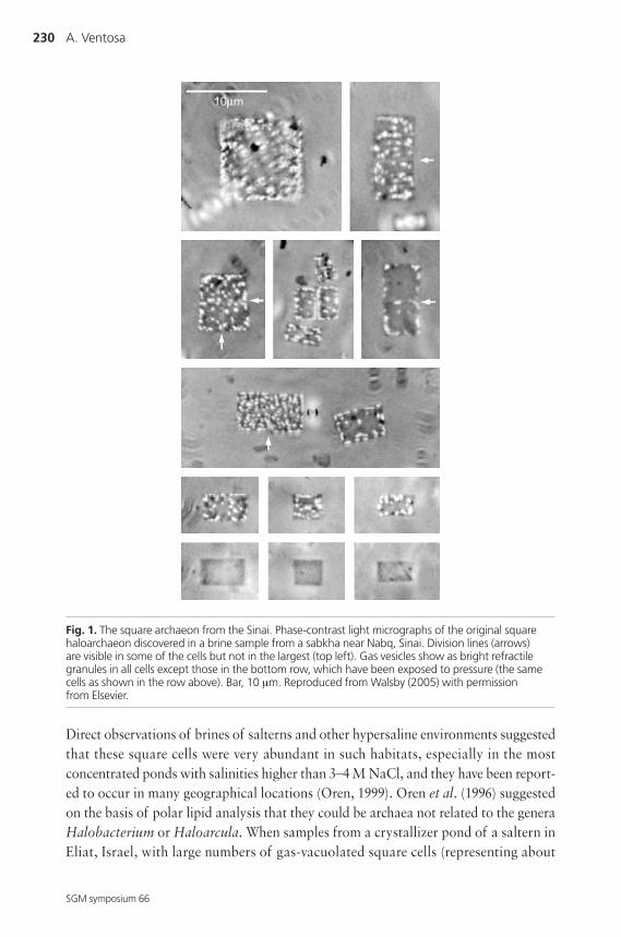
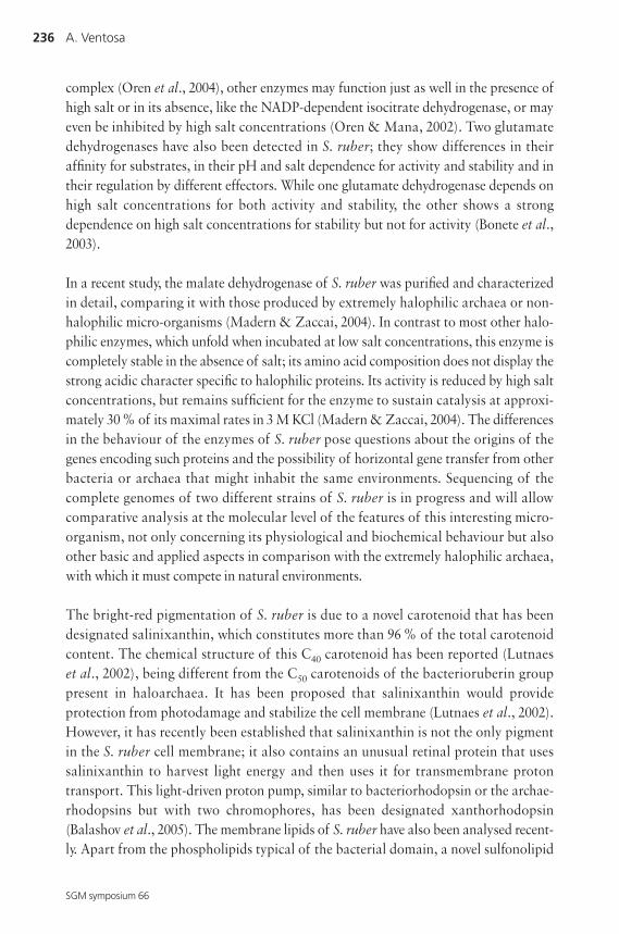
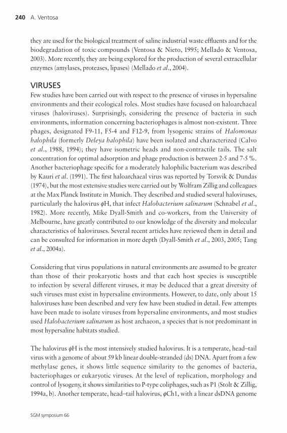
saturated brine pool, or sabkha, in the Sinai Peninsula (Walsby, 1980). These micro-
organisms were collected from the surface of the pool, had a large number of gas
vesicles and presented unique morphologies never observed previously in the microbial
world. The cells are squares and very thin, with sizes from 1.5 to 11 μm and a thickness
of about 0.2 μm (Fig. 1). Division planes were observed, with an arrangement indi-
cating that division occurred in two planes alternating at right angles, so that each
square grows to a rectangle which then divides into two equal squares, producing sheets
divided like postage stamps (Walsby, 1980; Parkes & Walsby, 1981). Also, different gas
vesicles were observed, from spindle-shaped to cylindrical with conical ends and, in
many cases, they were concentrated at the cell periphery. Both forms may occur within
the same cell, as had been observed previously in Halobacterium (Parkes & Walsby,
1981). Similar observations were made from other hypersaline environments in
different geographical areas, from salt ponds located in Baja California, Mexico, or San
Francisco Bay, California (Stoeckenius, 1981). Ultrastructure studies confirmed that the
square cells observed by Walsby were micro-organisms with a typical prokaryote
structure (Stoeckenius, 1981; Kessel & Cohen, 1982).
230 A. Ventosa
SGM symposium 66
Direct observations of brines of salterns and other hypersaline environments suggested
that these square cells were very abundant in such habitats, especially in the most
concentrated ponds with salinities higher than 3–4 M NaCl, and they have been report-
ed to occur in many geographical locations (Oren, 1999). Oren et al. (1996) suggested
on the basis of polar lipid analysis that they could be archaea not related to the genera
Halobacterium or Haloarcula. When samples from a crystallizer pond of a saltern in
Eliat, Israel, with large numbers of gas-vacuolated square cells (representing about
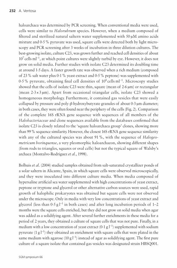
Fig. 1. The square archaeon from the Sinai. Phase-contrast light micrographs of the original squarehaloarchaeon discovered in a brine sample from a sabkha near Nabq, Sinai. Division lines (arrows) are visible in some of the cells but not in the largest (top left). Gas vesicles show as bright refractilegranules in all cells except those in the bottom row, which have been exposed to pressure (the samecells as shown in the row above). Bar, 10 μm. Reproduced from Walsby (2005) with permission from Elsevier.
Micro-organisms from hypersaline habitats 231
SGM symposium 66
55 % of the total population) were studied with respect to polar lipid composition,
Oren et al. (1996) observed that the most frequent lipids present were typical of halo-
philic archaea, members of the family Halobacteriaceae, but that the square archaea
may belong to a new genus. More recently, molecular techniques revealed that they
may represent a large proportion of the microbial population in many hypersaline
environments (Benlloch et al., 1996, 2001, 2002; Antón et al., 1999; Burns et al., 2004b).
Many attempts have been made to isolate these square micro-organisms, but only
recently have two independent studies reported their isolation and cultivation (Bolhuis
et al., 2004; Burns et al., 2004a). Previous studies based on the isolation of pure cultures
from different hypersaline environments reported the presence of halophilic archaea
with morphologies ranging from squares to discs and triangles, and not the typical rod
or spherical cell shapes that are observed within other bacterial groups. An early study
describing ‘box-shaped’ halophiles (Javor et al., 1982) did not permit the isolation of
square archaea, since the cells were pleomorphic and lacked the gas vesicles observed
in Walsby’s square cells. Most isolates studied in recent years that show pleomorphic
cells have been placed in the genera Haloferax (Ventosa, 2001b) or Haloarcula (Ventosa,
2001a). Most species of the genus Haloarcula have unique morphologies, such as
Haloarcula japonica, which typically shows many triangular and rhomboid cells
(Takashina et al., 1990), or Haloarcula quadrata (Oren et al., 1999). The latter species
was described based on a motile, square prokaryote isolated from the Gavish Sabkha in
the Sinai Peninsula. The square morphology and interesting mode of motility were
reported by Alam et al. (1984). The cells have a single or several flagella anchored from
a single or several locations that form a bundle. As in the case of other haloarchaea, this
species requires high salt concentrations for growth and for stabilization of the cells.
Optimal growth is obtained in the presence of 3.4–4.3 M NaCl and 0.1–0.5 M Mg2+.
It is interesting to note that at least 50–100 mM Mg2+ was required for maintenance of
the square morphology (Oren et al., 1999). However, its lack of gas vesicles and a more
variable morphology suggest that it does not correspond to the ‘square bacteria’
described by Walsby (1980).
The recent isolation of Walsby’s square haloarchaea, 25 years after their original
description, by two independent groups of microbiologists can be considered as an
important event. It required the use of new approaches that differed from traditional
culture methods, as well as extraordinary persistence.
Burns et al. (2004b) studied water samples from a crystallizer pond of a saltern located
in Geelong, Victoria, Australia, in which they observed by microscopy the abundance
of square cells. They used a combination of serial dilutions of the samples in liquid
media with different compositions (extinction cultures), and growth of the square
232 A. Ventosa
SGM symposium 66
haloarchaea was determined by PCR screening. When conventional media were used,
cells were similar to Halorubrum species. However, when a medium composed of
filtered and sterilized natural saltern water supplemented with 50 μM amino acids
mixture and 0.5 % pyruvate was used, square cells were detected both by light micro-
scopy and PCR screening after 3 weeks of incubation in three dilution cultures. The
best-growing isolate, culture C23, was grown further and reached cell densities of about
107 cells ml–1, at which point cultures were slightly turbid by eye. However, it does not
grow on solid media. Further studies with isolate C23 determined its doubling time
at around 1.5 days. A faster growth rate was observed when a rich medium (composed
of 23 % salt water plus 0.1 % yeast extract and 0.5 % peptone) was supplemented with
0.5 % pyruvate, obtaining final cell densities of 108 cells ml–1. Microscopy studies
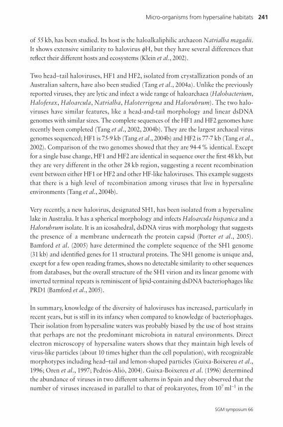
showed that the cells of isolate C23 were thin, square (mean of 2.6 μm) or rectangular
(mean 2.3 × 3 μm). Apart from occasional triangular cells, isolate C23 showed a
homogeneous morphology. Furthermore, it contained gas vesicles that were easily
collapsed by pressure and poly-β-hydroxybutyrate granules of about 0.3 μm diameter;
in both cases, they were often found near the periphery of the cells (Fig. 2). Comparison
of the complete 16S rRNA gene sequence with sequences of all members of the
Halobacteriaceae and clone sequences available from the databases confirmed that
isolate C23 is closely related to the ‘square haloarchaea group’ clones, showing more
than 99 % sequence similarity. However, the closest 16S rRNA gene sequence similarity
with any of the cultured species was about 91 %, with the sequence of Halogeo-
metricum borinquense, a very pleomorphic haloarchaeon, showing different shapes
(from rods to triangles, squares or oval cells) but not the typical square of Walsby’s
archaea (Montalvo-Rodríguez et al., 1998).
Bolhuis et al. (2004) studied samples obtained from salt-saturated crystallizer ponds of
a solar saltern in Alicante, Spain, in which square cells were observed microscopically,
and they were inoculated into different culture media. When media composed of
hypersaline artificial sea water supplemented with high concentrations of yeast extract,
peptone or tryptone and glycerol or other alternative carbon sources were used, rapid
growth of halophilic prokaryotes was obtained but square cells were not observed
under the microscope. Only in media with very low concentrations of yeast extract and
glycerol (less than 0.5 g l–1 in both cases) and after long incubation periods of 1–2
months were the square cells enriched, but they did not grow on solid media when agar
was added as a solidifying agent. After several further enrichments in these media for a
period of 2 years, they obtained a culture of square cells that was not pure. Finally, in a
medium with a low concentration of yeast extract (0.1 g l–1) supplemented with sodium
pyruvate (1 g l–1) they obtained an enrichment with square cells that were plated in the
same medium with agarose (10 g l–1) instead of agar as solidifying agent. The first pure
culture of a square isolate that contained gas vesicles was designated strain HBSQ001.
Micro-organisms from hypersaline habitats 233
SGM symposium 66
The cells were 2–5 μm wide and about 0.1–0.5 μm thick; in fresh cultures they observed
very large cells (20 × 20 to 40 × 40 μm and 0.1–0.5 μm thick) in which division walls were
not observed. This isolate required at least 3 M NaCl and showed an elevated tolerance
of high concentrations of MgCl2, being able to grow in media containing over 2 M
MgCl2 and 3.3 M NaCl.
When the 16S rRNA gene sequence of strain HBSQ001 was determined, phylogenetic
study showed that it was closely related to the SPhT sequence (99 % similarity) that had
been observed previously as related to Walsby’s square archaeon. Only 93 % 16S rRNA
gene sequence similarity was obtained with the haloarchaeon T1.3, a strain isolated
from an ancient salt deposit (McGenity et al., 2000), and lower values were seen with
haloarchaeal species. This low 16S rRNA gene sequence similarity suggests that the
new isolate would represent a new genus of the family Halobacteriaceae. Burns and co-
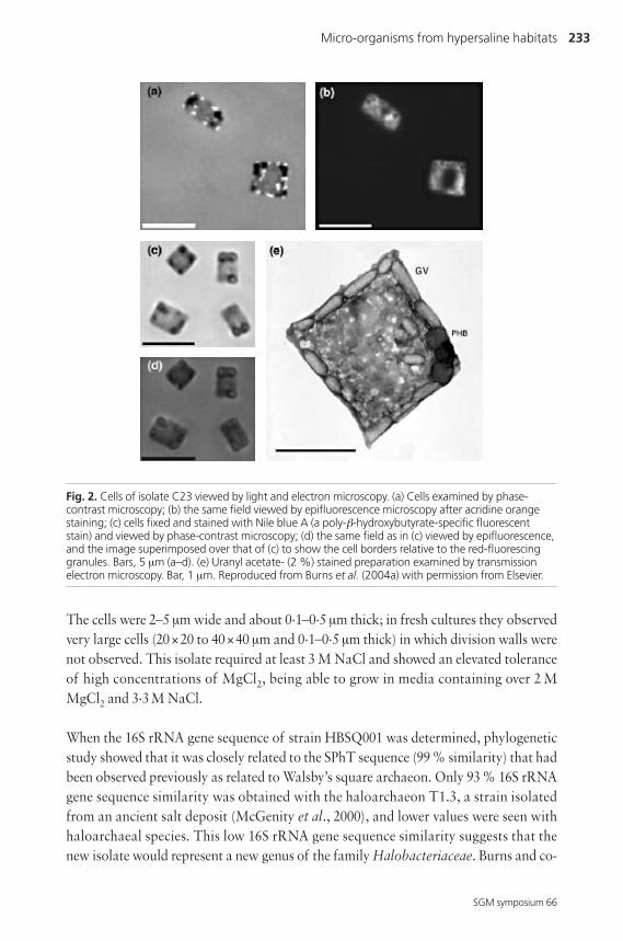
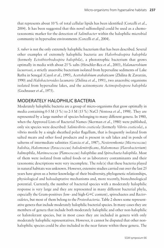
Fig. 2. Cells of isolate C23 viewed by light and electron microscopy. (a) Cells examined by phase-contrast microscopy; (b) the same field viewed by epifluorescence microscopy after acridine orangestaining; (c) cells fixed and stained with Nile blue A (a poly-β-hydroxybutyrate-specific fluorescentstain) and viewed by phase-contrast microscopy; (d) the same field as in (c) viewed by epifluorescence,and the image superimposed over that of (c) to show the cell borders relative to the red-fluorescinggranules. Bars, 5 μm (a–d). (e) Uranyl acetate- (2 %) stained preparation examined by transmissionelectron microscopy. Bar, 1 μm. Reproduced from Burns et al. (2004a) with permission from Elsevier.
234 A. Ventosa
SGM symposium 66
workers proposed the new name ‘Haloquadratum walsbyi’ if this isolate were to
constitute a new taxon (Bolhuis et al., 2004). However, a complete taxonomic char-
acterization would be required, including phenotypic as well as chemotaxonomic data
(such as polar lipid analysis). Furthermore, it would be interesting to determine the
sequence similarity between the two novel square archaea, strains HBSQ001 and C23,
in order to determine whether they are closely related, and to carry out a comparative
study to determine whether they are members of the same genus and species.
The reasons for the difficulties in isolating the square archaea can be attributed to
several factors, such as their sensitivity to agar and to high concentrations of yeast
extract, their slow growth rate (doubling time of 1–2 days under optimal conditions),
which permits rapid growth of other extremely halophilic micro-organisms in the
enriched cultures, and probably their requirement for relatively high concentrations of
MgCl2 to maintain the square morphology.
The isolation by two independent research groups of the square archaea introduces a
new methodology that permits the isolation of new fresh isolates of square archaea,
allowing us to establish whether they are represented by a single or several novel species.
Furthermore, it will be possible to determine their roles in the natural hypersaline
environments in which, on the basis of molecular ecology techniques, they represent a
large proportion of the microbial population, and to study other physiological or mole-
cular aspects such as their high tolerance of MgCl2 or the unique square morphology.
The complete sequence of the genome of these micro-organisms will also constitute a
highlight for comparative purposes with other organisms.
Recently, Walsby (2005) reviewed the features of the square archaea and the biological
significance of the square shape. This shape depends on the lack of turgor; most cells
are distended by turgor pressure, generated by the difference in water potential between
the external medium and the cell. In spherical or cylindrical cells, the pressure produces
stress in the cell wall. Square cells, like other haloarchaea, have no turgor pressure
(Walsby, 1980) and are free of stress in the cell wall, thus they need less complex cell
walls than other prokaryotes and might, in theory, adopt any shape. The shape of the
cell might be dependent on the shape and arrangement of the wall subunits. The cell
wall of square archaea is composed of regularly arranged subunits, forming a domin-
antly hexagonal lattice (Walsby, 1980; Parkes & Walsby, 1981; Stoeckenius, 1981; Kessel
& Cohen, 1982). The advantages of the thinness of the square cells could be related to
the large surface area, which would permit the uptake of nutrients as well as more
efficient light absorption, since it has been shown that the square archaea contain
purple membranes that are used for phototrophic growth. Furthermore, the gas vesicles
offer another clear advantage: they permit the cells to concentrate and rest parallel in a
Micro-organisms from hypersaline habitats 235
SGM symposium 66
horizontal position on the water surface. This movement to the surface has less ener-
getic cost in cells with gas vesicles than with flagella and could explain the slow growth
strategy and their dominance in saturated hypersaline environments (Walsby, 2005).
SALINIBACTER AND OTHER EXTREMELY HALOPHILICBACTERIAThe genus Salinibacter, with the single species Salinibacter ruber, was proposed by
Antón et al. (2002) for red-pigmented, motile, rod-shape extremely halophilic bacteria
isolated from saltern crystallizer ponds in Alicante and Mallorca (Spain). It grows
optimally between 20 and 30 % salt and requires at least 15 % salts. Therefore it is
as halophilic and salt-dependent as most extremely halophilic archaea belonging to
the family Halobacteriaceae (Grant et al., 2001). It is phylogenetically related to the
phylum Bacteroidetes but the closest cultivated relative is Rhodothermus marinus
(showing only about 89 % 16S rRNA gene sequence similarity), a thermophilic and
slightly halophilic bacterium. This bacterium constitutes a major component of the
community of hypersaline environments and was previously known only by its phylo-
type (designated ‘Candidatus Salinibacter’) (Antón et al., 2000). By using fluorescent
oligonucleotide probes, this organism has been shown to be abundant in the crystallizer
ponds of salterns, representing between 5 and 25 % of the total prokaryotic community
(Antón et al., 2000). They may be also responsible for the red colouration of saltern
crystallizer ponds (Oren & Rodriguez-Valera, 2001). However, their presence has not
been observed in other salterns studied recently (Bidle et al., 2005).
Despite its relatively recent isolation and description, new data about this interesting
bacterium, which can be considered as one of the most halophilic organisms belonging
to the Bacteria, have been accumulated in recent years. S. ruber has an extremely high
intracellular potassium content but very low concentrations of organic osmotic solutes
(glutamate, glycine betaine and N-α-acetyl lysine) (Oren et al., 2002b). Thus, it uses a
similar mode of haloadaptation to that of the haloarchaea, which also have high
intracellular K+ concentrations, and does not accumulate organic osmotic solutes such
as those used by most halophilic or halotolerant bacteria (Ventosa et al., 1998; Oren
et al., 2002b). Amino acid analysis of bulk protein of S. ruber showed a high content
of acidic amino acids, a low proportion of basic amino acids, a low content of hydro-
phobic amino acids and a high abundance of serine (Oren & Mana, 2002). When these
authors determined the levels of activity of several cytoplasmic enzymes at different
KCl and NaCl concentrations, they showed that these enzymes are adapted to function
in the presence of high salt concentrations (Oren & Mana, 2002). However, it seems
that the behaviour of each enzyme towards salt varies considerably. Thus, while some
enzymes are truly halophilic and require salt for activity and stability, such as the NAD-
dependent isocitrate dehydrogenase (Oren & Mana, 2002) or the fatty acid synthetase
236 A. Ventosa
SGM symposium 66
complex (Oren et al., 2004), other enzymes may function just as well in the presence of
high salt or in its absence, like the NADP-dependent isocitrate dehydrogenase, or may
even be inhibited by high salt concentrations (Oren & Mana, 2002). Two glutamate
dehydrogenases have also been detected in S. ruber; they show differences in their
affinity for substrates, in their pH and salt dependence for activity and stability and in
their regulation by different effectors. While one glutamate dehydrogenase depends on
high salt concentrations for both activity and stability, the other shows a strong
dependence on high salt concentrations for stability but not for activity (Bonete et al.,
2003).
In a recent study, the malate dehydrogenase of S. ruber was purified and characterized
in detail, comparing it with those produced by extremely halophilic archaea or non-
halophilic micro-organisms (Madern & Zaccai, 2004). In contrast to most other halo-
philic enzymes, which unfold when incubated at low salt concentrations, this enzyme is
completely stable in the absence of salt; its amino acid composition does not display the
strong acidic character specific to halophilic proteins. Its activity is reduced by high salt
concentrations, but remains sufficient for the enzyme to sustain catalysis at approxi-
mately 30 % of its maximal rates in 3 M KCl (Madern & Zaccai, 2004). The differences
in the behaviour of the enzymes of S. ruber pose questions about the origins of the
genes encoding such proteins and the possibility of horizontal gene transfer from other
bacteria or archaea that might inhabit the same environments. Sequencing of the
complete genomes of two different strains of S. ruber is in progress and will allow
comparative analysis at the molecular level of the features of this interesting micro-
organism, not only concerning its physiological and biochemical behaviour but also
other basic and applied aspects in comparison with the extremely halophilic archaea,
with which it must compete in natural environments.
The bright-red pigmentation of S. ruber is due to a novel carotenoid that has been
designated salinixanthin, which constitutes more than 96 % of the total carotenoid
content. The chemical structure of this C40 carotenoid has been reported (Lutnaes
et al., 2002), being different from the C50 carotenoids of the bacterioruberin group
present in haloarchaea. It has been proposed that salinixanthin would provide
protection from photodamage and stabilize the cell membrane (Lutnaes et al., 2002).
However, it has recently been established that salinixanthin is not the only pigment
in the S. ruber cell membrane; it also contains an unusual retinal protein that uses
salinixanthin to harvest light energy and then uses it for transmembrane proton
transport. This light-driven proton pump, similar to bacteriorhodopsin or the archae-
rhodopsins but with two chromophores, has been designated xanthorhodopsin
(Balashov et al., 2005). The membrane lipids of S. ruber have also been analysed recent-
ly. Apart from the phospholipids typical of the bacterial domain, a novel sulfonolipid
Micro-organisms from hypersaline habitats 237
SGM symposium 66
that represents about 10 % of total cellular lipids has been identified (Corcelli et al.,
2004). It has been suggested that this novel sulfonolipid could be used as a chemo-
taxonomic marker for the detection of Salinibacter within the halophilic microbial
community in hypersaline environments (Corcelli et al., 2004).
S. ruber is not the only extremely halophilic bacterium that has been described. Several
other examples of extremely halophilic bacteria are Halorhodospira halophila
(formerly Ectothiorhodospira halophila), a phototrophic bacterium that grows
optimally in media with about 25 % salts (Hirschler-Rea et al., 2003), Halanaerobium
lacusrosei, a strictly anaerobic bacterium isolated from hypersaline sediments of Lake
Retba in Senegal (Cayol et al., 1995), Acetohalobium arabaticum (Zhilina & Zavarzin,
1990) and Halobacteroides lacunaris (Zhilina et al., 1991), two anaerobic organisms
isolated from hypersaline lakes, and the actinomycete Actinopolyspora halophila
(Gochnauer et al., 1975).
MODERATELY HALOPHILIC BACTERIAModerately halophilic bacteria are a group of micro-organisms that grow optimally in
media containing 0.5 M (3 %) to 2.5 M (15 %) NaCl (Ventosa et al., 1998). They are
represented by a large number of species belonging to many different genera. In 1980,
when the Approved Lists of Bacterial Names (Skerman et al., 1980) were published,
only six species were described: Salinivibrio costicola (formerly Vibrio costicola), a
vibrio motile by a single sheathed polar flagellum, that is frequently isolated from
salted meats and other food products and is present in salt lakes and in ponds of
salterns of intermediate salinities (Garcia et al., 1987), Nesterenkonia (Micrococcus)
halobia, Halomonas (Paraccoccus) halodenitrificans, Halomonas (Flavobacterium)
halmophila, Marinococcus (Planococcus) halophilus and Spirochaeta halophila. Most
of them were isolated from salted foods or as laboratory contaminants and their
taxonomic descriptions were very incomplete. The role(s) that these bacteria played
in natural habitats was unknown. However, extensive studies carried out over the last 25
years have given us a better knowledge of their biodiversity, phylogenetic relationships,
physiological and haloadaptative mechanisms and, more recently, biotechnological
potential. Currently, the number of bacterial species with a moderately halophilic
response is very large and they are represented in many different bacterial phyla,
especially the Gram-positives (low- and high-G+C content), spirochaetes and Bacter-
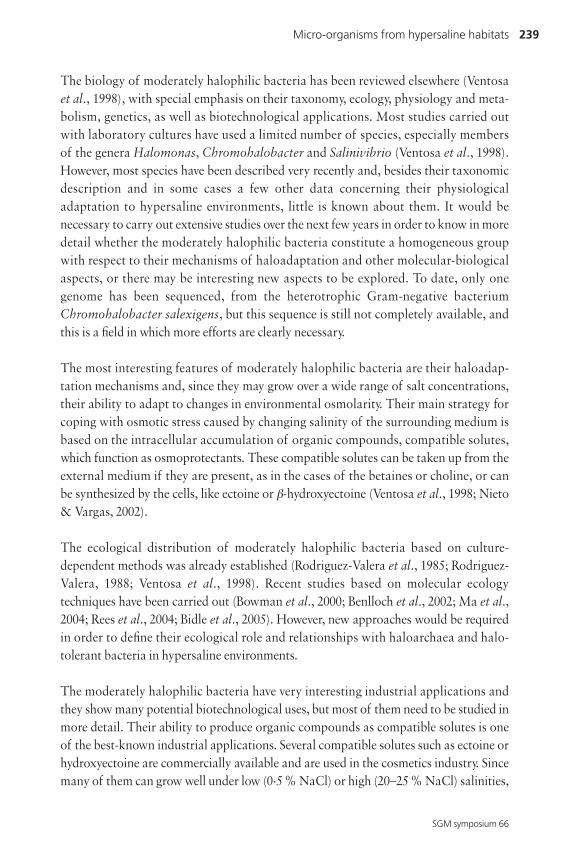
oidetes, but most of them belong to the Proteobacteria. Table 2 shows some represent-
ative genera that include moderately halophilic bacterial species. In many cases they are
members of genera that include both moderately halophilic and other non-halophilic
or halotolerant species, but in most cases they are included in genera with only
moderately halophilic representatives. However, it cannot be disputed that other non-
halophilic species could be also included in the near future within these genera. The
238 A. Ventosa
SGM symposium 66
close phylogenetic relationship of many moderately halophilic and non-halophilic
bacteria indicates that they are not evolutionary descendents of a single lineage and
that they evolved as extremophilic micro-organisms adapted to high saline environ-
ments by developing haloadaptation mechanisms that are similar to those of halo-
tolerant bacteria.
Table 2. Representative genera that include moderately halophilic species
Gram-positive Gram-negative
Alkalibacillus Algoriphagus Psychrobacter
Bacillus Alteromonas Rhodospirillum
Clostridium Arhodomonas Rhodothalassium
Gracilibacillus Chromohalobacter Rhodovibrio
Halobacillus Desulfocella Roseisalinus
Lentibacillus Desulfohalobium Salegentibacter
Marinococcus Desulfovibrio Salinimonas
Nocardiopsis Halanaerobacter Salinisphaera
Pontibacillus Halanaerobium Salinivibrio
Prauserella Halochromatium Salipiger
Saccharomonospora Haloincola Selenihalanaerobacter
Salinibacillus Halomonas Spirochaeta
Streptomonospora Halorhodospira Sporohalobacter
Tenuibacillus Halospina Staleya
Tetragenococcus Halothermothrix Sulfitobacter
Thalassobacillus Halothiobacillus Thiohalocapsa
Virgibacillus Halovibrio
Yania Idiomarina
Microbacterium Marinicola
Filobacillus Marinobacter
Dietzia Methylarcula
Marinibacillus Methylohalobius
Desulfotomaculum Muricauda
Salinicoccus Natroniella
Nesterenkonia Nitrincola
Jeotgalicoccus Orenia
Jeotgalibacillus Palleronia
Natronincola Pseudoalteromonas
Sporosarcina Psychroflexus
Micro-organisms from hypersaline habitats 239
SGM symposium 66
The biology of moderately halophilic bacteria has been reviewed elsewhere (Ventosa
et al., 1998), with special emphasis on their taxonomy, ecology, physiology and meta-
bolism, genetics, as well as biotechnological applications. Most studies carried out
with laboratory cultures have used a limited number of species, especially members
of the genera Halomonas, Chromohalobacter and Salinivibrio (Ventosa et al., 1998).
However, most species have been described very recently and, besides their taxonomic
description and in some cases a few other data concerning their physiological
adaptation to hypersaline environments, little is known about them. It would be
necessary to carry out extensive studies over the next few years in order to know in more
detail whether the moderately halophilic bacteria constitute a homogeneous group
with respect to their mechanisms of haloadaptation and other molecular-biological
aspects, or there may be interesting new aspects to be explored. To date, only one
genome has been sequenced, from the heterotrophic Gram-negative bacterium
Chromohalobacter salexigens, but this sequence is still not completely available, and
this is a field in which more efforts are clearly necessary.
The most interesting features of moderately halophilic bacteria are their haloadap-
tation mechanisms and, since they may grow over a wide range of salt concentrations,
their ability to adapt to changes in environmental osmolarity. Their main strategy for
coping with osmotic stress caused by changing salinity of the surrounding medium is
based on the intracellular accumulation of organic compounds, compatible solutes,
which function as osmoprotectants. These compatible solutes can be taken up from the
external medium if they are present, as in the cases of the betaines or choline, or can
be synthesized by the cells, like ectoine or β-hydroxyectoine (Ventosa et al., 1998; Nieto
& Vargas, 2002).
The ecological distribution of moderately halophilic bacteria based on culture-
dependent methods was already established (Rodriguez-Valera et al., 1985; Rodriguez-
Valera, 1988; Ventosa et al., 1998). Recent studies based on molecular ecology
techniques have been carried out (Bowman et al., 2000; Benlloch et al., 2002; Ma et al.,
2004; Rees et al., 2004; Bidle et al., 2005). However, new approaches would be required
in order to define their ecological role and relationships with haloarchaea and halo-
tolerant bacteria in hypersaline environments.
The moderately halophilic bacteria have very interesting industrial applications and
they show many potential biotechnological uses, but most of them need to be studied in
more detail. Their ability to produce organic compounds as compatible solutes is one
of the best-known industrial applications. Several compatible solutes such as ectoine or
hydroxyectoine are commercially available and are used in the cosmetics industry. Since
many of them can grow well under low (0.5 % NaCl) or high (20–25 % NaCl) salinities,
240 A. Ventosa
SGM symposium 66
they are used for the biological treatment of saline industrial waste effluents and for the
biodegradation of toxic compounds (Ventosa & Nieto, 1995; Mellado & Ventosa,
2003). More recently, they are being explored for the production of several extracellular
enzymes (amylases, proteases, lipases) (Mellado et al., 2004).
VIRUSESFew studies have been carried out with respect to the presence of viruses in hypersaline
environments and their ecological roles. Most studies have focused on haloarchaeal
viruses (haloviruses). Surprisingly, considering the presence of bacteria in such
environments, information concerning bacteriophages is almost non-existent. Three
phages, designated F9-11, F5-4 and F12-9, from lysogenic strains of Halomonas
halophila (formerly Deleya halophila) have been isolated and characterized (Calvo
et al., 1988, 1994); they have isometric heads and non-contractile tails. The salt
concentration for optimal adsorption and phage production is between 2.5 and 7.5 %.
Another bacteriophage specific for a moderately halophilic bacterium was described
by Kauri et al. (1991). The first haloarchaeal virus was reported by Torsvik & Dundas
(1974), but the most extensive studies were carried out by Wolfram Zillig and colleagues
at the Max Planck Institute in Munich. They described and studied several haloviruses,
particularly the halovirus φH, that infect Halobacterium salinarum (Schnabel et al.,
1982). More recently, Mike Dyall-Smith and co-workers, from the University of
Melbourne, have greatly contributed to our knowledge of the diversity and molecular
characteristics of haloviruses. Several recent articles have reviewed them in detail and
can be consulted for information in more depth (Dyall-Smith et al., 2003, 2005; Tang
et al., 2004a).
Considering that virus populations in natural environments are assumed to be greater
than those of their prokaryotic hosts and that each host species is susceptible
to infection by several different viruses, it may be deduced that a great diversity of
such viruses must exist in hypersaline environments. However, to date, only about 15
haloviruses have been described and very few have been studied in detail. Few attempts
have been made to isolate viruses from hypersaline environments, and most studies
used Halobacterium salinarum as host archaeon, a species that is not predominant in
most hypersaline habitats studied.
The halovirus φH is the most intensively studied halovirus. It is a temperate, head–tail
virus with a genome of about 59 kb linear double-stranded (ds) DNA. Apart from a few
methylase genes, it shows little sequence similarity to the genomes of bacteria,
bacteriophages or eukaryotic viruses. At the level of replication, morphology and
control of lysogeny, it shows similarities to P-type coliphages, such as P1 (Stolt & Zillig,
1994a, b). Another temperate, head–tail halovirus, φCh1, with a linear dsDNA genome
Micro-organisms from hypersaline habitats 241
SGM symposium 66
of 55 kb, has been studied. Its host is the haloalkaliphilic archaeon Natrialba magadii.
It shows extensive similarity to halovirus φH, but they have several differences that
reflect their different hosts and ecosystems (Klein et al., 2002).
Two head–tail haloviruses, HF1 and HF2, isolated from crystallization ponds of an
Australian saltern, have also been studied (Tang et al., 2004a). Unlike the previously
reported viruses, they are lytic and infect a wide range of haloarchaea (Halobacterium,
Haloferax, Haloarcula, Natrialba, Haloterrigena and Halorubrum). The two halo-
viruses have similar features, like a head-and-tail morphology and linear dsDNA
genomes with similar sizes. The complete sequences of the HF1 and HF2 genomes have
recently been completed (Tang et al., 2002, 2004b). They are the largest archaeal virus
genomes sequenced; HF1 is 75.9 kb (Tang et al., 2004b) and HF2 is 77.7 kb (Tang et al.,
2002). Comparison of the two genomes showed that they are 94.4 % identical. Except
for a single base change, HF1 and HF2 are identical in sequence over the first 48 kb, but
they are very different in the other 28 kb region, suggesting a recent recombination
event between either HF1 or HF2 and other HF-like haloviruses. This example suggests
that there is a high level of recombination among viruses that live in hypersaline
environments (Tang et al., 2004b).
Very recently, a new halovirus, designated SH1, has been isolated from a hypersaline
lake in Australia. It has a spherical morphology and infects Haloarcula hispanica and a
Halorubrum isolate. It is an icosahedral, dsDNA virus with morphology that suggests
the presence of a membrane underneath the protein capsid (Porter et al., 2005).
Bamford et al. (2005) have determined the complete sequence of the SH1 genome
(31 kb) and identified genes for 11 structural proteins. The SH1 genome is unique and,
except for a few open reading frames, shows no detectable similarity to other sequences
from databases, but the overall structure of the SH1 virion and its linear genome with
inverted terminal repeats is reminiscent of lipid-containing dsDNA bacteriophages like
PRD1 (Bamford et al., 2005).
In summary, knowledge of the diversity of haloviruses has increased, particularly in
recent years, but is still in its infancy when compared to knowledge of bacteriophages.
Their isolation from hypersaline waters was probably biased by the use of host strains
that perhaps are not the predominant microbiota in natural environments. Direct
electron microscopy of hypersaline waters shows that they maintain high levels of
virus-like particles (about 10 times higher than the cell population), with recognizable
morphotypes including head–tail and lemon-shaped particles (Guixa-Boixereu et al.,
1996; Oren et al., 1997; Pedrós-Alió, 2004). Guixa-Boixereu et al. (1996) determined
the abundance of viruses in two different salterns in Spain and they observed that the
number of viruses increased in parallel to that of prokaryotes, from 107 ml–1 in the
242 A. Ventosa
SGM symposium 66
lowest salinity ponds to 109 ml–1 in the most concentrated ponds (crystallizers), thus
maintaining a proportion of 10 virions per prokaryotic cell throughout the salinity
gradient. A lemon-shaped virus was found infecting square archaea; its abundance
increased along the salinity gradient together with the abundance of the square
archaea. In addition, many square cells were infected by viruses with other morphol-
ogies. Two additional studies (Díez et al., 2000; Sandaa et al., 2003) carried out in the
salterns of Alicante (Spain) determined the genetic diversity of the viruses by pulsed-
field gel electrophoresis. Sandaa et al. (2003) detected an increase in diversity from
sea water to intermediate salinity (15 %), followed by a decrease at higher salinities.
Oren et al. (1997) observed the presence of large numbers of virus-like particles by
electron-microscopy techniques in water samples of the Dead Sea. Up to 107 virus-
like particles ml–1 were detected, showing a variety of morphologies, from spindle-
shaped to polyhedral and tailed phages. The recent culture under laboratory conditions
of the square haloarchaea will permit the isolation of haloviruses that infect them and
will increase our knowledge of virus diversity in hypersaline environments. Further
studies are necessary in order to understand their role in the ecology and evolution
of haloarchaea.
OTHER HALOPHILIC ORGANISMSAccording to the information given earlier in this chapter, the organisms that inhabit
hypersaline environments are predominantly prokaryotes, but other eukaryotic
organisms may also be present, especially in habitats with lower salinities. They include
different species that are adapted to the high salt concentrations or may just survive
under these extreme conditions, such as algae, diatoms, protozoa or fungi. Several
publications have reviewed their biodiversity in hypersaline environments (Rodriguez-
Valera, 1988; Javor, 1989; Pedrós-Alió, 2004; Gunde-Cimerman et al., 2005a, b). In
saltern ponds, the change in species composition is associated with salinity and species
halotolerance or salt requirements. Thus, halotolerant species can be expected to be
progressively replaced by halophilic organisms as salinity increases; the presence of
different eukaryotic species and their abundance decrease continuously with increasing
salinity. Primary production is due to cyanobacteria and green algae; Dunaliella is the
unicellular green algae responsible for most of the primary production in hypersaline
environments. It is considered an excellent model organism for the study of salt
adaptation in algae. Besides, some Dunaliella strains can accumulate very large
amounts of β-carotene; this is produced commercially and constitutes a good example
of biotechnological applications of halophilic micro-organisms (Oren, 2005).
Rodriguez-Valera et al. (1985) and Rodriguez-Valera (1988) reported high productivity
values in saltern ponds between 10 and 30 % salinity, with a maximum at around 25 %
salts, which corresponded to the highest densities of Dunaliella. In ponds of salterns
Micro-organisms from hypersaline habitats 243
SGM symposium 66
and saline lakes with salinities higher than 10–15 %, large organisms disappear and
the brine shrimp Artemia salina and the larvae of the brine fly Ephydra are the only
macroscopic organisms that are observed.
Gunde-Cimerman and colleagues, from the University of Ljubljana, Slovenia, have
carried out several studies focused on the isolation and characterization of fungi
isolated from salterns in Slovenia and other geographical locations (Gunde-Cimerman
et al., 2004, 2005b). They observed a surprisingly rich diversity of fungi. Enumeration
of fungi in these habitats revealed their presence in relatively large numbers (up to
4 × 10⁴ ml–1), but the biodiversity appears to be limited to a small number of fungal
genera. The melanized fungi were represented by a yeast-like fungus called black yeast
and the related genus Cladosporium. Among the non-melanized fungi most frequently
observed were species of the genera Aspergillus, Penicillium with teleomorphic stages
and Wallemia, Scupolariopsis and Alternaria. Most species are halotolerant, but recent
data support the existence of halophilic species (Gunde-Cimerman et al., 2004; Kogej
et al., 2005). The black yeasts that were detected with the highest frequency just before
the increase in NaCl for the crystallization process were Hortaea werneckii, Phaeotheca
triangularis, Trimmatostroma salinum and Aureobasidium pullulans. Since Hortaea
werneckii, Phaeotheca triangularis and Trimmatostroma salinum are not known
outside saline environments, it is assumed that hypersaline waters are their natural
habitat (Gunde-Cimerman et al., 2000, 2004). More recently, non-melanized yeasts have
been isolated from several salterns worldwide and some salt lakes (Dead Sea, Great Salt
Lake). Among the species isolated from these environments were Pichia guilliermondii,
Debaryomyces hansenii, Yarrowia lipolytica and Candida parapsilosis, well-known
contaminants of low-water-activity food products, as well as other species not
previously related to hypersaline habitats nor known for their halotolerance, such as
Rhodosporidium sphaerocarpum, Rhodosporidium babjevae and Rhodotorula
larynges (Butinar et al., 2005).
Independently, an exhaustive study carried out by Buchalo et al. (2000) permitted the
taxonomic characterization of filamentous fungi isolated from the Dead Sea. They
included 26 species representing 13 different genera of Zygomycotina (Absidia glauca),
Ascomycotina (most representative were species of Aspergillus, Chaetomium, Clado-
sporium, Penicillium and Eurotium, as well as a new species of the genus Gymnascella,
designated Gymnascella marismortui) and mitosporic fungi (four species belonging to
the genera Acremonium, Stachybotrys and Ulocladium). It must be pointed out that
most fungal species isolated from the Dead Sea waters are common soil fungi and are
probably contaminant halotolerant organisms that may be adapted to live in this
hypersaline environment or are present as dormant spores (Buchalo et al., 2000).
244 A. Ventosa
SGM symposium 66
ACKNOWLEDGEMENTSI thank Cristina Sánchez-Porro for her assistance and critical reading of the manuscript and
Niall A. Logan for the review and suggested changes in the article. The author’s research was
supported by grants from the Quality of Life and Management of Living Resources Programme
of the European Commission (QLK3-CT-2002-01972), Spanish Ministerio de Educación y
Ciencia (BMC2003-1344) and Junta de Andalucía.
REFERENCES
Alam, M., Claviez, M., Oesterhelt, D. & Kessel, M. (1984). Flagella and motility behaviourof square bacteria. EMBO J 3, 2899–2903.
Antón, J., Llobet-Brossa, E., Rodriguez-Valera, F. & Amann, R. (1999). Fluorescence insitu hybridization analysis of the prokaryotic community inhabiting crystallizer ponds.Environ Microbiol 1, 517–523.
Antón, J., Rosselló-Mora, R., Rodríguez-Valera, F. & Amann, R. (2000). Extremelyhalophilic bacteria in crystallizer ponds from solar salterns. Appl Environ Microbiol 66, 3052–3057.
Antón, J., Oren, A., Benlloch, S., Rodríguez-Valera, F., Amann, R. & Rosselló-Mora, R.(2002). Salinibacter ruber gen. nov., sp. nov., a novel, extremely halophilic memberof the Bacteria from saltern crystallizer ponds. Int J Syst Evol Microbiol 52, 485–491.
Asker, D. & Ohta, Y. (2002). Haloferax alexandrinus sp. nov., an extremely halophiliccanthaxanthin-producing archaeon from a solar saltern in Alexandria (Egypt). Int JSyst Evol Microbiol 52, 729–738.
Balashov, S. P., Imasheva, E. S., Boichenko, V. A., Anton, J., Wang, J. M. & Lanyi, J. K.(2005). Xanthorhodopsin: a proton pump with a light-harvesting carotenoidantenna. Science 309, 2061–2064.
Baliga, N. S., Bonneau, R., Facciotti, M. T. & 12 other authors (2004). Genome sequenceof Haloarcula marismortui: a halophilic archaeon from the Dead Sea. Genome Res14, 2221–2234.
Bamford, D. H., Ravantti, J. J., Ronnholm, G., Laurinavicius, S., Kukkaro, P., Dyall-Smith, M., Somerharju, P., Kalkkinen, N. & Bamford, J. K. (2005). Constituentsof SH1, a novel lipid-containing virus infecting the halophilic euryarchaeonHaloarcula hispanica. J Virol 79, 9097–9107.
Benlloch, S., Acinas, S. G., Martinez-Murcia, A. J. & Rodriguez-Valera, F. (1996).Description of prokaryotic biodiversity along the salinity gradient of a multipond solarsaltern by direct PCR amplification 16S rDNA. Hydrobiologia 329, 19–31.
Benlloch, S., Acinas, S. G., Antón, J., Lopez-Lopez, A., Luz, S. P. & Rodriguez-Valera, F.(2001). Archaeal biodiversity in crystallizer ponds from a solar saltern: culture versusPCR. Microb Ecol 41, 12–19.
Benlloch, S., Lopez-Lopez, A., Casamayor, E. O. & 9 other authors (2002). Prokaryoticgenetic diversity throughout the salinity gradient of a coastal solar saltern. EnvironMicrobiol 4, 349–360.
Bidle, K., Amadio, W., Oliveira, P., Paulish, T., Hicks, S. & Earnest, C. (2005). A phylo-genetic analysis of haloarchaea found in a solar saltern. BIOS 76, 89–96.
Bolhuis, H., te Poele, E. M. & Rodriguez-Valera, F. (2004). Isolation and cultivation ofWalsby’s square archaeon. Environ Microbiol 6, 1287–1291.
Micro-organisms from hypersaline habitats 245
SGM symposium 66
Bonete, M. J., Perez-Pomares, F., Diaz, S., Ferrer, J. & Oren, A. (2003). Occurrence oftwo different glutamate dehydrogenase activities in the halophilic bacteriumSalinibacter ruber. FEMS Microbiol Lett 226, 181–186.
Bowman, J. P., McCammon, S. A., Rea, S. M. & McMeekin, T. A. (2000). The microbialcomposition of three limnologically disparate hypersaline Antarctic lakes. FEMSMicrobiol Lett 183, 81–88.
Brock, T. D. (1979). Ecology of saline lakes. In Strategies of Microbial Life in ExtremeEnvironments, pp. 29–47. Edited by M. Shilo. Weinheim: Verlag Chemie.
Buchalo, A. S., Nevo, E., Wasser, S. P. & Volz, P. A. (2000). Newly discovered halophilicfungi in the Dead Sea (Israel). In Journey to Diverse Microbial Worlds. Adaptation to Exotic Environments, pp. 241–252. Edited by J. Seckbach. Dordrecht: KluwerAcademic.
Burns, D. G., Camakaris, H. M., Janssen, P. H. & Dyall-Smith, M. L. (2004a). Cultivationof Walsby’s square haloarchaeon. FEMS Microbiol Lett 238, 469–473.
Burns, D. G., Camakaris, H. M., Janssen, P. H. & Dyall-Smith, M. L. (2004b). Combineduse of cultivation-dependent and cultivation-independent methods indicates thatmembers of most haloarchaeal groups in an Australian crystallizer pond are cultiv-able. Appl Environ Microbiol 70, 5258–5265.
Butinar, L., Santos, S., Spencer-Martins, I., Oren, A. & Gunde-Cimerman, N. (2005).Yeast diversity in hypersaline habitats. FEMS Microbiol Lett 244, 229–234.
Calvo, C., Garcia de la Paz, A. M., Bejar, V., Quesada, E. & Ramos-Cormenzana, A.(1988). Isolation and characterization of phage F9-11 from a lysogenic Deleyahalophila strain. Curr Microbiol 17, 49–53.
Calvo, C., Garcia de la Paz, A. M., Martinez-Checa, F. & Caba, M. A. (1994). Behaviourof two D. halophila bacteriophages with respect to salt concentrations and otherenvironmental factors. Toxicol Environ Chem 43, 85–93.
Castillo, A. M., Gutiérrez, M. C., Kamekura, M., Ma, Y., Cowan, D. A., Jones, B. E.,Grant, W. D. & Ventosa, A. (2006). Halovivax asiaticus gen. nov., sp. nov., a novel,extremely halophilic archaeon isolated from Inner Mongolia, China. Int J Syst EvolMicrobiol 56 (in press).
Cayol, J. L., Ollivier, B., Patel, B. K. C., Ageron, E., Grimont, P. A. D., Prensier, G. &Garcia, J. L. (1995). Haloanaerobium lacusroseus sp. nov., an extremely halophilicfermentative bacterium from the sediments of a hypersaline lake. Int J Syst Bacteriol45, 790–797.
Corcelli, A., Lattanzio, V. M., Mascolo, G., Babudri, F., Oren, A. & Kates, M. (2004).Novel sulfonolipid in the extremely halophilic bacterium Salinibacter ruber. ApplEnviron Microbiol 70, 6678–6685.
Denner, E. B. M., McGenity, T. J., Busse, H.-J., Grant, W. D., Wanner, G. & Stan-Lotter,H. (1994). Halococcus salifodinae sp. nov., an archaeal isolate from an Austrian saltmine. Int J Syst Bacteriol 44, 774–780.
Díez, B., Antón, J., Guixa-Boixereu, N., Pedrós-Alió, C. & Rodríguez-Valera, F. (2000).Pulsed-field gel electrophoresis analysis of virus assemblages present in a hypersalineenvironment. Int Microbiol 3, 159–164.
Dyall-Smith, M., Tang, S. L. & Bath, C. (2003). Haloarchaeal viruses: how diverse are they?Res Microbiol 154, 309–313.
Dyall-Smith, M. L., Burns, D. G., Camakaris, H. M., Janssen, P. H., Russ, B. E. & Porter, K.(2005). Haloviruses and their hosts. In Adaptation to Life at High Salt Concentrationsin Archaea, Bacteria, and Eukarya, pp. 553–564. Edited by N. Gunde-Cimerman, A. Oren & A. Plemenitas. Berlin: Springer.
ˆ
246 A. Ventosa
SGM symposium 66
Elazari-Volcani, B. (1957). Genus XII. Halobacterium. In Bergey’s Manual of Determin-ative Bacteriology, 7th edn, pp. 207–212. Edited by R. S. Breed, E. G. D. Murray & N. R. Smith. Baltimore: Williams & Wilkins.
Elshahed, M. S., Savage, K. N., Oren, A., Gutierrez, M. C., Ventosa, A. & Krumholz, L. R. (2004). Haloferax sulfurifontis sp. nov., a halophilic archaeon isolated from asulfide- and sulfur-rich spring. Int J Syst Evol Microbiol 54, 2275–2279.
Falb, M., Pfeiffer, F., Palm, P., Rodewald, K., Hickmann, V., Tittor, J. & Oesterhelt, D.(2005). Living with two extremes: conclusions from the genome sequence ofNatronomonas pharaonis. Genome Res 15, 1336–1343.
Fan, H., Xue, Y., Ma, Y., Ventosa, A. & Grant, W. D. (2004). Halorubrum tibetense sp.nov., a novel haloalkaliphilic archaeon from Lake Zabuye in Tibet, China. Int J SystEvol Microbiol 54, 1213–1216.
Feng, J., Zhou, P. J. & Liu, S. J. (2004). Halorubrum xinjiangense sp. nov., a novel halophileisolated from saline lakes in China. Int J Syst Evol Microbiol 54, 1789–1791.
Feng, J., Zhou, P., Zhou, Y. G., Liu, S. J. & Warren-Rhodes, K. (2005). Halorubrumalkaliphilum sp. nov., a novel haloalkaliphile isolated from a soda lake in Xinjiang,China. Int J Syst Evol Microbiol 55, 149–152.
Franzmann, P. D., Stackebrandt, E., Sanderson, K., Volkman, J. K., Cameron, D. E.,Stevenson, P. L., McMeekin, T. A. & Burton, H. R. (1988). Halobacteriumlacusprofundi sp. nov. a halophilic bacterium isolated from Deep Lake, Antarctica.Syst Appl Microbiol 11, 20–27.
Garcia, M. T., Ventosa, A., Ruiz-Berraquero, F. & Kocur, M. (1987). Taxonomic study and emended description of Vibrio costicola. Int J Syst Bacteriol 37, 251–256.
Gibbons, N. E. (1974). Family V. Halobacteriaceae fam. nov. In Bergey’s Manual ofDeterminative Bacteriology, 8th edn, pp. 269–273. Edited by R. E. Buchanan & N. E. Gibbons. Baltimore: Williams & Wilkins.
Gochnauer, M. B., Leppard, G. G., Komaratat, P., Kates, M., Novitsky, T. & Kushner, D. J. (1975). Isolation and characterization of Actinopolyspora halophila, gen. et sp. nov., an extremely halophilic actinomycete. Can J Microbiol 21, 1500–1511.
González, C., Gutierrez, C. & Ramírez, C. (1979). Halobacterium vallismortis sp. nov. anamylolytic and carbohydrate-metabolizing, extremely halophilic bacterium. Can JMicrobiol 24, 710–715.
Grant, W. D. (1990). General view of halophiles. In Superbugs. Microorganisms in ExtremeEnvironments, pp. 15–37. Edited by K. Horikoshi & W. D. Grant. Tokyo: Springer.
Grant, W. D. (2001a). Genus I. Halobacterium Elazari-Volcani 1957, 207,AL emend. Larsenand Grant 1989, 2222. In Bergey’s Manual of Systematic Bacteriology, 2nd edn, vol. 1, pp. 301–305. Edited by D. R. Boone, R. W. Castenholz & G. M. Garrity. NewYork: Springer.
Grant, W. D. (2001b). Genus IV. Halococcus Schoop 1935, 817AL. In Bergey’s Manual of Systematic Bacteriology, 2nd edn, vol. 1, pp. 311–314. Edited by D. R. Boone, R. W. Castenholz & G. M. Garrity. New York: Springer.
Grant, W. D., Kamekura, M., McGenity, T. J. & Ventosa, A. (2001). Class III. Halobacteriaclass. nov. In Bergey’s Manual of Systematic Bacteriology, 2nd edn, vol. 1, pp. 294–301. Edited by D. R. Boone, R. W. Castenholz & G. M. Garrity. New York: Springer.
Gruber, C., Legat, A., Pfaffenhuemer, M., Radax, C., Weidler, G., Busse, H. J. & Stan-Lotter, H. (2004). Halobacterium noricense sp. nov., an archaeal isolate from abore core of an alpine Permian salt deposit, classification of Halobacterium sp. NRC-1as a strain of H. salinarum and emended description of H. salinarum. Extremophiles8, 431–439.
Micro-organisms from hypersaline habitats 247
SGM symposium 66
Guixa-Boixereu, N., Calderón-Paz, J. I., Heldal, M., Bratbak, G. & Pedrós-Alió, C.(1996). Viral lysis and bacteriovory as prokaryotic loss factors along a salinitygradient. Aquat Microb Ecol 11, 215–227.
Gunde-Cimerman, N., Zalar, P., de Hoog, S. & Plemenitas, A. (2000). Hypersaline watersin salterns – natural ecological niches for halophilic black yeasts. FEMS Microbiol Ecol32, 235–240.
Gunde-Cimerman, N., Zalar, P., Petrovic, U., Turk, M., Kogej, T., de Hoog, G. S. &Plemenitas, A. (2004). Fungi in salterns. In Halophilic Microorganisms, pp. 103–113. Edited by A. Ventosa. Berlin: Springer.
Gunde-Cimerman, N., Oren, A. & Plemenitas, A. (editors) (2005a). Mikrosafari. TheBeautiful World of Microorganisms in the Salterns. Ljubljana: Drzavna ZalozbaSlovenije (in English and Slovenian).
Gunde-Cimerman, N., Oren, A. & Plemenitas, A. (editors) (2005b). Adaptation to Life at High Salt Concentrations in Archaea, Bacteria, and Eukarya. Dordrecht: Springer.
Gutierrez, M. C., Kamekura, M., Holmes, M. L., Dyall-Smith, M. L. & Ventosa, A.(2002). Taxonomic characterization of Haloferax sp. (“H. alicantei”) strain Aa 2.2:description of Haloferax lucentensis sp. nov. Extremophiles 6, 479–483.
Hezayen, F. F., Rehm, B. H. A., Tindall, B. J. & Steinbuechel, A. (2001). Transfer ofNatrialba asiatica B1T to Natrialba taiwanensis sp. nov. and description of Natrialbaaegyptiaca sp. nov., a novel extremely halophilic, aerobic, non-pigmented member of the Archaea from Egypt that produces extracellular poly(glutamic acid). Int J SystEvol Microbiol 51, 1133–1142.
Hezayen, F. F., Tindall, B. J., Steinbüchel, A. & Rehm, B. H. A. (2002). Characterization of a novel halophilic archaeon, Halobiforma haloterrestris gen. nov., sp. nov., andtransfer of Natronobacterium nitratireducens to Halobiforma nitratireducens comb.nov. Int J Syst Evol Microbiol 52, 2271–2280.
Hirschler-Rea, A., Matheron, R., Riffaud, C., Moune, S., Eatock, C., Herbert, R. A.,Willison, J. C. & Caumette, P. (2003). Isolation and characterization of spirilloidpurple phototrophic bacteria forming red layers in microbial mats of Mediterraneansalterns: description of Halorhodospira neutriphila sp. nov. and emendation of thegenus Halorhodospira. Int J Syst Evol Microbiol 53, 153–163.
Ihara, K., Watanabe, S. & Tamura, T. (1997). Haloarcula argentinensis sp. nov. andHaloarcula mukohataei sp. nov., two new extremely halophilic archaea collected inArgentina. Int J Syst Bacteriol 47, 73–77.
Itoh, T., Yamaguchi, T., Zhou, P. & Takashina, T. (2005). Natronolimnobius baerhuensisgen. nov., sp. nov. and Natronolimnobius innermongolicus sp. nov., novel haloalkali-philic archaea isolated from soda lakes in Inner Mongolia, China. Extremophiles 9,111–116.
Javor, B. J. (1989). Hypersaline Environments. Microbiology and Biogeochemistry. NewYork: Springer.
Javor, B., Requadt, C. & Stoeckenius, W. (1982). Box-shaped halophilic bacteria. J Bacteriol 151, 1532–1542.
Juez, G., Rodriguez-Valera, F., Ventosa, A. & Kushner, D. J. (1986). Haloarcula hispanicaspec. nov. and Haloferax gibbonsii spec. nov., two new species of extremelyhalophilic archaebacteria. Syst Appl Microbiol 8, 75–79.
Kamekura, M. & Dyall-Smith, M. L. (1995). Taxonomy of the family Halobacteriaceae andthe description of two new genera Halorubrobacterium and Natrialba. J Gen ApplMicrobiol 41, 333–350.
ˆ
ˆ
ˆ
ˆ
ˆ ˆ
248 A. Ventosa
SGM symposium 66
Kamekura, M., Dyall-Smith, M. L., Upasani, V., Ventosa, A. & Kates, M. (1997).Diversity of alkaliphilic halobacteria: proposals for transfer of Natronobacteriumvacuolatum, Natronobacterium magadii, and Natronobacterium pharaonis toHalorubrum, Natrialba, and Natronomonas gen. nov., respectively, as Halorubrumvacuolatum comb. nov., Natrialba magadii comb. nov., and Natronomonaspharaonis comb. nov., respectively. Int J Syst Bacteriol 47, 853–857.
Kanai, H., Kobayashi, T., Aono, R. & Kudo, T. (1995). Natronococcus amylolyticussp. nov., a haloalkaliphilic archaeon. Int J Syst Bacteriol 45, 762–766.
Kates, M. (1993). Membrane lipids of extreme halophiles: biosynthesis, function andevolutionary significance. Experientia (Basel) 49, 1027–1036.
Kauri, T., Ackerman, H.-W., Goel, U. & Kushner, D. J. (1991). A bacteriophage of amoderately halophilic bacterium. Arch Microbiol 156, 435–438.
Kessel, M. & Cohen, Y. (1982). Ultrastructure of square bacteria from a brine pool inSouthern Sinai. J Bacteriol 150, 851–860.
Klein, R., Baranyi, U., Rossler, N., Greineder, B., Scholz, H. & Witte, A. (2002). Natrialbamagadii virus φCh1: first complete nucleotide sequence and functional organizationof a virus infecting a haloalkaliphilic archaeon. Mol Microbiol 45, 851–863.
Kocur, M. & Hodgkiss, W. (1973). Taxonomic status of the genus Halococcus Schoop. Int JSyst Bacteriol 23, 151–156.
Kogej, T., Ramos, J., Plemenitas, A. & Gunde-Cimerman, N. (2005). The halophilicfungus Hortaea werneckii and the halotolerant fungus Aureobasidium pullulansmaintain low intracellular cation concentrations in hypersaline environments. ApplEnviron Microbiol 71, 6600–6605.
Kushner, D. J. & Kamekura, M. (1988). Physiology of halophilic eubacteria. In HalophilicBacteria, vol. I, pp. 109–138. Edited by F. Rodriguez-Valera. Boca Raton, FL: CRCPress.
Lanyi, J. K. (1995). Bacteriorhodopsin as a model for proton pumps. Nature 375, 461–463.
Lizama, C., Monteoliva-Sánchez, M., Suárez-García, A., Rosselló-Mora, R., Aguilera,M., Campos, V. & Ramos-Cormenzana, A. (2002). Halorubrum tebenquichensesp. nov., a novel halophilic archaeon isolated from the Atacama saltern, Chile. Int JSyst Evol Microbiol 52, 149–155.
Lutnaes, B. F., Oren, A. & Liaaen-Jensen, S. (2002). New C40-carotenoid acyl glycoside asprincipal carotenoid in Salinibacter ruber, an extremely halophilic eubacterium. J NatProd 65, 1340–1343.
Ma, Y., Zhang, W., Xue, Y., Zhou, P., Ventosa, A. & Grant, W. D. (2004). Bacterialdiversity of the Inner Mongolian Baer Soda Lake as revealed by 16S rRNA genesequence analyses. Extremophiles 8, 45–51.
Madern, D. & Zaccai, G. (2004). Molecular adaptation: the malate dehydrogenase from theextreme halophilic bacterium Salinibacter ruber behaves like a non-halophilic protein.Biochimie 86, 295–303.
Margesin, R. & Schinner, F. (2001). Potential of halotolerant and halophilic microorganismsfor biotechnology. Extremophiles 5, 73–83.
McGenity, T. J. & Grant, W. D. (1995). Transfer of Halobacterium saccharovorum,Halobacterium sodomense, Halobacterium trapanicum NRC 34021 andHalobacterium lacusprofundi to the genus Halorubrum gen. nov., as Halorubrumsaccharovorum comb. nov., Halorubrum sodomense comb. nov., Halorubrumtrapanicum comb. nov., and Halorubrum lacusprofundi comb. nov. Syst ApplMicrobiol 18, 237–243.
Micro-organisms from hypersaline habitats 249
SGM symposium 66
McGenity, T. J., Gemmell, R. T. & Grant, W. D. (1998). Proposal of a new halobacterialgenus Natrinema gen. nov., with two species Natrinema pellirubrum nom. nov. andNatrinema pallidum nom. nov. Int J Syst Bacteriol 48, 1187–1196.
McGenity, T. J., Gemmell, R. T., Grant, W. D. & Stan-Lotter, H. (2000). Origins of halophilic microorganisms in ancient salt deposits. Environ Microbiol 2, 243–250.
Mellado, E. & Ventosa, A. (2003). Biotechnological potential of moderately and extremelyhalophilic microorganisms. In Microorganisms for Health Care, Food and EnzymeProduction, pp. 233–256. Edited by J. L. Barredo. Kerala: Research Signpost.
Mellado, E., Sanchez-Porro, C., Martin, S. & Ventosa, A. (2004). Extracellular hydrolyticenzymes produced by moderately halophilic bacteria. In Halophilic Microorganisms,pp. 285–295. Edited by A. Ventosa. Berlin: Springer.
Montalvo-Rodríguez, R., Vreeland, R. H., Oren, A., Kessel, M., Betancourt, C. &López-Garriga, J. (1998). Halogeometricum borinquense gen. nov., sp. nov., anovel halophilic archaeon from Puerto Rico. Int J Syst Bacteriol 48, 1305–1312.
Montalvo-Rodríguez, R., López-Garriga, J., Vreeland, R. H., Oren, A., Ventosa, A. &Kamekura, M. (2000). Haloterrigena thermotolerans sp. nov., a halophilic archaeonfrom Puerto Rico. Int J Syst Evol Microbiol 50, 1065–1071.
Montero, C. G., Ventosa, A., Rodriguez-Valera, F., Kates, M., Moldoveanu, N. & Ruiz-Berraquero, F. (1989). Halococcus saccharolyticus sp. nov., a new species ofextremely halophilic non-alkaliphilic cocci. Syst Appl Microbiol 12, 167–171.
Mullakhanbhai, M. F. & Larsen, H. (1975). Halobacterium volcanii spec. nov., a Dead Sea halobacterium with a moderate salt requirement. Arch Microbiol 104, 207–214.
Mwatha, W. E. & Grant, W. D. (1993). Natronobacterium vacuolata sp. nov., ahaloalkaliphilic archaeon isolated from Lake Magadi, Kenya. Int J Syst Bacteriol 43, 401–404.
Ng, W. V., Kennedy, S. P., Mahairas, G. G. & 40 other authors (2000). Genome sequenceof Halobacterium species NRC-1. Proc Natl Acad Sci U S A 97, 12176–12181.
Nieto, J. J. & Vargas, C. (2002). Synthesis of osmoprotectants by moderately halophilicbacteria: genetic and applied aspects. Recent Res Devel Microbiol 6, 403–418.
Oren, A. (1983). Halobacterium sodomense sp. nov., a Dead Sea Halobacterium with anextremely high magnesium requirement. Int J Syst Bacteriol 33, 381–386.
Oren, A. (1999). The enigma of square and triangular halophilic archaea. In EnigmaticMicroorganisms and Life in Extreme Environments, pp. 337–355. Edited by J. Seckbach. Dordrecht: Kluwer.
Oren, A. (2005). A hundred years of Dunaliella research: 1905–2005. Saline Systems 1, 2.Oren, A. & Mana, L. (2002). Amino acid composition of bulk protein and salt relationships
of selected enzymes of Salinibacter ruber, an extremely halophilic bacterium.Extremophiles 6, 217–223.
Oren, A. & Rodriguez-Valera, F. (2001). The contribution of halophilic bacteria to the red coloration of saltern crystallizer ponds. FEMS Microbiol Ecol 36, 123–130.
Oren, A. & Ventosa, A. (1996). A proposal for the transfer of Halorubrobacteriumdistributum and Halorubrobacterium coriense to the genus Halorubrum asHalorubrum distributum comb. nov. and Halorubrum coriense comb. nov.,respectively. Int J Syst Bacteriol 46, 1180.
Oren, A., Ginzburg, M., Ginzburg, B. Z., Hochstein, L. I. & Volcani, B. E. (1990).Haloarcula marismortui (Volcani) sp. nov., nom. rev., an extremely halophilicbacterium from the Dead Sea. Int J Syst Bacteriol 40, 209–210.
250 A. Ventosa
SGM symposium 66
Oren, A., Gurevich, P., Gemmell, R. T. & Teske, A. (1995). Halobaculum gomorrense gen.nov., sp. nov., a novel extremely halophilic archaeon from the Dead Sea. Int J SystBacteriol 45, 747–754.
Oren, A., Duker, S. & Ritter, S. (1996). The polar lipid composition of Walsby’s squarebacterium. FEMS Microbiol Lett 138, 135–140.
Oren, A., Bratbak, G. & Heldal, M. (1997). Occurrence of virus-like particles in the DeadSea. Extremophiles 1, 143–149.
Oren, A., Ventosa, A., Gutiérrez, M. C. & Kamekura, M. (1999). Haloarcula quadratasp. nov., a square, motile archaeon isolated from a brine pool in Sinai (Egypt). Int JSyst Bacteriol 49, 1149–1155.
Oren, A., Elevi, R., Watanabe, S., Ihara, K. & Corcelli, A. (2002a). Halomicrobiummukohataei gen. nov., comb. nov., and emended description of Halomicrobiummukohataei. Int J Syst Evol Microbiol 52, 1831–1835.
Oren, A., Heldal, M., Norland, S. & Galinski, E. A. (2002b). Intracellular ion and organicsolute concentrations of the extremely halophilic bacterium Salinibacter ruber.Extremophiles 6, 491–498.
Oren, A., Rodriguez-Valera, F., Antón, J., Benlloch, S., Rosselló-Mora, R., Amann, R.,Coleman, J. & Russell, N. J. (2004). Red, extremely halophilic, but not archeal: thephysiology and ecology of Salinibacter ruber, a bacterium isolated from salterncrystallizer ponds. In Halophilic Microorganisms, pp. 63–74. Edited by A. Ventosa.Berlin: Springer.
Parkes, K. & Walsby, A. E. (1981). Ultrastructure of a gas-vacuolate square bacterium.J Gen Microbiol 126, 503–506.
Pasic, L., Bartual, S. G., Ulrih, N. P., Grabnar, M. & Velikonja, B. H. (2005). Diversity ofhalophilic archaea in the crystallizers of an Adriatic solar saltern. FEMS Microbiol Ecol54, 491–498.
Pedrós-Alió, C. (2004). Trophic ecology of solar salterns. In Halophilic Microorganisms, pp. 33–48. Edited by A. Ventosa. Berlin: Springer.
Petter, H. F. M. (1931). On bacteria of salted fish. Proc K Ned Akad Wet Amsterdam 34,1417–1423.
Porter, K., Kukkaro, P., Bamford, J. K., Bath, C., Kivela, H. M., Dyall-Smith, M. L. &Bamford, D. H. (2005). SH1: a novel, spherical halovirus isolated from an Australianhypersaline lake. Virology 335, 22–33.
Purdy, K. J., Cresswell-Maynard, T. D., Nedwell, D. B., McGenity, T. J., Grant, W. D.,Timmis, K. N. & Embley, T. M. (2004). Isolation of haloarchaea that grow at lowsalinities. Environ Microbiol 6, 591–595.
Rees, H. C., Grant, W. D., Jones, B. E. & Heaphy, S. (2004). Diversity of Kenyan soda lake alkaliphiles assessed by molecular methods. Extremophiles 8, 63–71.
Robb, F. T., Place, A. R., Sowers, K. R., Schreier, H. J., Dasarma, S. & Fleischmann, E. M.(editors) (1995). Archaea. a Laboratory Manual. Cold Spring Harbor, NY: ColdSpring Harbor Laboratory.
Rodriguez-Valera, F. (1988). Characteristics and microbial ecology of hypersalineenvironments. In Halophilic Bacteria, vol. 1, pp. 3–30. Edited by F. Rodriguez-Valera.Boca Raton, FL: CRC Press.
Rodriguez-Valera, F., Juez, G. & Kushner, D. J. (1983). Halobacterium mediterranei spec.nov., a new carbohydrate-utilizing extreme halophile. Syst Appl Microbiol 4, 369–381.
Rodriguez-Valera, F., Ventosa, A., Juez, G. & Imhoff, J. F. (1985). Variation of environ-mental features and microbial populations with salt concentrations in a multi-pondsaltern. Microb Ecol 11, 107–115.
Micro-organisms from hypersaline habitats 251
SGM symposium 66
Sandaa, R. A., Foss Skjoldal, E. & Bratbak, G. (2003). Virioplankton community structurealong a salinity gradient in a solar saltern. Extremophiles 7, 347–351.
Schnabel, H., Zillig, W., Pfaffle, M., Schnabel, R., Michel, H. & Delius, H. (1982).Halobacterium halobium phage φH. EMBO J 1, 87–92.
Schoop, G. (1935). Halococcus litoralis, ein obligat halphiler Farbstoffbildner. Dtsch TierarztlWochenschr 43, 817–820 (in German).
Skerman, V. B. D., McGowan, V. & Sneath, P. H. A. (1980). Approved lists of bacterialnames. Int J Syst Bacteriol 30, 225–420.
Soliman, G. S. H. & Trüper, H. G. (1982). Halobacterium pharaonis sp. nov., a newextremely haloalkaliphilic archaebacterium with a low magnesium requirement.Zentralbl Bakteriol Mikrobiol Hyg 1 Abt Orig C 3, 318–329.
Stan-Lotter, H., Pfaffenhuemer, M., Legat, A., Busse, H. J., Radax, C. & Gruber, C.(2002). Halococcus dombrowskii sp. nov., an archaeal isolate from a Permian alpinesalt deposit. Int J Syst Evol Microbiol 52, 1807–1814.
Stoeckenius, W. (1981). Walsby’s square bacterium: fine structure of an orthogonalprocaryote. J Bacteriol 148, 352–360.
Stolt, P. & Zillig, W. (1994a). Transcription of the halophage φH repressor gene is abolishedby transcription from an inversely oriented lytic promoter. FEBS Lett 344, 125–128.
Stolt, P. & Zillig, W. (1994b). Gene regulation in halophage φH; more than promoters. SystAppl Microbiol 16, 591–596.
Takashina, T., Hamamoto, T., Otozai, K., Grant, W. D. & Horikoshi, K. (1990). Halo-arcula japonica sp. nov., a new triangular halophilic archaebacterium. Syst ApplMicrobiol 13, 177–181.
Tang, S. L., Nuttall, S., Ngui, K., Fisher, C., Lopez, P., Dyall-Smith, M. (2002). HF2: adouble-stranded DNA tailed haloarchaeal virus with a mosaic genome. Mol Microbiol44, 283–296.
Tang, S. L., Fisher, C., Ngui, K., Nuttall, S. D. & Dyall-Smith, M. (2004a). Genomesequences of the head-tail haloviruses HF1 and HF2. In Halophilic Microorganisms,pp. 255–262. Edited by A. Ventosa. Berlin: Springer.
Tang, S. L., Nuttall, S. & Dyall-Smith, M. (2004b). Haloviruses HF1 and HF2: evidence for a recent and large recombination event. J Bacteriol 186, 2810–2817.
Tindall, B. J., Ross, H. N. M. & Grant, W. D. (1984). Natronobacterium gen. nov. andNatronococcus gen. nov., two new genera of haloalkaliphilic archaebacteria. SystAppl Microbiol 5, 41–57.
Tindall, B. J., Tomlinson, G. A. & Hochstein, L. I. (1989). Transfer of Halobacteriumdenitrificans (Tomlinson, Jahnke, and Hochstein) to the genus Haloferax as Haloferaxdenitrificans comb. nov. Int J Syst Bacteriol 39, 359–360.
Tomlinson, G. A. & Hochstein, L. I. (1976). Halobacterium saccharovorum sp. nov., acarbohydrate-metabolizing, extremely halophilic bacterium. Can J Microbiol 22,587–591.
Tomlinson, G. A., Jahnke, L. L. & Hochstein, L. I. (1986). Halobacterium denitrificans sp.nov., an extremely halophilic denitrifying bacterium. Int J Syst Bacteriol 36, 66–70.
Torreblanca, M., Rodriguez-Valera, F., Juez, G., Ventosa, A., Kamekura, M. & Kates,M. (1986). Classification of non-alkaliphilic halobacteria based on numericaltaxonomy and polar lipid composition, and description of Haloarcula gen. nov. andHaloferax gen. nov. Syst Appl Microbiol 8, 89–99.
Torsvik, T. & Dundas, I. D. (1974). Bacteriophage of Halobacterium salinarium. Nature 248,680–681.
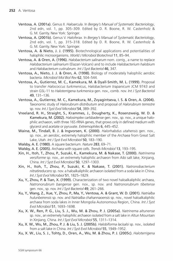
252 A. Ventosa
SGM symposium 66
Ventosa, A. (2001a). Genus II. Haloarcula. In Bergey’s Manual of Systematic Bacteriology,2nd edn, vol. 1, pp. 305–309. Edited by D. R. Boone, R. W. Castenholz & G. M. Garrity. New York: Springer.
Ventosa, A. (2001b). Genus V. Haloferax. In Bergey’s Manual of Systematic Bacteriology,2nd edn, vol. 1, pp. 315–318. Edited by D. R. Boone, R. W. Castenholz & G. M. Garrity. New York: Springer.
Ventosa, A. & Nieto, J. J. (1995). Biotechnological applications and potentialities ofhalophilic microorganisms. World J Microbiol Biotechnol 11, 85–94.
Ventosa, A. & Oren, A. (1996). Halobacterium salinarum nom. corrig., a name to replaceHalobacterium salinarium (Elazari-Volcani) and to include Halobacterium halobiumand Halobacterium cutirubrum. Int J Syst Bacteriol 46, 347.
Ventosa, A., Nieto, J. J. & Oren, A. (1998). Biology of moderately halophilic aerobicbacteria. Microbiol Mol Biol Rev 62, 504–544.
Ventosa, A., Gutierrez, M. C., Kamekura, M. & Dyall-Smith, M. L. (1999). Proposal to transfer Halococcus turkmenicus, Halobacterium trapanicum JCM 9743 and strain GSL-11 to Haloterrigena turkmenica gen. nov., comb. nov. Int J Syst Bacteriol49, 131–136.
Ventosa, A., Gutierrez, M. C., Kamekura, M., Zvyagintseva, I. S. & Oren, A. (2004).Taxonomic study of Halorubrum distributum and proposal of Halorubrum terrestresp. nov. Int J Syst Evol Microbiol 54, 389–392.
Vreeland, R. H., Straight, S., Krammes, J., Dougherty, K., Rosenzweig, W. D. &Kamekura, M. (2002). Halosimplex carlsbadense gen. nov., sp. nov., a unique halo-philic archaeon, with three 16S rRNA genes, that grows only in defined medium withglycerol and acetate or pyruvate. Extremophiles 6, 445–452.
Wainø, M., Tindall, B. J. & Ingvorsen, K. (2000). Halorhabdus utahensis gen. nov., sp. nov., an aerobic, extremely halophilic member of the Archaea from Great SaltLake, Utah. Int J Syst Evol Microbiol 50, 183–190.
Walsby, A. E. (1980). A square bacterium. Nature 283, 69–71.Walsby, A. E. (2005). Archaea with square cells. Trends Microbiol 13, 193–195.Xin, H., Itoh, T., Zhou, P., Suzuki, K., Kamekura, M. & Nakase, T. (2000). Natrinema
versiforme sp. nov., an extremely halophilic archaeon from Aibi salt lake, Xinjiang,China. Int J Syst Evol Microbiol 50, 1297–1303.
Xin, H., Itoh, T., Zhou, P., Suzuki, K. & Nakase, T. (2001). Natronobacteriumnitratireducens sp. nov. a haloalkaliphilic archaeon isolated from a soda lake in China.Int J Syst Evol Microbiol 51, 1825–1829.
Xu, Y., Zhou, P. & Tian, X. (1999). Characterization of two novel haloalkaliphilic archaea,Natronorubrum bangense gen. nov., sp. nov. and Natronorubrum tibetensegen. nov., sp. nov. Int J Syst Bacteriol 49, 261–266.
Xu, Y., Wang, Z., Xue, Y., Zhou, P., Ma, Y., Ventosa, A. & Grant, W. D. (2001). Natrialbahulunbeirensis sp. nov. and Natrialba chahannaoensis sp. nov., novel haloalkaliphilicarchaea from soda lakes in Inner Mongolia Autonomous Region, China. Int J SystEvol Microbiol 51, 1693–1698.
Xu, X. W., Ren, P. G., Liu, S. J., Wu, M. & Zhou, P. J. (2005a). Natrinema altunensesp. nov., an extremely halophilic archaeon isolated from a salt lake in Altun Mountainin Xinjiang, China. Int J Syst Evol Microbiol 55, 1311–1314.
Xu, X. W., Wu, M., Zhou, P. J. & Liu, S. J. (2005b). Halobiforma lacisalsi sp. nov., isolatedfrom a salt lake in China. Int J Syst Evol Microbiol 55, 1949–1952.
Xu, X. W., Liu, S. J., Tohty, D., Oren, A., Wu, M. & Zhou, P. J. (2005c). Haloterrigena
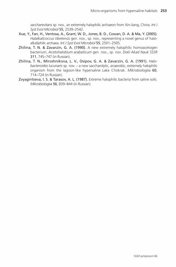
Micro-organisms from hypersaline habitats 253
SGM symposium 66
saccharevitans sp. nov., an extremely halophilic archaeon from Xin-Jiang, China. Int JSyst Evol Microbiol 55, 2539–2542.
Xue, Y., Fan, H., Ventosa, A., Grant, W. D., Jones, B. D., Cowan, D. A. & Ma, Y. (2005).Halalkalicoccus tibetensis gen. nov., sp. nov., representing a novel genus of halo-alkaliphilic archaea. Int J Syst Evol Microbiol 55, 2501–2505.
Zhilina, T. N. & Zavarzin, G. A. (1990). A new extremely halophilic homoacetogenbacterium, Acetohalobium arabaticum gen. nov., sp. nov. Dokl Akad Nauk SSSR311, 745–747 (in Russian).
Zhilina, T. N., Miroshnikova, L. V., Osipov, G. A. & Zavarzin, G. A. (1991). Halo-bacteroides lucunaris sp. nov. – a new saccharolytic, anaerobic, extremely halophilicorganism from the lagoon-like hypersaline Lake Chokrak. Mikrobiologiia 60,714–724 (in Russian).
Zvyagintseva, I. S. & Tarasov, A. L. (1987). Extreme halophilic bacteria from saline soils.Mikrobiologiia 56, 839–844 (in Russian).


![[NOIP-JPO] Procedures to JPO · The JPO application, which validly claims priority to multiple NOIP or direct PCT applications, or which is the divisional application validly based](https://img.dokumen.tips/doc/110x75/6039855ee68fd879637d1412/noip-jpo-procedures-to-jpo-the-jpo-application-which-validly-claims-priority.jpg)