Embed Size (px)
Citation preview

University of Pennsylvania Dental Medicine
MRGPRX2 SIGNALING ON MAST CELL-MEDIATED
PSEUDOALLERGY AND NEUROIMMUNE INTERACTION
Chalatip Chompunud Na Ayudhya
A DISSERTATION
Presented to the Faculty of Penn Dental Medicine
in Partial Fulfillment of the Requirements for the
Degree of Doctor of Science in Dentistry
2021
Supervisor of Dissertation
_________________________________
Hydar Ali, PhD
Professor of Pathology, Department of Basic and Translational Sciences
Director of Faculty Advancement and Diversity
Chair of Dissertation Committee
_________________________________
Claire H. Mitchell, PhD
Professor of Anatomy and Cell Biology, Department of Basic and Translational Sciences
Director of DScD Curriculum and Training

Dissertation Committee
_________________________________
Faizan Alawi, DDS
Professor of Pathology, Department of Basic and Translational Sciences
Associate Dean for Academic Affairs
Director of Penn Oral Pathology Services
_________________________________
Sunday O. Akintoye, BDS, DDS, MS
Associate Professor, Department of Oral Medicine
Director of Oral Medicine Research Program
_________________________________
Flavia Teles, DDS, MS, DMSc
Associate Professor, Department of Basic and Translational Sciences
MRGPRX2 SIGNALING ON MAST CELL-MEDIATED PSEUDOALLERGY AND NEUROIMMUNE INTERACTION
COPYRIGHT
2021
Chalatip Chompunud Na Ayudhya

iv
ACKNOWLEDGEMENT
First and foremost, I would like to sincerely thank my mentor, Dr. Hydar Ali, PhD for his
tremendous support and advice throughout my time at Penn. I have been very fortunate to be a
member of his lab and have learned the best example from him. Thank you for providing me
research opportunities and for guiding, inspiring and shaping me to grow as a researcher.
I deeply thank my committee members, Drs. Claire Mitchell (chair), Flavia Teles, Sunday
Akintoye and Faizan Alawi, for providing insightful comments and guidance from the beginning to
the end of my program. Their knowledge and expertise in the field of research were tremendously
valuable to enrich my work and career goals.
I would like to sincerely express my gratitude to the Ananda Mahidol Foundation for giving
me an opportunity to pursue my doctoral degree and providing me with the necessary financial
support throughout my doctoral study.
I gratefully thank my family for their unconditional love and endless support physically,
emotionally and financially throughout my life. You are truly the wind beneath my wings. Also, being
far away from home, from my beloved family and friends in Thailand, to a new country with totally
different culture and people was not an easy thing. I could not have succeeded thus far without all
the help, support and love from everyone around me. Thank you to all my friends for encouraging
me and believing in me when I could not even believe in myself. Moreover, I would not have finished
this work without the help from our present and past lab members, Aetas Amponnawarat, Monica
Thapaliya, Ibrahim Alkanfari, Anirban Ganguly, Shaswati Chaki and Saptarshi Roy. Thank you for
your suggestions, contributions and friendships.
Give thanks to the Lord, for He is good; for his love endures forever.
(Psalm 107:1)

v
ABSTRACT
MRGPRX2 SIGNALING ON MAST CELL-MEDIATED PSEUDOALLERGY AND NEUROIMMUNE INTERACTION
Chalatip Chompunud Na Ayudhya, DDS
Hydar Ali, PhD
In addition to high affinity IgE receptor, human mast cells (MCs) express a newly identified
receptor known as Mas-related G protein-coupled receptor X2 (MRGPRX2; mouse ortholog
MrgprB2). This receptor is predominantly expressed on only one subtype of MCs and it can be
activated by a diverse group of cationic agonists including host defense peptides, Food and Drug
Administration (FDA)-approved drugs associated with pseudoallergy and neuropeptides secreted
from sensory nerve endings. Not surprisingly, MRGPRX2 has been implicated in several MC-
mediated health and disease, ranging from host defense and wound healing to drug-induced
pseudoallergic reactions, neurogenic inflammation and pain. However, there is a controversy
regarding its role on rocuronium-induced hypersensitivity. Furthermore, the molecular mechanisms
underlying MRGPRX2 regulation remain largely unknown. In this dissertation, we first investigated
the role of MRGPRX2 on rocuronium-induced hypersensitivity. The effect of MRGPRX2 mutations
(M196I, L226P and L237P) identified in a patient with rocuronium hypersensitivity were also tested.
We found that rocuronium induced degranulation in murine and human MCs via MrgprB2 and
MRGPRX2, respectively, but with different affinities, indicating important functional differences
between these receptors. This indicates that mice expressing MrgprB2 may not be a suitable model
to study human MRGPRX2 function and highlights the need to develop better animal models.
It is now realized that activation of MCs by substance P (SP) via MRGPRX2 contributes to
neurogenic inflammation, pain and itch. We sought to identify the mechanisms underlying
MRGPRX2 signaling and regulation on SP-activated MC responses. Using pertussis toxin and YM-
254890, we demonstrated that SP induces MRGPRX2-mediated Ca2+ mobilization and
degranulation via both Gαi and Gαq. Next, we utilized information obtained from both structural
modeling and naturally occurring MRGPRX2 missense variants to identify putative G protein

vi
coupling regions. In addition, several gain- and loss-of-function missense single nucleotide
polymorphisms (SNPs) in MRGPRX2 have been discovered.
Finally, we demonstrated that SP can activate β-arrestin recruitment and receptor
internalization. A tyrosine residue in the highly conserved NPxxY motif of MRGPRX2 (Tyr279) is
crucial for SP-induced β-arrestin recruitment and receptor internalization. This study reveals the
novel findings that activation of MRGPRX2/B2 by SP is regulated by β-arrestins and that a highly
conserved tyrosine residue is responsible for MRGPRX2 signaling and regulation.

vii
TABLE OF CONTENTS ACKNOWLEDGEMENT ................................................................................................................. IV ABSTRACT ...................................................................................................................................... V LIST OF ILLUSTRATIONS ............................................................................................................. IX LIST OF ABBREVIATIONS ............................................................................................................. X CHAPTER 1: INTRODUCTION ........................................................................................................ 1
1.1. AN OVERVIEW OF MAST CELLS .................................................................................................. 2 1.1.1. Mast cell development ................................................................................................... 2 1.1.2. Mast cell heterogeneity and plasticity ............................................................................ 2 1.1.3. Mast cell activation and mediator release...................................................................... 4
1.2. MAS-RELATED G PROTEIN-COUPLED RECEPTOR X2 (MRGPRX2) ............................................. 5 1.2.1. Discovery of MRGPRX2 as a mast cell receptor for basic secretagogues ................... 5 1.2.2. Identification of MrgprB2 as the mouse ortholog of human MRGPRX2 ........................ 5
1.3. ROLE OF MRGPRX2 IN DRUG HYPERSENSITIVITY REACTIONS ................................................... 7 1.4. ROLE OF MRGPRX2 IN NEUROIMMUNE INTERACTIONS AND SKIN HOMEOSTASIS ......................... 8 1.5. MRGPRX2 SIGNALING AND REGULATION: IMPLICATION FROM OTHER GPCRS.......................... 10 1.6. DISSERTATION AIMS ............................................................................................................... 14
CHAPTER 2: MRGPRX2 ACTIVATION BY ROCURONIUM: INSIGHTS FROM STUDIES WITH HUMAN SKIN MAST CELLS AND MISSENSE VARIANTS .......................................................... 16
2.1. ABSTRACT ............................................................................................................................. 17 2.2. INTRODUCTION....................................................................................................................... 18 2.3. MATERIALS AND METHODS ..................................................................................................... 19 2.4. RESULTS ............................................................................................................................... 23
2.4.1. Rocuronium activates murine PMCs via MrgprB2. ...................................................... 23 2.4.2. Rocuronium induces MRGPRX2-mediated degranulation in human MCs.................. 24 2.4.3. MRGPRX2 variants presented in a patient with POH to rocuronium do not display gain-of-function phenotype for rocuronium-induced MC degranulation. ............................... 25
2.5. DISCUSSION .......................................................................................................................... 26 2.6. CONCLUSION ......................................................................................................................... 29 2.7. ACKNOWLEDGMENTS ............................................................................................................. 29
CHAPTER 3: IDENTIFICATION OF GAIN AND LOSS OF FUNCTION MISSENSE VARIANTS IN MRGPRX2’S TRANSMEMBRANE AND INTRACELLULAR DOMAINS FOR MAST CELL ACTIVATION BY SUBSTANCE P .................................................................................................. 34
3.1. ABSTRACT ............................................................................................................................. 35 3.2. INTRODUCTION....................................................................................................................... 36 3.3. MATERIALS AND METHODS ..................................................................................................... 39 3.4. RESULTS ............................................................................................................................... 42
3.4.1. MRGPRX2 mediates SP-induced MC activation via both Gαi and Gαq. ..................... 42 3.4.2. Mutations of the Highly Conserved Residues 3x46, 6x37, and 7x53 in MRGPRX2 Lead to a Significant Reduction in SP-Induced MC Activation. ............................................. 43 3.4.3. Naturally Occurring Missense MRGPRX2 Variants at or Near the Conserved Residues, V123F and V282M, Display Loss of Function Phenotype for SP-Induced MC Activation. ............................................................................................................................... 44 3.4.4. Naturally Occurring Missense MRGPRX2 Variants at the Second Intracellular Loop, R138C and R141C, Display Loss of Function Phenotype for SP-Induced MC Activation. ... 44 3.4.5. Mutations in Potential Phosphorylation Sites of MRGPRX2 Leads to Enhanced MC Activation in Response to SP. ................................................................................................ 45
viii
3.4.6. Naturally Occurring Missense MRGPRX2 Variants at its Carboxyl-Terminus, S325L and L329Q, Display Gain of Function Phenotype for SP-Induced MC Activation. ................ 45
3.5. DISCUSSION .......................................................................................................................... 45 3.6. ACKNOWLEDGEMENTS ........................................................................................................... 49
CHAPTER 4: SUBSTANCE P SERVES AS A BALANCED AGONIST FOR MRGPRX2 AND A SINGLE TYROSINE RESIDUE IS REQUIRED FOR Β-ARRESTIN RECRUITMENT AND RECEPTOR INTERNALIZATION ................................................................................................... 56
4.1. ABSTRACT ............................................................................................................................. 57 4.2. INTRODUCTION....................................................................................................................... 58 4.3. MATERIALS AND METHODS ..................................................................................................... 60 4.4. RESULTS ............................................................................................................................... 64
4.4.1. SP is a balanced agonist for MRGPRX2. .................................................................... 64 4.4.2. SP-induced β-arrestin recruitment and MRGPRX2 internalization are G protein-independent............................................................................................................................ 65 4.4.3. β-arrestin2 regulates SP/MrgprB2-mediated MC degranulation. ................................ 66 4.4.4. Mutation of a highly conserved tyrosine residue of MRGPRX2 (Y279A) abolishes SP-induced β-arrestin recruitment. .............................................................................................. 66 4.4.5. Tyrosine residue in MRGPRX2 (Y279) is required for SP-mediated receptor internalization. ........................................................................................................................ 67
4.5. DISCUSSION .......................................................................................................................... 68 4.6. ACKNOWLEDGEMENTS ........................................................................................................... 70
CHAPTER 5: CONCLUSION AND FUTURE DIRECTIONS .......................................................... 77 LISTS OF PUBLICATIONS ............................................................................................................ 81 REFERENCES ............................................................................................................................... 82
ix
LIST OF ILLUSTRATIONS Figure 1-1. Human MRGPRX2 and its mouse ortholog MrgprB2 can be activated by diverse
groups of ligands.
Figure 1-2. Activation of mast cells by substance P (SP) via MRGPRX2 contributes to neurogenic inflammation.
Figure 1-3. Snake diagram of secondary structure of MRGPRX2.
Figure 1-4. Pattern of residue contacts upon receptor activation in class A GPCRs.
Figure 1-5. Canonical GPCR signaling paradigm.
Figure 2-1. Rocuronium activates mouse peritoneal mast cells via MrgprB2.
Figure 2-2. Rocuronium activates LAD2 cells and primary human skin mast cells to cause degranulation.
Figure 2-3. Rocuronium induces MRGPRX2 internalization in LAD2 cells and primary human skin mast cells.
Figure 2-4. MRGPRX2 mutations rendered the receptor unresponsiveness to rocuronium.
Figure 3-1. Effects of pertussis toxin (PTx) and YM-254890 on SP-induced degranulation and Ca2+ mobilization in RBL-2H3 cells stably expressing MRGPRX2 (RBL-MRGPRX2).
Figure 3-2. Effects of mutations at MRGPRX2’s highly conserved positions within transmembrane domains (V123A, I225A, and Y279A) on cell surface expression, SP-induced Ca2+ mobilization, and degranulation in transiently transfected RBL-2H3 cells.
Figure 3-3. Effects of naturally occurring MRGPRX2 variants at the receptor’s conserved transmembrane domains (V123F, T224A, and V282M) on SP-induced responses in transiently transfected RBL-2H3 cells.
Figure 3-4. Effects of naturally occurring MRGPRX2 variants at the receptor’s intracellular loops (Y137H, R138C, R140C, and R141C) on SP-induced responses in transiently transfected RBL-2H3 cells.
Figure 3-5. Effects of Ser/Thr residues on MRGPRX2’s carboxyl-terminus on cell surface expression, SP-induced Ca2+ mobilization, and degranulation in transiently transfected RBL-2H3 cells.
Figure 3-6. Effects of naturally occurring MRGPRX2 variants within the receptor’s carboxyl-terminus (Q305R, D311H, S325L, and L329Q) on SP-induced responses in transiently transfected RBL-2H3 cells.
Figure 4-1. Substance P (SP) is a balanced agonist for MRGPRX2.
Figure 4-2. Pertussis toxin (PTx) inhibits SP-induced mast cell (MC) degranulation, but has no effect on β-arrestin recruitment and MRGPRX2 internalization.
Figure 4-3. β-arrestin2 regulates MrgprB2-mediated MC degranulation in response to SP.
Figure 4-4. Y279A mutation of MRGPRX2 abolishes β-arrestin recruitment in response to SP.
Figure 4-5. Y279A mutation of MRGPRX2 impairs SP-induced receptor internalization.
Figure 4-6. Y279A mutation of MRGPRX2 displays resistance to SP-induced receptor trafficking.

x
LIST OF ABBREVIATIONS MC Mast cell MCTC Mast cells containing tryptase and chymase MCT Mast cells containing tryptase CTMCs Connective tissue mast cells MMCs Mucosal mast cells BMMCs Bone marrow-derived mast cells PMCs Peritoneal mast cells Ig Immunoglobulin FcεRI High affinity immunoglobulin E receptor GPCR G protein-coupled receptor MRGPRX2 Mas-Related G protein-coupled receptor X2 MrgprB2 Mas-Related G protein-coupled receptor B2 HDP Host defense peptide NP Neuropeptide FDA Food and Drug Administration SCF Stem cell factor TLR Toll-like receptor SP Substance P WT Wild-type NMBDs Neuromuscular blocking drugs POH Perioperative hypersensitivity BAT Basophil activation test IDT Intradermal skin test TM Transmembrane ECL Extracellular loop ICL Intracellular loop PTx Pertussis toxin MFI Mean fluorescent intensity LAMP-1 Lysosomal-associated membrane protein 1 RBL-2H3 Rat basophilic leukemia-2H3 cells RBL-MRGPRX2 RBL-2H3 cells stably expressing MRGPRX2 HTLA Engineered HEK-293T cells stably expressing a β-arrestin2–tobacco
etch virus fusion gene HTLA-MRGPRX2 HTLA cells stably expressing MRGPRX2-Tango Tango assay Transcriptional activation following arrestin translocation assay SNPs Single nucleotide polymorphisms

1
CHAPTER 1: Introduction
Mast cells (MCs) are multifunctional granulated immune cells of the myeloid lineage that
are widely located at the host-environment interface such as the skin and mucosal tissues. While
best known as effectors cells in anaphylaxis and allergic and inflammatory diseases, MCs also play
important roles in diverse physiological processes including tissue homeostasis, host defense, and
wound healing (1-3). They are widely distributed throughout the body in most vascularized tissues
and are particularly found at the host-environment interfaces, such as the skin, respiratory tract,
and gastrointestinal tract. Therefore, MCs are regarded as professional sentinels and first
responders against infectious pathogens and other potential insults (3-5).
Classical MC activation occurs via the cross-linking of high affinity immunoglobulin E (IgE)
receptor (FcεRI) with antigen. Besides FcεRI, MCs express a variety of receptors that enable them
to respond to a broad range of stimuli. A major breakthrough in MC research has been the discovery
of a novel G protein-coupled receptor (GPCR) known as Mas-related GPCR-X2 (MRGPRX2).
Unique features of MRGPRX2 are that this receptor is expressed predominantly on one subtype of
human MCs and that it is responsive to a wide spectrum of cationic ligands including host defense
peptides (HDPs), neuropeptides (NPs) and many Food and Drug Administration (FDA)-approved
drugs (6-10). Unsurprisingly, activation of MCs via MRGPRX2 has been implicated in multiple
physiological and pathological conditions, including host defense against microbial infection,
pseudoallergy, neurogenic inflammation, chronic inflammatory diseases, and pain (9-15). Better
understanding of the molecular mechanisms underlying MRGPRX2 activation and regulation will
not only enhance our fundamental knowledge of MC responses, but will also lead to the
development of novel therapeutic approaches for the modulation of allergic and inflammatory
diseases.

2
1.1. An overview of mast cells
1.1.1. Mast cell development
MCs are tissue-resident granulocytes derived from hematopoietic stem cells in the bone
marrow (16). Unlike other immune cells, MCs do not terminally differentiate in the bone marrow,
but circulate in the bloodstream as immature progenitors. They complete their differentiation and
maturation in peripheral tissues under the influence of the cytokine milieu and tissue
microenvironment (16-18). Mature MCs are long-lived and can undergo repeated rounds of
activation in response to stimuli (19, 20), while other granulocytes, such as neutrophils and
basophils, have short lifespan and undergo apoptosis after their recruitment and activation in the
tissues (21).
Stem cell factor (SCF), the ligand for the CD117/c-Kit, is a major regulator of MC biology
(22). Activation of c-Kit signaling through SCF is crucial for MC development. Furthermore, SCF/c-
Kit signaling also play a role in the migration, proliferation, survival and activation of MCs (22, 23).
Mouse strains bearing mutations in the genes for the c-Kit receptor (KitW/W-v and KitW-sh) or its ligand
SCF (Sl/Sld) have profoundly deficient numbers of MCs, emphasizing an essential role of c-Kit and
SCF in MC development (24, 25). In addition to SCF, various cytokines, such as interleukin (IL)-3,
IL-4, IL-9, IL-10, transforming growth factor-β, and nerve growth factor, also modulate MC
differentiation and proliferation (22, 26).
1.1.2. Mast cell heterogeneity and plasticity
MC maturation is largely influenced by microenvironmental conditions. As such, mature
MCs display considerable heterogeneity as a direct consequence of different development patterns
(18). Moreover, phenotypic and functional characteristics of MCs can be dynamically modulated or
“tuned” by many genetic and environmental factors they encounter (27). For example, the
expression profile of proteases in jejunal MCs can change during Trichinella spiralis infection, which
results in the alteration of MC function (28).
In humans, MCs are generally categorized into two major subtypes based on the protease
content of their secretory granules: MCs that contain both tryptase and chymase are known as

3
MCTC, and those that contain only tryptase are known as MCT (29, 30) (Table 1-1). MCTC are
predominantly found in connective tissues such as the skin and gingiva, while MCT are more
prominent in the mucosal tissues such as respiratory and gastrointestinal mucosa (17, 31, 32). In
rodents, two subtypes of MCs have been described: connective tissue MCs (CTMCs) and mucosal
MCs (MMCs). CTMCs are found in the skin and peritoneum, and express predominantly mouse
MC protease (MCP)-4, -5, -6, and carboxypeptidase A, whereas MMCs reside in the lung and gut,
and express MCP-1 and -2 (18, 29). Rodent CTMCs share characteristics with human MCTC, while
MMCs resemble human MCT (17, 18) (Table 1-1).
MCTC and CTMCs are sometimes considered as innate/constitutive MCs as they are
constitutively present in connective tissues and are generally unaffected by T cells. By contrast,
MCT and MMCs are referred to as adaptive/induced MCs as their maturation are induced in a T-
cell-dependent manner (31, 33). Intriguingly, while both MC subtypes in humans and mice can be
activated by antigen/IgE via FcεRI, only MCTC and CTMCs are responsive to a diverse range of
cationic substances, collectively called basic secretagogues, such as compound 48/80,
mastoparan, peptidergic drugs, NPs, and HDPs (6, 8) (Table 1-1).
Table 1-1. Major subtypes of MCs and their phenotypic/functional characteristics.
Characteristics Mast Cell Subtypes
Human MCs MCTC MCT
Rodent MCs CTMCs MMCs
Tissue localization Skin, serosal cavities, and gingiva Gastrointestinal and respiratory mucosa
Protease content
Human: Contain both tryptase and chymase Rodent: Contain MCP-4, -5, -6, and carboxypeptidase A
Human: Contain only tryptase Rodent: Contain MCP-1 and -2
Type Innate Adaptive
T cell dependence No Yes
Stimulants Antigens and basic secretagogues (for example, NPs and HDPs) Antigens

4
1.1.3. Mast cell activation and mediator release
Effector functions of MCs are dependent on their ability to release a plethora of biologically
active mediators upon activation (1). MCs can be activated by a broad range of stimuli, such as
antigens, infectious organisms, and endogenous inflammatory factors, via the receptors present on
their cell surfaces. In addition to the classical and most studied FcεRI, MCs also express numerous
receptors including immunoglobulin G receptors (FcγRs), Toll-like receptors (TLRs e.g. TLR2 and
TLR4), complement receptors (e.g. C3aR and C5aR), and recently identified MRGPRX2, allowing
MCs to recognize and react directly to pathogenic stimuli (4, 5, 8, 10, 34, 35).
Upon activation, MCs can release their preformed and de novo synthesized mediators in
a phasic manner (4). An early phase of MC response involves degranulation, a process by which
preformed mediators stored in their secretory granules can be immediately released into the
extracellular environment following stimulation (36, 37). This process is mediated by intracellular
calcium mobilization, which activates a cascade of downstream events that trigger exocytosis of
secretory granules (38). Pre-stored mediators, such as histamine, proteases, vascular endothelial
growth factor (VEGF), and tumor necrosis factor-alpha (TNF-α), have marked effects on endothelial
cells and smooth muscle, and nerves, leading to increased vascular permeability, vasodilation,
smooth muscle contraction, and stimulation of afferent neurons (1, 37, 39, 40). In addition to
releasing preformed mediators, MC activation also leads to de novo production and secretion of
lipid mediators, such as leukotrienes, prostaglandins and platelet-activating factor, as well as an
array of cytokines and chemokines, which accounts as the late MC response (1, 4, 37). MC-derived
cytokines and chemokines play critical roles in the differentiation and activation of several immune
cells, contributing to the development and homeostasis of the immune system. Furthermore, they
promote various cell recruitment to the sites of inflammation, contributing to the pathophysiology of
inflammatory responses (4, 5, 41).

5
1.2. Mas-related G protein-coupled receptor X2 (MRGPRX2)
1.2.1. Discovery of MRGPRX2 as a mast cell receptor for basic secretagogues
Besides activation of MCs by antigen/IgE, connective tissue-type MCs can respond to
basic secretagogues via an IgE-independent pathway. These cationic peptides were previously
proposed to trigger MC degranulation in a receptor-independent manner by directly activating
pertussis toxin-sensitive G proteins (Gαi2 and Gαi3) (42-44). Until in 2006, Tatemoto et al. (8)
provided the first demonstration that MRGPRX2 (formerly known as MrgX2) is expressed at high
level on human skin MCTC but not on lung MCT, and is an endogenous receptor for basic
secretagogues. A number of basic secretagogues, including compound 48/80, MC-degranulating
peptide (MCDP), and substance P (SP), increased reporter gene expression in PC12h cells
transiently transfected with MRGPRX2 and induced dose-dependent calcium mobilization in HEK-
293 cells stably expressing MRGPRX2, but not parental cells (8). Furthermore, they demonstrated
that MCDP and SP activate G proteins in membranes prepared from HEK-293 cells expressing
MRGPRX2, suggesting that MRGPRX2 is a link between the activation of basic secretagogues and
G proteins (8).
1.2.2. Identification of MrgprB2 as the mouse ortholog of human MRGPRX2
MRGPRs are recently identified GPCRs that belong to the rhodopsin-like class A GPCR
family. They can be divided into nine subfamilies (A–H and X), with subfamily X is specific to
primates including humans, while subfamilies A, B, C, and H exist only in rodents (45).
Despite many in vitro studies on MRGPRX2, no murine model for in vivo study was initially
employed due to difficulty to identify the putative mouse ortholog of human MRGPRX2. Unlike the
human genome, which encodes for only four MRGPRs (MRGPRX1 – 4), there are 22 potential
coding genes for MRGPRs in mice (9, 10). In 2015, a landmark study by McNeil et al. (10) revealed
that mouse peritoneal MCs (PMCs) solely express MrgprB2 mRNA and MrgprB2 expression is
restricted to connective tissue‐type MCs. By generating mice with a 4-base pair deletion in the
MrgprB2 coding region (MrgprB2MUT mice), MCs obtained from these mice show dramatic reduction
of calcium mobilization and histamine release in response to MRGPRX2 ligands, including

6
compound 48/80, SP, and peptidergic drugs, in vitro. Furthermore, MrgprB2MUT mice demonstrate
remarkably reduced paw edema in vivo when compared to the wild-type (WT) mice (10). Of note,
MrgprB2 mutation in MCs does not impair their development and IgE-mediated response. These
findings strongly indicate that MrgprB2 is the mouse basic secretagogue receptor and the ortholog
of human MRGPRX2. This discovery paved the way for the in vivo studies of MrgprB2-mediated
MC activation in physiology and diseases, which substantially advance our understanding of
MRGPRX2’s functional roles. Since then, an ever-increasing number of MRGPRX2/B2 ligands,
ranging from HDPs and bacterial quorum-sensing molecules (QSMs) to NPs and peptidergic drugs
associated with pseudoallergic reactions, have been discovered (6, 10, 46-50) (Figure 1-1).
Figure 1-1. Human MRGPRX2 and its mouse ortholog MrgprB2 can be activated by a diverse group of ligands, including endogenous NPs and HDPs, bacterial quorum-sensing peptides, and peptidergic drugs. MC activation by different MRGPRX2/B2 agonist results in distinct mediator release and biological responses. (Galli SJ et al., 2020).
It is worth noting that although MrgprB2 is regarded as the mouse counterpart of
MRGPRX2, these two receptors share only ~53% sequence similarity and thus demonstrate
significant species-specificity with respect to the concentrations of ligands required to activate each
receptor (10, 51). EC50 values (concentration of ligand required to induce half-maximal response)
of most ligands for MrgprB2 are significantly higher than those for MRGPRX2. For example, SP
activates MrgprB2 and MRGPRX2 with an EC50 value of 54 μM and 152 nM, respectively. By
contrast, rocuronium, a widely used neuromuscular blocking drug (NMBD), has an EC50 value of
22.2 µg/mL for MrgprB2, whereas an EC50 value for MRGPRX2 is 261 µg/mL (10).

7
1.3. Role of MRGPRX2 in drug hypersensitivity reactions
Anaphylaxis to FDA-approved drugs, including antibiotics, opioids, iodinated contrast
media, and neuromuscular blocking drugs (NMBDs), have increased in recent years. These
potentially life-threatening reactions represent a diagnostic challenge for allergists as the
underlying mechanisms of allergic reactions to many drugs remain elusive. While it is generally
thought to be mediated via a cross-linking of drug-specific IgE and FcεRI presented on the MC
surfaces, some reactions are mediated independently of IgE. In 2015, McNeil et al. (10)
demonstrated that certain drugs containing a tetrahydroisoquinoline (THIQ) motif or similar
structure, such as NMBDs and fluoroquinolone antibiotics, induce MC degranulation and mediators
release, subsequently resulting in allergic reactions, via MRGPRX2 and its mouse ortholog
MrgprB2. MrgprB2MUT mice exhibited substantially reduced hind paw swelling and less drop in their
body temperature compared to the WT mice after administration of peptidergic drugs. Of note, the
deletion of the receptor in MrgprB2MUT mice did not alter the IgE-mediated pathway (10). These
findings strongly indicate that MC activation through MRGPRX2/B2 is distinct from IgE-mediated
pathway.
NMBDs, such as mivacurium, atracurium, cisatracurium, and rocuronium, while are
routinely used during general anesthesia and surgery to reduce unwanted muscle movement,
account for nearly 60% of anaphylactic reactions in a surgical setting (52). Of all the NMBDs, the
incidence rates of anaphylaxis are found to be higher after rocuronium administration than those of
the other NMBDs (53, 54). Spoerl et al. (55) reported three cases of rocuronium-induced
hypersensitivity which displayed positive skin test reaction, despite having negative rocuronium-
specific IgE and basophil activation test (BAT). Based on these findings, it was proposed that this
rocuronium hypersensitivity is mediated via the direct activation of MRGPRX2 (56). On the contrary,
a large clinical study conducted with 140 patients suspected of perioperative hypersensitivity to
rocuronium demonstrated that hypersensitivity to rocuronium mainly results from IgE-mediated MC
activation (57).

8
More intriguingly, the possibility that rocuronium activates human MCs via MRGPRX2 has
been subject of controversy. While McNeil et al. (10) showed that rocuronium activates MRGPRX2
transfected in HEK293 cells with an EC50 value of 261 µg/mL, Lansu et al. (51) could not reproduce
this finding. Furthermore, rocuronium, even at high concentration (up to 2 mg/mL), did not induce
degranulation in either human MC line LAD2 cells or CD34+-derived human MCs that endogenously
express MRGPRX2 (58, 59). This lack of effect of rocuronium on MRGPRX2 is surprising given
that it induces massive degranulation in mouse PMCs via MrgprB2 and could reflect the differences
between mouse MrgprB2 and human MRGPRX2 (10, 51).
1.4. Role of MRGPRX2 in neuroimmune interactions and skin homeostasis
The close relationship between MCs and nerves has been well established in most tissues
including the skin, gastrointestinal mucosa, and respiratory tract. MCs are particularly found in close
proximity to sensory nerve endings, and the frequency of anatomic associations between MCs and
nerves is significantly increased at sites of inflammation (3). Furthermore, MCs and nerves
demonstrate bidirectional communication that is mediated through shared ligands, such as
cytokines and NPs, and their cognate receptors (3, 60). These close anatomic localization and
functional interaction of MCs and sensory nerves provide a significant link between the immune
and nervous systems and play important protective roles in maintaining tissue homeostasis and
responding to external challenges. However, dysregulation of MC–nerve interaction has been
suggested as a major contributor in the pathogenesis of neurogenic inflammation, pain, and allergic
skin diseases (61-63).
Substance P (SP) is an 11-amino acid NP that belongs to the tachykinin family (64). It
exerts a wide range of physiological as well as pathological functions, with well recognition for its
roles in inflammation and pain perception (64, 65). SP is one of the most comprehensively studied
NPs that mediates crosstalk between neurons and MCs. SP released from the sensory nerve
endings can induce MC degranulation, resulting in the release of multiple pro-inflammatory
mediators (64-66). In turn, MC-derived mediators, such as histamine, tryptase, and leukotrienes,
can activate their specific receptors expressed on sensory nerves to cause further SP release (67-

9
69). Activation of MCs by SP has been implicated in the pathogenesis of a number of
neuroinflammatory conditions such as sickle cell disease (70), chronic urticaria (6), and atopic
dermatitis (15).
The neurokinin-1 receptor (NK-1R) is a canonical receptor for SP that was previously
thought to be responsible for SP-induced MC activation. Murine MMCs such as bone marrow-
derived MCs (BMMCs) do not generally express NK-1R, but when cultured in the presence of SCF
and IL-4, they displayed significant expression of NK-1R (71). These SCF/IL-4-treated BMMCs was
activated in response to SP and the activation was significantly diminished by an NK-1R antagonist
(71), suggesting that SP may activate MCs via this receptor. The discovery that SP contributes to
neurogenic inflammation and pain led to the development of multiple NK-1R antagonists as
potential therapies for inflammatory conditions and pain over the past three decades. However,
these compounds, while shown to be effective in animal models, have failed to demonstrate anti-
inflammatory and analgesic effects in human clinical trials (72, 73).
In addition to NK-1R, human skin MCs express MRGPRX2 which has been shown to be a
receptor for basic secretagogues including SP. Fujisawa et al. (6) demonstrated that SP-induced
MC degranulation and prostaglandin D2 generation are significantly depleted in MRGPRX2-
silenced human skin MCs. Additionally, conventional NK-1R antagonists have been shown an off-
target effect on the mouse MrgprB2 (74). These findings therefore raise the possibility that the
nociceptive and proinflammatory actions of SP may be mediated via alternative mechanisms,
presumably MRGPRX2/B2, rather than the interaction with NK-1R.
The speculation that SP mediates neurogenic inflammation and pain via MRGPRX2/B2
has been recently confirmed by a well-integrated study by Green et al. (13). Using two inflammatory
pain models, postoperative incision model and Complete Freund’s Adjuvant (CFA) model, they
demonstrated that activation of MrgprB2 by SP is required for inflammatory mechanical and thermal
hyperalgesia. MrgprB2-deficient (MrgprB2−/−) mice, but not NK-1R−/− mice, had significant
reductions in swelling, pain hypersensitivity, and immune cell infiltration when compared to WT
mice (13). They also showed that knockdown of MRGPRX2 expression in human MC line LAD2

10
cells results in substantially reduction of cytokines and chemokines, such as TNF-α, CCL2 and
CCL3, released upon SP stimulation, whereas there is no decrease in LAD2 treated with an NK-
1R antagonist (13). Taken together, these findings strongly indicate that MRGPRX2/B2 activation
are responsible for the release of proinflammatory cytokines/chemokines and the recruitment of
innate immune cells, including neutrophils and monocytes, to the site of injury following SP
stimulation (Figure 1-2). Thus, targeting MRGPRX2 could therefore serve as novel therapeutic
approach for the management of diseases associated with neurogenic inflammation and pain.
However, the molecular mechanism of MRGPRX2 activation by SP has yet to be elucidated.
Figure 1-2. During tissue injury, substance P (SP) released from peripheral sensory nerve endings contributes to neurogenic inflammation through the activation of MCs via MRGPRX2/B2. Activation of MRGPRX2/B2 by SP leads to cytokine release and recruitment of immune cells, which facilitate inflammatory responses and peripheral nerve sensitization. (Meixiong J et al., 2020)
1.5. MRGPRX2 signaling and regulation: Implication from other GPCRs
MRGPRX2 is a member of class A GPCRs. Thus, it shares a common structure of seven
transmembrane (TM) α-helices connected by three extracellular loops (ECLs) and three
intracellular loops (ICLs), with an extracellular amino-terminal tail (N-terminus) and an intracellular
carboxy-terminal tail (C-terminus). The extracellular part of the receptor is responsible for ligand
binding, whereas the intracellular part is involved in binding downstream effectors such as
heterotrimeric G proteins (Gαβγ) and β-arrestins (75) (Figure 1-3).

11
Figure 1-3. Snake diagram of secondary structure of MRGPRX2 obtained from the GPCR database (GPCRdb; www.gpcrdb.org) (76). Each circle represents amino acid residue with one letter code. The extracellular part, including extracellular loops (ECLs) and transmembrane domains (TMs), and N-terminus, is responsible for binding ligands. The intracellular part, including intracellular loops (ICLs) and TMs, and C-terminus, is involved in binding downstream effectors and initiating signaling cascades.
GPCRs, as their name implies, mainly interact with ‘heterotrimeric G proteins’ to translate
the signal from extracellular ligands into intracellular responses. Binding of ligands at the “ligand
binding cradle” on the extracellular region of GPCRs leads to conformational rearrangements of
the cytoplasmic side to facilitate the binding of G proteins, leading to G protein activation and
initiating downstream signaling cascades (75, 77). Closely related GPCRs exhibit high degree of
conserved sequence motifs and a common activation pathway, especially in the regions implicated
in ligand binding and G protein coupling (78).
Venkatakrishnan et al. (79) analyzed crystal structures of 27 class A GPCRs in inactive
and active states to investigate the activation pathways across class A GPCRs and reported a
common pattern of residue contact rearrangement involved TM3 (3x46), TM6 (6x37), and TM7
(7x53) in all class A receptors. (In this GPCR numbering system, the first number denotes the helix
(1-7) and the second number indicates the residue position relative to the most conserved position,
which is assigned the number 50. Thus, 7x53 denotes a residue in TM7, which is at 3 positions
after the most conserved residue (7x50). Similarly, 3x46, denotes a residue in TM3, which is at 4
positions before the most conserved residue). In the inactive state, the residue at 6x37 is in contact
with a conserved hydrophobic residue at position 3x46 (36). Upon activation, the residue at position

12
3x46 breaks the contact with 6x37 and forms a new contact with Tyr7x53 within the highly conserved
NPxxY motif of TM7 (35-38) (Figure 1-4). Based on molecular modeling and naturally occurring
missense mutations, studies from our and other labs led to the identification of the ligand binding
cradle for a number of MRGPRX2 agonists (17-19). However, the possibility that residues 3x46,
6x37 and 7x53 in MRGPRX2 couple to G proteins to cause MC degranulation has not been
investigated.
Figure 1-4. Pattern of residue contacts upon receptor activation in class A GPCRs. Dotted circles around 6x37 and 7x53 denote the movement of TM6 and TM7 upon activation. Figure is adapted from Venkatakrishnan et al., 2016.
There are four major subtypes of heterotrimeric G proteins according to the functional and
structural homologies of the Gα subunits: Gαs, Gαi/o, Gαq/11, and Gα12/13. Interaction of GPCRs with
different Gα subunits elicit distinct downstream signaling cascades and cellular responses (80). For
example, Gαq/11 proteins activate phospholipase C, which hydrolyzes phosphatidylinositol
phosphate to diacyl glycerol (DAG) and inositol 1,4,5-trisphosphate (IP3), resulting in protein kinase
C (PKC) activation and intracellular calcium mobilization (81). Previous studies on the mechanisms
of MRGPRX2-mediated MC responses have shown that basic secretagogues cause MC
degranulation via pertussis toxin (PTx)-sensitive G protein-dependent signaling pathway (8, 46,
48). PTx catalyzes the ADP-ribosylation of the α subunits of the heterotrimeric Gαi/o protein family
(Gαo, Gαi1, Gαi2 and Gαi3) (82), of which MCs express Gαi2 and Gαi3 (44). However, while
degranulation in response to MRGPRX2 agonists, such as cathelicidin LL-37 and human β-

13
defensin-3, is inhibited by PTx, calcium mobilization is not (46, 48). These findings suggest that
MRGPRX2 signaling pathway is mediated via both PTx-dependent (Gαi) and -independent
(presumably Gαq) G proteins in order to induce human MC activation and responses. However, the
molecular mechanisms in which specific G protein mediates MRGPRX2 signaling to induce MC
activation and degranulation has not been investigated.
In addition to G proteins, most GPCRs signal via an additional pathway that involves the
recruitment of adapter proteins known as β-arrestins (83, 84). This pathway has been implicated in
GPCR desensitization (uncoupling of the G protein from the cognate receptor), endocytosis, and
internalization in order to prevent further G protein coupling and overactivation of the receptor (85).
Furthermore, recent evidence shows that β-arrestins also play an important role in G protein-
independent downstream signaling for cell migration, growth, and differentiation (86-88) (Figure 1-
5).
Figure 1-5. In the canonical GPCR signaling paradigm, ligand binding to the receptor leads to G protein activation, resulting in the initiation of distinct downstream signaling cascades and cell responses such as the release of intracellular calcium from the endoplasmic reticulum (ER). This G protein signaling is regulated by adapter proteins, β‐arrestins. Binding of β‐arrestins can sterically block further G protein activation (desensitization) and lead to GPCR internalization/trafficking. Furthermore, β‐arrestins serve as signal transducers in several G protein-independent signaling pathways.
Two isoforms of β-arrestins, β-arrestin1 and β-arrestin2, are ubiquitously expressed in all
tissues and can differentially regulate GPCR signaling and trafficking (89-91). Our lab previously
demonstrated that β-arrestin2 is expressed at high levels and contributes to GPCR desensitization

14
in human MCs (91, 92). Furthermore, Roy et al., recently showed that mice with MC-specific
deletion of β-arrestin2 display enhanced MC degranulation in vitro and vascular permeability in vivo
in response to ciprofloxacin when compared to littermate controls (92). Of note, absence of β-
arrestin2 had no effect on FcεRI/c-Kit cell surface receptor expression or MC number in these mice,
indicating that β-arrestin2 has no effect on the development and maturation of MCs (92). These
findings suggest that targeting β-arrestin2-associated pathway might represent a potential
therapeutic strategy to modulate allergic and inflammatory diseases caused by MRGPRX2-
mediated MC activation.
1.6. Dissertation aims
Since the discovery of MRGPRX2, this receptor has gained tremendous significance in
MC-mediated health and disease. As the multiligand receptor that can be activated by various
endogenous and exogenous stimuli, including SP and several FDA-approved drugs, MRGPRX2
has been suggested to modulate both pseudoallergic drug reactions and inflammatory responses.
Thus, targeting MRGPRX2 might therefore represent promising therapeutic approaches for the
prevention and management of drug hypersensitivity and inflammatory diseases. The specific aims
of the works presented in this dissertation are as follows:
Specific Aim 1: To determine the role of MRGPRX2 in rocuronium-induced hypersensitivity
reactions and anaphylaxis.
The demonstration that many NMBDs induce degranulation in MRGPRX2 transfected cells
led to the hypothesis that this receptor contributes to clinically relevant NMBD-induced
hypersensitivity reactions. However, other studies have raised doubts regarding the role of
MRGPRX2 in rocuronium-induced hypersensitivity. In Aim 1, the ability of rocuronium to cause
human MC degranulation was investigated. Furthermore, the potential effect of recently identified
MRGPRX2 mutations on rocuronium-induced hypersensitivity was tested.
Specific Aim 2: To identify the MRGPRX2-G protein interaction in response to MC activation by
SP.

15
In Aim 2, I first identified the specific G proteins (Gαi and Gαq) and their interactions with
MRGPRX2 that participate in SP-mediated MC responses. The specific amino acid residues on
MRGPRX2 that are responsible for SP-induced MC activation and regulation were identified using
the information obtained from both structural modeling and naturally occurring MRGPRX2
missense variants. The effect of these mutations on G protein-mediated MC activation in response
to SP was tested.
Specific Aim 3: To investigate the MRGPRX2-β-arrestin2 interaction on modulating MC activation
by SP.
In Aim 3, I first determined whether SP can induce β-arrestin-mediated signaling, including
MRGPRX2 internalization and desensitization, and whether these responses are independent of
G proteins. The biological role of β-arrestin2 on SP-induced MC degranulation was investigated.
Finally, the structural component of MRGPRX2 that is responsible for receptor internalization and
regulation was identified.

16
CHAPTER 2: MRGPRX2 Activation by Rocuronium: Insights
from Studies with Human Skin Mast Cells and Missense Variants
Chalatip Chompunud Na Ayudhya1, Aetas Amponnawarat1, Saptarshi Roy1,
Carole A. Oskeritzian2, and Hydar Ali1,*
1 Department of Basic and Translational Sciences, University of Pennsylvania, School of Dental
Medicine, Philadelphia, PA-19104, USA
2 Department of Pathology, Microbiology and Immunology, University of South Carolina School
of Medicine, Columbia, SC-20209, USA
* Correspondence: [email protected].; Tel.: +1-215-573-1993
This chapter has been originally published in a special issue “Mast Cell Development, Activation
and Contribution to Health and Disease” of Cells, 2021.
Chompunud Na Ayudhya C, Amponnawarat A, Roy S, Oskeritzian CA, Ali H. MRGPRX2 Activation by Rocuronium: Insights from Studies with Human Skin Mast Cells and Missense Variants. Cells. 2021 Jan 15;10(1):156. doi: 10.3390/cells10010156. PMID: 33467419; PMCID: PMC7830812.

17
2.1. Abstract
Perioperative hypersensitivity (POH) to the neuromuscular blocking drug (NMBD) rocuronium was
previously thought to be IgE and mast cell (MC)-mediated. However, the recent seminal
observation that rocuronium induces degranulation in murine peritoneal MCs (PMCs) via Mas-
related G protein-coupled receptor B2 (MrgprB2) led to the idea that POH to this drug involves the
activation of MRGPRX2 (human ortholog of MrgprB2). Furthermore, based on the demonstration
that a patient with POH to rocuronium displayed three missense mutations (M196I, L226P and
L237P) in MRGPRX2's transmembrane domains, it was proposed that this hypersensitivity reaction
resulted from aberrant activation of this receptor. We found that rocuronium at 20 µg/mL caused
degranulation in mouse PMCs via MrgprB2 but required at least 500 µg/mL to induce degranulation
in human MCs via MRGPRX2. Furthermore, RBL-2H3 cells transiently expressing M196I, L226P
and L237P variants did not display enhanced degranulation in response to rocuronium when
compared to the wild-type receptor. These findings provide the first demonstration that rocuronium
induces degranulation in human MCs via MRGPRX2. Furthermore, the important differences
between MrgprB2 and MRGPRX2 and the inability of rocuronium to induce enhanced response in
cells expressing MRGPRX2 variants suggest that the mechanism of its POH is more complex than
previously thought.
Keywords: MRGPRX2; MrgprB2; anaphylaxis; mast cells; missense mutation; rocuronium.
18
2.2. Introduction
Anaphylactic reactions to drugs used during general anesthesia are rare, yet seriously life-
threatening conditions that can occur with a mortality rate ranging from 3% to 9% (93). Rocuronium
is a widely used neuromuscular blocking drug (NMBD) that is frequently accounted for perioperative
hypersensitivity (POH) (57). Recent evidence has shown that besides immunoglobulin E (IgE)/high-
affinity IgE receptor (FcεRI)-dependent mast cell (MC) activation, rocuronium causes degranulation
in murine peritoneal mast cells (PMCs) via the activation of a novel G protein-coupled receptor
(GPCR) known as Mas-related GPCR-B2 (MrgprB2; human ortholog MRGPRX2) (10). Additionally,
some patients with rocuronium-induced POH displayed positive skin tests to irritating
concentrations of rocuronium (1 – 10 mg/mL), despite demonstrating negative specific-IgE (sIgE)
and/or basophil activation test (BAT) (55, 94). Based on these findings, it has been proposed that
MRGPRX2 plays a critical role in rocuronium-induced POH in humans (56).
Despite one report showing that rocuronium induces degranulation in mouse PMCs via
MrgprB2, evidence that it induces degranulation in human MCs via MRGPRX2 is lacking. McNeil
et al. (10) showed that rocuronium causes intracellular Ca2+ mobilization in transfected HEK293
cells with EC50 values of 22.2 µg/mL and 261 µg/mL, for mouse MrgprB2 and human MRGPRX2,
respectively. Although these authors showed that rocuronium induces robust degranulation in
mouse PMCs via MrgprB2, its effect on human MCs was not reported (10). More recent studies
have shown that rocuronium, up to a concentration of 2 mg/mL, induces a small and transient Ca2+
mobilization in human peripheral CD34+ cell-derived MCs and a human MC line LAD2 cells via
MRGPRX2 but this response does not provide sufficient signal for degranulation (58, 59, 95). The
reason for the discrepancy between MrgprB2 and MRGPRX2 is not known but could reflect the fact
that there is only ~53% sequence homology between these two receptors, resulting in different
ligand binding affinities (9, 10). Although high concentrations of rocuronium causes irritative skin
reactions, the possibility that this is mediated via the activation of MRGPRX2 in human skin MCs
has not been determined.

19
Suzuki et al. (94) recently reported a patient who experienced severe POH to rocuronium.
Intradermal injection of undiluted rocuronium (10 mg/mL) in this patient resulted in a positive skin
reaction but a total IgE level was within normal limits and no sIgE to rocuronium was detected.
Based on these findings, this POH reaction was diagnosed as rocuronium-induced non-IgE-
mediated anaphylaxis. Sequence analysis of genomic DNA from the blood of this patient revealed
the presence of three missense mutations (M196I, L226P and L237P) in MRGPRX2’s 5th and 6th
transmembrane domains (94). Rocuronium did not induce degranulation in rat basophilic leukemia
(RBL-2H3) cells transiently expressing MRGPRX2 (94). It was proposed that mutations found in
MRGPRX2 of this patient with rocuronium-induced reaction would render the receptor more
susceptible to activation. However, this hypothesis has not been tested experimentally.
The goals of the present study were to determine if rocuronium causes degranulation in
mouse and human MCs and to test the hypothesis that missense mutations (M196I, L226P and
L237P) in MRGPRX2’s transmembrane domains result in gain-of-function phenotype for MC
degranulation by rocuronium. The data presented herein confirms the previous report that
rocuronium induces degranulation in mouse PMCs via MrgprB2 but provides the first demonstration
that it causes degranulation in human MCs via MRGPRX2 but requiring at least 25-fold higher
concentration to do so. Furthermore, functional studies with MRGPRX2 mutants found in a patient
with rocuronium-induced POH suggest that mechanism of its hypersensitivity is more complex than
previously thought.
2.3. Materials and Methods
2.3.1. Reagents
All cell culture reagents were purchased from Invitrogen (Carlsbad, CA). Amaxa
transfection kit (Kit V) was obtained from Lonza (Gaithersburg, MD). Recombinant mouse
interleukin-3 (IL-3) and stem cell factor (SCF) and recombinant human SCF (rhSCF) were
purchased from Peprotech (Rocky Hill, NJ). Rocuronium bromide (CAS no. 119302-91-9, Item no.
23698) was purchased from Cayman Chemical (Ann Arbor, MI). p-nitrophenyl-N-acetyl-β-D-
glucosamine (PNAG) was from Sigma-Aldrich (St. Louis, MO). Fluorescein isothiocyanate (FITC)-

20
conjugated anti-human LAMP-1 and phycoerythrin (PE)-conjugated anti-human MRGPRX2
antibodies were from BioLegend (San Diego, CA). Plasmid encoding hemagglutinin (HA)-tagged
human MRGPRX2 in pReceiver-MO6 vector was obtained from GeneCopoeia (Rockville, MD).
2.3.2. Mice
C57BL/6 (wild-type; WT) mice were obtained from the Jackson Laboratory (Bar Harbor,
ME). MrgprB2−/− mice in C57BL/6 background generated using CRISPR-Cas9 mediated gene
deletion of MrgprB2 were obtained from CRISPR-Cas9 core facility of the University of
Pennsylvania (96). Mice were housed in pathogen-free cages on autoclaved hardwood bedding.
Eight- to twelve-weeks-old male and female mice were used in this study. All experiments were
approved by the Institutional Animal Care and Use Committee at The University of Pennsylvania.
2.3.3. Cell line cultures
The human MC line LAD2 was provided by Dr. A. Kirshenbaum and Dr. D. Metcalfe
(Laboratory of Allergic Diseases, National Institute of Allergy and Infectious Diseases, National
Institutes of Health, Bethesda, MD) and was maintained in complete StemPro-34 medium
supplemented with l-glutamine (2 mM), penicillin (100 IU/mL), streptomycin (100 μg/mL), and
rhSCF (100 ng/mL). Hemidepletions were performed weekly with media containing rhSCF (97).
RBL-2H3 cells were maintained as monolayer cultures in Dulbecco’s modified Eagle’s medium
(DMEM) supplemented with 10% FBS, L-glutamine (2 mM), penicillin (100 IU/mL) and streptomycin
(100 µg/mL) at 37°C with 5% CO2 (98). RBL-2H3 cells stably expressing MRGPRX2 (RBL-
MRGPRX2) were used and maintained similarly in the presence of G-418 (1 mg/mL) (46, 99).
2.3.4. Human skin-derived mast cell isolation and culture
Surgical skin samples were collected from the Cooperative Human Tissue Network of the
National Cancer Institute, as approved by the Internal Review Board at the University of South
Carolina. Skin MCs were harvested and cultured from 3 human donors as previously described
(100). Briefly, subcutaneous fat was removed by blunt dissection, and residual tissue was cut into
1- to 2-mm fragments and digested with type 2 collagenase (1.5 mg/mL), hyaluronidase (0.7

21
mg/mL), and type 1 DNase (0.3 mg/mL) in Hank’s Balanced Salt Solution (HBSS) for 2 h at 37°C.
The dispersed cells were collected by filtering through a No. 80 mesh sieve and resuspended in
HBSS containing 1% fetal calf serum (FCS) and 10 mM HEPES. Cells were resuspended in HBSS
and layered over 75% Percoll in an HBSS cushion and centrifuged at 700 × g at room temperature
for 20 min. Nucleated cells were collected from the buffer/Percoll interface. Percoll gradient-
enriched cells were resuspended at a concentration of 1 × 10⁶ cells/mL in serum-free X-VIVO 15
medium containing 100 ng/mL rhSCF. MCs were used after 6-10 weeks of culture, when purity was
nearly 100%, as confirmed with toluidine blue staining.
2.3.5. Peritoneal mast cell isolation and culture
PMCs were purified from WT and MrgprB2−/− mice as described previously (92). Briefly,
the peritoneal cavity was lavaged with 10 mL of HBSS supplemented with 3% FCS and 10 mM
HEPES. The cells were cultured in Iscove’s Modified Dulbecco’s Medium (IMDM) supplemented
with 10% FCS, murine IL-3 (10 ng/mL), and murine SCF (30 ng/mL). After 48 h, non-adherent cells
were removed and adherent cells were cultured in fresh medium for an additional 10-14 days.
Suspension cells were used for experiments as PMCs.
2.3.6. Mast cell staining
Toluidine blue and Alcian/Safranin staining were performed on the cultured PMCs (5 × 10⁴)
as described previously (101). Images were captured using a Nikon E600 microscope with a digital
camera attached at 40X magnification and analyzed using Nikon NIS Elements software.
2.3.7. RNA isolation and PCR
RNA isolation and PCR was performed as described previously (92). Briefly, total mRNA
was isolated from WT and MrgprB2−/− PMCs using the RNeasy Plus Mini Kit (Qiagen) as per
manufacturer’s instructions. cDNA was prepared using High Capacity cRNA Reverse Transcriptase
Kit (Applied Biosystem). PCR was performed using the MrgprB2 (Forward 5’-GTCACAGACCAGTT
TAACACTTCC-3’ and Reverse 5’-CAGCCATAGCCAGGTTGAGAA-3’) and GAPDH (Forward 5’-
CCATGACAACTTTGGCATTG-3’ and Reverse 5’-CCTGCTTCACCACCTTCTTG-3’) primers.

22
PCR product was run in 2% agarose gel and image was acquired in iBright™ 1500 Imaging System
(Thermo Scientific).
2.3.8. Generation of RBL-2H3 cells transiently expressing MRGPRX2 and its variants
Q5 site-directed mutagenesis kit (New England BioLabs) was used to generate MRGPRX2
variants in pReceiver-MO6 vector. To confirm the correct nucleotide sequences, each mutant was
verified by DNA sequencing prior to transfection. The forward and reverse primers used for new
variants are listed below.
M196I: Forward: 5’-TTTTATTCATCGTTCTCTGTGGGTCC-3’
Reverse: 5’-AAATCAGCCACGCTGCAG-3’;
L226P: Forward: 5’-CTGACCATCCCGCTCACAGTG-3’
Reverse: 5’-GTACAGCCTGGTCAGTGG-3’;
L237P: Forward: 5’-CTCTGCGGCCCGCCCTTTGGC-3’
Reverse: 5’-GAGGAACACCAGCACTGTGAGC-3’;
RBL-2H3 cells (2 x 10⁶) were transiently transfected with 2 µg of HA-tagged plasmid using
the Amaxa Nucleofector Device and Amaxa Kit V according to the manufacturer’s protocol. Cells
were used within 16 – 20 h after transfection.
2.3.9. MRGPRX2 expression and internalization using flow cytometry
To induce MRGPRX2 internalization, cells (5 x 10⁵) were stimulated with rocuronium (2
mg/mL) for 30 min at 37°C. To detect cell surface MRGPRX2 expression, cells were washed with
FACS buffer (PBS containing 2% FCS and 0.02% sodium azide), incubated with the PE-conjugated
anti-MRGPRX2 antibody for 30 min at 4°C in the dark, and fixed in 1.5% paraformaldehyde. The
samples were acquired in a BD LSR II flow cytometer (San Jose, CA) and analyzed by WinList
software. The adjusted mean fluorescent intensity (MFI) was calculated as MFI of sample/MFI of
isotype control.
2.3.10. Degranulation measured by β-hexosaminidase release assay
RBL-2H3 cells (5 × 10⁴), LAD2 cells (1 × 10⁴), human skin-derived MCs (5 × 10³) and PMCs
(1 × 10⁴) were seeded into a 96-well, white, clear-bottom cell culture plate in a total volume of 50

23
μl HEPES buffer containing 0.1% bovine serum albumin (BSA). Experimental groups were
stimulated with different concentrations of rocuronium for 30 min at 37°C. Cells without treatment
were designated as controls. To determine the total β-hexosaminidase release, unstimulated cells
were lysed in 50 μl of 0.1% Triton X-100. Aliquots (20 μl) of supernatants or cell lysates were
incubated with 20 μl of 1 mM p-nitrophenyl-N-acetyl-β-D-glucosamine (PNAG) for 1 h at 37°C. The
reaction was stopped by adding 250 μl of stop buffer (0.1 M Na2CO3/0.1 M NaHCO3). The β-
hexosaminidase release was assessed by measuring absorbance at 405 nm using Versamax
microplate spectrophotometer (San Jose, CA).
2.3.11. Degranulation measured by the surface expression of lysosomal-associated
membrane protein 1 (LAMP-1)
Degranulation was also assessed by flow cytometric measurement of the surface
expression of LAMP-1. Cells (5 × 10⁵) were stimulated with rocuronium (2 mg/mL) for 5 min,
washed and exposed to FITC-conjugated anti-LAMP-1 antibody for 30 min at 4°C. Cell surface
expression of LAMP-1 was determined by flow cytometry as described above.
2.3.12. Statistical analysis
Data shown are mean ± standard error of the mean (SEM) values derived from at least
three independent experiments. Statistical significance was determined using t-test and one- or
two-way ANOVA. Differences were considered as statistically significant at a value * p < 0.05, ** p
< 0.01, *** p < 0.001, and **** p < 0.0001. Data were analyzed by GraphPad Prism version 6.07.
2.4. Results
2.4.1. Rocuronium activates murine PMCs via MrgprB2.
McNeil et al. (10) showed that EC50 value for rocuronium-induced Ca2+ mobilization in
HEK293 cells expressing MrgprB2 is 22.2 µg/mL. They also demonstrated that rocuronium at a
concentration of 500 µg/mL induces substantial degranulation in mouse PMCs and that this
response was abolished in cells obtained from MrgprB2−/− mice (10). Due to the recent reports that
rocuronium, even at a concentration of 2 mg/mL, does not induce degranulation in human MCs

24
(58, 59, 95), we first sought to validate the activity of rocuronium used in the present study by
testing its ability to activate mouse MrgprB2. For this, we utilized MrgprB2−/− mice generated by
CRISPR-Cas9-mediated gene deletion and confirmed that the absence of the receptor transcript
in PMCs (Figure 2-1;A) had no effect on their differentiation and maturation as determined by
Toluidine Blue and Alcian/Safranin staining (Figure 2-1;B). Rocuronium, at concentration of 20
μg/mL, induced ~30% degranulation in PMCs obtained from the wild-type (WT) mice but this
response was absent in cells obtained from MrgprB2−/− mice (Figure 2-1;C). It is noteworthy that
McNeil et al. (10) showed that 500 µg/mL of rocuronium induced >80% degranulation in mouse
PMCs. Thus, the data presented in Figure 2-1 is consistent with previously reported EC50 value for
rocuronium-induced Ca2+ mobilization in transfected HEK293 cells and degranulation in mouse
PMCs.
2.4.2. Rocuronium induces MRGPRX2-mediated degranulation in human MCs.
Given that the EC50 for value for rocuronium-induced Ca2+ mobilization in HEK293 cells
expressing MRGPRX2 is 261 µg/mL, we expected this concentration of the drug to induce
degranulation in LAD2 cells naturally expressing the receptor (10). However, we found that the
lowest concentration of rocuronium that induced degranulation, as measured by β-hexosaminidase
release, was 500 µg/mL and maximal response was obtained at 2 mg/mL (Figure 2-2;A). We did
not use rocuronium at concentration higher than 2 mg/mL because of its reported cytotoxic effect
at higher concentrations (59). Rocuronium-induced degranulation in LAD2 cells was further
confirmed by assessing the upregulation of LAMP-1 on the cell surface. Consistent with β-
hexosaminidase release, rocuronium induced an increase in cell surface LAMP-1 expression on
LAD2 cells as detected by flow cytometry (Figure 2-2;B,C). To confirm the biological relevance of
our studies with LAD2 cells, we cultured MCs isolated from the human skin of 3 different donors.
We found that rocuronium (2 mg/mL) triggered degranulation as measured by β-hexosaminidase
release and LAMP-1 expression (Figure 2-2;D – F) but these responses were lower in magnitude
than those observed with LAD2 cells.
25
Cell surface expression of MRGPRX2 on LAD2 cells and primary skin MCs was determined
by flow cytometry using PE-conjugated anti-MRGPRX2 antibody. As shown in Figure 2-3;A,B,
MRGPRX2 is expressed at a significantly lower level on skin MCs than LAD2 cells. This finding is
consistent with lower level of degranulation in skin MCs when compared to LAD2 cells (Figure 2-
2). Codeine induces degranulation in human skin MCs via MRGPRX2 and it also causes receptor
internalization as measured by loss of cell surface expression by flow cytometry (102). To
determine if rocuronium also causes MRGPRX2 internalization, LAD2 cells and skin MCs were
exposed to rocuronium (2 mg/mL for 30 min) and cell surface receptor expression was determined
by flow cytometry. As shown in Figure 2-3;C – F, rocuronium caused significant loss of cell surface
receptor expression in both LAD2 and skin MCs. These findings suggest that similar to codeine
(102), rocuronium induces degranulation in human MCs via MRGPRX2 and causes receptor
internalization.
2.4.3. MRGPRX2 variants presented in a patient with POH to rocuronium do not display
gain-of-function phenotype for rocuronium-induced MC degranulation.
Suzuki et al. (94) recently reported that a patient with severe rocuronium-induced POH
harbors three missense mutations (M196I, L226P and L237P) in MRGPRX2’s 5th and 6th
transmembrane domains (Figure 2-4;A). We have previously utilized RBL-2H3 cells transiently
expressing missense variants of MRGPRX2 to determine the impact of single nucleotide
polymorphism on receptor function in response to a variety of agonists (103, 104). To assess if
rocuronium induces degranulation in RBL-2H3 cells, we utilized cells stably expressing MRGPRX2
(RBL-MRGPRX2) (46, 99). We found that similar to the situation in LAD2 cells (Figure 2-2;A),
rocuronium caused dose-dependent degranulation (500 µg/mL – 2 mg/mL) in RBL-MRGPRX2 but
not in untransfected cells (Figure 2-4;B,C).
To test the possibility that MRGPRX2 mutations reported in this patient render the receptor
more susceptible to rocuronium-induced MC degranulation, we first constructed cDNAs encoding
MRGPRX2 mutants; M196I, L226P and L237P, and generated separate transient transfectants
expressing each variant in RBL-2H3 cells. Flow cytometry analysis demonstrated that while M196I

26
and L226P variants expressed on the cell surface at a level similar to WT MRGPRX2, L237P variant
displayed reduced expression (Figure 2-4;D). Surprisingly, we found that cells expressing L226P
and L237P variants showed loss-of-function phenotype for MC degranulation in response to
rocuronium, while cells expressing M196I variant responded similarly to the WT receptor (Figure
2-4;E). Rocuronium at the concentration of 1 mg/mL evoked nearly peak response in cells
expressing WT-MRGPRX2 (Figure 2-4;B), thus it would not be able to identify variants with
enhanced activity. To further validate if M196I variant possibly displays a gain-of-function
phenotype by rendering the mutated receptor more responsive at lower concentrations of the drug,
we performed a dose response of rocuronium-induced degranulation in cells expressing M196I
variant and the WT receptor. We found that rocuronium at different concentrations induces similar
degranulation in cells expressing M196I variant and the WT receptor.
It is possible that the patient who displayed rocuronium-induced POH harbors more than
one mutation in the same MRGPRX2 allele (94). Because we do not have any information on the
MRGPRX2 status of the patient’s parents, we are unable to determine that possibility. However,
we constructed cDNAs encoding double (M196I, L226P) and triple (M196I, L226P, L237P) variants,
generated transient transfectants in RBL-2H3 cells and determined cell surface expression by flow
cytometry. We found that the double mutant expressed on the surface of RBL-2H3 cells but the
triple mutant did not (Figure 2-4;D). However, both double and triple mutants were resistant to
rocuronium-induced degranulation (Figure 2-4;E). Thus, the demonstration that an individual with
severe rocuronium-induced POH harbors MRGPRX2 mutations that renders loss-of-function
phenotype for MC degranulation does not support the hypothesis that MRGPRX2 participates in
rocuronium hypersensitivity in this patient.
2.5. Discussion
The seminal observation that rocuronium activates murine PMCs via MrgprB2 (10) led to
the idea that POH to this drug involves the activation of MRGPRX2. However, attempts to
demonstrate rocuronium-induced degranulation in MRGPRX2-expressing human MCs have not
been successful to date (58, 59, 95). A number of patients with rocuronium-induced POH displayed

27
positive intradermal skin test (IDT) to rocuronium at irritative concentrations (1 mg/mL and 10
mg/mL), but were negative for both sIgE and BAT (55, 94). These individuals were diagnosed with
non-IgE-mediated anaphylaxis and it was proposed that MRGPRX2 plays a critical role in
rocuronium-induced POH without any evidence of MC degranulation by MRGPRX2. The novel
findings of the present study are that rocuronium induces degranulation in human and murine MCs
with different efficacies, and that missense mutations found in MRGPRX2 of a patient with
rocuronium hypersensitivity do not render the receptor more susceptible to activation. These
findings suggest that mechanism of rocuronium-induced POH is more complex than previously
thought.
The data presented herein provides the first demonstration that irritating concentrations of
rocuronium that were reported to induce skin reactions in patients with drug hypersensitivity (55,
94) induce degranulation in LAD2 cells, MRGPRX2-expressing RBL-2H3 cells and primary human
skin MCs. The reason for the difference between the present study and previous reports is not clear
but could reflect differences in receptor expression on different cell types and concentrations of
rocuronium used. For example, Fernandopulle et al. (95) showed that while 200 µM rocuronium
(~120 µg/mL) induces a small but significant Ca2+ response, this was not associated with
degranulation in LAD2 cells. The demonstration in the present study that this concentration of
rocuronium does not induce degranulation in LAD2 and transfected RBL-2H3 cells is consistent
with previous report and suggest that small increase in Ca2+ mobilization does not provide sufficient
signal for MC degranulation.
We found that although rocuronium caused degranulation in human skin MCs, the
magnitude of the response was lower than that observed in LAD2 cells and transfected RBL-2H3
cells and this difference is associated with differences in cell surface receptor expression. Elst et
al. (59) developed an MRGPRX2 knockdown strategy in primary human peripheral CD34+ cell-
derived MCs to study MRGPRX2-mediated drug reactions in vitro. These MCs consist of almost
equal numbers of MRGPRX2+ and MRGPRX2- cells. The authors reported that high non-toxic
concentration of rocuronium (1640 µM; ~1 mg/mL) induced a small but transient Ca2+ mobilization

28
via MRGPRX2 but this response was not associated with MC degranulation (59). This probably
reflects low level of receptor expression, which provides sufficient signal for a small and transient
Ca2+ mobilization but not for degranulation. The report by Navinés-Ferrer et al. (58) that rocuronium
at a concentration of 2 mg/mL does not induce degranulation in LAD2 cells is inconsistent with our
finding in this study and is difficult to explain. One possibility is that these authors utilized an inactive
preparation of the drug as they did not perform control experiments to validate its activity using
mouse PMCs or HEK293 cells expressing MRGPRX2 or MrgprB2.
The transfection and functional studies performed in this study were based on MRGPRX2
mutations present in one patient with rocuronium-induced POH. The hypothesis that these
mutations would render the receptor more responsive to rocuronium was not realized (94). To more
clearly define the roles of these MRGPRX2 mutations on the responsiveness to rocuronium, it will
be important to generate MCs from CD34+ cells of this individual and to compare the expression of
MRGPRX2 and their responsiveness to rocuronium with MCs derived from the CD34+ cells of
normal individuals. For this patient, the diagnosis of rocuronium-induced POH was made based on
a positive IDT with undiluted rocuronium (10 mg/mL). However, in the present study, we
demonstrated that rocuronium at a concentration of 2 mg/mL induces degranulation in normal
human skin MCs. These findings suggest that skin reactions induced by irritative concentrations of
rocuronium are normal part of skin MC activation via MRGPRX2 and are unrelated to
hypersensitivity. Furthermore, a negative rocuronium sIgE result does not rule out an IgE-mediated
response to rocuronium or cross-reactivity with other drugs used in this patient (105, 106).
It is, however, possible that other mutations in MRGPRX2 can render the receptor more
susceptible to activation by rocuronium and other drugs, making it an important target for
hypersensitivity reactions. Examination of the GPCRdb database (GPCRdb.org) (107) reveals the
presence of 72 missense MRGPRX2 variants with possible deleterious effects. It is therefore
possible that one or more of these or other mutations positively modulate MRGPRX2 expression,
ligand binding affinity and signaling to promote hypersensitivity in the absence of IgE (104, 108).
Of interest, Lansu et al. (51) identified an MRGPRX2 mutation in its ligand binding pocket (E164D)

29
that increases the receptor affinity for drugs used in anesthesia. Sequence alignment predicts that
MrgprB2’s E171 is the residue likely to “sits” in the MRGPRX2 E164 position (109). Taken together,
these findings suggest that individuals harboring missense mutations in MRGPRX2’s ligand binding
pocket that makes the receptor function similar to MrgprB2 may display rocuronium-induced POH
via MRGPRX2. Therefore, analysis of MRGPRX2 polymorphisms in the genome of suspected
patients together with studies to determine their expression level and susceptibility to degranulation
may enable us to determine specific MRGPRX2 variants that are involved in POH to rocuronium
and other drugs used during general anesthesia.
2.6. Conclusion
In summary, the present study demonstrated that rocuronium induces degranulation in
murine and human MCs via MrgprB2 and MRGPRX2, respectively, but with different affinities,
indicating important functional differences between these receptors. MRGPRX2 mutations recently
reported in a patient with POH to rocuronium displayed loss-of-function phenotype, thus disputing
the role of MRGPRX2 in rocuronium-induced POH and suggesting that mechanism of rocuronium-
induced POH is more complex than previously thought.
2.7. Acknowledgments
We thank Drs. Arnold Kirshenbaum and Dean Metcalfe (National Institute of Allergy and
Infectious Diseases/NIH) for providing LAD2 cells. We thank the FACS core facility of the School
of Dental Medicine, University of Pennsylvania for flow cytometry acquisition and analysis. We also
thank the University of Pennsylvania’s CRISPR/Cas9 Targeting Core and Transgenic and Chimeric
mouse facility for generating the MrgprB2−/− mice for our laboratory. This work was supported by
National Institutes of Health grants R01-AI124182, R01-AI143185 and R01-AI149487 to Hydar Ali,
and R21-AR067996 and P20 GM-103641 to Carole A. Oskeritzian.

30
Figure 2-1. Rocuronium activates mouse PMCs via MrgprB2. (A) PCR analysis was performed to determine the mRNA expression of MrgprB2 in WT and MrgprB2−/− PMCs. GAPDH was used for normalization. (B) WT and MrgprB2−/− PMCs were stained with Toluidine blue and Alcian/Safranin. The images were acquired at 40X resolution. Bars = 10 and 20 µm. (C) WT and MrgprB2−/− PMCs were stimulated with rocuronium (20 µg/mL) for 30 min, and β-hexosaminidase release was determined. Data are expressed as mean r SEM. Statistical significance was determined by two-way ANOVA with Tukey post-hoc test. **** p < 0.0001 compared to the control, #### p < 0.0001 compared between WT and MrgprB2−/− groups.

31
Figure 2-2. Rocuronium activates LAD2 cells and primary human skin MCs to cause degranulation. (A) LAD2 cells were exposed to buffer (control) or different concentrations of rocuronium for 30 min, and β-hexosaminidase release was determined. (B) Cells were stimulated with rocuronium (2 mg/mL) for 5 min, and LAMP-1 expression was determined by flow cytometry. Representative histograms of three independent experiments are shown. (C) The adjusted MFI levels of LAMP-1 expression are shown. Adjusted MFI was calculated as MFI of sample/MFI of isotype control. (D) Primary skin MCs were isolated and cultured from the human skin of 3 different donors. Skin-derived MCs were used to determine rocuronium-induced β-hexosaminidase release, and (E-F) LAMP-1 expression. All data points are the mean ± SEM of at least three experiments. For comparisons of two samples, two-tailed unpaired t–test was used. For comparisons of multiple samples to a control group, one–way ANOVA with Dunnett's post-hoc test was used. * p < 0.05, ** p < 0.01 and **** p < 0.0001.

32
Figure 2-3. Rocuronium induces MRGPRX2 internalization in LAD2 cells and primary human skin MCs. (A) Flow cytometry histograms for MRGPRX2 cell surface expression (solid black line) and isotype (dotted gray line) are shown. Histograms are representative of three independent experiments. (B) The comparison of MRGPRX2 expression levels between human MC line LAD2 cells and primary human skin MCs. (C) LAD2 cells were stimulated with rocuronium (2 mg/mL) for 30 min, and MRGPRX2 internalization was determined by flow cytometry. (D) The adjusted MFI levels of MRGPRX2 cell surface expression are shown. (E-F) Rocuronium-induced MRGPRX2 internalization and adjusted MFI levels of MRGPRX2 cell surface expression were determined in primary human skin MCs. All data points are the mean r SEM of at least three experiments. Statistical significance was determined by two-tailed unpaired t-test. * p < 0.05.

33
Figure 2-4. MRGPRX2 mutations rendered the receptor unresponsiveness to rocuronium. (A) Snake diagram of MRGPRX2 indicating three missense mutations identified in the patient. (B) RBL-MRGPRX2 were stimulated with different concentrations of rocuronium for 30 min, and β-hexosaminidase release was determined. (C) Untransfected WT RBL-2H3 cells were stimulated with rocuronium (2 mg/mL) for 30 min, and E-hexosaminidase release was determined. (D) Cell surface expression of WT MRGPRX2 and its variants was determined by flow cytometry. (E) Cells expressing WT MRGPRX2 and its variants were exposed to buffer (control) or rocuronium (1 mg/mL) for 30 mins, and β-hexosaminidase release was determined. (F) Dose response of rocuronium-induced β-hexosaminidase release was determined in cells expressing WT MRGPRX2 and M196I. Data are the mean r SEM of at least three experiments. For comparisons of two samples, two-tailed unpaired t–test was used. For comparisons of multiple samples to a control group, one–way ANOVA with Dunnett's post-hoc test was used. ** p < 0.01, *** p < 0.001 and **** p < 0.0001.

34
CHAPTER 3: Identification of Gain and Loss of Function
Missense Variants in MRGPRX2’s Transmembrane and
Intracellular Domains for Mast Cell Activation by Substance P
Chalatip Chompunud Na Ayudhya1, Saptarshi Roy1, Ibrahim Alkanfari1,2, Anirban Ganguly1,
and Hydar Ali1,*
1 Department of Basic and Translational Sciences, University of Pennsylvania, School of
Dental Medicine, Philadelphia, PA-19104, USA
2 Current Address: Faculty of Dentistry, King AbdulAziz University, Jeddah, Saudi Arabia
* Correspondence: [email protected].; Tel.: +1-215-573-1993
This chapter has been originally published in a special issue “Activation and Modulation of Mast
Cells” of the International Journal of Molecular Sciences, 2019.
Chompunud Na Ayudhya C, Roy S, Alkanfari I, Ganguly A, Ali H. Identification of Gain and Loss of Function Missense Variants in MRGPRX2's Transmembrane and Intracellular Domains for Mast Cell Activation by Substance P. Int J Mol Sci. 2019 Oct 23;20(21):5247. doi: 10.3390/ijms20215247. PMID: 31652731; PMCID: PMC6862462.

35
3.1. Abstract
The neuropeptide substance P (SP) contributes to neurogenic inflammation through the activation
of human mast cells via MAS-related G protein-coupled receptor-X2 (MRGPRX2). Using pertussis
toxin and YM-254890, we demonstrated that SP induces MRGPRX2-mediated Ca2+ mobilization
and degranulation via both Gαi and Gαq. To determine the roles of MRGPRX2’s transmembrane
(TM) and intracellular domains on SP-induced responses, we utilized information obtained from
both structural modeling and naturally occurring MRGPRX2 missense variants. We found that
highly conserved residues in TM6 (I225) and TM7 (Y279) of MRGPRX2 are essential for SP-
induced Ca2+ mobilization and degranulation in transiently transfected rat basophilic leukemia
(RBL-2H3) cells. Cells expressing missense variants in the receptor’s conserved residues (V123F
and V282M) as well as intracellular loops (R138C and R141C) failed to respond to SP. By contrast,
replacement of all five Ser/Thr residues with Ala and missense variants (S325L and L329Q) in
MRGPRX2’s carboxyl-terminus resulted in enhanced mast cell activation by SP when compared to
the wild-type receptor. These findings suggest that MRGPRX2 utilizes conserved residues in its
TM domains and intracellular loops for coupling to G proteins and likely undergoes desensitization
via phosphorylation at Ser/Thr residues in its carboxyl-terminus. Furthermore, identification of gain
and loss of function MRGPRX2 variants has important clinical implications for SP-mediated
neurogenic inflammation.
Keywords: Mast cells; MRGPRX2; Missense Variants; Substance P; Neurogenic inflammation

36
3.2. Introduction
Mast cells (MCs) are tissue-resident granulocytes of hematopoietic origin that play a pivotal
role in the inflammatory processes due to their ability to release a wide array of proinflammatory
mediators and recruit various immune cells upon stimulation (110-112). They are widely distributed
throughout the body and found in close proximity to peripheral nerve endings in various tissues
including skin, gastrointestinal mucosa, and respiratory tract (60). Besides close anatomic
localization, accumulating evidence suggests bidirectional functional communication between MCs
and neurons, providing a significant link between the immune and nervous system (3, 60). MC-
derived mediators such as histamine and tryptase activate receptors on sensory nerve endings,
resulting in the release of neuropeptides including substance P (SP) which, in turn, evokes further
MC activation (3, 61, 66, 67). Activation of MCs by SP leads to their degranulation, resulting in
vasodilation, plasma extravasation, and the recruitment of immune cells including lymphocytes,
neutrophils, and macrophages (3, 63, 64). Immune cell recruitment further amplifies local
inflammatory responses and facilitates peripheral nerve sensitization, which are critical
characteristics of neurogenic inflammation (63). SP-induced neurogenic inflammation has been
implicated in the pathogenesis of pain and many chronic inflammatory diseases such as sickle cell
disease (70), atopic dermatitis (15), and chronic idiopathic urticaria (6).
The biological effects of SP were previously thought to be mediated via its canonical
neurokinin-1 receptor (NK-1R) (64, 72, 113). Several antagonists of this receptor have been
developed as potential therapies for a variety of conditions including chemotherapy-induced
nausea, inflammation, and pain. While NK-1R antagonists are effective in the treatment of
chemotherapy-induced nausea and vomiting, they fail to demonstrate significant anti-inflammatory
and analgesic effects (72, 113). This raises the interesting possibility that the nociceptive and
proinflammatory actions of SP may be mediated via alternative mechanisms. Recent studies have
demonstrated that SP activates human and murine MCs via MRGPRX2 and Mrgprb2, respectively
(8, 10). Expression of MRGPRX2 is upregulated in human skin MCs of patients with chronic
idiopathic urticaria when compared to healthy individuals (6). Recent study by Serhan et al. (15)

37
demonstrated that SP released from nociceptors activates murine skin MCs and contributes to the
development of allergic skin diseases, such as atopic dermatitis, through activation of Mrgprb2.
Furthermore, Green et al. (13) showed that inflammatory and thermal hyperalgesia requires
Mrgprb2-mediated recruitment of immune cells at the injury site. They also demonstrated that SP
promotes the release of multiple pro-inflammatory cytokines and chemokines from human MCs via
activation of MRGPRX2 (13). Taken together, these findings suggest that MRGPRX2/Mrgprb2
participate in neurogenic inflammation, chronic urticaria, atopic dermatitis, and pain (6, 13, 15, 114).
However, the molecular mechanism by which MRGPRX2 is activated in response to SP has not
been determined.
All G protein-coupled receptors (GPCRs) are structurally similar containing 7-
transmembrane (TM) α-helices. Binding of ligands to the receptor from the extracellular site
promotes the opening of TM6, which results in conformational changes in the cytoplasmic side of
membrane, leading to allosteric activation of G proteins (75, 77, 79). Venkatakrishnan et al. (79)
analyzed the pattern of contact between structurally equivalent residues from the crystal structures
of 27 class A GPCRs. From this analysis, it became clear that, upon receptor activation, there is a
highly conserved reorganization of residue contacts in TM3 (3x46), TM6 (6x37), and TM7 (7x53)
(79). In this GPCR numbering scheme, the first number denotes the TM domains (1-7) and the
second number indicates the residue position relative to the most conserved position, which is
assigned the number 50 (76, 115). Thus, 3x46 denotes a residue in TM3, which is at 4 positions
before the most conserved residue (3x50). Similarly, 7x53 denotes a residue in TM7, which is at 3
positions after the most conserved residue (7x50). Mutations of residues 3x46, 6x37, and 7x53 in
a number of class A GPCRs result in significant reduction of G protein activation and downstream
signaling, confirming the roles of these positions for the activation of different G proteins (79, 80).
In addition to TM domains, conserved residues present in the 2nd intracellular loop (ICL2) of a
number of class A GPCRs are involved in coupling to G proteins (84, 116). MRGPRX2 is a member
of class A GPCR family, but the possibility that residues 3x46, 6x37 and 7x53 and conserved
residues present in its ICL2 couple to G proteins to cause MC activation has not been tested.

38
In addition to G proteins, most class A GPCRs signal via another pathway that involves
phosphorylation of the receptors at Ser/Thr residues in their carboxyl-terminus by GPCR kinases
and the recruitment of adapter proteins known as β-arrestins (83, 117-119). This pathway has been
implicated in the regulation of GPCR desensitization (uncoupling of the G protein from the cognate
receptor), endocytosis, and internalization (119). GPCR agonists that preferentially activate G
proteins are known as G protein biased and those activate β-arrestin are known as β-arrestin
biased agonists. However, agonists that activate both pathways are known as balanced agonists
(120). Our original studies using host defense peptide LL-37 as a ligand for MRGPRX2
demonstrated that the receptor is resistant to agonist-induced phosphorylation and desensitization,
indicating that it acts as a G protein-biased agonist for the receptor (46). However, our more recent
studies demonstrated that distinct ligands act as balanced or G protein biased agonists for
MRGPRX2 (120). The carboxyl terminus of MRGPRX2 contains five Ser/Thr residues. However,
the possibility that these potential phosphorylation sites contribute to receptor regulation by SP has
not been determined.
Molecular modeling and mutagenesis studies led to the identification of ligand binding
pocket for a number of MRGPRX2 agonists (51, 103, 109). We recently demonstrated that naturally
occurring missense variants in MRGPRX2’s predicted ligand binding pocket result in loss of
function phenotype of MC activation in response to a diverse groups of ligands including the
neuropeptide SP (103). The goal of the present study was to utilize both structural information
derived crystal structures of other GPCRs and naturally occurring MRGPRX2 missense variants to
determine the roles of MRGPRX2’s TM and IC domains on MC activation by SP. The data
presented herein identify a number of gain and loss of function of missense variants of MRGPRX2.
These findings have important clinical implications with regard to resistance and susceptibility for
developing MC-mediated neurogenic inflammation, pain, atopic dermatitis, and chronic urticaria (6,
13, 15, 114).

39
3.3. Materials and Methods
3.3.1. Materials
All cell culture reagents were obtained from Invitrogen (Gaithersburg, MD). Amaxa
transfection kit (Kit V) was obtained from Lonza (Gaithersburg, MD). Q5 Site-Directed Mutagenesis
Kit was from New England BioLabs (Ipswich, MA). Substance P (SP) was from AnaSpec (Fremont,
CA). Pertussis toxin (PTx) was from List Biological Laboratories (Campbell, CA). YM-254890
compound was from Wako Chemicals USA (Richmond, VA). p-nitrophenyl-N-acetyl-β-D-
glucosamine (PNAG) was from Sigma-Aldrich (St. Louis, MO). Fura-2 acetoxymethyl ester was
from Abcam (Cambridge, MA). PE-conjugated anti-MRGPRX2 antibody was from BioLegend (San
Diego, CA). MRGPRX2 plasmid encoding hemagglutinin (HA)-tagged human MRGPRX2 in
pReceiver-MO6 vector was obtained from GeneCopoeia (Rockville, MD).
3.3.2. Cell culture
Rat basophilic leukemia (RBL-2H3) cells were maintained as monolayer cultures in
Dulbecco’s modified Eagle’s medium (DMEM) supplemented with 10% FBS, L-glutamine (2 mM),
penicillin (100 IU/ml) and streptomycin (100 µg/ml) at 37°C with 5% CO2 (98). RBL-2H3 cells stably
expressing MRGPRX2 (RBL-MRGPRX2) were maintained similarly in the presence of G-418 (1
mg/ml) (121).
3.3.3. Construction of MRGPRX2 variants
Q5 site-directed mutagenesis kit (New England BioLabs) was used to generate MRGPRX2
variants in HA-tagged plasmid. To confirm the correct nucleotide sequences, each mutant was
verified by DNA sequencing prior to transfection. The forward and reverse primers used for each
variant are listed below.
V123A: Forward: 5’-CTGAGCACCGCCAGCACCGAG-3’
Reverse: 5’- CATGCTCAGGCCTGCAAG-3’;
V123F: Forward: 5’-GCTGAGCACCTTCAGCACCGA-3’
Reverse: 5’-ATGCTCAGGCCTGCAAGG-3’;
Y137H: Forward: 5’-GCCCATCTGGCATCGCTGCCG-3’

40
Reverse: 5’-CACAGGACGGACAGGCAG-3’;
R138C: Forward: 5’-CATCTGGTATTGCTGCCGCCG-3’
Reverse: 5’-GGCCACAGGACGGACAGG-3’;
R140C: Forward: 5’-GTATCGCTGCtGCCGCCCCAG-3’
Reverse: 5’-CAGATGGGCCACAGGACGG-3’;
R141C: Forward: 5’-TCGCTGCCGCTGCCCCAGACA-3’
Reverse: 5’-TACCAGATGGGCCACAGGACGG-3’;
T224A: Forward: 5’-GCTGTACCTGGCCATCCTGCT-3’
Reverse: 5’-CTGGTCAGTGGCAGACCC-3;
I225A: Forward: 5’- GTACCTGACCGCCCTGCTCACAGTGC-3’
Reverse: 5’- AGCCTGGTCAGTGGCAGA-3’;
Y279A: Forward: 5’- CCCCATCATTGCCTTCTTCGTGG-3’
Reverse: 5’-TTGGCACTGCTGTTAAGAG-3’;
V282M: Forward: 5’- TTACTTCTTCATGGGCTCTTTTAGG-3’
Reverse: 5’-ATGATGGGGTTGGCACTG-3’;
Q305R: Forward: 5’-AGGGCTCTGCGGGACATTGCT-3’
Reverse: 5’-CTGGAGAGCCAGCTTGAG-3’;
D311H: Forward: 5’-TGCTGAGGTGCATCACAGTGAAG-3’
Reverse: 5’-ATGTCCTGCAGAGCCCTC-3’;
S325L: Forward: 5’-CCGGAGATGTTGAGAAGCAGTCTG-3’
Reverse: 5’-GGTGCCCTGACGGAAGCA-3’;
L329Q: Forward: 5’-AGAAGCAGTCAGGTGTAGCTCGAG-3’
Reverse: 5’- CGACATCTCCGGGGTGCC-3’
MRGPRX2 phosphorylation-deficient mutant (Ser/Thr residues mutated to Ala; 'ST-
MRGPRX2) was generated by PCR. Construct was verified by DNA sequencing prior to
transfection. The forward and reverse primers used are listed below.
'ST-MRGPRX2: Forward: 5’-ACATCCGCGGACCATGTACCCTTACGACGTCCCAGACTACGCT
GATCCAACCACCCCGGCCTGGGGAACAGAA-3’
Reverse: 5’-ACATCTCGAGCTACACCAGAGCGGCTCTCGCCATCTCCGGGGCGCCCTGACGG
AAGCATCCTTCAGCGTGATC-3’
3.3.4. Generation of cells transiently expressing MRGPRX2 and its variants
RBL-2H3 cells transiently expressing MRGPRX2 or its missense variants were generated
as described previously (103). Briefly, cells (2 x 10⁶) were transfected with 2 µg of HA-tagged
41
plasmid using the Amaxa Nucleofector Device and Amaxa Kit V according to the manufacturer’s
protocol. Cells were used within 16 – 20 h after transfection.
To detect cell surface MRGPRX2 and its variants expression, transfected RBL-2H3 cells
(0.5 x 10⁶) were incubated with PE-conjugated anti-MRGPRX2 antibody for 30 mins at 4°C in the
dark, washed in FACS buffer (PBS containing 2 % FCS and 0.02% sodium azide), and fixed in
1.5% paraformaldehyde. Cells were acquired using a BD LSR II flow cytometer (San Jose, CA).
Results were analyzed using WinList software, version 8.
3.3.5. Degranulation assay
The degranulation was measured by β-hexosaminidase release as described previously
(98). Briefly, transfected RBL-2H3 cells (5 x 10⁴ cells per well) were seeded into a 96-well, white,
clear-bottom cell culture plate and incubated overnight in a 37°C incubator with 5% CO2. To
determine the inhibitory effects of PTx and YM-254890 on MC degranulation, cells were pretreated
with PTx (100 ng/ml, 16 h) and/or YM-254890 (10 μM, 5 mins) prior to stimulation with SP. Cells
were then washed twice and suspended in a total volume of 50 μl HEPES buffer containing 0.1%
bovine serum albumin (BSA). Experimental groups were stimulated with SP for 30 mins at 37°C.
Cells without treatment were designated as controls. To determine the total β-hexosaminidase
release, unstimulated cells were lysed in 50 Pl of 0.1% Triton X-100. Aliquots (20 μl) of supernatants
or cell lysates were incubated with 20 μl of 1 mM p-nitrophenyl-N-acetyl-β-D-glucosamine (PNAG)
for 1 h at 37°C. The reaction was stopped by adding 250 μl of stop buffer (0.1 M Na2CO3/0.1 M
NaHCO3). The β-hexosaminidase release was assessed by measuring absorbance at 405 nm
using Versamax microplate spectrophotometer (San Jose, CA).
3.3.6. Calcium mobilization assay
Transfected RBL-2H3 cells (2 x 10⁶) were loaded with 1 μM Fura-2 acetoxymethyl ester
for 30 mins at 37°C, followed by de-esterification in HEPES-buffered saline for additional 15 mins
at room temperature. Cells were washed, resuspended in 1.5 ml of HEPES-buffered saline
containing 0.1% BSA, and then stimulated with SP. In some experiments, cells were treated with

42
PTx (100 ng/ml, 16 h) and/or YM-254890 (10 μM, 5 mins), and then stimulated with SP. Ca2+
mobilization was determined using a Hitachi F-2700 Fluorescence Spectrophotometer with dual
excitation wavelength of 340 and 380 nm, and an emission wavelength of 510 nm.
3.3.7. Statistical analysis
Data shown are mean r standard error of the mean (SEM) values derived from at least
three independent experiments. GraphPad Prism scientific software version 6.07 was used for
statistical analysis. Statistical significance was determined using two-tailed unpaired t-test and two-
way ANOVA. Differences were considered as statistically significant at a value * p < 0.05, ** p <
0.01, *** p < 0.001, and **** p < 0.0001.
3.4. Results
3.4.1. MRGPRX2 mediates SP-induced MC activation via both Gαi and Gαq.
In addition to SP, amphipathic peptides such as the cathelicidin LL-37 and human β-
defensin-3 activate human MCs via MRGPRX2 (46, 48). We previously showed that while
degranulation in response to these agonists is blocked by pertussis toxin (PTx), Ca2+ mobilization
is not (46, 48). These findings suggest that MRGPRX2 may couple to both PTx-sensitive (Gαi) and
insensitive (Gαq) G proteins. To determine the G protein specificity for SP-induced MRGPRX2-
mediated responses, we utilized a pharmacological approach using a Gαi-specific inhibitor (PTx)
and a Gαq-specific inhibitor (YM-254890) (122). Rat basophilic leukemia (RBL-2H3), a commonly
used model for MC activation, does not endogenously express MRGPRX2. We therefore utilized
RBL-2H3 cells stably expressing MRGPRX2 (RBL-MRGPRX2) to determine the effects of SP on
MC activation (46, 48, 121).
SP has been shown to induce MRGPRX2-mediated MC degranulation in a dose-
dependent manner (8). We found that at a low concentration of SP (0.1 μM), PTx caused
substantial inhibition of MC degranulation. However, at higher concentrations of SP, only about
50% of MC degranulation was inhibited by PTx (Figure 3-1;A). A similar inhibitory profile was also
observed for the Gαq inhibitor, YM-254890, but the extent of inhibition was lower at high

43
concentrations of SP (1 and 10 μM) (Figure 3-1;A). However, SP-induced degranulation was
abolished in cells treated with both PTx and YM-254890 (Figure 3-1;A) We also tested the effects
of PTx and YM-254890 alone and in combination on SP-induced Ca2+ mobilization. Similar to
degranulation, we found that PTx or YM-254890 caused partial inhibition of the SP response but a
combination of both inhibitors resulted in almost complete inhibition of SP-induced Ca2+ response
(Figure 3-1;B). Taken together, these findings suggest that MRGPRX2 utilizes both the Gαi and
Gαq families of G proteins for SP-induced MC degranulation.
3.4.2. Mutations of the Highly Conserved Residues 3x46, 6x37, and 7x53 in MRGPRX2 Lead
to a Significant Reduction in SP-Induced MC Activation.
Based on structural and computational studies, it was proposed that positions 3x46, 6x37,
and 7x53 are conserved among class A GPCRs and likely participate in G protein coupling (79).
Amino acids at these positions in MRGPRX2 were identified from the GPCR database (GPCRdb)
(76). Residues at positions 3x46, 6x37, and 7x53 in MRGPRX2 are Val, Ile, and Tyr, respectively.
Notably, these residues are either large hydrophobic or aromatic residues which are likely to fulfill
the van der Waals criterion and facilitate contact formation during the receptor conformational
rearrangement (79).
To determine if these residues in MRGPRX2 contribute to SP-induced MC activation, we
first constructed single Ala substitution mutations at these positions, namely V123A, I225A, and
Y279A, respectively (Figure 3-2;A,B). We then generated transient transfectants in RBL-2H3 cells.
Flow cytometry analysis using phycoerythrin (PE)-conjugated anti-MRGPRX2 antibody showed
that these point mutations did not adversely affection cell surface receptor expression (Figure 3-
2;C). Interestingly, cells expressing V123A mutant responded normally to SP for Ca2+ mobilization
but degranulation was inhibited by ~50% when compared to the wild-type (WT) receptor (Figure
3-2;D,E). Although the mutants I225A and Y279A expressed normally on the cell surface (Figure
3-2;C), they did not respond to SP for Ca2+ mobilization or degranulation (Figure 3-2;D,E).

44
3.4.3. Naturally Occurring Missense MRGPRX2 Variants at or Near the Conserved Residues,
V123F and V282M, Display Loss of Function Phenotype for SP-Induced MC Activation.
Next, we searched the GPCRdb (76) to determine if there were any missense MRGPRX2
variants present in the human population with mutations at or near position 3x46, 6x36, or 7x53.
We identified three MRGPRX2 variants, namely V123F (3x46), T224A (6x36), and V282M (7x56)
(Figure 3-3;A,B). Allele frequency for each variant is shown in Figure 3-3;B. We used the site-
directed mutagenesis approach to generate cDNAs encoding each of these variants, which were
then transiently transfected in RBL-2H3 cells. Flow cytometry analysis demonstrated that
MRGPRX2 and all its variants were expressed on the cell surface (Figure 3-3;C). SP-induced
Ca2+ mobilization was partially reduced in cells expressing the variant V123F when compared to
the WT receptor, but degranulation was completely inhibited (Figure 3-3;D,E). However, cells
expressing the variant T224A responded normally to SP for Ca2+ mobilization and degranulation
(Figure 3-3;D,E). By contrast, V282M variant was resistant to both SP-induced Ca2+ mobilization
and degranulation (Figure 3-3;D,E).
3.4.4. Naturally Occurring Missense MRGPRX2 Variants at the Second Intracellular Loop,
R138C and R141C, Display Loss of Function Phenotype for SP-Induced MC Activation.
Apart from conformational changes in TM helices, recent crystallography and spectroscopy
studies on GPCR-heterotrimeric G protein complexes have shown that intracellular loops of the
receptors also interact with G proteins and are important for G protein activation (116, 123). Thus,
we further searched for naturally occurring missense MRGPRX2 variants in the receptor’s
intracellular loops and were able to identify four missense variants within ICL2 (Figure 3-4;A,B).
cDNAs encoding these variants were generated and transiently transfected in RBL-2H3 cells. Flow
cytometry analysis demonstrated that all four variants expressed on the cell surface (Figure 3-
4;C). We found that Y137H and R140C variants responded to SP for Ca2+ mobilization and
degranulation similar to the WT receptor (Figure 3-4;D,E). By contrast, SP failed to activate these
responses in cells expressing R138C and R141C variants (Figure 3-4;D,E).

45
3.4.5. Mutations in Potential Phosphorylation Sites of MRGPRX2 Leads to Enhanced MC
Activation in Response to SP.
Phosphorylation of GPCRs by GPCR kinases provides an important mechanism for their
desensitization (83, 117, 118). Human MRGPRX2 possesses five potential phosphorylation sites
at its carboxyl-terminus. To determine the role of MRGPRX2 phosphorylation on SP-induced
responses, we generated cDNAs encoding an MRGPRX2 mutant in which all Ser/Thr residues
were replaced with alanine ('ST-MRGPRX2) (Figure 3-5;A,B). Transiently transfected RBL-2H3
cells demonstrated reduced cell surface expression of 'ST-MRGPRX2, when compared to the WT
receptor (Figure 3-5;C). Despite this, SP induced greater Ca2+ mobilization and degranulation in
cells expressing 'ST-MRGPRX2 when compared to the WT receptor (Figure 3-5;D,E).
3.4.6. Naturally Occurring Missense MRGPRX2 Variants at its Carboxyl-Terminus, S325L
and L329Q, Display Gain of Function Phenotype for SP-Induced MC Activation.
Search of the GPCRdb (76) led to the identification of four missense variants in the
carboxyl-terminus of MRGPRX2 (Figure 3-6;A,B), of which one variant results in the replacement
of Ser with Leu (S325L). Flow cytometry analysis of transfected RBL-2H3 cells demonstrated
equivalent cell surface expression of all variants (Figure 3-6;C). Cells expressing Q305R and
D311H variants responded similarly to SP for Ca2+ mobilization and degranulation when compared
to the WT receptor (Figure 3-6;D,E). By contrast, S325L and L329Q variants displayed higher
responses to SP for both Ca2+ mobilization and degranulation (Figure 3-6;D,E).
3.5. Discussion
Unique features of MRGPRX2 that differentiate it from other class A GPCRs are that it is
expressed predominantly in one subtype of MCs and responds to a variety of cationic ligands,
including SP (9, 13, 14, 50). Structure-based computational modeling and site directed
mutagenesis approach have been used to show that negatively charged residues Glu164 (E164)
in TM4 (4x60) and Asp184 (D184) in TM5 (5x36) are important for binding opioids and SP (51, 103,
109). We recently showed that missense variants in the MRGPRX2’s ligand-binding pocket (G165E
and D184H) fail to respond to a variety of cationic ligands including SP, human β-defensin-3, and

46
icatibant (bradykinin B2 receptor antagonist) for receptor activation (103). In the present study, we
utilized information derived from the comparison of crystal structures of a number of class A
GPCRs, as well as naturally occurring missense variants in MRGPRX2’s predicted G protein
coupling domains and potential phosphorylation sites to identify a number of gain and loss of
function variants. These findings have important implications for SP/MRGPRX2-mediated
conditions such as neurogenic inflammation, pain, atopic dermatitis, and chronic idiopathic urticaria
(6, 13, 15, 114).
In the inactive state of class, A GPCRs, the residue at 6x37 is in contact with a conserved
hydrophobic residue at position 3x46 (79). Upon receptor activation, this interaction is rearranged
so that residue at 3x46 breaks contact with residue 637 and forms a new contact with a tyrosine
residue, Tyr7x53, within the highly conserved NPxxY motif of TM7 (77, 79, 84). This rearrangement
results in the activation of G proteins. Accordingly, Ala substitution of each of these residues (3x46,
6x37, and 7x53) of the vasopressin V2 receptor results in its uncoupling from Gαs and Gαq (79).
We showed that MRGPRX2 coupled to both Gαi and Gαq families of G proteins for Ca2+ mobilization
and degranulation in response to SP. Thus, PTx (a Gαi-specific inhibitor) in combination with YM-
254890 compound (a Gαq-specific inhibitor) completely inhibited SP-induced MC activation. By
contrast, using either PTx or YM-254890 alone was unable to abolish Ca2+ and degranulation
responses to SP. Of note, many GPCRs have been shown to display distinct intracellular signaling
and cellular responses depend on agonist concentrations (124). It is possible that low-dose SP
induces MRGPRX2 to preferentially couple to either Gαi or Gαq, whereas a high concentration of
SP mediates MRGPRX2 conformational change to couple to both G proteins. The data presented
herein suggest that similar to other class A GPCRs, residues 3x46, 6x37, and 7x53 in MRGPRX2
contribute to coupling to Gαi and Gαq families of G proteins and that naturally occurring missense
variants within or near some of these highly conserved residues may contribute to loss of function
phenotype for MC activation by SP. One interesting finding of the present study was that while
V123A (3x46) mutation resulted in partial inhibition of SP-induced degranulation, the missense
variant V123F (3x46) failed to respond to SP for Ca2+ mobilization or degranulation. These findings

47
suggest that the presence of a bulky Phe group in the missense variant V123F less effectively
breaks the interaction of 3x46 with 6x37 or blocks the formation of new contact Tyr residue at 7x53.
Another interesting finding was that while cells expressing I225A mutation (6x37) were resistant to
SP-induced Ca2+ mobilization and degranulation, a missense mutation T224A responded normally
to SP. However, a missense V282M mutation three amino acids way from the Tyr7x53 in the
conserved NPxxY motif resulted in complete loss of function phenotype for SP-induced MC
degranulation. This finding likely emphasizes the importance of this region of MRGPRX2 for
coupling to G proteins.
Additionally, we identified four missense MRGPRX2 variants at the predicted G protein
coupling regions within its ICL2. Crystallography and cryogenic electron microscopy studies of
GPCR-heterotrimeric G protein complexes have provided evidence that this ICL interacts with Gα
subunit to promote GDP dissociation and subsequent GTP binding, resulting in activation of G
proteins (116, 123). Mutations of ICL2 in β2 adrenergic receptor have been shown to impair G
protein coupling (125). Here, we found that cells expressing R138C and R141C variants in this
region displayed loss of function phenotype in response to SP. By contrast, other MRGPRX2
variants (Y137H and R140C) had no effect on SP-induced MC activation. Intriguingly, this region
has also been identified as a cholesterol recognition amino acid consensus (CRAC) motif of
MRGPRX2. Cholesterol-rich microdomains (lipid rafts) are membrane microdomains enriched in
cholesterol and glycerophospholipids that mediate organization and function of many membrane
receptors and biomolecules including GPCRs (126). The orientation and organization of membrane
proteins present in the lipid raft allow greater efficiency and specificity of signal transduction by
facilitating protein–protein interactions and preventing crosstalk between competing pathways
(126). Given that MRGPRX2 contains the CRAC motif, it is possible that lipid rafts also contribute
to MRGPRX2 activation and G protein coupling. Positively charged Arg residue of MRGPRX2
(R138 and R141) might be necessary to interact with negatively-charged hydroxyl group of
cholesterol for proper MRGPRX2 functioning. Substitution of this amino acid with neutral amino
acid Cys may disrupt the interaction with lipid raft domains, resulting in loss of function phenotype.

48
The interaction between MRGPRX2 and lipid rafts will be the subject of further investigation to
delineate the role of lipid rafts in MRGPRX2 signaling.
While GPCR signaling is essential for regulating physiological function of cells,
overstimulation can be deleterious and contributes to pathologic conditions. Thus, following their
activation, GPCRs undergo desensitization via phosphorylation of Ser/Thr residues at their
carboxyl-terminus (83, 84). Binding of β-arrestin to phosphorylated GPCRs has been implicated in
receptor desensitization, endocytosis, and internalization (83, 84, 119). It also initiates a distinct
downstream signaling pathway known as β-arrestin-mediated activation (83, 84, 119). Here, we
showed that mutation of all possible phosphorylation sites of MRGPRX2 ('ST-MRGPRX2) leads
to significantly higher SP-induced MC activation. We further examined the effects of naturally
occurring missense MRGPRX2 mutations within the carboxyl-terminus. Of these, we identified one
missense variant in which a potential phosphorylation site is mutated, S325L. Interestingly, cells
expressing this variant exhibited gain of function phenotype for MC degranulation in response to
SP. These findings are consistent with previous studies in β2 adrenergic receptor that demonstrated
the importance of distal phosphorylation residues for high-affinity β-arrestin binding and receptor
desensitization (127). Distinct GPCR phosphorylation sites have been proposed to be targeted by
different GPCR kinases and establish a specific barcode that imparts distinct conformations to the
recruited β-arrestin, thus regulating different functional activities, such as desensitization,
internalization, and downstream signaling (127). It is possible that the S325 of MRGPRX2 is
responsible for receptor desensitization, thus mutation in this position leads to enhanced SP-
induced responses due to impaired desensitization.
In addition, the carboxyl-terminus of MRGPRX2 contains a class I PDZ (PSD-95/Dlg/Zo1)
recognition motif S/T-X-φ (where ‘‘φ’’ indicates hydrophobic amino acid and ‘‘X’’ indicates any
amino acid). PDZ proteins have been implicated in regulating receptor desensitization,
internalization, and signaling for several GPCRs such as β2 adrenergic receptor, parathyroid
hormone receptor, and opioid receptors (128). For example, the PDZ protein, Na+/H+ exchanger
regulatory factor 1 (NHERF1) has been shown to regulate type 1 parathyroid hormone receptor

49
signaling by anchoring the receptor to the plasma membrane, thus restricting its desensitization
and internalization (129). Our lab previously demonstrated that these proteins also promote C3a-
induced degranulation in human MCs (130). Given that MRGPRX2 possesses a class I PDZ motif,
it is possible that PDZ proteins such as NHERF1 contributes to the regulation of MRGPRX2. It is
also possible that missense mutations in the receptor’s PDZ motif may enhance the interaction with
PDZ proteins, resulting in gain of function phenotype. Altogether, our findings herein indicate the
significance of carboxyl-terminal residues for MRGPRX2 regulation and activation. The MRGPRX2
mutations at its carboxyl-terminus may lead to gain of function phenotype for SP-induced MC
degranulation due to impaired receptor desensitization, enhanced interaction with PDZ proteins, or
both.
Taken together, the data presented herein have identified mutations in MRGPRX2 at the
regions involved in the receptor activation pathway. Missense MRGPRX2 mutations in the G
protein-coupling regions in TMs and ICL2 fail to activate MC in response to SP, presumably due to
impaired G protein coupling. By contrast, MRGPRX2 variants at the carboxyl-terminus, which is
responsible for receptor phosphorylation and desensitization, lead to higher responses for MC
activation. Thus, individuals with loss of function MRGPRX2 mutation, V123F, R138C, R141C, or
V282M may display resistance to developing neurogenic inflammation and chronic inflammatory
diseases. By contrast, individuals who harbor the gain of function variant, S325L or L329Q, may
be more susceptible to develop these conditions.
3.6. Acknowledgements
We thank the FACS core facilities of the School of Dental Medicine, University of
Pennsylvania, for flow cytometry data acquisition and analysis. We also thank Dr. Hariharan
Subramanian for generating of MRGPRX2 phosphorylation-deficient mutant ('ST-MRGPRX2).
This work was supported by National Institutes of Health Grant R01-AI124182 to Hydar Ali.
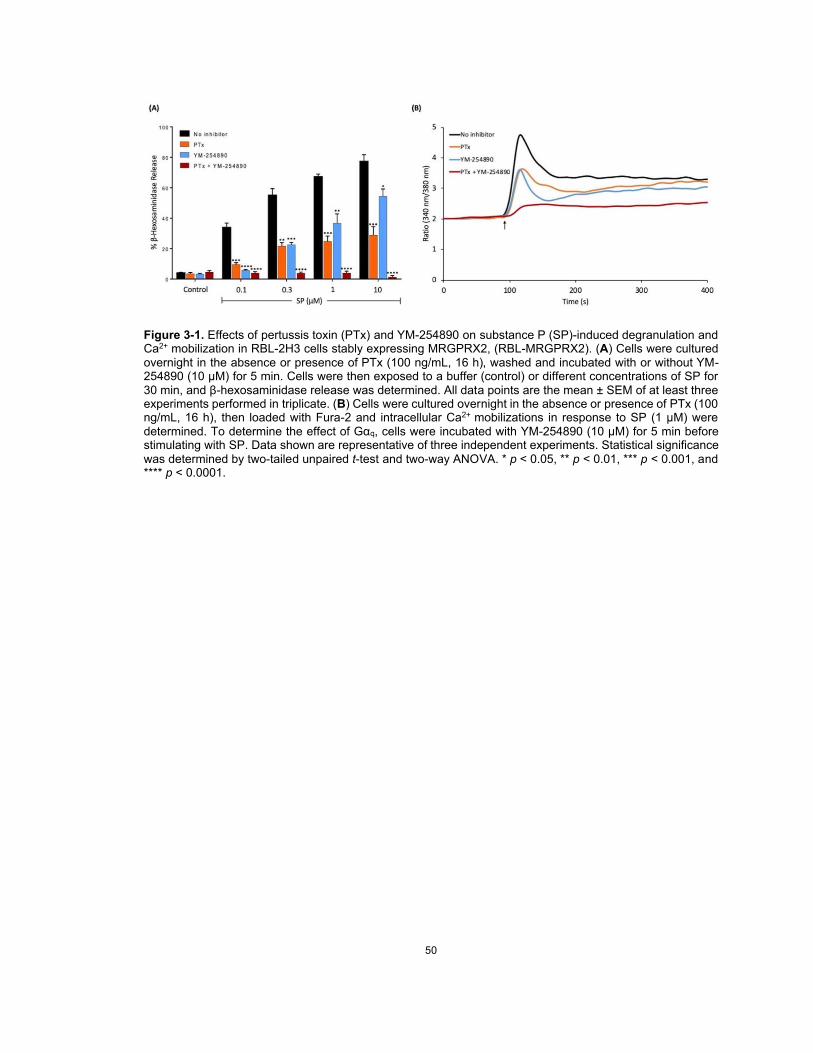
50
Figure 3-1. Effects of pertussis toxin (PTx) and YM-254890 on substance P (SP)-induced degranulation and Ca2+ mobilization in RBL-2H3 cells stably expressing MRGPRX2, (RBL-MRGPRX2). (A) Cells were cultured overnight in the absence or presence of PTx (100 ng/mL, 16 h), washed and incubated with or without YM-254890 (10 μM) for 5 min. Cells were then exposed to a buffer (control) or different concentrations of SP for 30 min, and β-hexosaminidase release was determined. All data points are the mean ± SEM of at least three experiments performed in triplicate. (B) Cells were cultured overnight in the absence or presence of PTx (100 ng/mL, 16 h), then loaded with Fura-2 and intracellular Ca2+ mobilizations in response to SP (1 μM) were determined. To determine the effect of Gαq, cells were incubated with YM-254890 (10 μM) for 5 min before stimulating with SP. Data shown are representative of three independent experiments. Statistical significance was determined by two-tailed unpaired t-test and two-way ANOVA. * p < 0.05, ** p < 0.01, *** p < 0.001, and **** p < 0.0001.

51
Figure 3-2. Effects of mutations at MRGPRX2’s highly conserved positions within transmembrane domains (V123A, I225A, and Y279A) on cell surface expression, SP-induced Ca2+ mobilization, and degranulation in transiently transfected RBL-2H3 cells. (A) Snake diagram of secondary structure of MRGPRX2. Each circle represents amino acid residue with one letter code. Solid red, yellow, and blue backgrounds denote the residues at positions 3x46 (V123), 6x37 (I225), and 7x53 (Y279), respectively. (B) amino acid change for each MRGPRX2 mutant. (C) RBL-2H3 cells transiently expressing wild-type (WT)-MRGPRX2 and its mutants were incubated with phycoerythrin (PE)-anti-MRGPRX2 antibody and cell surface receptor expression was determined by flow cytometry. Representative histograms for WT/mutant (black line) and control untransfected cells (blue line) are shown. (D) cells expressing WT-MRGPRX2 and its mutants were loaded with Fura-2 and intracellular Ca2+ mobilization in response to SP (1 μM) was determined. Data shown are representative of three independent experiments. (E) cells were exposed to a buffer (control) or SP (1 μM) for 30 min, and β-hexosaminidase release was determined. All data points are the mean ± SEM of at least three experiments performed in triplicate. Statistical significance was determined by a two-tailed unpaired t-test. *** p < 0.001 and **** p < 0.0001.

52
Figure 3-3. Effects of naturally occurring MRGPRX2 variants at the receptor’s conserved transmembrane domains (V123F, T224A, and V282M) on SP-induced responses in transiently transfected RBL-2H3 cells. (A) Snake diagram of secondary structure of MRGPRX2. Each circle represents amino acid residue with one letter code. Solid red, yellow, and blue backgrounds denote the naturally occurring MRGPRX2 variants V123F, T224A, and V282M, respectively. (B) amino acid change for each MRGPRX2 variant with allele frequency. (C) cell surface expression of WT-MRGPRX2 and its variants was determined by flow cytometry using PE-anti-MRGPRX2 antibody. Representative histograms for WT/variant (black line) and control untransfected cells (blue line) are shown. (D) cells expressing WT-MRGPRX2 and its variants were loaded with Fura-2 and intracellular Ca2+ mobilization in response to SP (1 μM) was determined. Data shown are representative of three independent experiments. (E) cells were exposed to a buffer (control) or SP (1 μM) for 30 min, and β-hexosaminidase release was determined. All data points are the mean ± SEM of at least three experiments performed in triplicate. Statistical significance was determined by a two-tailed unpaired t test. **** p < 0.0001.

53
Figure 3-4. Effects of naturally occurring MRGPRX2 variants at the receptor’s intracellular loops (Y137H, R138C, R140C, and R141C) on SP-induced responses in transiently transfected RBL-2H3 cells. (A) Snake diagram of secondary structure of MRGPRX2. Each circle represents amino acid residue with one letter code. Solid yellow, red, purple, and pink backgrounds denote the naturally occurring MRGPRX2 variants. (B) amino acid change for each MRGPRX2 variant with allele frequency. (C) cell surface expression of WT-MRGPRX2 and its variants was determined by flow cytometry using PE-anti MRGPRX2 antibody. Representative histograms for WT/variant (black line) and control untransfected cells (blue line) are shown. (D) cells expressing WT-MRGPRX2 and its variants were loaded with Fura-2 and intracellular Ca2+ mobilization in response to SP (1 μM) was determined. Data shown are representative of three independent experiments. (E) cells were exposed to a buffer (control) or SP (1 μM) for 30 min, and β-hexosaminidase release was determined. All data points are the mean ± SEM of at least three experiments performed in triplicate. Statistical significance was determined by a two-tailed unpaired t-test. **** p < 0.0001.

54
Figure 3-5. Effects of Ser/Thr residues on MRGPRX2’s carboxyl-terminus on cell surface expression, SP-induced Ca2+ mobilization, and degranulation in transiently transfected RBL-2H3 cells. (A) Snake diagram of secondary structure of MRGPRX2. Each circle represents amino acid residue with one letter code. Solid red backgrounds denote Ser/Thr residues. (B) schematic representation of the carboxyl-terminus of MRGPRX2 (WT) and a phosphorylation-deficient mutant in which all Ser/Thr were replaced with Ala ('ST-MRGPRX2). (C) cell surface expression of WT and 'ST-MRGPRX2 was determined by flow cytometry using PE-anti-MRGPRX2 antibody. Representative histograms for WT-MRGPRX2 (black line), 'ST-MRGPRX2 (red line), and control untransfected cells (blue line) are shown. (D) cells expressing WT and 'ST-MRGPRX2 were loaded with Fura-2 and intracellular Ca2+ mobilization in response to SP (1 μM) was determined. Data shown are representative of three independent experiments. (E) cells were exposed to a buffer (control) or SP (1 μM) for 30 min, and β-hexosaminidase release was determined. All data points are the mean ± SEM of at least three experiments performed in triplicate. Statistical significance was determined by a two-tailed unpaired t-test. * p < 0.05.

55
Figure 3-6. Effects of naturally occurring MRGPRX2 variants within the receptor’s carboxyl-terminus (Q305R, D311H, S325L, and L329Q) on SP-induced responses in transiently transfected RBL-2H3 cells. (A) Snake diagram of secondary structure of MRGPRX2. Each circle represents amino acid residue with one letter code. Solid purple, orange, pink, and red backgrounds denote the naturally occurring missense variants Q305R, D311H, S325L, and L329Q, respectively. (B) amino acid change for each MRGPRX2 variant with allele frequency. (C) cell surface expression of WT-MRGPRX2 and its variants was determined by flow cytometry using PE-anti MRGPRX2 antibody. Representative histograms for WT/variant (black line) and control untransfected cells (blue line) are shown. (D) cells expressing WT-MRGPRX2 and its variants were loaded with Fura-2 and intracellular Ca2+ mobilization in response to SP (1 μM) was determined. Data shown are representative of three independent experiments. (E) cells were exposed to buffer (control) or SP (1 μM) for 30 min, and β-hexosaminidase release was determined. All data points are the mean ± SEM of at least three experiments performed in triplicate. Statistical significance was determined by a two-tailed unpaired t-test. * p < 0.05 and ** p < 0.01.

56
CHAPTER 4: Substance P serves as a balanced agonist for
MRGPRX2 and a single tyrosine residue is required for β-
arrestin recruitment and receptor internalization
Chalatip Chompunud Na Ayudhya1,2, Aetas Amponnawarat1,3, Hydar Ali1,*
1 Department of Basic and Translational Sciences, University of Pennsylvania, School of Dental
Medicine, Philadelphia, PA-19104, USA
2 Department of Oral Diagnosis, Faculty of Dentistry, Naresuan University, Phitsanulok 65000,
Thailand
3 Department of Family and Community Dentistry, Faculty of Dentistry, Chiang Mai University
50200, Chiang Mai, Thailand
* Correspondence: [email protected].; Tel.: +1-215-573-1993
This chapter has been originally published in a special issue “Mast Cells in Human Health and
Diseases” of the International Journal of Molecular Sciences, 2021.
Chompunud Na Ayudhya C, Amponnawarat A, Ali H. Substance P Serves as a Balanced Agonist for MRGPRX2 and a Single Tyrosine Residue Is Required for β-Arrestin Recruitment and Receptor Internalization. Int J Mol Sci. 2021; 22(10):5318. https://doi.org/10.3390/ijms22105318.

57
4.1. Abstract
The neuropeptide substance P (SP) mediates neurogenic inflammation, pain and contributes to
atopic dermatitis in mice through the activation of mast cells (MCs) via Mas-related G protein-
coupled receptor (GPCR)-B2 (MrgprB2, human ortholog MRGPRX2). In addition to G proteins,
certain MRGPRX2 agonists activate an additional signaling pathway that involves the recruitment
of E-arrestins, which contributes to receptor internalization and desensitization (balanced agonists).
We found that SP caused β-arrestin recruitment, MRGPRX2 internalization and desensitization.
These responses were independent of G proteins, indicating that SP serves as a balanced agonist
for MRGPRX2. A tyrosine residue in the highly conserved NPxxY motif contributes to the activation
and internalization of many GPCRs. We have previously shown that Tyr279 of MRGPRX2 is
essential for G protein-mediated signaling and degranulation. To assess its role in β-arrestin-
mediated MRGPRX2 regulation, we replaced Tyr279 in the NPxxY motif of MRGPRX2 with Ala
(Y279A). Surprisingly, we found that unlike the wild-type receptor, Y279A mutant of MRGPRX2
was resistant to SP-induced β-arrestin recruitment and internalization. This study reveals the novel
findings that activation of MRGPRX2 by SP is regulated by β-arrestins and that a highly conserved
tyrosine residue within MRGPRX2’s NPxxY motif contributes to both G protein- and β-arrestin-
mediated responses.
Keywords: Mast cells; MRGPRX2; MrgprB2; Tyrosine; Substance P; Signaling; Internalization

58
4.2. Introduction
Mast cells (MCs) are key sentinel immune cells that are often found in close proximity to
sensory nerve endings in various tissues, including the skin, gastrointestinal mucosa, and
respiratory tract (3). The role of MC-neuron interaction as a regulatory unit in both physiology and
disease has recently gained prominence (13, 15, 62, 131-133). Substance P (SP) has long been
established as an inflammatory neuropeptide and is known to activate a variety of cell types via the
neurokinin-1 receptor (NK-1R). Mouse bone marrow-derived MCs (BMMCs) express NK-1R and
its expression level is upregulated upon FcεRI stimulation (134). However, emerging evidence
suggests that SP contributes to neurogenic inflammation and pain in mice through MC activation
via a novel G protein-coupled receptor (GPCR) known as Mas-related GPCR-B2 (MrgprB2; human
ortholog MRGPRX2) (13, 15). Activation of MrgprB2 by SP is required for regulating inflammatory
hyperalgesia via the release of pro-inflammatory cytokines and chemokines, and the recruitment
of immune cells at the injury site, which facilitates inflammatory responses and peripheral
sensitization (13). Furthermore, Serhan et al. recently showed that SP released from nociceptors
activates murine skin MCs and contributes to the development of atopic dermatitis, a type 2 allergic
skin disease, through the activation of MrgprB2 (15). These findings challenge our current
understanding of SP-mediated neuroinflammatory diseases and pain, and raise an interesting
possibility that targeting MRGPRX2 might represent a promising therapeutic approach for the
management of these neuroinflammatory associated conditions in humans. However, the
mechanisms involved in the activation and regulation of MRGPRX2 by SP has yet to be fully
elucidated.
As a member of class A GPCR family, MRGPRX2 shares a common structure of seven
transmembrane (TM) α-helices. The extracellular part is responsible for ligand binding, whereas
the intracellular part is involved in binding downstream effectors such as heterotrimeric G proteins
(75). Besides G protein-mediated signaling, most GPCRs signal via an additional pathway that
involves the recruitment of adapter proteins known as β-arrestins (83, 84). The recruitment of β-
arrestins results in uncoupling of the receptor from G proteins and termination of receptor activation
(desensitization) (83, 135). They further target the receptor internalization/endocytosis, which

59
downregulates signaling as the receptor is physically removed from the cell surface (86, 136, 137).
In addition to receptor desensitization and internalization, it is now established that β-arrestins also
play important roles in various G protein-independent downstream signaling to promote chemotaxis
and to modulate inflammation (86-88). Thus, β-arrestins regulate nearly all aspects of receptor
activity, including desensitization, downregulation, trafficking and signaling.
While most GPCR agonists target signaling pathways mediated by both G proteins and β-
arrestins (“balanced agonists”), some agonists preferentially activate only particular pathway
(“biased agonists”) (80, 138). GPCR agonists that preferentially activate G proteins are known as
G-protein-biased and those activate β-arrestins are known as β-arrestin-biased agonists. For
MRGPRX2, compound 48/80 and codeine activate both G proteins and β-arrestins (balanced
agonists) and can cause receptor internalization, which is associated with functional desensitization
(120, 139). On the contrary, host defense peptides, such as cathelicidin LL-37 and angiogenic
peptide AG-30/5C, activate only G proteins but not β-arrestins (G protein-biased), and do not
induce receptor desensitization and internalization (46, 120, 140). Biased signaling has gained
important therapeutic implications in several GPCRs (80). However, whether SP acts as a balanced
or biased agonist for MRGPRX2 remains unknown.
Closely related GPCRs exhibit high degree of conserved sequence motifs and a common
activation pathway, especially in the regions implicated in ligand binding and G protein coupling
(78). Recent computer-based structural modeling, sequence analysis and mutagenesis studies
have led to the identification of residues in MRGPRX2 that are responsible for ligand binding and
G protein coupling (51, 103, 104, 109). One of the most conserved GPCR sequences is the NPxxY
motif located in TM7. A tyrosine residue Tyr7x53 in this motif is pivotal for receptor activation for all
class A GPCRs (79). Structural modeling studies in β2 adrenergic receptor, rhodopsin and M2
muscarinic acetylcholine receptors revealed that upon ligand binding, Tyr7x53 undergoes substantial
rotamer conformations and provides activation switch through formation of a water-mediated
hydrogen bond (141, 142). This favors an outward movement in the cytoplasmic end of TM6 and
allows the receptor coupling to G proteins and other signal transducers, which represents a
hallmark of GPCR activation. Besides its importance in receptor activation, Tyr7x53 is suggested to

60
regulate agonist-induced receptor internalization in many GPCRs (143-146). In MRGPRX2, the
corresponding tyrosine residue is Tyr279. Our previous study has shown that this tyrosine residue
is required for G protein-mediated MRGPRX2 activation in response to SP (104). However, the role
of this tyrosine residue on β-arrestin-mediated MRGPRX2 signaling has not been determined.
The goals of the current study were to determine whether SP serves as a balanced or
biased agonist for MRGPRX2 and to investigate the potential effects of MRGPRX2’s Tyr7x53 (Tyr279)
residue on β-arrestin recruitment and receptor internalization. The data presented in herein suggest
that SP serves as a balanced agonist for MRGPRX2 and that Tyr7x53 contributes to both G protein-
dependent signaling for degranulation and G-protein independent signaling for β-arrestin
recruitment and receptor internalization.
4.3. Materials and Methods
4.3.1. Materials
All cell culture and Lipofectamine 2000 transfection reagents were obtained from Invitrogen
(Gaithersburg, MD). Amaxa Nucleofector kit (Kit V) was purchased from Lonza (Gaithersburg, MD).
Phycoerythrin (PE)-conjugated (Cat.#359004) and purified unconjugated anti-MRGPRX2
antibodies (Cat.#359002) were from BioLegend (San Diego, CA). Donkey anti-mouse Alexa Fluor
488 (Cat.#A21202) and 647 (Cat.#A31571) conjugated IgG secondary antibodies were from
Invitrogen (Gaithersburg, MD). p-nitrophenyl-N-acetyl-β-D-glucosamine (PNAG) was from Sigma-
Aldrich (St. Louis, MO). Fura-2 acetoxymethyl ester was from Abcam (Cambridge, MA). SP was
purchased from AnaSpec (Fremont, CA). Pertussis toxin (PTx) was from List Biological
Laboratories (Campbell, CA). Plasmid encoding hemagglutinin (HA)-tagged human MRGPRX2 in
pReceiver-MO6 vector was obtained from GeneCopoeia (Rockville, MD). MRGPRX2 Y279A
mutant in HA-tagged plasmid was reported previously (104). MRGPRX2-Tango plasmid (Addgene
no. 66440) was a gift from Dr. Bryan Roth. MRGPRX2 Y279A mutant in Tango plasmid was
generated by Penn Genomics Analysis Core (Philadelphia, PA).
61
4.3.2. Mice
C57BL/6 (wild-type; WT) and β-arrestin2 knockout (βArr2−/−) mice were obtained from the
Jackson Laboratory (Bar Harbor, ME). Mice were housed in pathogen-free cages on autoclaved
hardwood bedding. Eight-to-twelve-week-old male and female mice were used in this study. All
experiments were approved by the Institutional Animal Care and Use Committee of the University
of Pennsylvania.
4.3.3. Cell culture
Rat basophilic leukemia (RBL-2H3) cells were maintained as monolayer cultures in
Dulbecco’s modified Eagle’s medium (DMEM) supplemented with 10% FBS, L-glutamine (2 mM),
penicillin (100 IU/mL) and streptomycin (100 µg/mL) at 37°C with 5% CO2 (98). RBL-2H3 cells
stably expressing MRGPRX2 (RBL-MRGPRX2) were used and maintained similarly in the
presence of G-418 (1 mg/mL) (46, 99).
HTLA (engineered HEK-293T cells stably expressing a E-arrestin2–tobacco etch virus
fusion gene) cells and HTLA cells stably expressing MRGPRX2-Tango (HTLA-MRGPRX2) were
maintained in DMEM supplemented with 10% FBS, L-glutamine (2 mM), penicillin (100 IU/ml),
streptomycin (100 µg/ml), hygromycin B (200 µg/ml), puromycin (5 mg/ml), and G-418 (500 µg/ml)
(51, 147).
Peritoneal MCs (PMCs) were purified from WT and βArr2−/− mice as described previously
(92). Briefly, the peritoneal cavity was lavaged with 10 mL of HBSS supplemented with 3% FCS
and 10 mM HEPES. The cells were cultured in Iscove’s Modified Dulbecco’s Medium (IMDM)
supplemented with 10% FCS, murine IL-3 (10 ng/mL), and murine SCF (30 ng/mL). After 48 h,
non-adherent cells were removed and adherent cells were cultured in fresh medium for an
additional 10-14 days. Suspension cells were then determined for MC receptor expression and
function, and were used for experiments as PMCs.

62
4.3.4. Generation of cells transiently expressing WT MRGPRX2 and its variant
RBL-2H3 cells (2 x 10⁶) were transiently transfected with 2 µg of HA-tagged plasmid using
the Amaxa Nucleofector Device and Amaxa Kit V according to the manufacturer’s protocol. Cells
were used within 16 – 20 h after transfection (103).
For HTLA cells transiently expressing WT MRGPRX2 or its missense variant, cells (1 x 10⁶
cells per well) were plated in a 6-well plate in antibiotic-free medium (DMEM supplemented with
10% FBS and L-glutamine) and incubated overnight at 37oC with 5% CO2. The following day, cells
were transfected with 2 µg of MRGPRX2 or its missense variants in Tango plasmids using the
Lipofectamine 2000 DNA transfection reagent according to the manufacturer’s protocol. Cells were
incubated overnight at 37oC with 5% CO2 in antibiotic-free medium and were used within 16 – 48 h
after transfection (51).
4.3.5. Receptor expression and internalization using flow cytometry
Cells expressing either WT MRGPRX2 or its mutants (5 x 10⁵) were stimulated with either
SP or buffer for indicated time at 37oC. Cells were washed twice with ice-cold FACS buffer (PBS
containing 2 % FCS and 0.02% sodium azide) and incubated with PE-conjugated anti-MRGPRX2
antibody for 30 min at 4oC in the dark. Cells were then washed with FACS buffer and fixed in 1.5%
paraformaldehyde. Cells were acquired using a BD LSR II flow cytometer (San Jose, CA) and
MRGPRX2 expression were analyzed using WinList software, version 8.
4.3.6. Degranulation
The degranulation was measured by E-hexosaminidase release as described previously
(98). Briefly, RBL-2H3 cells (5 x 10⁴ cells) or PMCs (1 x 10⁴ cells) were seeded into a 96-well, white,
clear-bottom cell culture plate and incubated overnight in a 37°C incubator with 5% CO2. Cells were
then washed twice and suspended in a total volume of 50 Pl HEPES buffer containing 0.1% bovine
serum albumin (BSA). Experimental groups were stimulated with SP for 30 min at 37°C. Cells
without treatment were designated as controls. To determine the total E-hexosaminidase release,
unstimulated cells were lysed in 50 Pl of 0.1% Triton X-100. Aliquots (20 Pl) of supernatants or cell

63
lysates were incubated with 20 Pl of 1 mM p-nitrophenyl-N-acetyl-β-D-glucosamine (PNAG) for 1 h
at 37°C. The reaction was stopped by adding 250 Pl of stop buffer (0.1 M Na2CO3/0.1 M NaHCO3).
The β-hexosaminidase release was assessed by measuring absorbance at 405 nm using
Versamax microplate spectrophotometer (San Jose, CA).
4.3.7. Transcriptional activation following arrestin translocation (TANGO) assay
HTLA cells expressing either WT MRGPRX2 or its variants (5 x 10⁴ cells per well) were
plated into a 96-well plate in triplicates in a total volume of 160 Pl antibiotic-free medium and
incubated for 6 h at 37oC to allow attachment. After 6 h, the medium was aspirated, and cells were
incubated with MRGPRX2 ligands in 160 Pl antibiotic-free medium for additional 16 h at 37oC. The
medium and ligands were then aspirated and 100 Pl of Bright-Glo solution (Promega) was added
to each well. Relative luminescence unit was measured in a Thermo Labsystems Luminoskan
Ascent 392 Microplate Luminometer (51, 120).
4.3.8. β-arrestin translocation by live imaging confocal microscopy
RBL-2H3 cells were co-transfected with green fluorescent protein-tagged β-arrestin2
plasmid (βArr2-GFP) and WT-MRGPRX2 or its mutants with a ratio of 3:1 for β-arrestin2:receptor
using Amaxa Kit V as described above. Transfected cells were plated onto a 35-mm glass bottom
dish. Cell surface receptor was determined by incubating with purified anti-MRGPRX2 antibody,
followed by Alexa Fluor 647-conjugated secondary antibody. Cells were then stimulated with SP
and live images of β-arrestin2 translocation as indicated by green fluorescence images were
collected using Nikon A1R confocal microscope.
4.3.9. Receptor trafficking using immunofluorescence microscopy
Receptor trafficking after SP stimulation was modified from previously described antibody
feeding assay (148). RBL-2H3 cells expressing either MRGPRX2 or its variants were plated onto
sterilized glass coverslips (2x10⁵ cells/12-mm diameter coverslip in 24-well plate) and incubated
overnight at 37oC with 5% CO2 to allow attachment. Cells were rinsed with PBS and blocked with
blocking buffer (PBS with 2% BSA) for 30 min at room temperature. Primary antibody incubation

64
was performed using purified anti-MRGPRX2 antibody (1:250 dilution) for 1 h at 4°C to label the
cell surface expressed receptors. Cells were then stimulated with SP for 30 min at 37oC to allow
internalization, followed by fixed with 4% paraformaldehyde for 15 min at 4°C. Labeled surface
receptors were detected by incubating with saturated Alexa Fluor 647-conjugated secondary
antibody (red) for 1 h at 4°C. Cells were then permeabilized by using 0.2% Triton X-100 in blocking
buffer for 30 min and internalized receptors were detected by incubating with Alexa Fluor 488-
conjugated secondary antibody (green) for 30 min at 4°C. Then, cells were mounted onto the glass
slides using ProLong Gold Antifade mounting medium (Invitrogen) and images were visualized
using a Nikon Eclipse Ni microscope.
4.3.10. Statistical analysis
Data shown are mean r standard error of the mean (SEM) values derived from at least
three independent experiments. GraphPad Prism scientific software version 6.07 was used for
statistical analysis. Statistical significance was determined using unpaired two-tailed t-test and one-
or two-way ANOVA. Differences were considered as statistically significant at a value * p < 0.05, **
p < 0.01, *** p < 0.001, and **** p < 0.0001.
4.4. Results
4.4.1. SP is a balanced agonist for MRGPRX2.
For many GPCRs, biased signaling has important therapeutic implications as it can elicit
distinct physiological responses (80, 138, 149). Certain MRGPRX2 ligands, including compound
48/80, codeine and HDP AG-30/5C, have recently been identified as either balanced or G-protein-
biased agonists (120, 139). SP is a well-known MRGPRX2 agonist; however, whether SP is a
balanced or biased agonist for MRGPRX2 has yet to be investigated. We have previously shown
that SP induces Ca2+ mobilization and degranulation in a G protein-dependent manner (104). Thus,
we first sought to determine if SP can also trigger β-arrestin recruitment. For this, we utilized an
assay known as transcriptional activation following arrestin translocation (TANGO) using HTLA
cells (engineered HEK-293T cells stably expressing a β-arrestin2–tobacco etch virus fusion gene)
stably expressing human MRGPRX2 (HTLA-MRGPRX2) (51, 120). Cells were exposed to either

65
buffer (control) or SP at different concentrations for 16 h and β-arrestin-mediated gene expression
(indicative of β-arrestin recruitment) was measured. We found that SP (30 PM and 100 PM)
significantly induced β-arrestin-mediated gene expression (Figure 4-1;A).
For most GPCRs, the recruitment of β-arrestins is involved in receptor internalization and
desensitization. Compound 48/80 and codeine trigger robust β-arrestin recruitment, MRGPRX2
internalization and inhibition of degranulation in response to subsequent stimulation by the same
ligand (desensitization) (120, 139). Given that SP induced β-arrestin recruitment, we hypothesized
that it could also cause receptor internalization and desensitization. Rat basophilic leukemia (RBL-
2H3) cell line, a commonly used model for MC activation, does not endogenously express Mrgpr
receptors (46, 150). We therefore utilized RBL-2H3 cells stably expressing human MRGPRX2
(RBL-MRGPRX2) to determine the effects of SP on MRGPRX2 internalization and desensitization.
For receptor internalization, RBL-MRGPRX2 cells were stimulated with different concentrations of
SP for different time-points and incubated with PE-conjugated anti-MRGPRX2 antibody to
determine cell surface receptor expression by flow cytometry. Consistent with β-arrestin
recruitment, we found that SP triggered MRGPRX2 internalization and that this response was dose-
dependent (Figure 4-1;B). Furthermore, preincubation of RBL-MRGPRX2 with 30 PM of SP
overnight resulted in nearly complete inhibition of degranulation on second stimulation by the same
ligand (Figure 4-1;C). Taken together, these studies suggest that SP serves as a balanced agonist
for MRGPRX2.
4.4.2. SP-induced β-arrestin recruitment and MRGPRX2 internalization are G protein-
independent.
SP has been shown to induce MRGPRX2-mediated Ca2+ mobilization and MC
degranulation in a G protein-dependent manner (104). We therefore asked if β-arrestin recruitment
and MRGPRX2 internalization in response to SP are also mediated in a G protein-dependent
manner. We utilized a G protein inhibitor, pertussis toxin (PTx), and confirmed that pretreatment of
RBL-MRGPRX2 cells with PTx (100 ng/ml, 16 h) attenuated degranulation in response to SP
(Figure 4-2;A). By contrast, PTx had no effect on SP-induced β-arrestin-mediated gene expression

66
and MRGPRX2 internalization (Figure 4-2;B,C). These findings indicate that β-arrestin recruitment
and MRGPRX2 internalization in response to SP are mediated independently of G proteins.
4.4.3. β-arrestin2 regulates SP/MrgprB2-mediated MC degranulation.
MrgprB2 has been identified as the mouse ortholog of human MRGPRX2 [38]. Previous
studies showed that SP activates murine MCs to cause degranulation and inflammatory responses
via MrgprB2 [3,38]. Of note, there are significant differences in agonist affinities between mouse
MrgprB2 and human MRGPRX2 receptors. While SP activates MRGPRX2 with an EC50 of 152
nM, it activates MrgprB2 with a higher EC50 value of 54 µM [38]. To investigate the biological role
of β-arrestin2 on SP/MrgprB2-mediated MC responses, we utilized peritoneal MCs (PMCs)
obtained from wild-type (WT) and β-arrestin2 knockout (βArr2−/−) mice [39]. Absence of β-arrestin2
had no effect on the development and maturation of MCs as shown by similar levels of cell surface
FcεRI and c-Kit expression (Figure 4-3;A). However, degranulation in response to SP was
significantly enhanced in PMCs generated from βArr2−/− mice when compared to cells obtained
from WT mice (Figure 4-3;B). These findings suggest that β-arrestin2 expressed in MCs
contributes to MrgprB2 desensitization in response to SP.
4.4.4. Mutation of a highly conserved tyrosine residue of MRGPRX2 (Y279A) abolishes SP-
induced β-arrestin recruitment.
The highly conserved NPxxY motif is important for GPCR activation and regulation (78,
79). Consistent with the findings in other GPCRs (79, 151), the tyrosine residue located in the
NPxxY motif of MRGPRX2 (Tyr279) has been previously shown to be essential for both SP-induced
Ca2+ mobilization and degranulation (104). We sought to determine whether this residue also
mediates β-arrestin signaling. Since we demonstrated that SP induced β-arrestin pathway
independently of G proteins, our initial hypothesis was that it might not contribute to the β-arrestin
signaling. To assess this, we first constructed Y279A mutant in the MRGPRX2-Tango plasmid and
generated transient transfectants in HTLA cells. We found that Y279A mutant showed similar level
of cell surface expression as the WT receptor (Figure 4-4;A). Surprisingly, however, Y279A

67
mutation resulted in complete loss of β-arrestin-mediated gene expression in response to SP
(Figure 4-4;B).
One limitation of TANGO assay is that it measures β-arrestin-mediated gene expression
after an overnight stimulation with agonist, thus not reflecting the typical β-arrestin recruitment
which occurs rapidly within minutes (152). Therefore, we utilized a green fluorescent protein-tagged
β-arrestin2 plasmid (βArr2-GFP) to detect changes in SP-induced β-arrestin recruitment by
confocal microscopy. βArr2-GFP were co-expressed transiently with either WT-MRGPRX2 or
Y279A mutant in RBL-2H3 cells. As shown in Figure 4-4;C, SP caused a rapid translocation (within
a minute) of βArr2-GFP from cytoplasm to membrane in RBL-2H3 cells expressing the WT
receptor. By contrast, this response was not observed in cells expressing Y279A mutant. These
findings indicate that mutation of a highly conserved tyrosine residue of MRGPRX2 is required for
SP-mediated β-arrestin recruitment.
4.4.5. Tyrosine residue in MRGPRX2 (Y279) is required for SP-mediated receptor
internalization.
We next examined the effect of Y279A mutation on SP-induced MRGPRX2 internalization.
HTLA cells expressing the WT-MRGPRX2 exhibited reduced cell surface receptor expression
following SP stimulation as determined by flow cytometry, whereas SP was unable to trigger
receptor internalization in cells expressing Y279A mutant (Figure 4-5;A,B).
We next performed immunofluorescence study to confirm and visualize the MRGPRX2
receptor internalization in RBL-2H3 cells. The differential labeling of cell-surface and internalized
receptors technique was used (148). For this, MRGPRX2 expressed on cell surface were labeled
with 1o antibody (unconjugated anti-MRGPRX2 antibody). Then, the receptor internalization was
initiated by stimulating with SP for 30 min. The remaining surface receptors were labeled with Alexa
Fluor 647-conjugated 2o antibody (red). Cells were then permeabilized and the internalized
receptors were labeled with Alexa Fluor 488-conjugated 2o antibody (green) (Figure 4-6;A). We
found that following SP stimulation, cell surface expression of WT-MRGPRX2 was significantly
reduced and displayed punctate pattern, while there was an increase number of internalized

68
receptor, as indicated by an increased green signal. On the contrary, cells expressing Y279A
mutant did not display receptor internalization and the expression of cell surface remained
unchanged (Figure 4-6;B). Taken together, these findings indicate that the conserved tyrosine
residue in NPxxY motif of MRGPRX2 plays an important role on the receptor regulation and
internalization upon stimulation by SP.
4.5. Discussion
Activation of MCs by SP has been implicated in the pathogenesis of neurogenic
inflammation, pain, and itch via MRGPRX2 and its mouse counterpart MrgprB2 (13, 15, 153). In
addition to G proteins, most GPCRs have been shown to interact with β-arrestins for receptor
desensitization and internalization. MRGPRX2 can undergo β-arrestin-mediated internalization in
response to some, but not all agonists. For example, compound 48/80 and codeine act as balanced
agonists for MRGPRX2 that activate both G proteins and β-arrestins, and thus can cause receptor
internalization, which is associated with functional receptor desensitization (120, 139). On the
contrary, host defense peptides (HDPs), such as cathelicidin LL-37 and angiogenic peptide AG-
30/5C, activate only G proteins, but not β-arrestins (G protein-biased), and do not induce receptor
desensitization and internalization (46, 120, 140). Biased signaling has gained important
therapeutic implications for several GPCRs (80). However, whether SP is balanced or biased
agonist for MRGPRX2 remains unknown. In this study, we demonstrated that SP serves as a
balanced agonist for MRGPRX2 and that Tyr7x53 both contributes to G protein-dependent signaling
for degranulation and promotes β-arrestin recruitment and MRGPRX2 internalization. Furthermore,
β-arrestin2 negatively regulates MrgprB2-mediated MC degranulation in response to SP. Thus,
targeting Tyr7x53 and β-arrestin2 could provide novel therapeutic modalities for modulating
SP/MRGPRX2-mediated inflammatory diseases.
Based on the analysis of common activation pathway in 234 structures from 45 class A
GPCRs, several conserved residues and key motifs have been identified that are involved in
receptor activation and regulation (78). Of these, the NPxxY sequence at the cytoplasmic end of
the TM7 domain is one of the most highly conserved motifs among class A GPCRs. A study by

69
Venkatakrishnan et al. suggested that Tyr7x53 in the NPxxY motif is essential for class A receptor
activation and regulation (79). We previously demonstrated that substitution of the corresponding
tyrosine residue (Tyr279) in MRGPRX2 to Ala (Y279A) diminished SP-induced Ca2+ mobilization
and degranulation responses (104). Besides G protein signaling, mutations in the NPxxY motif of
D1B-adrenergic and β2-adrenergic receptors have been associated with diminished agonist-
mediated β-arrestin recruitment (154). Here, we found that β-arrestin recruitment following SP
stimulation was abolished in Y279A mutant when compared with the WT receptor, indicating that
this tyrosine residue is also important for β-arrestin-mediated signaling via MRGPRX2.
The NPxxY motif has also been suggested as a common endocytic motif for GPCRs. The
role of the tyrosine residue in the highly conserved NPxxY motif in receptor endocytosis and
regulation has been established in certain GPCRs, including β2-adrenergic receptor (143), N-formyl
peptide receptor (145), and NK-1R (146). For example, mutation of the corresponding tyrosine
residue of the β2-adrenergic receptor to alanine (Y326A) abolishes agonist-induced receptor
phosphorylation, internalization, and desensitization (143, 155). Similarly, formyl peptide receptor
mutation Y301A results in a complete loss of agonist-induced receptor internalization (145).
However, while this conserved tyrosine residue appears to be essential for agonist-induced
receptor internalization of some GPCRs, it is not required for the internalization of angiotensin II
receptor (156, 157) or gastrin-releasing peptide receptor (158). Thus, it is possible that the role of
a highly conserved tyrosine residue in the NPxxY motif on agonist-induced receptor internalization
is receptor specific. Here, we found that MRGPRX2 Y279A mutation impaired receptor
internalization in response to SP and caused retention of receptors at the cell surface. These
findings suggest that the tyrosine residue in the NPxxY motif of MRGPRX2 is important for SP-
induced receptor internalization. It is uncertain whether Tyr279 affects MRGPRX2 phosphorylation,
and thus contributes to receptor internalization. It will be important to further investigate the effect
of this tyrosine residue and NPxxY motif on the receptor phosphorylation. Furthermore, the
mechanistic pathway responsible for MRGPRX2 internalization should be further identified.

70
It is worth noting that, in this study, we mostly utilized HTLA and RBL-2H3 cell lines
transfected with human MRGPRX2 to investigate the effects of SP on MRGPRX2 regulation. While
recent evidence demonstrates that ectopically expressed MRGPRX2 retains its authentic
mechanisms for MC activation and degranulation as of human MCs endogenously expressed
MRGPRX2 (150), further studies in human-derived MCs are warranted to confirm these findings.
In summary, this study extends our previous observations on the pivotal role of tyrosine
residue Tyr279 in the NPxxY motif of MRGPRX2 on G protein activation (104). The data presented
herein demonstrate the novel finding that this single Tyr279 residue contributes to both G protein
and β-arrestin signaling in response to SP. Substitution of Tyr279 to alanine (Y279A) abolishes SP-
induced MC degranulation, β-arrestin recruitment, and MRGPRX2 internalization. This tyrosine
residue may contribute to the regulation of MRGPRX2 activation and internalization, presumably
by maintaining receptor conformation, thus controlling G protein coupling and activation as well as
the internalization process. These findings have an important clinical implication as individuals
harboring this mutation may become resistant to developing neurogenic inflammation and
inflammatory diseases.
4.6. Acknowledgements
We thank the FACS and confocal microscopy core facilities of the School of Dental
Medicine, University of Pennsylvania for data acquisition and analysis. We also thank the Penn
Genomics Analysis Core, University of Pennsylvania for generating MRGPRX2 Y279A mutant in
Tango plasmid. Moreover, we would like to thank Dr. Saptarshi Roy for generating HTLA-
MRGPRX2 cells used in this study. This work was funded by National Institutes of Health grants
R01-AI124182, R01-AI143185 and R01-AI149487 to Hydar Ali.

71
Figure 4-1. Substance P (SP) is a balanced agonist for MRGPRX2. (A) HTLA cells stably expressing MRGPRX2 (HTLA-MRGPRX2) were exposed to different concentrations of SP for 16 h, and β-arrestin–mediated gene expression was determined. (B) Rat basophilic leukemia (RBL) cells stably expressing MRGPRX2 (RBL-MRGPRX2) were stimulated with different concentrations of SP for the times indicated, and level of receptor internalization was determined by flow cytometry. (C) RBL-MRGPRX2 cells were cultured in the absence (untreated) or presence of SP (30 µM) for 16 h. Cells were subsequently stimulated with different concentrations of SP for 30 min, and percent degranulation was determined by β-hexosaminidase release assay. All data points are the mean ± SEM of at least three experiments. For comparisons of two samples, two-tailed unpaired t-test was used. For comparisons of multiple samples to a control group, one–way analysis of variance (ANOVA) with Dunnett's post-hoc test was used. *** p < 0.001 and **** p < 0.0001.

72
Figure 4-2. Pertussis toxin (PTx) inhibits SP-induced mast cell (MC) degranulation, but has no effect on β-arrestin recruitment and MRGPRX2 internalization. RBL-MRGPRX2 cells were cultured in the absence or presence of PTx (100 ng/ml, 16 h), and the effects of SP on (A) degranulation, (B) β-arrestin-mediated gene expression, and (C) MRGPRX2 internalization were determined. All data points are the mean ± SEM of at least three experiments. Statistical significance was determined by two-tailed unpaired t-test. **** p < 0.0001.

73
Figure 4-3. β-arrestin2 regulates MrgprB2-mediated MC degranulation in response to SP. (A) Peritoneal MCs (PMCs) obtained from wild type (WT) and βArr2−/− mice displayed similar levels of surface c-Kit and FcεRI expression as determined by flow cytometry. (B) Cells were exposed to either buffer (control) or SP (50 µM) for 30 mins, and E-hexosaminidase release was determined. All data points are the mean r SEM of at least three experiments performed in triplicate. Statistical significance was determined by two-tailed unpaired t-test. *p < 0.05.

74
Figure 4-4. Y279A mutation of MRGPRX2 abolishes β-arrestin recruitment in response to SP. (A) HTLA cells were transiently transfected with cDNA encoding either WT-MRGPRX2 or its Y279A mutant, and cell surface receptor expression was determined by flow cytometry using PE-anti-MRGPRX2 antibody. (B) Cells were stimulated with SP (30 µM) for 16 h, and β-arrestin–mediated gene expression was measured. All data points are the mean ± SEM of at least three experiments. Two-tailed unpaired t-test was used. ** p < 0.01. (C) RBL cells co-expressing WT MRGPRX2 or Y279A and βArr2-GFP were stimulated with SP (30 µM) for 1 min and β-arrestin translocation was investigated by confocal microscopy. Scale bar = 10 µm.

75
Figure 4-5. Y279A mutation of MRGPRX2 impairs SP-induced receptor internalization. (A) HTLA transiently expressing WT-MRGPRX2 or its Y279A mutant were stimulated with SP (30 µM) for 30 min, and the receptor internalization was determined by flow cytometry. Representative histogram for cell surface receptor expression before (black line) and after SP stimulation (blue line) are shown. (B) The percentage of receptor internalization after SP stimulation was calculated. All data points are the mean ± SEM of at least three experiments. Two-tailed unpaired t-test was used. **p < 0.001.
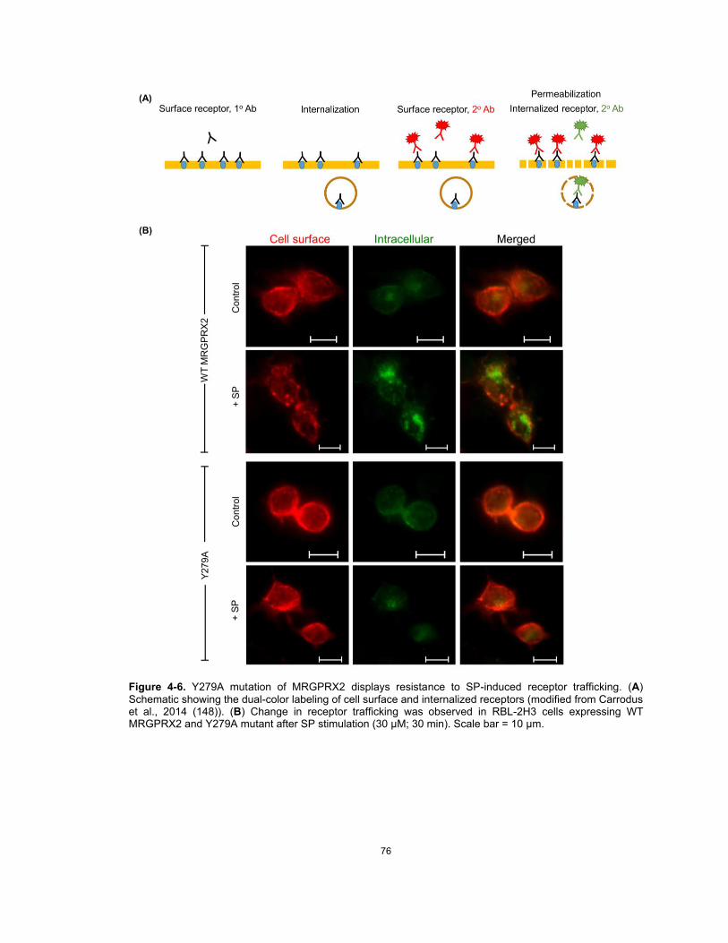
76
Figure 4-6. Y279A mutation of MRGPRX2 displays resistance to SP-induced receptor trafficking. (A) Schematic showing the dual-color labeling of cell surface and internalized receptors (modified from Carrodus et al., 2014 (148)). (B) Change in receptor trafficking was observed in RBL-2H3 cells expressing WT MRGPRX2 and Y279A mutant after SP stimulation (30 µM; 30 min). Scale bar = 10 µm.

77
CHAPTER 5: Conclusion and Future Directions
In addition to the classical IgE-mediated activation, human MCs also respond to a diverse
group of basic secretagogues, including host defense peptides (HDPs), neuropeptides (NPs), and
a number of FDA-approved peptidergic drugs, via the newly identified MRGPRX2 receptor (mouse
ortholog MrgprB2) (10). Recent studies have greatly emphasized the role of MRGPRX2 in both
health and disease, ranging from host defense and wound healing to drug-induced pseudoallergic
reactions, neurogenic inflammation, pain and itch (10, 13, 14, 50, 159). However, some studies
have doubted the role of MRGPRX2 on neuromuscular blocking drug (NMBD)-induced
hypersensitivity, especially for rocuronium (51, 57, 58, 106, 160). Furthermore, the molecular
mechanisms involved in the regulation of MRGPRX2/B2 function remain largely unknown.
In CHAPTER 2, we addressed the discrepancy regarding the biological relevance of
MRGPRX2 on rocuronium-induced hypersensitivity. Our study provided the first demonstration that
rocuronium can induce degranulation in human MCs via MRGPRX2 and that the ability of
rocuronium to induce degranulation depends on the level of MRGPRX2 expression. It is worth
noting that the minimal concentration of rocuronium needed to stimulate human MC degranulation
is significantly higher than that required for mouse MrgprB2 activation. These distinct differences
between MrgprB2 and MRGPRX2 demonstrate the challenge in translating animal studies into
humans. This highlights the need to develop better animal models to study MRGPRX2 function in
vivo.
Furthermore, we tested the possibility whether MRGPRX2 mutations (M196I, L226P, and
L237P) reported in a patient with rocuronium-induced perioperative anaphylaxis render the
receptor more susceptible to rocuronium hypersensitivity (161). We found that cells expressing the
mutated receptors (M196I, L226P, and L237P) either respond normally to rocuronium or display
loss-of-function phenotype. These findings do not support the role of MRGPRX2 on rocuronium
hypersensitivity in this patient. Given that MRGPRX2 is expressed predominantly in human skin
MCs, it is plausible that erythema observed at injection site in this patient likely reflects normal
degranulation process unrelated to anaphylaxis. On the other hand, anaphylaxis might be a result

78
from IgE/FcHRI-mediated activated of MCs that are present in other locations such as the
gastrointestinal and respiratory tracts. However, it is worth noting that although MRGPRX2
expression is usually low in normal lung and colon MCs, it is upregulated in individuals with severe
asthma and ulcerative colitis. Furthermore, MRGPRX2 expression is upregulated in cutaneous
MCs of patients with skin inflammatory diseases, such as chronic spontaneous urticaria. Thus, it is
possible that in those particular individuals, MRGPRX2 may also contribute to drug-induced
pseudoallergy.
In CHAPTER 3 and 4, we delineated the mechanisms underlying MRGPRX2 signaling and
regulation on SP-activated MC responses. It is now realized that SP released from sensory neurons
also activate cutaneous MCs via MRGPRX2 to induce degranulation and chemokine/cytokine
generation, contributing to neurogenic inflammation, pain, and itch. Thus, targeting MRGPRX2 and
its signaling might therefore represent novel therapeutic approaches for the prevention and/or
treatment of neurogenic inflammatory diseases and pain. In CHAPTER 3, we focused on the
MRGPRX2-G protein interaction, while CHAPTER 4 the receptor-β-arrestin2 interaction was
investigated.
First, we identified the specific G proteins that couple to MRGPRX2 in response to MC
activation by SP. Previous studies on the mechanisms of MRGPRX2-mediated MC responses
demonstrated that PTx inhibits MC degranulation in response to MRGPRX2 agonists, including
compound 48/80, cathelicidin LL-37 and human β-defensin-3, but not calcium mobilization. These
findings lead to the possibility that MRGPRX2 couple to both PTx-sensitive (Gαi) and insensitive
(Gαq) families of G proteins, in order to induce human MC activation. To test if SP activates
MRGPRX2 via both Gαi and Gαq to induce Ca2+ mobilization and MC degranulation, I utilized a
pharmacological approach using a Gαi-specific inhibitor (PTx) and a Gαq-specific inhibitor (YM-
254890). Using PTx or YM-254890 alone caused partial inhibition of Ca2+ mobilization and MC
degranulation by SP, but a combination of both inhibitors abolished the responses. These findings
suggest that, as expected, MRGPRX2 couples to both Gαi and Gαq families of G proteins to induce
Ca2+ mobilization and degranulation in response to SP.

79
Next, the information obtained from both structural modeling (79) and naturally occurring
MRGPRX2 missense variants were gathered to identify residues in the TM and intracellular
domains of MRGPRX2 that regulate G protein-mediated MC activation by SP. Putative G protein
coupling regions have been established. In addition, several gain- and loss-of-function missense
single nucleotide polymorphisms (SNPs) in MRGPRX2 have been identified. We found that SNPs
in MRGPRX2’s G protein-coupling regions (V123F, V282M, R138C and R141C) failed to activate
MC in response to SP. By contrast, MRGPRX2 variants at the carboxyl-terminus that is responsible
for receptor phosphorylation and desensitization (S325L and L329Q) showed higher SP-induced
responses, presumably by preventing MRGPRX2 phosphorylation (104). The effects of these
MRGPRX2 variants on modulating each G protein coupling and activation should be further
investigated. Additionally, future studies will need to be conducted to determine impact of these
SNPs on MRGPRX2/MC-mediated conditions.
Lastly, we provided the first demonstration that SP serves as a balanced agonist for
MRGPRX2 and can induce β-arrestin-dependent signaling. A tyrosine residue in the highly
conserved NPxxY motif of MRGPRX2, Tyr279, has been identified as the residue not only important
for G protein activation, but also for β-arrestin activation and receptor internalization. These findings
altogether have an important clinical relevance as individuals with loss-of-function mutations in
MRGPRX2 may display resistance to developing neurogenic inflammation and chronic
inflammatory diseases. By contrast, individuals who harbor the gain-of-function variants may be
more susceptible to develop these conditions. Determining the structure and understanding the
molecular mechanisms of MRGPRX2 activation will not only enhance our fundamental knowledge
of MRGPRX2-mediated MC responses, but will also provide great translational potentials for the
development of novel therapeutics for SP-induced inflammatory conditions and pain.
Ultimately, future in vivo studies targeting either G protein or β-arrestin signaling of
MRGPRX2 should be conducted in the context of pathological conditions. Previous studies in mice
demonstrated that SP contributes to neurogenic inflammation through the activation of MCs via
MrgprB2 (mouse counterpart of human MRGPRX2) (13). However, MrgprB2 shares only ~53%
80
sequence identity with MRGPRX2 (9). Thus, in vivo studies using MrgprB2-/- mice may not
accurately reflect mast cell biological responses in humans. To overcome the translational
challenge of using MrgprB2-modified mice, several approached of humanized mice with
MRGPRX2 expressing human MCs has been utilized to study the MRGPRX2 function (162, 163).
Mice expressing human MRGPRX2 variants and/or β-arrestin knockout should be used to
investigate the effects of MRGPRX2 signaling modulation on neurogenic inflammation in vivo.
Atopic dermatitis (AD), also known as atopic eczema, is a chronic and relapsing T-helper-
2 (TH2)-associated inflammatory skin disease characterized by eczematous lesions and severe
pruritus (164). Dysregulation between MC-neuron interaction have been implicated in the
pathogenesis of AD (165-167). Recently, Serhan et al. provided a strong evidence that the
MrgprB2+ MCs establish functional clusters in the skin with a subpopulation of SP-producing
sensory neurons expressing the transient receptor potential cation channel subfamily V number 1
(TRPV1) and Tac1, the gene encoding the SP precursor (TRPV1+Tac1+ nociceptors) (15). These
MC-neuron clusters are required for the development and pathophysiology of AD. Hence, targeting
MRGPRX2 and its signaling could be a promising therapeutic modality for the prevention and/or
management of chronic itch and inflammation in human AD as well as other diseases mediated by
this receptor.
MRGPRX2-expressing MCs are predominantly present in the skin as well as gingiva, and
contribute to skin and gingival homeostasis. Our lab has previously shown that patients with severe
periodontitis display higher number of MCs and increased MRGPRX2 expression. Oral medicine
usually involves the diagnosis and management of patients with orofacial disorders and oral
manifestations of systemic diseases. As several oral lesions represent the early/first signs of
mucocutaneous disorders, it is possible that MRGPRX2 may be implicated in the
pathogenesis/progression of these mucocutaneous disorders and the patients with those
conditions may have upregulated MRGPRX2 expression. This putative role of MRGPRX2 on oral
mucocutaneous lesions should be further investigated.

81
Lists of Publications
Original Research
• Chompunud Na Ayudhya C, Amponnawarat A, Roy S, Oskeritzian CA, Ali H. MRGPRX2 Activation by Rocuronium: Insights from Studies with Human Skin Mast Cells and Missense Variants. Cells. 2021 Jan 15;10(1):156.
• Chompunud Na Ayudhya C, Roy S, Alkanfari I, Ganguly A, Ali H. Identification of Gain and Loss of Function Missense Variants in MRGPRX2's Transmembrane and Intracellular Domains for Mast Cell Activation by Substance P. Int J Mol Sci. 2019;20(21):5247.
• Chompunud Na Ayudhya C, Amponnawarat A, Ali H. Substance P Serves as a Balanced Agonist for MRGPRX2 and a Single Tyrosine Residue is Required for β-arrestin Recruitment and Receptor Internalization. Int J Mol Sci. 2021; 22(10):5318. https://doi.org/10.3390/ijms22105318.
• Amponnawarat A, Chompunud Na Ayudhya C, Ali H. Murepavadin, a Small Molecule Host Defense Peptide Mimetic, Activates Mast Cells via Mas-related G Protein-Coupled Receptors. Front Immunol. 2021. (Accepted; in press)
Review Article
• Chompunud Na Ayudhya C, Roy S, Thapaliya M, Ali H. Roles of a Mast Cell-Specific Receptor MRGPRX2 in Host Defense and Inflammation. J Dent Res. 2020;99(8):882-890.
• Thapaliya M, Chompunud Na Ayudhya C, Amponnawarat A, Roy S, Ali H. Mast Cell-Specific MRGPRX2: a Key Modulator of Neuro-Immune Interaction in Allergic Diseases. Curr Allergy Asthma Rep. 2021;21(1):3.
• Roy S, Chompunud Na Ayudhya C, Thapaliya M, Deepak V, Ali H. Multifaceted MRGPRX2: New Insight into the Role of Cutaneous Mast Cells in Health and Disease. J Allergy Clin Immunol. 2021 May;S0091-6749(21)00721-1. doi: 10.1016/j.jaci.2021.03.049. Online ahead of print.
Clinical Case Report
• Chompunud Na Ayudhya C, Granquist EJ, Mupparapu M, Sollecito TP, Stoopler ET. Advanced maxillofacial imaging for temporomandibular disorder in special needs patients. Spec Care Dentist. 2018;38(3):150-154.
• Chompunud Na Ayudhya C, Alawi F, Akintoye SO. Unusual oral multifocal epithelial hyperplasia in an adult African-American lung transplant patient. Transpl Infect Dis. 2021 Apr;23(2):e13497.

82
REFERENCES
1. da Silva EZ, Jamur MC, Oliver C. Mast cell function: a new vision of an old cell. J Histochem Cytochem. 2014;62(10):698-738. 2. Galli SJ, Gaudenzio N, Tsai M. Mast Cells in Inflammation and Disease: Recent Progress and Ongoing Concerns. Annu Rev Immunol. 2020;38:49-77. 3. Forsythe P. Mast Cells in Neuroimmune Interactions. Trends Neurosci. 2019;42(1):43-55. 4. Abraham SN, St John AL. Mast cell-orchestrated immunity to pathogens. Nat Rev Immunol. 2010;10(6):440-52. 5. St John AL, Abraham SN. Innate immunity and its regulation by mast cells. J Immunol. 2013;190(9):4458-63. 6. Fujisawa D, Kashiwakura J, Kita H, Kikukawa Y, Fujitani Y, Sasaki-Sakamoto T, et al. Expression of Mas-related gene X2 on mast cells is upregulated in the skin of patients with severe chronic urticaria. J Allergy Clin Immunol. 2014;134(3):622-33 e9. 7. Varricchi G, Pecoraro A, Loffredo S, Poto R, Rivellese F, Genovese A, et al. Heterogeneity of Human Mast Cells With Respect to MRGPRX2 Receptor Expression and Function. Front Cell Neurosci. 2019;13:299. 8. Tatemoto K, Nozaki Y, Tsuda R, Konno S, Tomura K, Furuno M, et al. Immunoglobulin E-independent activation of mast cell is mediated by Mrg receptors. Biochem Biophys Res Commun. 2006;349(4):1322-8. 9. Subramanian H, Gupta K, Ali H. Roles of Mas-related G protein-coupled receptor X2 on mast cell-mediated host defense, pseudoallergic drug reactions, and chronic inflammatory diseases. J Allergy Clin Immunol. 2016;138(3):700-10. 10. McNeil BD, Pundir P, Meeker S, Han L, Undem BJ, Kulka M, et al. Identification of a mast-cell-specific receptor crucial for pseudo-allergic drug reactions. Nature. 2015;519(7542):237-41. 11. Ali H. Emerging Roles for MAS-Related G Protein-Coupled Receptor-X2 in Host Defense Peptide, Opioid, and Neuropeptide-Mediated Inflammatory Reactions. Adv Immunol. 2017;136:123-62. 12. Arifuzzaman M, Mobley YR, Choi HW, Bist P, Salinas CA, Brown ZD, et al. MRGPR-mediated activation of local mast cells clears cutaneous bacterial infection and protects against reinfection. Sci Adv. 2019;5(1):eaav0216. 13. Green DP, Limjunyawong N, Gour N, Pundir P, Dong X. A Mast-Cell-Specific Receptor Mediates Neurogenic Inflammation and Pain. Neuron. 2019;101(3):412-20 e3. 14. Meixiong J, Anderson M, Limjunyawong N, Sabbagh MF, Hu E, Mack MR, et al. Activation of Mast-Cell-Expressed Mas-Related G-Protein-Coupled Receptors Drives Non-histaminergic Itch. Immunity. 2019;50(5):1163-71.e5. 15. Serhan N, Basso L, Sibilano R, Petitfils C, Meixiong J, Bonnart C, et al. House dust mites activate nociceptor-mast cell clusters to drive type 2 skin inflammation. Nat Immunol. 2019;20(11):1435-43. 16. Dahlin JS, Hallgren J. Mast cell progenitors: origin, development and migration to tissues. Mol Immunol. 2015;63(1):9-17. 17. Gurish MF, Austen KF. Developmental origin and functional specialization of mast cell subsets. Immunity. 2012;37(1):25-33. 18. Moon TC, St Laurent CD, Morris KE, Marcet C, Yoshimura T, Sekar Y, et al. Advances in mast cell biology: new understanding of heterogeneity and function. Mucosal Immunol. 2010;3(2):111-28. 19. Xiang Z, Block M, Lofman C, Nilsson G. IgE-mediated mast cell degranulation and recovery monitored by time-lapse photography. J Allergy Clin Immunol. 2001;108(1):116-21. 20. Suurmond J, Habets KLL, Tatum Z, Schonkeren JJ, Hoen PAC, Huizinga TWJ, et al. Repeated FcepsilonRI triggering reveals modified mast cell function related to chronic allergic responses in tissue. J Allergy Clin Immunol. 2016;138(3):869-80. 21. Geering B, Stoeckle C, Conus S, Simon HU. Living and dying for inflammation: neutrophils, eosinophils, basophils. Trends Immunol. 2013;34(8):398-409.

83
22. Okayama Y, Kawakami T. Development, migration, and survival of mast cells. Immunol Res. 2006;34(2):97-115. 23. Reber L, Da Silva CA, Frossard N. Stem cell factor and its receptor c-Kit as targets for inflammatory diseases. Eur J Pharmacol. 2006;533(1-3):327-40. 24. Galli SJ, Kitamura Y. Genetically mast-cell-deficient W/Wv and Sl/Sld mice. Their value for the analysis of the roles of mast cells in biologic responses in vivo. Am J Pathol. 1987;127(1):191-8. 25. Grimbaldeston MA, Chen CC, Piliponsky AM, Tsai M, Tam SY, Galli SJ. Mast cell-deficient W-sash c-kit mutant Kit W-sh/W-sh mice as a model for investigating mast cell biology in vivo. Am J Pathol. 2005;167(3):835-48. 26. Hu ZQ, Zhao WH, Shimamura T. Regulation of mast cell development by inflammatory factors. Curr Med Chem. 2007;14(28):3044-50. 27. Galli SJ, Kalesnikoff J, Grimbaldeston MA, Piliponsky AM, Williams CM, Tsai M. Mast cells as "tunable" effector and immunoregulatory cells: recent advances. Annu Rev Immunol. 2005;23:749-86. 28. Friend DS, Ghildyal N, Gurish MF, Hunt J, Hu X, Austen KF, et al. Reversible expression of tryptases and chymases in the jejunal mast cells of mice infected with Trichinella spiralis. J Immunol. 1998;160(11):5537-45. 29. Welle M. Development, significance, and heterogeneity of mast cells with particular regard to the mast cell-specific proteases chymase and tryptase. J Leukoc Biol. 1997;61(3):233-45. 30. Irani AA, Schechter NM, Craig SS, DeBlois G, Schwartz LB. Two types of human mast cells that have distinct neutral protease compositions. Proc Natl Acad Sci U S A. 1986;83(12):4464-8. 31. Reber LL, Sibilano R, Mukai K, Galli SJ. Potential effector and immunoregulatory functions of mast cells in mucosal immunity. Mucosal Immunol. 2015;8(3):444-63. 32. Gupta K, Idahosa C, Roy S, Lee D, Subramanian H, Dhingra A, et al. Differential Regulation of Mas-Related G Protein-Coupled Receptor X2-Mediated Mast Cell Degranulation by Antimicrobial Host Defense Peptides and Porphyromonas gingivalis Lipopolysaccharide. Infect Immun. 2017;85(10). 33. Dwyer DF, Barrett NA, Austen KF. Expression profiling of constitutive mast cells reveals a unique identity within the immune system. Nat Immunol. 2016;17(7):878-87. 34. Okayama Y, Kirshenbaum AS, Metcalfe DD. Expression of a functional high-affinity IgG receptor, Fc gamma RI, on human mast cells: Up-regulation by IFN-gamma. J Immunol. 2000;164(8):4332-9. 35. Ali H. Regulation of human mast cell and basophil function by anaphylatoxins C3a and C5a. Immunol Lett. 2010;128(1):36-45. 36. Espinosa E, Valitutti S. New roles and controls of mast cells. Curr Opin Immunol. 2018;50:39-47. 37. Moon TC, Befus AD, Kulka M. Mast cell mediators: their differential release and the secretory pathways involved. Front Immunol. 2014;5:569. 38. Holowka D, Wilkes M, Stefan C, Baird B. Roles for Ca2+ mobilization and its regulation in mast cell functions: recent progress. Biochem Soc Trans. 2016;44(2):505-9. 39. Wernersson S, Pejler G. Mast cell secretory granules: armed for battle. Nat Rev Immunol. 2014;14(7):478-94. 40. Pejler G, Ronnberg E, Waern I, Wernersson S. Mast cell proteases: multifaceted regulators of inflammatory disease. Blood. 2010;115(24):4981-90. 41. Mukai K, Tsai M, Saito H, Galli SJ. Mast cells as sources of cytokines, chemokines, and growth factors. Immunol Rev. 2018;282(1):121-50. 42. Mousli M, Bronner C, Bockaert J, Rouot B, Landry Y. Interaction of substance P, compound 48/80 and mastoparan with the alpha-subunit C-terminus of G protein. Immunol Lett. 1990;25(4):355-7. 43. Mousli M, Bueb JL, Bronner C, Rouot B, Landry Y. G protein activation: a receptor-independent mode of action for cationic amphiphilic neuropeptides and venom peptides. Trends Pharmacol Sci. 1990;11(9):358-62.

84
44. Ferry X, Eichwald V, Daeffler L, Landry Y. Activation of betagamma subunits of G(i2) and G(i3) proteins by basic secretagogues induces exocytosis through phospholipase Cbeta and arachidonate release through phospholipase Cgamma in mast cells. J Immunol. 2001;167(9):4805-13. 45. Solinski HJ, Gudermann T, Breit A. Pharmacology and signaling of MAS-related G protein-coupled receptors. Pharmacol Rev. 2014;66(3):570-97. 46. Subramanian H, Gupta K, Guo Q, Price R, Ali H. Mas-related gene X2 (MrgX2) is a novel G protein-coupled receptor for the antimicrobial peptide LL-37 in human mast cells: resistance to receptor phosphorylation, desensitization, and internalization. J Biol Chem. 2011;286(52):44739-49. 47. Kamohara M, Matsuo A, Takasaki J, Kohda M, Matsumoto M, Matsumoto S, et al. Identification of MrgX2 as a human G-protein-coupled receptor for proadrenomedullin N-terminal peptides. Biochem Biophys Res Commun. 2005;330(4):1146-52. 48. Subramanian H, Gupta K, Lee D, Bayir AK, Ahn H, Ali H. beta-Defensins activate human mast cells via Mas-related gene X2. J Immunol. 2013;191(1):345-52. 49. Gupta K, Kotian A, Subramanian H, Daniell H, Ali H. Activation of human mast cells by retrocyclin and protegrin highlight their immunomodulatory and antimicrobial properties. Oncotarget. 2015;6(30):28573-87. 50. Pundir P, Liu R, Vasavda C, Serhan N, Limjunyawong N, Yee R, et al. A Connective Tissue Mast-Cell-Specific Receptor Detects Bacterial Quorum-Sensing Molecules and Mediates Antibacterial Immunity. Cell Host Microbe. 2019;26(1):114-22 e8. 51. Lansu K, Karpiak J, Liu J, Huang XP, McCorvy JD, Kroeze WK, et al. In silico design of novel probes for the atypical opioid receptor MRGPRX2. Nat Chem Biol. 2017;13(5):529-36. 52. Mertes PM, Alla F, Tréchot P, Auroy Y, Jougla E. Anaphylaxis during anesthesia in France: an 8-year national survey. J Allergy Clin Immunol. 2011;128(2):366-73. 53. Petitpain N, Argoullon L, Masmoudi K, Fedrizzi S, Cottin J, Latarche C, et al. Neuromuscular blocking agents induced anaphylaxis: Results and trends of a French pharmacovigilance survey from 2000 to 2012. Allergy. 2018;73(11):2224-33. 54. Reddy JI, Cooke PJ, van Schalkwyk JM, Hannam JA, Fitzharris P, Mitchell SJ. Anaphylaxis is more common with rocuronium and succinylcholine than with atracurium. Anesthesiology. 2015;122(1):39-45. 55. Spoerl D, D'Incau S, Roux-Lombard P, Harr T, Czarnetzki C. Non-IgE-Dependent Hypersensitivity to Rocuronium Reversed by Sugammadex: Report of Three Cases and Hypothesis on the Underlying Mechanism. Int Arch Allergy Immunol. 2016;169(4):256-62. 56. Spoerl D, Nigolian H, Czarnetzki C, Harr T. Reclassifying Anaphylaxis to Neuromuscular Blocking Agents Based on the Presumed Patho-Mechanism: IgE-Mediated, Pharmacological Adverse Reaction or "Innate Hypersensitivity"? Int J Mol Sci. 2017;18(6). 57. Van Gasse AL, Elst J, Bridts CH, Mertens C, Faber M, Hagendorens MM, et al. Rocuronium Hypersensitivity: Does Off-Target Occupation of the MRGPRX2 Receptor Play a Role? J Allergy Clin Immunol Pract. 2019;7(3):998-1003. 58. Navinés-Ferrer A, Serrano-Candelas E, Lafuente A, Muñoz-Cano R, Martín M, Gastaminza G. MRGPRX2-mediated mast cell response to drugs used in perioperative procedures and anaesthesia. Sci Rep. 2018;8(1):11628. 59. Elst J, Sabato V, Faber MA, Bridts CH, Mertens C, Van Houdt M, et al. MRGPRX2 and Immediate Drug Hypersensitivity: Insights from Cultured Human Mast Cells. J Investig Allergol Clin Immunol. 2020:0. 60. Kleij HP, Bienenstock J. Significance of Conversation between Mast Cells and Nerves. Allergy Asthma Clin Immunol. 2005;1(2):65-80. 61. Gupta K, Harvima IT. Mast cell-neural interactions contribute to pain and itch. Immunol Rev. 2018;282(1):168-87. 62. Siiskonen H, Harvima I. Mast Cells and Sensory Nerves Contribute to Neurogenic Inflammation and Pruritus in Chronic Skin Inflammation. Front Cell Neurosci. 2019;13:422. 63. Ren K, Dubner R. Interactions between the immune and nervous systems in pain. Nat Med. 2010;16(11):1267-76.

85
64. Mashaghi A, Marmalidou A, Tehrani M, Grace PM, Pothoulakis C, Dana R. Neuropeptide substance P and the immune response. Cell Mol Life Sci. 2016;73(22):4249-64. 65. O'Connor TM, O'Connell J, O'Brien DI, Goode T, Bredin CP, Shanahan F. The role of substance P in inflammatory disease. J Cell Physiol. 2004;201(2):167-80. 66. Kulka M, Sheen CH, Tancowny BP, Grammer LC, Schleimer RP. Neuropeptides activate human mast cell degranulation and chemokine production. Immunology. 2008;123(3):398-410. 67. Shim WS, Oh U. Histamine-induced itch and its relationship with pain. Mol Pain. 2008;4:29. 68. Steinhoff M, Vergnolle N, Young SH, Tognetto M, Amadesi S, Ennes HS, et al. Agonists of proteinase-activated receptor 2 induce inflammation by a neurogenic mechanism. Nat Med. 2000;6(2):151-8. 69. Taylor-Clark TE, Nassenstein C, Undem BJ. Leukotriene D4 increases the excitability of capsaicin-sensitive nasal sensory nerves to electrical and chemical stimuli. Br J Pharmacol. 2008;154(6):1359-68. 70. Sagi V, Mittal A, Gupta M, Gupta K. Immune cell neural interactions and their contributions to sickle cell disease. Neurosci Lett. 2019;699:167-71. 71. van der Kleij HP, Ma D, Redegeld FA, Kraneveld AD, Nijkamp FP, Bienenstock J. Functional expression of neurokinin 1 receptors on mast cells induced by IL-4 and stem cell factor. J Immunol. 2003;171(4):2074-9. 72. Borsook D, Upadhyay J, Klimas M, Schwarz AJ, Coimbra A, Baumgartner R, et al. Decision-making using fMRI in clinical drug development: revisiting NK-1 receptor antagonists for pain. Drug Discov Today. 2012;17(17-18):964-73. 73. Hill R. NK1 (substance P) receptor antagonists--why are they not analgesic in humans? Trends Pharmacol Sci. 2000;21(7):244-6. 74. Azimi E, Reddy VB, Shade KC, Anthony RM, Talbot S, Pereira PJS, et al. Dual action of neurokinin-1 antagonists on Mas-related GPCRs. JCI Insight. 2016;1(16):e89362. 75. Katritch V, Cherezov V, Stevens RC. Diversity and modularity of G protein-coupled receptor structures. Trends Pharmacol Sci. 2012;33(1):17-27. 76. Munk C, Isberg V, Mordalski S, Harpsøe K, Rataj K, Hauser AS, et al. GPCRdb: the G protein-coupled receptor database - an introduction. Br J Pharmacol. 2016;173(14):2195-207. 77. Venkatakrishnan AJ, Deupi X, Lebon G, Tate CG, Schertler GF, Babu MM. Molecular signatures of G-protein-coupled receptors. Nature. 2013;494(7436):185-94. 78. Zhou Q, Yang D, Wu M, Guo Y, Guo W, Zhong L, et al. Common activation mechanism of class A GPCRs. Elife. 2019;8. 79. Venkatakrishnan AJ, Deupi X, Lebon G, Heydenreich FM, Flock T, Miljus T, et al. Diverse activation pathways in class A GPCRs converge near the G-protein-coupling region. Nature. 2016;536(7617):484-7. 80. Wootten D, Christopoulos A, Marti-Solano M, Babu MM, Sexton PM. Mechanisms of signalling and biased agonism in G protein-coupled receptors. Nat Rev Mol Cell Biol. 2018;19(10):638-53. 81. Kamato D, Thach L, Bernard R, Chan V, Zheng W, Kaur H, et al. Structure, Function, Pharmacology, and Therapeutic Potential of the G Protein, Gα/q,11. Front Cardiovasc Med. 2015;2:14. 82. Mangmool S, Kurose H. G(i/o) protein-dependent and -independent actions of Pertussis Toxin (PTX). Toxins (Basel). 2011;3(7):884-99. 83. Gurevich VV, Gurevich EV. GPCR Signaling Regulation: The Role of GRKs and Arrestins. Front Pharmacol. 2019;10:125. 84. Hilger D, Masureel M, Kobilka BK. Structure and dynamics of GPCR signaling complexes. Nat Struct Mol Biol. 2018;25(1):4-12. 85. Cahill TJ, 3rd, Thomsen AR, Tarrasch JT, Plouffe B, Nguyen AH, Yang F, et al. Distinct conformations of GPCR-β-arrestin complexes mediate desensitization, signaling, and endocytosis. Proc Natl Acad Sci U S A. 2017;114(10):2562-7. 86. Shenoy SK, Lefkowitz RJ. β-Arrestin-mediated receptor trafficking and signal transduction. Trends Pharmacol Sci. 2011;32(9):521-33.

86
87. Smith JS, Nicholson LT, Suwanpradid J, Glenn RA, Knape NM, Alagesan P, et al. Biased agonists of the chemokine receptor CXCR3 differentially control chemotaxis and inflammation. Sci Signal. 2018;11(555). 88. Lefkowitz RJ, Shenoy SK. Transduction of receptor signals by beta-arrestins. Science. 2005;308(5721):512-7. 89. Kohout TA, Lin FS, Perry SJ, Conner DA, Lefkowitz RJ. beta-Arrestin 1 and 2 differentially regulate heptahelical receptor signaling and trafficking. Proc Natl Acad Sci U S A. 2001;98(4):1601-6. 90. Paing MM, Stutts AB, Kohout TA, Lefkowitz RJ, Trejo J. beta -Arrestins regulate protease-activated receptor-1 desensitization but not internalization or Down-regulation. J Biol Chem. 2002;277(2):1292-300. 91. Vibhuti A, Gupta K, Subramanian H, Guo Q, Ali H. Distinct and shared roles of β-arrestin-1 and β-arrestin-2 on the regulation of C3a receptor signaling in human mast cells. PLoS One. 2011;6(5):e19585. 92. Roy S, Gupta K, Ganguly A, Ali H. β-Arrestin2 expressed in mast cells regulates ciprofloxacin-induced pseudoallergy and IgE-mediated anaphylaxis. J Allergy Clin Immunol. 2019;144(2):603-6. 93. Volcheck GW, Hepner DL. Identification and Management of Perioperative Anaphylaxis. J Allergy Clin Immunol Pract. 2019;7(7):2134-42. 94. Suzuki Y, Liu S, Kadoya F, Takasaki Y, Yorozuya T, Mogi M. Association between mutated Mas-related G protein-coupled receptor-X2 and rocuronium-induced intraoperative anaphylaxis. Br J Anaesth. 2020. 95. Fernandopulle NA, Zhang SS, Soeding PF, Mackay GA. MRGPRX2 activation in mast cells by neuromuscular blocking agents and other agonists: Modulation by sugammadex. Clin Exp Allergy. 2020. 96. Alkanfari I, Freeman KB, Roy S, Jahan T, Scott RW, Ali H. Small-Molecule Host-Defense Peptide Mimetic Antibacterial and Antifungal Agents Activate Human and Mouse Mast Cells via Mas-Related GPCRs. Cells. 2019;8(4). 97. Kirshenbaum AS, Akin C, Wu Y, Rottem M, Goff JP, Beaven MA, et al. Characterization of novel stem cell factor responsive human mast cell lines LAD 1 and 2 established from a patient with mast cell sarcoma/leukemia; activation following aggregation of FcepsilonRI or FcgammaRI. Leuk Res. 2003;27(8):677-82. 98. Ali H, Richardson RM, Tomhave ED, DuBose RA, Haribabu B, Snyderman R. Regulation of stably transfected platelet activating factor receptor in RBL-2H3 cells. Role of multiple G proteins and receptor phosphorylation. J Biol Chem. 1994;269(40):24557-63. 99. Subramanian H, Kashem SW, Collington SJ, Qu H, Lambris JD, Ali H. PMX-53 as a dual CD88 antagonist and an agonist for Mas-related gene 2 (MrgX2) in human mast cells. Mol Pharmacol. 2011;79(6):1005-13. 100. Kambe N, Kambe M, Kochan JP, Schwartz LB. Human skin-derived mast cells can proliferate while retaining their characteristic functional and protease phenotypes. Blood. 2001;97(7):2045-52. 101. Malbec O, Roget K, Schiffer C, Iannascoli B, Dumas AR, Arock M, et al. Peritoneal cell-derived mast cells: an in vitro model of mature serosal-type mouse mast cells. J Immunol. 2007;178(10):6465-75. 102. Babina M, Wang Z, Roy S, Guhl S, Franke K, Artuc M, et al. MRGPRX2 Is the Codeine Receptor of Human Skin Mast Cells: Desensitization through β-Arrestin and Lack of Correlation with the FcεRI Pathway. J Invest Dermatol. 2020. 103. Alkanfari I, Gupta K, Jahan T, Ali H. Naturally Occurring Missense MRGPRX2 Variants Display Loss of Function Phenotype for Mast Cell Degranulation in Response to Substance P, Hemokinin-1, Human β-Defensin-3, and Icatibant. J Immunol. 2018;201(2):343-9. 104. Chompunud Na Ayudhya C, Roy S, Alkanfari I, Ganguly A, Ali H. Identification of Gain and Loss of Function Missense Variants in MRGPRX2's Transmembrane and Intracellular Domains for Mast Cell Activation by Substance P. Int J Mol Sci. 2019;20(21).

87
105. Elst J, Sabato V, Mertens C, Garvey LH, Ebo DG. Association between mutated Mas-related G protein-coupled receptor-X2 and rocuronium-induced intraoperative anaphylaxis. Comment on Br J Anaesth 2020. Br J Anaesth. 2020. 106. Ebo DG, Van der Poorten ML, Elst J, Van Gasse AL, Mertens C, Bridts C, et al. Immunoglobulin E cross-linking or MRGPRX2 activation: clinical insights from rocuronium hypersensitivity. Br J Anaesth. 2020. 107. Pándy-Szekeres G, Munk C, Tsonkov TM, Mordalski S, Harpsøe K, Hauser AS, et al. GPCRdb in 2018: adding GPCR structure models and ligands. Nucleic Acids Res. 2018;46(D1):D440-d6. 108. Porebski G, Kwiecien K, Pawica M, Kwitniewski M. Mas-Related G Protein-Coupled Receptor-X2 (MRGPRX2) in Drug Hypersensitivity Reactions. Front Immunol. 2018;9:3027. 109. Reddy VB, Graham TA, Azimi E, Lerner EA. A single amino acid in MRGPRX2 necessary for binding and activation by pruritogens. J Allergy Clin Immunol. 2017;140(6):1726-8. 110. Theoharides TC, Alysandratos KD, Angelidou A, Delivanis DA, Sismanopoulos N, Zhang B, et al. Mast cells and inflammation. Biochim Biophys Acta. 2012;1822(1):21-33. 111. Choi HW, Suwanpradid J, Kim IH, Staats HF, Haniffa M, MacLeod AS, et al. Perivascular dendritic cells elicit anaphylaxis by relaying allergens to mast cells via microvesicles. Science. 2018;362(6415). 112. Karhausen J, Abraham SN. How mast cells make decisions. J Clin Invest. 2016;126(10):3735-8. 113. Garcia-Recio S, Gascon P. Biological and Pharmacological Aspects of the NK1-Receptor. Biomed Res Int. 2015;2015:495704. 114. Navratilova E, Porreca F. Substance P and Inflammatory Pain: Getting It Wrong and Right Simultaneously. Neuron. 2019;101(3):353-5. 115. Isberg V, de Graaf C, Bortolato A, Cherezov V, Katritch V, Marshall FH, et al. Generic GPCR residue numbers - aligning topology maps while minding the gaps. Trends Pharmacol Sci. 2015;36(1):22-31. 116. Rasmussen SG, DeVree BT, Zou Y, Kruse AC, Chung KY, Kobilka TS, et al. Crystal structure of the beta2 adrenergic receptor-Gs protein complex. Nature. 2011;477(7366):549-55. 117. Gupta K, Subramanian H, Klos A, Ali H. Phosphorylation of C3a receptor at multiple sites mediates desensitization, beta-arrestin-2 recruitment and inhibition of NF-kappaB activity in mast cells. PLoS One. 2012;7(10):e46369. 118. Guo Q, Subramanian H, Gupta K, Ali H. Regulation of C3a receptor signaling in human mast cells by G protein coupled receptor kinases. PLoS One. 2011;6(7):e22559. 119. Cahill TJ, 3rd, Thomsen AR, Tarrasch JT, Plouffe B, Nguyen AH, Yang F, et al. Distinct conformations of GPCR-beta-arrestin complexes mediate desensitization, signaling, and endocytosis. Proc Natl Acad Sci U S A. 2017;114(10):2562-7. 120. Roy S, Ganguly A, Haque M, Ali H. Angiogenic Host Defense Peptide AG-30/5C and Bradykinin B(2) Receptor Antagonist Icatibant Are G Protein Biased Agonists for MRGPRX2 in Mast Cells. J Immunol. 2019;202(4):1229-38. 121. Gupta K, Subramanian H, Ali H. Modulation of host defense peptide-mediated human mast cell activation by LPS. Innate Immun. 2016;22(1):21-30. 122. Takasaki J, Saito T, Taniguchi M, Kawasaki T, Moritani Y, Hayashi K, et al. A novel Galphaq/11-selective inhibitor. J Biol Chem. 2004;279(46):47438-45. 123. Zhang Y, Sun B, Feng D, Hu H, Chu M, Qu Q, et al. Cryo-EM structure of the activated GLP-1 receptor in complex with a G protein. Nature. 2017;546(7657):248-53. 124. Civciristov S, Ellisdon AM, Suderman R, Pon CK, Evans BA, Kleifeld O, et al. Preassembled GPCR signaling complexes mediate distinct cellular responses to ultralow ligand concentrations. Sci Signal. 2018;11(551). 125. Moro O, Lameh J, Högger P, Sadée W. Hydrophobic amino acid in the i2 loop plays a key role in receptor-G protein coupling. J Biol Chem. 1993;268(30):22273-6. 126. Villar VA, Cuevas S, Zheng X, Jose PA. Localization and signaling of GPCRs in lipid rafts. Methods Cell Biol. 2016;132:3-23.
88
127. Nobles KN, Xiao K, Ahn S, Shukla AK, Lam CM, Rajagopal S, et al. Distinct phosphorylation sites on the β(2)-adrenergic receptor establish a barcode that encodes differential functions of β-arrestin. Sci Signal. 2011;4(185):ra51. 128. Ardura JA, Friedman PA. Regulation of G protein-coupled receptor function by Na+/H+ exchange regulatory factors. Pharmacol Rev. 2011;63(4):882-900. 129. Wang B, Yang Y, Abou-Samra AB, Friedman PA. NHERF1 regulates parathyroid hormone receptor desensitization: interference with beta-arrestin binding. Mol Pharmacol. 2009;75(5):1189-97. 130. Subramanian H, Gupta K, Ali H. Roles for NHERF1 and NHERF2 on the regulation of C3a receptor signaling in human mast cells. PLoS One. 2012;7(12):e51355. 131. Forsythe P, Bienenstock J. The mast cell-nerve functional unit: a key component of physiologic and pathophysiologic responses. Chem Immunol Allergy. 2012;98:196-221. 132. Buhner S, Schemann M. Mast cell-nerve axis with a focus on the human gut. Biochim Biophys Acta. 2012;1822(1):85-92. 133. Meixiong J, Basso L, Dong X, Gaudenzio N. Nociceptor-Mast Cell Sensory Clusters as Regulators of Skin Homeostasis. Trends Neurosci. 2020;43(3):130-2. 134. Sumpter TL, Ho CH, Pleet AR, Tkacheva OA, Shufesky WJ, Rojas-Canales DM, et al. Autocrine hemokinin-1 functions as an endogenous adjuvant for IgE-mediated mast cell inflammatory responses. J Allergy Clin Immunol. 2015;135(4):1019-30.e8. 135. Sente A, Peer R, Srivastava A, Baidya M, Lesk AM, Balaji S, et al. Molecular mechanism of modulating arrestin conformation by GPCR phosphorylation. Nat Struct Mol Biol. 2018;25(6):538-45. 136. Lefkowitz RJ, Whalen EJ. beta-arrestins: traffic cops of cell signaling. Curr Opin Cell Biol. 2004;16(2):162-8. 137. Tian X, Kang DS, Benovic JL. β-arrestins and G protein-coupled receptor trafficking. Handb Exp Pharmacol. 2014;219:173-86. 138. Smith JS, Lefkowitz RJ, Rajagopal S. Biased signalling: from simple switches to allosteric microprocessors. Nat Rev Drug Discov. 2018;17(4):243-60. 139. Babina M, Wang Z, Roy S, Guhl S, Franke K, Artuc M, et al. MRGPRX2 Is the Codeine Receptor of Human Skin Mast Cells: Desensitization through beta-Arrestin and Lack of Correlation with the FcepsilonRI Pathway. J Invest Dermatol. 2020. 140. Chen E, Chuang LS, Giri M, Villaverde N, Hsu NY, Sabic K, et al. Inflamed Ulcerative Colitis Regions Associated With MRGPRX2-Mediated Mast Cell Degranulation and Cell Activation Modules, Defining a New Therapeutic Target. Gastroenterology. 2021;160(5):1709-24. 141. Yuan S, Filipek S, Palczewski K, Vogel H. Activation of G-protein-coupled receptors correlates with the formation of a continuous internal water pathway. Nat Commun. 2014;5:4733. 142. Kruse AC, Ring AM, Manglik A, Hu J, Hu K, Eitel K, et al. Activation and allosteric modulation of a muscarinic acetylcholine receptor. Nature. 2013;504(7478):101-6. 143. Barak LS, Tiberi M, Freedman NJ, Kwatra MM, Lefkowitz RJ, Caron MG. A highly conserved tyrosine residue in G protein-coupled receptors is required for agonist-mediated beta 2-adrenergic receptor sequestration. J Biol Chem. 1994;269(4):2790-5. 144. Kramer HK, Andria ML, Kushner SA, Esposito DH, Hiller JM, Simon EJ. Mutation of tyrosine 318 (Y318F) in the delta-opioid receptor attenuates tyrosine phosphorylation, agonist-dependent receptor internalization, and mitogen-activated protein kinase activation. Brain Res Mol Brain Res. 2000;79(1-2):55-66. 145. He R, Browning DD, Ye RD. Differential roles of the NPXXY motif in formyl peptide receptor signaling. J Immunol. 2001;166(6):4099-105. 146. Böhm SK, Khitin LM, Smeekens SP, Grady EF, Payan DG, Bunnett NW. Identification of potential tyrosine-containing endocytic motifs in the carboxyl-tail and seventh transmembrane domain of the neurokinin 1 receptor. J Biol Chem. 1997;272(4):2363-72. 147. Kroeze WK, Sassano MF, Huang XP, Lansu K, McCorvy JD, Giguère PM, et al. PRESTO-Tango as an open-source resource for interrogation of the druggable human GPCRome. Nat Struct Mol Biol. 2015;22(5):362-9.

89
148. Carrodus NL, Teng KS, Munro KM, Kennedy MJ, Gunnersen JM. Differential labeling of cell-surface and internalized proteins after antibody feeding of live cultured neurons. J Vis Exp. 2014(84):e51139. 149. Violin JD, Crombie AL, Soergel DG, Lark MW. Biased ligands at G-protein-coupled receptors: promise and progress. Trends Pharmacol Sci. 2014;35(7):308-16. 150. Lazki-Hagenbach P, Ali H, Sagi-Eisenberg R. Authentic and Ectopically Expressed MRGPRX2 Elicit Similar Mechanisms to Stimulate Degranulation of Mast Cells. Cells. 2021;10(2). 151. Gabilondo AM, Krasel C, Lohse MJ. Mutations of Tyr326 in the beta 2-adrenoceptor disrupt multiple receptor functions. Eur J Pharmacol. 1996;307(2):243-50. 152. Spillmann M, Thurner L, Romantini N, Zimmermann M, Meger B, Behe M, et al. New Insights into Arrestin Recruitment to GPCRs. Int J Mol Sci. 2020;21(14). 153. Meixiong J, Dong X. Mas-Related G Protein-Coupled Receptors and the Biology of Itch Sensation. Annu Rev Genet. 2017;51:103-21. 154. Ragnarsson L, Andersson Å, Thomas WG, Lewis RJ. Mutations in the NPxxY motif stabilize pharmacologically distinct conformational states of the α(1B)- and β(2)-adrenoceptors. Sci Signal. 2019;12(572). 155. Barak LS, Ménard L, Ferguson SS, Colapietro AM, Caron MG. The conserved seven-transmembrane sequence NP(X)2,3Y of the G-protein-coupled receptor superfamily regulates multiple properties of the beta 2-adrenergic receptor. Biochemistry. 1995;34(47):15407-14. 156. Hunyady L, Bor M, Baukal AJ, Balla T, Catt KJ. A conserved NPLFY sequence contributes to agonist binding and signal transduction but is not an internalization signal for the type 1 angiotensin II receptor. J Biol Chem. 1995;270(28):16602-9. 157. Laporte SA, Servant G, Richard DE, Escher E, Guillemette G, Leduc R. The tyrosine within the NPXnY motif of the human angiotensin II type 1 receptor is involved in mediating signal transduction but is not essential for internalization. Mol Pharmacol. 1996;49(1):89-95. 158. Slice LW, Wong HC, Sternini C, Grady EF, Bunnett NW, Walsh JH. The conserved NPXnY motif present in the gastrin-releasing peptide receptor is not a general sequestration sequence. J Biol Chem. 1994;269(34):21755-61. 159. Zimmermann C, Troeltzsch D, Gimenez-Rivera VA, Galli SJ, Metz M, Maurer M, et al. Mast cells are critical for controlling the bacterial burden and the healing of infected wounds. Proc Natl Acad Sci U S A. 2019;116(41):20500-4. 160. Elst J, Sabato V, Mertens C, Garvey LH, Ebo DG. Association between mutated Mas-related G protein-coupled receptor-X2 and rocuronium-induced intraoperative anaphylaxis. Comment on Br J Anaesth 2020; 125: e446-e448. Br J Anaesth. 2020;125(6):e448-e50. 161. Suzuki Y, Liu S, Kadoya F, Takasaki Y, Yorozuya T, Mogi M. Association between mutated Mas-related G protein-coupled receptor-X2 and rocuronium-induced intraoperative anaphylaxis. Br J Anaesth. 2020;125(6):e446-e8. 162. Mencarelli A, Gunawan M, Yong KSM, Bist P, Tan WWS, Tan SY, et al. A humanized mouse model to study mast cells mediated cutaneous adverse drug reactions. J Leukoc Biol. 2020. 163. Gaudenzio N, Sibilano R, Starkl P, Tsai M, Galli SJ, Reber LL. Analyzing the Functions of Mast Cells In Vivo Using 'Mast Cell Knock-in' Mice. J Vis Exp. 2015(99):e52753. 164. Weidinger S, Novak N. Atopic dermatitis. Lancet. 2016;387(10023):1109-22. 165. Kawakami T, Ando T, Kimura M, Wilson BS, Kawakami Y. Mast cells in atopic dermatitis. Curr Opin Immunol. 2009;21(6):666-78. 166. Jarvikallio A, Harvima IT, Naukkarinen A. Mast cells, nerves and neuropeptides in atopic dermatitis and nummular eczema. Arch Dermatol Res. 2003;295(1):2-7. 167. Yosipovitch G, Berger T, Fassett MS. Neuroimmune interactions in chronic itch of atopic dermatitis. J Eur Acad Dermatol Venereol. 2020;34(2):239-50.