Embed Size (px)
DESCRIPTION
Understanding Anatomy and Physiology
Citation preview

PART I I I
Regulation and
Integration of the body

CHAPTER OUTLINEOverview of the Nervous System
Divisions of the Nervous System
Nervous System Cells
Repair of Nerve Fibers
Impulse Conduction
Synapses
Structure of the Spinal Cord
Spinal Nerves
Somatic Reflexes
General Structures of the Brain
Divisions of the Brain
Functions of the Cerebral Cortex
Cranial Nerves
Visceral Reflexes
Structure of the Autonomic Nervous System
Divisions of the Autonomic Nervous System
Effects of the ANS on Target Organs
LEARNING OUTCOMESOverview of the Nervous System
1. Describe the two divisions of the nervous
system.
2. Name the two types of cells that make up the
nervous system and describe the function of
each.
3. List the basic parts of a neuron.
4. Recall the structure and function of the myelin
sheath.
5. Explain the process of impulse conduction in
both myelinated and unmyelinated nerve fibers.
6. Discuss how a nerve impulse is transmitted
from one neuron to another.
7. Describe the anatomy of the spinal cord.
8. Define the structure and general function of
spinal nerves.
9. Identify the categories of spinal nerves.
10. Recall the four components of a reflex arc.
The Brain and Cranial Nerves
11. Describe the major subdivisions of the brain
and the functions of each.
12. Identify the location of gray and white matter
in the brain.
13. Name the layers of the meninges and relate its
function.
14. Summarize the production and circulation of
cerebrospinal fluid.
15. Summarize the function of the reticular
activating system.
16. List the 12 cranial nerve, using name and
number and identify the functions of each.
Autonomic Nervous System
17. Describe how visceral reflexes differ from
somatic reflexes.
18. Compare the structure and function of the
autonomic and somatic nervous system.
19. Identify the differences in structure and
function between the sympathetic and
parasympathetic divisions of the autonomic
nervous system.

10chapter NERVOUSSYSTEMThere are more nerve cells in the human body than there are
stars in the Milky Way.
To remain in balance (homeostasis), the various organ systems of the body must work together. Even an act as simple aseating lunch requires input from multiple body systems, including the endocrine system (which senses a drop in bloodglucose levels and triggers the sensation of hunger), the muscular system (which allows you to chew your food), and thedigestive system (which processes the food and eliminates the waste). The nervous system coordinates these systems so eachknows exactly what to do and when to do it.
The nervous system—consisting of the brain, spinal cord, and nerves—constantly receives signals about changes withinthe body as well the external environment. It then processes the information, decides what action needs to occur, and sendselectrical and chemical signals to the cells, telling them how to respond. The nervous system also powers our ability to learn,feel, create, and experience emotion. Of all the body’s systems, the nervous system is the most complex.
1 The nervous system uses
sense organs and nerve
endings to detect changes both
inside and outside the body.
2 The nervous system
processes the information
received, relates it to past
experiences, and determines
what response is appropriate.
3 The nervous system issues
commands to muscles and
glands to initiate changes based
on its information.
FAST FACTDuring fetal development,neurons grow at a rate of about250,000 neurons per minute.
The body has two organ systems dedicated to coordinating the activities of the trillions of cells making up the human form.One of those systems—the endocrine system—employs chemical messengers called hormones to communicate with cells.In contrast, the nervous system uses electrical signals to transmit messages at lightning speed.
The nervous system has three essential roles:
Overview of the Nervous System

PA
RT
III Re
gu
latio
n a
nd
Inte
gra
tion
of th
e B
od
y
160
Nerves
Ganglia
Brain
Spinal cord
In brief, the peripheral nervous system consists of everything outside of the brain and spinal cord. However, because thenervous system performs so many different functions, it’s helpful to further subdivide the peripheral nervous system, asshown in the flowchart below.� � � � � � � � � � � � � � � � � � � � � � � � � � � � � � � � � �� � � � � � � � � � � � � � � � � � � � � � � � � � � � � � � � � � � � � �
Carries signals from nerve
endings to CNS
� � � � � � � � � � � � � � � � � � � � � �Transmits information from
CNS to rest of body� � � � � � � � � � � �Carries signals from
skin, bones, joints,
and muscles
� � � � � � � � � � � �Carries signals from
viscera of heart, lungs,
stomach, and bladder
� � � � � � � � � � � �Allows voluntary
movements of
skeletal muscles
� � � � � � � � � � � � �Provides “automatic”
activities such as control
of blood pressure and
heart rate� � � � � � � � � � � � � � � � � �Arouses the body for action
� � � � � � � � � � � � � � � � � � � �Has a calming effect
The nervous system contains two main divisions: the central nervous system (CNS) and the peripheral nervous system(PNS).
Divisions of the Nervous System
The central nervous
system consists of the
brain and spinal cord.
The peripheral nervous
system consists of the vast
network of nerves throughout
the body.
FAST FACTThe study of the nervous systemis called neurobiology.
The Body AT WORKStar-shaped astrocytes—the most numerous of all glial
cells—are pervasive throughout the brain. A tiny “foot”
exists at the end of each of the astrocyte’s star-like
projections. Some of the feet latch onto a capillary while
others connect with a neuron. This arrangement allows the
astrocyte to funnel glucose from the bloodstream to the
neuron for nourishment. What’s more, the feet of the
astrocytes join with the endothelial cells lining the walls of
capillaries to create a semi-permeable membrane called the
blood-brain barrier (BBB). The BBB, which exists
throughout the brain, allows small molecules (like oxygen,
carbon dioxide, and water) to diffuse across to the brain but
blocks larger molecules. This helps protect the brain from
foreign substances. However, it also prevents most
medications from reaching brain tissue, making treating
disorders of the brain challenging.
Two types of cells make up the nervous system: neurons and neuroglia. Neurons are the excitable, impulse-conducting cellsthat perform the work of the nervous system, while neuroglia protect the neurons.
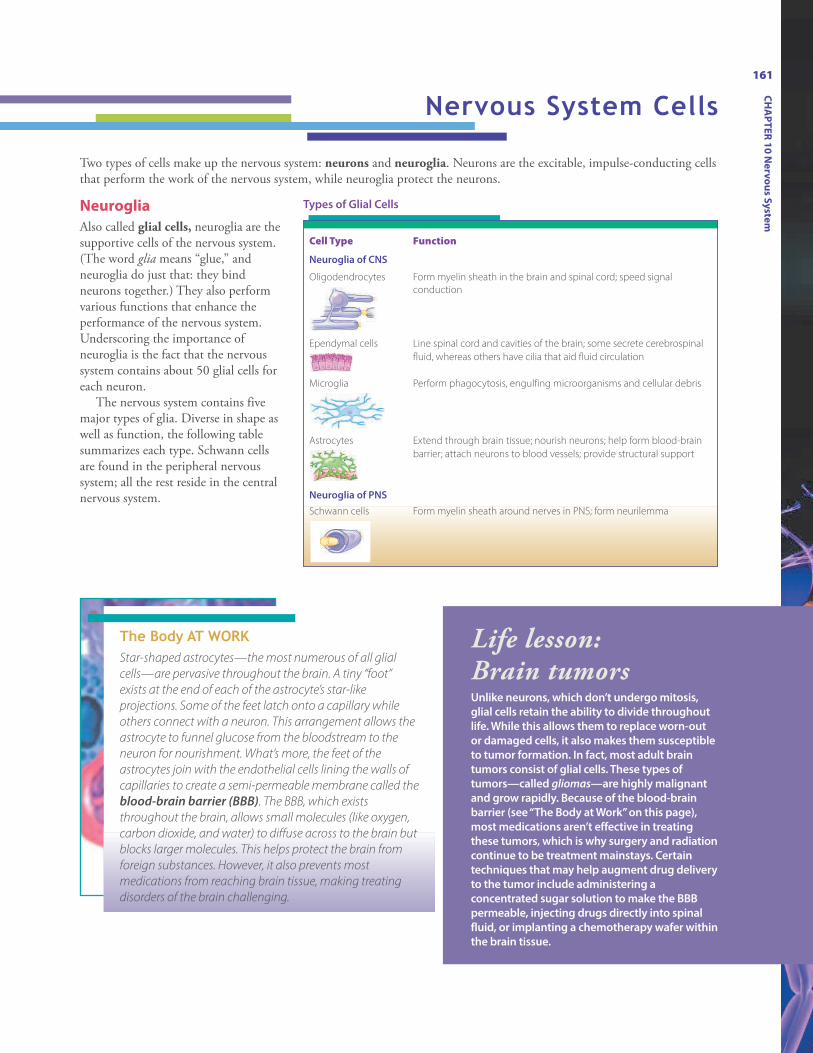
Neuroglia
Also called glial cells, neuroglia are thesupportive cells of the nervous system.(The word glia means “glue,” andneuroglia do just that: they bindneurons together.) They also performvarious functions that enhance theperformance of the nervous system.Underscoring the importance ofneuroglia is the fact that the nervoussystem contains about 50 glial cells foreach neuron.
The nervous system contains fivemajor types of glia. Diverse in shape aswell as function, the following tablesummarizes each type. Schwann cellsare found in the peripheral nervoussystem; all the rest reside in the centralnervous system.
Cell Type Function
Neuroglia of CNS
Oligodendrocytes Form myelin sheath in the brain and spinal cord; speed signal
conduction
Ependymal cells Line spinal cord and cavities of the brain; some secrete cerebrospinal
fluid, whereas others have cilia that aid fluid circulation
Microglia Perform phagocytosis, engulfing microorganisms and cellular debris
Astrocytes Extend through brain tissue; nourish neurons; help form blood-brain
barrier; attach neurons to blood vessels; provide structural support
Neuroglia of PNS
Schwann cells Form myelin sheath around nerves in PNS; form neurilemma
Life lesson: Brain tumorsUnlike neurons, which don’t undergo mitosis,
glial cells retain the ability to divide throughout
life. While this allows them to replace worn-out
or damaged cells, it also makes them susceptible
to tumor formation. In fact, most adult brain
tumors consist of glial cells. These types of
tumors—called gliomas—are highly malignant
and grow rapidly. Because of the blood-brain
barrier (see “The Body at Work” on this page),
most medications aren’t effective in treating
these tumors, which is why surgery and radiation
continue to be treatment mainstays. Certain
techniques that may help augment drug delivery
to the tumor include administering a
concentrated sugar solution to make the BBB
permeable, injecting drugs directly into spinal
fluid, or implanting a chemotherapy wafer within
the brain tissue.
Nervous System Cells
Types of Glial Cells
161
CH
AP
TE
R 1
0 N
erv
ou
s Sy
stem

PA
RT
III Re
gu
latio
n a
nd
Inte
gra
tion
of th
e B
od
y
162
Neurons
Nerve cells called neurons handle the nervous system’s role of communication. There are three classes of neurons: sensory(afferent) neurons, interneurons, and motor (efferent) neurons. Each neuron type fulfills one of the three general functionsof the nervous system.
Sensory neurons
Sensory (afferent) neurons
detect stimuli—such as touch,
pressure, heat, cold, or chemicals—
and then transmit information
about the stimuli to the CNS.
Interneurons
Interneurons, which are found only in the CNS, connect
the incoming sensory pathways with the outgoing motor
pathways. Besides receiving, processing, and storing
information, the connections made by these neurons
make each of us unique in how we think, feel, and act.
Motor neurons
Motor (efferent) neurons relay
messages from the brain (which the
brain emits in response to
stimuli) to the muscle or gland cells.
Dendrite
Axon branch
Dendrite
Axon branch
Axon branch Axon branch
Types of Neurons
Neurons vary greatly in both size and shape. They also vary according to the type, number, and length of projections.
Multipolar neurons
Multipolar neurons have one axon and multiple dendrites. This is
the most common type of neuron and includes most neurons of the
brain and spinal cord.
Bipolar neurons
Bipolar neurons have two processes: an axon and a dendrite with
the cell body in between the two processes. These neurons can be
found in the retina of the eye and olfactory nerve in the nose.
Unipolar neurons
Unipolar neurons have one process—an axon—that extends from
the cell body before branching in a T shape. These neurons mostly
reside in the sensory nerves of the peripheral nervous system.
FAST FACTAbout 90% of the body’s neurons are interneurons.

163
CH
AP
TE
R 1
0 N
erv
ou
s Sy
stem
Neuron Structure
Neurons are perhaps the most diverse of all body cells, assuming a variety of shapes and sizes. In general, though, neuronshave three basic parts: a cell body and two extensions called an axon and a dendrite.
Nucleus
FAST FACTThe sciatic nerve contains thelongest axon in the body; itextends from the base of thespine to the big toe in each foot.
The cell body (also called the soma) is the
control center of the neuron and contains
the nucleus.
Dendrites, which look like the bare
branches of a tree, receive signals
from other neurons and conduct the
information to the cell body. Some neurons
have only one dendrite; others have
thousands.
The axon, which carries nerve signals away
from the cell body, is longer than the
dendrites and contains few branches.
Nerve cells have only one axon; however,
the length of the fiber can range from a
few millimeters to as much as a meter.
The axons of many (but not all) neurons are
encased in a myelin sheath. Consisting
mostly of lipid, myelin acts to insulate the
axon. In the peripheral nervous system,
Schwann cells form the myelin sheath. In
the CNS, oligodendrocytes assume this role.
(For more information, see “Myelin” on the
next page.)
Gaps in the myelin sheath, called nodes of
Ranvier, occur at evenly spaced intervals.
The end of the axon branches extensively,
with each axon terminal ending in a
synaptic knob. Within the synaptic knobs
are vesicles containing a neurotransmitter.
PA
RT
III Re
gu
latio
n a
nd
Inte
gra
tion
of th
e B
od
y
164
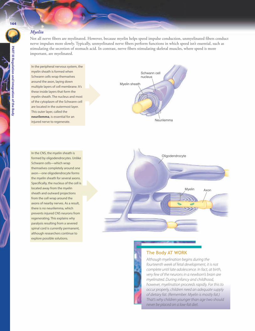
Myelin
Not all nerve fibers are myelinated. However, because myelin helps speed impulse conduction, unmyelinated fibers conductnerve impulses more slowly. Typically, unmyelinated nerve fibers perform functions in which speed isn’t essential, such asstimulating the secretion of stomach acid. In contrast, nerve fibers stimulating skeletal muscles, where speed is moreimportant, are myelinated.
Schwann cell nucleus
Neurilemma
Myelin sheath
Oligodendrocyte
Myelin Axon
In the peripheral nervous system, the
myelin sheath is formed when
Schwann cells wrap themselves
around the axon, laying down
multiple layers of cell membrane. It’s
these inside layers that form the
myelin sheath. The nucleus and most
of the cytoplasm of the Schwann cell
are located in the outermost layer.
This outer layer, called the
neurilemma, is essential for an
injured nerve to regenerate.
In the CNS, the myelin sheath is
formed by oligodendrocytes. Unlike
Schwann cells—which wrap
themselves completely around one
axon—one oligodendrocyte forms
the myelin sheath for several axons.
Specifically, the nucleus of the cell is
located away from the myelin
sheath and outward projections
from the cell wrap around the
axons of nearby nerves. As a result,
there is no neurilemma, which
prevents injured CNS neurons from
regenerating. This explains why
paralysis resulting from a severed
spinal cord is currently permanent,
although researchers continue to
explore possible solutions.
The Body AT WORKAlthough myelination begins during the
fourteenth week of fetal development, it is not
complete until late adolescence. In fact, at birth,
very few of the neurons in a newborn’s brain are
myelinated. During infancy and childhood,
however, myelination proceeds rapidly. For this to
occur properly, children need an adequate supply
of dietary fat. (Remember: Myelin is mostly fat.)
That’s why children younger than age two should
never be placed on a low-fat diet.

165
CH
AP
TE
R 1
0 N
erv
ou
s Sy
stem
When nerves are injured (such as from a cut, crushing injury, or some other type of trauma), their ability to repairthemselves depends upon the extent of the injury as well as their location. Nerves in the peripheral nervous system canregenerate as long as the soma and neurilemma are intact. Because nerves in the central nervous system lack a neurilemma,they cannot regenerate. Therefore, most injuries to the brain and spinal cord cause permanent damage. The followingfigures illustrate the repair process in a somatic motor neuron.
Site of injury
MacrophagesDegenerating
Schwann cells
New Schwann
cells
Muscle fiber
Regeneration
tunnel
Growth
processes
1When a nerve fiber is cut, the distal portion of the axon
is separated from its source of nutrition. Consequently, it
begins to degenerate along with the myelin sheath and
Schwann cells. Macrophages move in to clean up the resulting
debris.
2 Because the muscle fibers normally innervated by the
nerve are deprived of nervous input, they begin to
atrophy, or shrink. Meanwhile, the severed portion of the axon
sprouts new growth processes. At the same time, the
neurilemma forms a tunnel near the site of the injury; new
Schwann cells grow within the tunnel.
3When one of the new growth processes finds its way into
the tunnel, it begins to grow rapidly (3 to 5 mm/day). At
that point, the other growth processes begin to retract.
4 The new fiber continues to grow, guided by the tunnel,
until it reestablishes contact with the muscle. After that
occurs, the reinnervated muscle fibers regrow.
Repair of Nerve Fibers
Life lesson: Nerve injuriesWhen a peripheral nerve is severed, neurosurgeons may try to realign the nerveends surgically. If the severed ends aren’t adjacent to one another, the surgeonmay use a nerve or vein graft to bridge the gap. Success with these techniques isvariable, however. Another method currently being researched is the use ofsynthetic guidance channels to help direct newly growing axons. The channelsmay be implanted empty, or they may be filled with growth factors or neuralcells.

PA
RT
III Re
gu
latio
n a
nd
Inte
gra
tion
of th
e B
od
y
166
To relay messages to organs and tissues throughout the body, nerves must initiate and then transmit signals from one neuronto the next neuron at lightning speed. Signal transmission occurs through an electrical current, which, like all electricalcurrents, results from the flow of charged particles from one point to another.
In the body, whenever ions with opposite electrical charges are separated by a membrane, the potential exists for them tomove toward one another (depending, of course, upon the permeability of the membrane). This is called membranepotential. A membrane that exhibits membrane potential—an excess of positive ions on one side of the membrane and anexcess of negative ions on the other side—is said to be polarized.
+–
+–
+–
+–
+–
+–
+–
+–
+–
+–
+–
+–
+–
+–
+–
+–
+–
+–
+–
+–
+–
+–
+–
+–
+–
+–
+–
+–
+–
+–
+–
+–
+–
+–
+–
+–
Na+
When a neuron is not conducting an electrical signal, its interior has a negative electrical charge, while the charge on theoutside is positive. The outside of the cell is rich with sodium ions (Na1) while the inside contains an abundance ofpotassium ions (K1). The interior of the cell contains other ions as well, particularly large, negatively charged proteins andnucleic acids. These additional particles give the cell’s interior its overall negative charge. Because of the membrane’spermeability, a certain amount of sodium and potassium ions leak across the membrane. However, the sodium-potassiumpump constantly works to restore the ions to the appropriate side. (For more information on the sodium-potassium pump,see Chapter 3, Cells.) This state of being inactive and polarized is called resting potential. The neuron is resting, but it hasthe potential to react if a stimulus comes along.
1 Resting potential
• Inside of cell has negative charge;
outside has positive charge
• Exterior rich in Na1; interior rich
in K1
2Depolarization
• Stimulus causes Na1 to enter cell
• Region of interior changes from
negative to positive
When a stimulus (such as chemicals, heat, or mechanical pressure) comes along, channels on the resting neuron’s membraneopen and the Na1 from outside the membrane rushes into the cell. The addition of all these positively charged ions changesthe charge of a region of the cell’s interior from negative to positive. As the membrane becomes more positive, it is said todepolarize.
Impulse Conduction
ANIMATION

+–
+–
+–
+–
+–
+–
+–
+–
+–
+–
+–
+–
+–
+–
+–
+–
+–
+–
Na+
If the depolarization is strong enough—in other words, if the stimulus goes above what’s known as the threshold level—adjacent channels also open, allowing even more Na1 to flood the cell’s interior. This creates an action potential, meaningthat the neuron has become active as it conducts an impulse along the axon. Another term for action potential is nerveimpulse. The action potential continues down the axon as one segment stimulates the segment next to it.
3Action potential
• Channels in adjacent areas open
and more Na1 enters the cell
• Nerve impulse continues down the
length of the axon
+–
+–
+–
+–
+–
+–
+–
+–
+–
+–
+–
+–
+–
+–
+–
+–
+–
+–
Na+
K+
K+
Meanwhile, the sudden influx of Na1 triggers the opening of other channels to allow K1 to flow out of the cell. Soon afterK1 begins to exit, the Na1 channels shut to prevent any more Na1 from flowing into the cell. This repolarizes the cell;however, Na1 and K1 are now flip-flopped, with the outside containing more K1 and the inside containing more Na1.
4 Repolarization
• K1 flows out of cell
• Electrical balance restored: interior
has negative charge and exterior
has positive charge
+–
+–
+–
+–
+–
+–
+–
+–
+–
+–
+–
+–
+–
+–
+–
+–
+–
+–
K+K+
Na+
Na+
Na+
Na+
Although the membrane is polarized, the neuron won’t respond to a new stimulus as long as the Na1 and K1 are on thewrong sides of the membrane. This is known as the refractory period. The sodium-potassium pump works to return Na1
to the outside and K1 to the inside. When this is completed, the nerve is again polarized and in resting potential until itreceives another stimulus.
5 Refractory period
• Membrane is polarized, but Na1
and K1 are on wrong sides of
membrane
• Sodium-potassium pump works to
restore ions to rightful sides
The Body AT WORKAction potential is an “all or nothing” event. When a stimulus reaches a threshold and
depolarizes the neuron, the neuron fires at its maximum voltage. If the stimulus doesn’t reach
the threshold, the neuron doesn’t fire at all. What’s more, a stronger stimulus doesn’t produce
a stronger response. In this way, as each neuron segment triggers firing in the segment next
to it, the nerve impulse continues at the same strength all the way to the synaptic knobs.
167
CH
AP
TE
R 1
0 N
erv
ou
s Sy
stem

PA
RT
III Re
gu
latio
n a
nd
Inte
gra
tion
of th
e B
od
y
168
FAST FACTNeurons can transmitimpulses amazingly fast: upto 120 meters per second(268 miles per hour).
+–
+–
+–
+– +
–
+–
+–
+–
+–
+–
Electrical changes occur at the nodes of Ranvier, creating an action potential. Thecurrent flows under the myelin sheath to the next node, where it triggers anotheraction potential.
Impulse Conduction in Myelinated Fibers
Nerve impulses move through unmyelinated fibers as previously described. In myelinated fibers, however, the thick layer ofmyelin encasing the axons of most nerve fibers blocks the free movement of ions across the cell membrane. The only placeion exchange can occur is at the nodes of Ranvier: the evenly spaced gaps in myelin. The following illustration shows how anerve impulse travels down a myelinated fiber.
+–
+–
+–
+–+
–
+–
+–
+–
+–
+–
This process continues as the signal moves down the axon. Because the actionpotentials occur only at the nodes, the impulse seems to “leap” from node to node.This type of signal conduction is called saltatory conduction. (The word saltatorycomes from the Latin word saltare, which means “to leap.”)
Life lesson: Multiple sclerosisMultiple sclerosis (MS) is a disease in which the myelin sheaths surrounding the nerves of the CNS deteriorate and arereplaced by hard scar tissue (called plaques). These changes disrupt nerve conduction and cause symptoms that vary,depending upon which nerves are affected. Common symptoms include visual disturbances (such as blindness ordouble vision), weakness, loss of coordination, and speech disturbances. The disease progresses over many years;symptoms typically improve and then worsen in unpredictable cycles. MS tends to strike women between the ages of20 and 40 years. Although the exact cause is unknown, experts speculate that a virus may trigger an autoimmunereaction in which the patient’s immune system attacks the myelin of the CNS. There is no known cure.
ANIMATION

169
CH
AP
TE
R 1
0 N
erv
ou
s Sy
stem
FAST FACTOne neuron can have multiple synapses. In fact, in aparticular portion of the brain, one neuron can have up to100,000 synapses.
Nerve impulses usually travel through several different neurons before reaching their target organ or tissue. For this tohappen, the impulse must have some way of transferring from one neuron to the next. The area where this occurs is called asynapse. Chapter 9 discussed the synapses that occur between nerves and muscles; now we’ll examine the synapses thatoccur between two neurons.
Some synapses (such as those between cardiac muscle cells and certain types of smooth tissue cells) are electrical. In theseinstances, adjacent neurons touch, which allows an action potential to pass smoothly from one neuron to the next. Morecommonly, synapses are chemical. In these instances, the two neurons don’t touch. Instead, a chemical called aneurotransmitter bridges a very narrow gap (the synaptic cleft) to carry the message from the first neuron (the presynapticneuron) to the next (the postsynaptic neuron). Although greatly simplified, here’s what basically happens:
Ca2+
Action potentialAxon DendriteAction potential+–
+–
+–
+–
+–
+–
Na+
1When an action potential
reaches a synaptic knob, the
membrane depolarizes. This causes
ion channels to open, which allows
calcium ions to enter the cell.
2 The infusion of calcium causes
vesicles to fuse with the plasma
membrane and then release their
store of a neurotransmitter into the
synapse.
3Once released, the neurotransmitter
binds to receptors on the postsynaptic
membrane. Each neurotransmitter has a
specific receptor. (For example, the
neurotransmitter epinephrine can bind
only to receptors specific to epinephrine.)
4 The specific neurotransmitter
determines whether the impulse
continues (called excitation) or whether it
is stopped (called inhibition). If the
neurotransmitter is excitatory—as shown
here—Na1 channels open, the membrane
becomes depolarized, and the impulse
continues. If the impulse is inhibitory, K1
channels open, and the impulse stops.
5 The receptor then releases the neuro-
transmitter, after which it is reabsorbed
by the synaptic knobs and recycled or
destroyed by enzymes (as shown here).
FAST FACTScientists have discovered more than 100 differentneurotransmitters in the human body. Some commonneurotransmitters include acetylcholine, epinephrine,norepinephrine, serotonin, dopamine, and histamine.
Synapses
ANIMATION
PA
RT
III Re
gu
latio
n a
nd
Inte
gra
tion
of th
e B
od
y
170
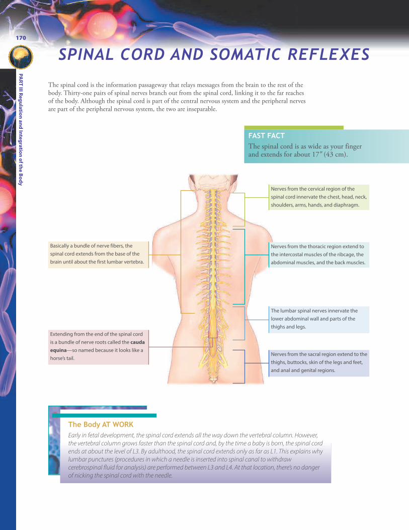
The spinal cord is the information passageway that relays messages from the brain to the rest of thebody. Thirty-one pairs of spinal nerves branch out from the spinal cord, linking it to the far reachesof the body. Although the spinal cord is part of the central nervous system and the peripheral nervesare part of the peripheral nervous system, the two are inseparable.
Basically a bundle of nerve fibers, the
spinal cord extends from the base of the
brain until about the first lumbar vertebra.
Extending from the end of the spinal cord
is a bundle of nerve roots called the cauda
equina—so named because it looks like a
horse’s tail.
Nerves from the cervical region of the
spinal cord innervate the chest, head, neck,
shoulders, arms, hands, and diaphragm.
Nerves from the thoracic region extend to
the intercostal muscles of the ribcage, the
abdominal muscles, and the back muscles.
The lumbar spinal nerves innervate the
lower abdominal wall and parts of the
thighs and legs.
Nerves from the sacral region extend to the
thighs, buttocks, skin of the legs and feet,
and anal and genital regions.
FAST FACTThe spinal cord is as wide as your fingerand extends for about 1799(43 cm).
The Body AT WORKEarly in fetal development, the spinal cord extends all the way down the vertebral column. However,
the vertebral column grows faster than the spinal cord and, by the time a baby is born, the spinal cord
ends at about the level of L3. By adulthood, the spinal cord extends only as far as L1. This explains why
lumbar punctures (procedures in which a needle is inserted into spinal canal to withdraw
cerebrospinal fluid for analysis) are performed between L3 and L4. At that location, there’s no danger
of nicking the spinal cord with the needle.
SPINAL CORD AND SOMATIC REFLEXES

171
CH
AP
TE
R 1
0 N
erv
ou
s Sy
stem
Vertebral body
Anterior horn
Posterior horn
Spinal nerve
The spinal cord sits inside a protective, bony tunnel created by the stacked vertebrae. A cross section clearly shows the twotypes of nervous tissue (white matter and gray matter) that make up the spinal cord.
Gray matter—which appears gray
because of its lack of myelin—
contains mostly the cells bodies of
motor neurons and interneurons.
This H-shaped mass is divided into
two sets of horns: the posterior
(dorsal) horns and the ventral
(anterior) horns.
White matter appears white
because of its abundance of myelin.
It contains bundles of axons (called
tracts) that carry impulses from one
part of the nervous system to
another.
A minute opening called the central
canal carries cerebrospinal fluid
through the spinal cord.
A small space—called the epidural
space—lies between the outer
covering of the spinal cord and the
vertebrae; it contains a cushioning
layer of fat as well as blood vessels
and connective tissue.
The dorsal (posterior) nerve
root contains fibers that carry
sensory information into the
spinal cord. It enters the dorsal
horn of the spinal cord.
Cell bodies of the dorsal
neurons are clustered in a
knot-like structure called a
ganglion.
A spinal nerve is a single nerve
resulting from the fusion of the
dorsal and ventral nerve roots.
Because the nerve contains
both sensory and motor
fibers—meaning it can
transmit impulses in two
directions—it’s called a mixed
nerve.
Fibers in the ventral (anterior)
nerve roots exit from the
ventral horn to carry motor
information out of the spinal
cord.
The pia mater is the innermost
layer. This transparent membrane
clings to the outer surface of the
brain and spinal cord. It also
contains blood vessels.
The subarachnoid space lies
between the arachnoid mater and
the pia mater. It is filled with
cerebrospinal fluid.
The arachnoid mater—a delicate
layer resembling a cobweb—lies
between the dura mater and the
pia mater.
The dura mater is the tough
outer layer.
Attachment of Spinal Nerves
Spinal nerves travel through gaps between the vertebrae(which are held apart by intervertebral discs) and attach tothe spinal cord by way of two roots: the dorsal and theventral roots.
Meninges of the Spinal Cord
In addition to the bony protection offered by the vertebrae,the spinal cord is further protected by three layers of fibrousconnective tissue, called the meninges. (The meninges alsocovers the brain.) The three layers of the meninges, fromthe inside out, are the pia mater, the arachnoid mater, andthe dura mater.
Structure of the Spinal Cord

PA
RT
III Re
gu
latio
n a
nd
Inte
gra
tion
of th
e B
od
y
172
That Makes SenseTo remember the difference between ascending and
descending tracts, think about this: When you step on a
nail, the sensation of pain ascends to your brain.
(Ascending: sensory.) In response, your brain issues an
impulse that travels down to your foot, telling you to
move. (Descending: motor.)
!
Spinal Tracts
Within the white matter of the spinal cord are bundles of axons called tracts that serve as the routes of communication toand from the brain. All the nerve fibers of a single tract have a similar origination, destination, and function. As an example,the fibers of the spinothalamic tract originate in the spinal cord (spino-) and end in the thalamus (thalamic). In addition,they all convey sensations of pain, touch, and temperature to the thalamus in the brain.
Some of the most important tracts are highlighted in the figure below. Note: All tracts exist on both sides of the spinalcord, but, in this illustration, the ascending tracts are highlighted on the left and the descending tracts on the right.
The dorsal column relays
sensations of deep pressure and
vibration as well as those needed
to create awareness of the body’s
position (proprioception).
The spinocerebellar tract is
responsible for proprioception.
The spinothalamic tract relays
sensations of temperature,
pressure, pain, and touch.
The corticospinal tracts (also
called the pyramidal tracts)
are responsible for fine
movements of hands, fingers,
feet, and toes on the opposite
side of the body.
The extrapyramidal tracts are
a group of tracts associated
with balance and muscle tone.
Ascending tracts convey sensory signals (such as pain) up the
spinal cord to the brain.
Descending tracts conduct motor impulses down the spinal
cord to skeletal muscles.
The Body AT WORKMost of the spinal cord tracts cross from one side
of the body to the other in the brainstem. This is
called decussation. For example, sensory signals
from the right side of the body are sent to the left
side of the brain. Also, motor signals being sent to
the right side of the body originate on the left side
of the brain. This is why someone who suffers a
stroke affecting motor centers in the left side of the
brain will have weakness or paralysis on the right
side of the body and vice versa.
FAST FACTAll the axons in a given tractserve one general function.

173
CH
AP
TE
R 1
0 N
erv
ou
s Sy
stem
Life lesson: Spinal cord injuryOver 10,000 people in the United States suffer from spinal cord injuries each year. Males between the ages of 16and 30 have the greatest risk, mainly because of their tendency for high-risk behaviors. Most injuries result fromcar and motorcycle accidents.
If the spinal cord is severed—often because of a vertebral fracture—it causes a loss of movement and sensationbelow the level of the injury. For example, a spinal cord injury between the levels of T1 and L1 causes paralysis inthe legs ( paraplegia); an injury above the C5 vertebra causes paralysis in all the limbs (quadriplegia). An injuryabove C4 is especially serious because this is where the phrenic nerve exits the spinal cord. Because the phrenicnerve innervates the diaphragm, an injury here can cause respiratory failure.
Spinal nerves (part of the peripheral nervous system) relay information from the spinal cord to the rest of the body.
That Makes SenseTo make sense of the terms used for nerves and nerve tracts, think about this: Sensation
always travels toward the CNS. Sensory nerves transmit impulses toward the spinal cord;
once there, they travel up the spinal cord (ascend) along the ascending tract. As a further
hint, remember that afferent (sensory) nerves link to the ascending tract. Motor nerves
carry messages about movement; therefore, those impulses leave (or exit) the spinal cord
along efferent (motor) nerves.
!
Blood vessels
Axons
Myelin
sheath
Connective tissue
Most nerves contain both sensory and motor fibers and are called mixed nerves. These nerves can transmit signals in twodirections. A few nerves (such as the optic nerves) are sensory nerves and contain only sensory (afferent) fibers. They carrysensations toward the spinal cord. Others are motor nerves and contain only motor (efferent) fibers and carry messages tomuscles and glands.
A nerve consists of many nerve fibers
(axons) encased by connective tissue. The
number of nerve fibers contained in a
single nerve varies from a few to as many
as a million. (Remember: A neuron is a
nerve cell; a nerve contains many neurons.)
Nerve fibers are gathered together in
bundles called fascicles; in turn, several
fascicles are grouped together—along
with blood vessels—and wrapped in a
dense connective tissue.
Spinal Nerves

PA
RT
III Re
gu
latio
n a
nd
Inte
gra
tion
of th
e B
od
y
174
C1
C2C3
T4
T10
L2
L3
L4
L5
S1
S2
S4
S5
S3
Co1
L1
T12
T11
T9
T8
T7
T6
T5
T3T2
T1C8C7C6C5
C4
Phrenic nerve
Axillary
nerve
Radial
nerve
Median
nerve
Ulnar
nerve
Femoral
nerve
Sciatic
nerve
Categories of Spinal Nerves
Thirty-one pairs of spinal nerves connect to the spinal cord. They include:
l 8 cervical nerves (C1-C8)l 12 thoracic nerves (T1-T12)l 5 lumbar nerves (L1-L5)l 5 sacral nerves (S1-S5)l 1 coccygeal nerve (Co)
The first cervical nerve exits the spinal cord between the skull and theaxis. The other nerves pass through holes in the vertebra (intervertebralforamina).
Once outside the spinal column, each spinal nerve forms several largebranches. Some of these branches subdivide further to form nervenetworks called plexuses. The four major plexuses are the cervical plexus,the brachial plexus, the lumbar plexus, and the sacral plexus.
The cervical plexus contains nerves that supply the muscles and
skin of the neck, tops of the shoulders, and part of the head. The
phrenic nerve, which stimulates the diaphragm for breathing, is
located here.
The brachial plexus innervates the lower part of the shoulder and
the arm. Key nerves traveling into the arm from this region include
the axillary nerve (which passes close to the armpit, making it
susceptible to damage from the use of crutches), the radial nerve,
the ulnar nerve, and the median nerve.
The lumbar plexus—derived from the fibers of the first four
lumbar vertebrae—supplies the thigh and leg. A key nerve in this
region is the large femoral nerve.
The sacral plexus is formed from fibers from nerves L4, L5, and S1
through S4. (Because of the co-mingling of fibers of the sacral
plexus with those of the lumbar plexus, these two plexuses are
often referred to as the lumbosacral plexus.) The sciatic nerve, the
largest nerve in the body, arises here and runs down the back of the
thigh. Irritation of this nerve causes severe pain down the back of
the leg, a condition called sciatica.

175
CH
AP
TE
R 1
0 N
erv
ou
s Sy
stem
Dermatomes
Each spinal nerve (except for C1) innervates a specific area of the skin.These areas are called dermatomes. Clinicians use this information toidentify the location of a nerve abnormality by testing a patient’s responseto pinpricks in the different areas.
Reflexes are a quick, involuntary, predictable response to a stimulus. Reflexes employ a neural circuit called a reflex arc,which bypasses regions of the brain where conscious decisions are made. That’s why someone becomes aware of a reflex onlyafter it’s occurred. Some reflexes—called autonomic (visceral) reflexes—involve secretion from glands or the contraction ofsmooth muscle (such as dilation of the pupil). These reflexes are governed by autonomic neurons, which will be discussedlater in this chapter.
Somatic reflexes involve the contraction of a skeletal muscle after being stimulated by a somatic motor neuron. Somaticreflexes often help protect the body against harm—such as causing you to withdraw your hand from a hot stove. Otherreflexes help you maintain your posture. (See “The Body at Work” on this page.) Several reflexes (such as the patellar reflex,described below) are commonly tested during physical exams to identify certain diseases.
1 Somatic receptors (located
in the skin, a muscle, or a
tendon) detect a sensation, such
as the stretching of the thigh
muscle when the patellar
tendon is tapped.
2 Afferent (sensory) nerve fibers send
a signal directly to the spinal cord. 3The impulse immediately
passes to a motor neuron.
4 The motor neuron initiates an
impulse back to the muscle,
causing it to contract, producing a
slight kick in the lower leg.
The Body AT WORKYour ability to stand, walk, and correct your balance can all be
attributed to reflexes. Specifically, skeletal muscles contain
sensory receptors that send messages to the brain about the
amount of stretch in a muscle as well as the movement of body
parts. This allows the brain to emit signals to correct muscle
tone and control movement; it also allows it to trigger a reflex
to correct posture. For example, keeping your balance can be
attributed to the reflexive contracting and relaxing of various
muscles—all without your awareness.
Somatic Reflexes
C2C3C4
C7
T1
T9T8
T7T6T5
T4T3T2
L2L1T12T11
T10
S2
S1
L5
L4
L3
S3
Cervical nerves
Thoracic nerves
Lumbar nerves
Sacral nerves
C5
T1
C5C6C8
ANIMATION

PA
RT
III Re
gu
latio
n a
nd
Inte
gra
tion
of th
e B
od
y
176
The brain is divided into four major regions: the cerebrum, the diencephalon, the cerebellum, and the brainstem.
THE BRAIN AND CRANIAL NERVESThe brain is the site for thought, learning, reasoning, memory, and creativity. Indeed, the brain performs numerous amazingfunctions, many of which remain beyond our grasp.
Gyri Sulci
Cerebral
hemispheres
The cerebrum is the largest portion of the
brain. Its surface is marked by thick ridges
called gyri (singular: gyrus). Shallow
grooves called sulci (singular: sulcus) divide
the gyri. Deep sulci are called fissures.
The diencephalon sits between the
cerebrum and the midbrain.
The cerebellum is the second largest
region of the brain. Although smaller than
the cerebrum, it contains more neurons
than the rest of the brain combined.
The brainstem makes up the rest of the
brain. It consists of three structures:
• Midbrain
• Pons
• Medulla oblongataA deep groove called the
longitudinal fissure divides the
cerebrum into right and left
cerebral hemispheres. A thick
bundle of nerves called the corpus
callosum runs along the bottom of
the fissure and serves to connect
the two hemispheres.
Gray and White Matter
Like the spinal cord, the brain contains both gray and white matter. Unlike the spinalcord (in which gray matter forms the interior), in the brain, gray matter forms thesurface. Specifically, gray matter (consisting of cell bodies and interneurons) covers thecerebrum and cerebellum in a layer called the cortex. Underneath the cortex is whitematter, although gray matter exists in patches called nuclei throughout the whitematter. The white matter contains bundles of axons that connect one part of the brainto another.
The Body AT WORKBecause the brain’s gray matter (the part charged with thought, learning, and
reasoning) is located at its surface, the folds allow more gray matter to be
packed into the small area of the skull. (As an analogy, think of fitting a large
piece of paper into a small space: it becomes possible if you crunch it into a
ball.) Scientists have long thought that the brain’s folds explain why humans
are more intelligent than species with smoother brains. Recent discoveries
have revealed that the folding pattern varies with each individual, making a
person’s brain folds as unique as his fingerprints. What’s more, scientists have
discovered abnormal folding patterns in those suffering from a variety of
mental and neurodevelopment disorders, ranging from depression to autism.
General Structures of the Brain

177
CH
AP
TE
R 1
0 N
erv
ou
s Sy
stem
FAST FACTMen have larger brains than women.However, brain size is proportional to body size and does not reflect intelligence.
Brain:
Gray matter
White matter
Arachnoid villus
Meninges of the Brain
Like the spinal cord, meninges covers the outside surface of the brain, offering protection. Nearby bone also helps protectthe brain from trauma.
The bones of the skull provide the
outermost protection.
Inside the skull, three layers of
meninges cover the brain. The
layers are the same as in the
spinal cord:
• The dura mater consists of
two layers: the outer layer
(the periosteal layer) is
attached to the inner surface
of the skull; the inner
meningeal layer forms the
outer covering of the brain
and continues as the dura
mater of the spinal cord.
• The arachnoid mater is the
middle layer.
• The pia mater clings tightly
to the surface of the brain.
In some locations, the dura mater
separates to create spaces called dural
sinuses. These sinuses collect blood
that has passed through the brain and
is on its way back to the heart.
A subdural space separates the
dura from the arachnoid mater.
A subarachnoid space
separates the arachnoid
mater from the pia
mater.
In some places, the dura mater extends
inward and separates major portions of the
brain. The falx cerebri, shown here, dips into
the longitudinal fissure to separate the
right and left hemispheres. Elsewhere, the
tentorium cerebella extends over the top of
the cerebellum, separating it from the
cerebrum.
Life lesson: MeningitisInfection or inflammation of the meninges is called meningitis. Infection may be caused by several differentbacteria or viruses that gain entry to the central nervous system by spreading from other locations in the body,such as from an ear or sinus infection. Bacterial meningitis occurs less frequently than viral meningitis, but it canbe life-threatening without immediate treatment.
Symptoms of meningitis include fever, stiff neck, irritability, headache, drowsiness, and seizures. Infants withmeningitis may have different symptoms, including poor feeding, bulging fontanelles, and a high-pitched cry.Viral meningitis usually causes milder symptoms, such as those similar to a cold or the flu. In fact, viral meningitisoften goes undiagnosed because the symptoms are so mild.
To diagnose meningitis, a sample of cerebrospinal fluid is obtained through a lumbar puncture. The fluid isthen examined for bacteria and white blood cells, a sign of inflammation. Viral meningitis usually resolves on itsown in 7 to 10 days, while bacterial meningitis requires hospitalization and treatment with intravenous antibiotics.

PA
RT
III Re
gu
latio
n a
nd
Inte
gra
tion
of th
e B
od
y
178
Ventricles
The brain contains four chambers, called ventricles.
Central canal
Cerebrospinal Fluid
A clear, colorless fluid called cerebrospinal fluid (CSF) fills the ventricles and central canal; it also bathes the outside of thebrain and spinal cord. CSF is formed from blood by the choroid plexus (a network of blood vessels lining the floor or wallof each ventricle).
Two lateral ventricles arch through the cerebral
hemispheres: one in the right hemisphere and one in the
left.
Each of the lateral ventricles connects to a third ventricle.
A canal then leads to the fourth ventricle. This space
narrows to form the central canal, which extends through
the spinal cord.
Life lesson: HydrocephalusIf the flow of CSF becomes blocked anywhere on its route, the fluid accumulates in the brain’s ventricles. Thiscondition is called hydrocephalus, or, more commonly, “water on the brain. ” The accumulating CSF causes theventricles to expand. In an infant whose cranial bones haven’t fused, the entire head expands. An adult, however,has no such “release valve.” In this situation, the expanding ventricles compress the brain tissue against the sidesof the skull and intracranial pressure rises. Untreated, the condition can prove fatal. It can, however, besuccessfully treated by inserting a tube, or shunt, to drain fluid from the ventricles into a vein in the neck.
FAST FACTAlthough the brain constitutes only 2%of an adult’s body weight, it receives15% of the blood and consumes 20% ofthe body’s oxygen and glucose.
The Body AT WORKThe brain produces about 500 ml of CSF each day; however,
much of that is reabsorbed. At any one time, an adult brain
contains about 140 ml of CSF. CSF is not stagnant; rather, it
constantly flows through the central nervous system, providing
nourishment in the form of glucose and protein and helping to
remove metabolic wastes.
CSF plays other roles as well. For example, CSF helps protect
the brain against minor trauma: When the head is jolted, CSF
acts as a cushion to keep the brain from striking the inside of the
skull. In addition, CSF plays a role in the maintenance of
homeostasis. Specifically, the brain monitors the level of CO2 in
CSF and triggers responses as needed to help the body regain
equilibrium.

179
CH
AP
TE
R 1
0 N
erv
ou
s Sy
stem
6
5
1
2
4
3
The choroid plexus in each lateral ventricle
secretes CSF.
The CSF flows into the third ventricle, where
another choroid plexus adds more fluid.
Some of the CSF moves into the central
canal of the spinal cord. Most flows
through two tiny openings (foramina) into
a space leading to the subarachnoid space.
It then flows into the fourth ventricle,
where still more CSF is added by the
choroid plexus in that ventricle.
The CSF then flows through the
subarachnoid space, up the back of the
brain, down around the spinal cord, and up
the front of the brain.
The CSF is reabsorbed into the venous
bloodstream by projections of the
arachnoid mater into the dural sinuses
(called arachnoid villi).
FAST FACTAn interruption in the flow of blood tothe brain for as little as 10 secondscauses unconsciousness; an interruptionfor 4 minutes produces irreparable braindamage.
Blood-Brain Barrier
The brain demands a high volume of blood to function properly.However, blood also contains substances such as antibodies andmacrophages that would harm the brain. As a means of protection, ablood-brain barrier serves to restrict what substances can pass fromthe bloodstream into the tissue fluid of the brain. This barrier consistsmainly of capillaries formed by tightly joined endothelial cells (asopposed to the loosely overlapping cells that form the capillaries ingeneral circulation). A thick basement membrane adds to the barrier,as do the feet of astrocytes, which reach out and surround theendothelial cells. This arrangement creates a semi-permeablemembrane throughout the brain. As discussed previously, themembrane allows small molecules (like oxygen, carbon dioxide, andwater) to diffuse across to the brain but blocks larger molecules. Othersubstances that can diffuse across the barrier include alcohol, nicotine,caffeine, and anesthetics. Trauma or inflammation can damage theblood-brain barrier and allow pathogens to enter. (For moreinformation, see “The Body at Work” under the section on neurogliaon page 161.)
123
4
5
6
Formation and Flow of CSF
FAST FACTThe circulation of CSF is aidedby pulsations in the choroidplexus and by the motion of thecilia of ependymal cells.
ANIMATION

The divisions of the brain, starting at the bottom, are the brainstem, cerebellum, diencephalon, and cerebrum.
Brainstem
The brainstem consists of the midbrain, pons, and medulla oblongata.
Thalamus
Spinal cord
The midbrain contains tracts that relay
sensory and motor impulses. It also
contains centers for auditory and visual
reflexes as well as clusters of neurons
integral to muscle control.
The pons contains tracts that convey
signals to and from different parts of the
brain. Several cranial nerves arise from this
area; they include cranial nerves V
(trigeminal), VI (abducens), VII (facial), and
VIII (vestibulocochlear). (The cranial nerves
will be discussed later in this chapter.)
The medulla oblongata attaches the brain to the spinal cord.
Besides relaying sensory and motor signals between the brain
and spinal cord, the medulla contains nuclei that perform
functions vital to human life. These include:
• The cardiac center, which regulates heart rate
• The vasomotor center, which controls blood vessel
diameter, which, in turn, affects blood pressure
• Two respiratory centers, which regulate breathing
The medulla also houses reflex centers for coughing, sneezing,
swallowing, and vomiting. Several cranial nerves (cranial nerve
IX [glossopharyngeal], X [vagus], XI [accessory], and XII
[hypoglossal]) either begin or end in the medulla.
FAST FACTBecause the medulla contains centers that regulateheart rate, blood pressure, and breathing, an injuryhere—such as from a blow to the base of the skull—can prove fatal.
Cerebellum
About the size of a fist, the cerebellum houses more neurons than the rest of the
brain combined. Connected to the cerebral cortex by approximately 40 million
neurons, the cerebellum receives, and processes, messages from all over the
brain. Long known to play a key role in motor functions, recent discoveries show
that the cerebellum assumes a powerful role in sensory, cognitive, and even
emotional functions as well. In brief, the cerebellum:
• Joins forces with the cerebral cortex to monitor body movements and send
messages crucial for balance, coordination, and posture
• Stores the information necessary for muscle groups to work together to
perform smooth, efficient, and coordinated movements
• Evaluates sensory input, such as touch, spatial perception, and sound
People with cerebellar dysfunction (such as from a tumor, hemorrhage, or
trauma) have a spastic gait, poor balance, jerky movements, and tremors. They
also tend to have poor impulse control and overreact emotionally.
PA
RT
III Re
gu
latio
n a
nd
Inte
gra
tion
of th
e B
od
y
180
Divisions of the Brain

The Body AT WORKScattered throughout the brainstem is a set of
interconnected nuclei called the reticular
formation. Fibers extend from there to many
parts of the cerebrum, the cerebellum, and the
spinal cord. One component of the reticular
formation is the reticular activating system
(RAS). Charged with maintaining a state of
wakefulness and alertness, the RAS receives
sensory input from the eyes and ears. After
filtering out insignificant signals (such as routine
noise), it sends impulses to the cerebral cortex so
the mind remains conscious and alert. Drugs
that depress the reticular activating system
induce sleep.
The reticular formation also has tracts
extending into the spinal cord that are involved in
posture and equilibrium. Other components of
the reticular formation include the cardiac and
vasomotor centers of the medulla oblongata,
which are responsible for heart rate and blood
pressure.
Thalamus
Shaped like two eggs sitting side by side, the thalamus resides
on the top of the brainstem. It acts as a gateway for nearly every
sensory impulse (including smell, sight, taste, pain, pressure, heat,
cold, and touch) travelling to the cerebral cortex. The thalamus
processes and filters these impulses, transmitting some, but not
all, to the cerebral cortex.
The thalamus plays other roles as well. For example, it relays
messages regarding certain complex movements; it also is
involved in memory and emotion.
Hypothalamus
The hypothalamus’ small size belies its crucial function. In fact,
this tiny area of the brain extends its influence to nearly every
organ of the body. The hypothalamus plays a key role in
numerous functions. For example, it:
• Controls the autonomic nervous system (which is responsible
for such vital functions as heart rate and blood pressure)
• Contains centers responsible for hunger, thirst, and temperature
regulation
• Controls the pituitary gland—often called the “master gland”
because of its influence on most endocrine glands (such as the
thyroid, testes, ovaries, and adrenal glands)
• Is involved in multiple emotional responses, including fear,
anger, pleasure, and aggression
Visual input
Reticular formation
Ascending sensory tracts
Descending motor tractsto spinal cord
Auditory input
Radiation to cerebral cortex
181
CH
AP
TE
R 1
0 N
erv
ou
s Sy
stem
Diencephalon
The diencephalon is a region deep inside the brain consisting ofseveral structures, with the chief ones being the thalamus andthe hypothalamus.

PA
RT
III Re
gu
latio
n a
nd
Inte
gra
tion
of th
e B
od
y
182
Central sulcus
Lateral sulcus
Precentral gyrus Postcentral
gyrus
Frontal lobe
• Central sulcus forms the posterior
boundary
• Governs voluntary movements,
memory, emotion, social judgment,
decision making, reasoning, and
aggression; is also the site for certain
aspects of one’s personality
Parietal lobe
• Central sulcus forms the anterior boundary
• Concerned with receiving and interpreting bodily
sensations (such as touch, temperature, pressure,
and pain); also governs proprioception (the
awareness of one’s body and body parts in space
and in relation to each other)
Occipital lobe
• Concerned with analyzing and
interpreting visual information
Temporal lobe
• Separated from the parietal lobe by the
lateral sulcus
• Governs hearing, smell, learning,
memory, emotional behavior, and visual
recognition
Insula
• Hidden behind the lateral sulcus
• Plays a role in many different functions,
including perception, motor control, self-
awareness, and cognitive functioning
Life lesson: Brain lesionsThe symptoms resulting from injuries to key areas of the brain have been a primary source of information aboutthe role those areas play. Following are some examples of symptoms that may occur following trauma or stroke tospecific brain regions.
• Parietal lobe lesion: Dysfunction in this part of the brain causes people to ignore objects on the opposite sideof the body—even their own arm and leg. Patients may dress only half their body and even deny that theopposite arm or leg belongs to them.
• Temporal lobe lesion: An injury here can impair the ability to identify familiar objects. Some may not evenrecognize their own face. In other instances, the person may lose the ability to differentiate between sounds,causing him to lose any appreciation of music.
• Frontal lobe lesion: A lesion or injury here can result in severe personality disorders and cause sociallyinappropriate behavior.
FAST FACTIf the brain’s surface were flattened, it wouldmeasure about 465 square inches (3000 cm2), orabout the size of an opened newspaper.
Cerebrum
The largest, and most obvious, portion of the brain is the cerebrum. Your ability to think, remember, feel, use judgment,and move can be credited to the cerebrum.
Some of the more obvious sulci (grooves) divide the cerebrum into five distinct lobes. Each lobe is named for the bonesof the skull that lie directly over them.

183
CH
AP
TE
R 1
0 N
erv
ou
s Sy
stem
Inside the Cerebrum
The Limbic System
Sometimes called the “emotional brain,” the limbic system is the seat of emotion and learning. It’s formed by a complex setof structures that encircle the corpus callosum and thalamus. In brief, it links areas of the lower brainstem (which controlautomatic functions) with areas in the cerebral cortex associated with higher mental functions. Two key structures of thelimbic system include the hippocampus and amygdala.
Feelings of anger, fear, sexual feelings, sorrow, and pleasure only result because of a functioning limbic system. However,to ensure that those feelings are expressed in socially acceptable ways, other parts of the cerebral cortex must also be engaged.Limbic system activity, without the moderating influence of other parts of the cerebrum, leads to attacks of uncontrollablerage.
Brainstem
Thalamus
The bulk of the cerebrum is white
matter, which consists of bundles of
myelinated nerve fibers, called tracts.
Tracts carry impulses from one part of
the cerebrum to the other, or from the
cerebrum to other parts of the brain or
spinal cord.
Most of the tracts that pass from one
hemisphere to the other travel
through a large “bridge” called the
corpus callosum. This arrangement
allows the brain’s two hemispheres to
communicate with each other.
The surface of the cerebrum, called the
cerebral cortex, consists of a thin layer
of gray matter. (Even though the layer
is thin, gray matter actually makes up
about 40% of the brain’s mass.)
Masses of gray matter—called basal
nuclei, or, sometimes, basal
ganglia—lie deep within the
cerebrum. These structures play a
role in the control of movement.
Other tracts carry information back
and forth between the brain and the
spinal cord. These tracts are extensions
of the ascending (sensory)
spinothalamic tracts and the
descending (motor) corticospinal
tracts. Note how the tracts cross in the
brainstem, with the right side of the
brain sending impulses to the left side
of the body (and vice versa).
Hippocampus: Charged with converting
short-term memory into long-term
memory, making it crucial for memory
and learning.
Amygdala: Two almond-shaped masses of
neurons on either side of the thalamus;
concerned with emotions such as anger,
jealousy, and fear; it also stores, and can
recall, emotions from past events. This
explains why a current event can trigger
emotions from a previous experience,
such as feeling pleasure when viewing
a picture of a favorite vacation spot or
crying in grief on the anniversary of a loved
one’s death.

PA
RT
III Re
gu
latio
n a
nd
Inte
gra
tion
of th
e B
od
y
184
The Body AT WORKThe illustration shown here, called a
homunculus, maps the parts of the
cerebral cortex dedicated to specific
regions of the body. The size of the
body parts in the illustration reflects
the amount of cortical tissue
dedicated to sending signals to, and
processing information from, those
areas. For example, the hands
perform many intricate movements
and can detect a variety of sensations.
Therefore, they demand a large
amount of brain tissue. In contrast,
the back performs few movements
and has limited sensitivity;
consequently, it commands a much
smaller area of the cortex.
Even though specific areas of the brain focus on certain functions, these areas are not absolute and can vary amongindividuals. They can also shift within an individual to compensate for an injury, a characteristic known as plasticity. Keep inmind that no area of the brain acts alone. Normal brain function requires multiple structures of the central nervous systemto work together. Many of the brain’s roles require the integration of both sensation and movement.
Central sulcus
Central sulcus
Sensory Functions of the Cerebral Cortex
Sensory nerve fibers transmit signals up the spinal cord tothe thalamus, which forwards them to the postcentral gyrus.
Motor Functions of the Cerebral Cortex
The primary somatic motor area is the precentral gyrus.
The postcentral gyrus is the
primary somatic sensory
area of the brain. It receives
impulses of heat, cold, and
touch from receptors all over
the body. (Because of
decussation, the right
postcentral gyrus receives
signals from the left side of the
body and the left gyrus
receives signals from the right.)
Adjacent to the postcentral
gyrus is the somatic sensory
association area. This area
allows us to pinpoint the
location of pain, identify a
texture, and be aware of how
our limbs are positioned.
Movement begins with the
intention to move. Neurons in
the motor association area
determine which movements
are required to perform a
specific task. It then sends the
appropriate signals to the
precentral gyrus.
In response, neurons in the
precentral gyrus send
impulses through the motor
tracts in the brainstem and
spinal cord. The impulses travel
to the skeletal muscles, and
movement occurs.
Toes
Lips
Face
Eye
Brow
Neck
Thumb
Fingers
Wrist
Arm
Tru
nk
Hip
Knee
Ankle
Sw
allo
win
g
Tong
ueJaw
Toes
LipsFace
Eye
Neck
Thumb
Fingers
Hand
Arm
Tru
nk H
ipKne
e
Leg
Teeth and gums
NoseGenitals
TongueJaw
Output:
Motor cortex
Input:
Sensory cortex
Functions of the Cerebral Cortex

Language
Each aspect of language—which includes the ability to read, write, speak, and understand—is handled by a different regionof the cerebral cortex.
1Written words stimulate
the primary visual cortex.
2 The angular gyrus translates
the written words into a form
that can be spoken.
3Wernicke’s area formulates the
words into phrases that comply
with learned grammatical rules.
4 Broca’s area plans the
muscle movements required
of the larynx, tongue, cheeks, and
lips to form the words; it then
sends the appropriate impulses to
the primary motor cortex.
5 The primary motor cortex
sends impulses to the muscles
necessary to pronounce the word.
The Body AT WORKBecause Wernicke’s area (located in the left temporal lobe) is responsible for language comprehension, patients
suffering an injury to this part of the brain have difficulty comprehending what others are saying. They will also have
difficulty forming their own sentences, to the extent that they may make no sense. This condition is called Wernicke’s
aphasia.
Because Broca’s area (located in the left frontal lobe) controls the muscle movements required for speech, those
suffering an injury to this area will understand what’s being said, but they’ll find it difficult, or even impossible, to
speak. This condition is called Broca’s aphasia.
FAST FACTOnly about 4% of brain cells are activeat any given time.
185
CH
AP
TE
R 1
0 N
erv
ou
s Sy
stem

PA
RT
III Re
gu
latio
n a
nd
Inte
gra
tion
of th
e B
od
y
186
The primary gustatory complex handles
the interpretation and sensation of taste.
The primary visual cortex is responsible
for sight. It governs the recognition of size,
color, light, motion, and dimension.
The visual association area interprets the
information acquired through the primary
visual cortex. This area allows us to
recognize familiar objects.
The primary auditory complex
is responsible for hearing.
The auditory association area
gives us the ability to recognize
familiar sounds, including a
person’s voice or the name of a
piece of music.
The olfactory association area
interprets the sense of smell.
Sleep
The body, as well as the brain, requires periods of sleep, and even dreaming, to function optimally. However, scientists still havemuch to learn about both sleep and dreaming. What is known, however, is that sleep occurs in repetitive phases called stages.
FAST FACTNext to each primary sensory area of the cerebral cortexis an association area that identifies and interpretssensory information so that it meaningful and useful.
Stage Brain Waves Characteristics
Stage 1 This is a period of drowsiness. People will awaken easily from this stage
when stimulated. Brain waves are active.
Stage 2 This is a time of light sleep. Brain waves show occasional spikes in amplitude,
a reflection of the interaction of the thalamus and cerebral cortex.
Rapid-eye
movement (REM)
sleep
About five times a night, a sleeper will backtrack from stage 3 to stage 2, at
which time REM sleep begins. In this stage, the eyes move rapidly back and
forth and dreaming occurs. This stage lasts from 10 minutes to an hour.
Stage 3 This stage of moderate to deep sleep begins about 20 minutes after stage 1.
Muscles relax, the heart rate slows, and blood pressure drops.
Stage 4 This is called slow-wave sleep (SWS) because of the rhythmic brain waves.
The muscles are very relaxed, and the sleeper will be difficult to awaken.
Stages of Sleep
Special Senses
The primary senses of taste, smell, vision, and hearing are handled by specialized regions of the brain, as shown below. (Formore information on the senses, see Chapter 11, Sense Organs.)

187
CH
AP
TE
R 1
0 N
erv
ou
s Sy
stem
Memory
The ability to store and retrieve information is one of the brain’s key functions. The cerebral cortexhandles two types of memory: short-term memory—in which information is stored briefly and thenforgotten—and long-term memory—in which information is stored for days or years. Many parts ofthe cerebral cortex participate in creating memory. Memory most likely results when repeatedstimulation permanently changes the synapses of a specific circuit of neurons. Although still poorlyunderstood, experts think the synaptic changes facilitate impulse transmission.
Experts also know that the limbic system as a whole, and the hippocampus in particular, plays arole in memory. If the hippocampus is damaged, the ability to form new memories is impaired. Infact, the hippocampus is one of the first regions to suffer damage in Alzheimer’s disease, although itcan also be damaged due to hypoxia, encephalitis, or temporal lobe epilepsy.
Cerebral Lateralization
While the two hemispheres of the brain may look identical, they handle different functions. In brief,the left hemisphere is the more analytical side; it focuses on language and the types of reasoning usedin math and science. The right hemisphere is more concerned with creativity and spatial ability.
In a normal brain, the two hemispheres communicate with each other via the corpus callosum,allowing for the smooth integration of information. While neither hemisphere is “dominant,” the lefthemisphere is usually considered the categorical hemisphere, although this varies somewhat withhand dominance. For example, the left hemisphere is dominant for speech in 95% of those who areright-handed, while the right hemisphere is dominant for speech in only 4% of right-handers. Incontrast, among left-handers, the right hemisphere is dominant for speech in about 15%; the lefthemisphere, in 70%; and neither, in 15%.
The following illustration highlights some of the specializations of each hemisphere.
• Motor control of right side of body
• Language
• Analytical thought
• Logical
• Concrete
• Science and math
• Motor control of left side of body
• “Big picture”
• Creativity
• Emotion
• Imagination
• Art and music
Left hemisphere Right hemisphere
FAST FACTCerebral lateralization, or dominance, seems to be strongerin men than in women. In fact, when the left hemisphereis damaged, men are three times as likely to suffer aphasia.Although the reason for this isn’t completely clear,scientists speculate that women have more communicationbetween their right and left hemispheres.

PA
RT
III Re
gu
latio
n a
nd
Inte
gra
tion
of th
e B
od
y
188
I
VIII
VII
X
Olfactory nerve (I, sensory)
• Governs sense of smell
• Terminates in olfactory bulbs in the cribriform plate, just above the nasal
cavity
• Impairment results in an impaired sense of smell (which may be linked to a
loss of taste)
• To test: Ask person to smell substances such as vanilla or coffee
Facial nerve (VII, mixed)
• Sensory portion concerned with taste; motor portion controls facial
expression and secretion of tears and saliva
• Damage causes sagging facial muscles and a distorted sense of taste
• To test: Check sense of taste on anterior two-thirds of tongue; test ability to
smile, frown, whistle, and raise eyebrows
Vestibulocochlear nerve (VIII, sensory)
• Concerned with hearing and balance
• Damage results in deafness, dizziness, nausea, and loss of balance
• To test: Test hearing, balance, and ability to walk a straight line
Vagus nerve (X, mixed)
• Longest and most widely distributed cranial nerve
• Supplies organs in the head and neck as well as those in the thoracic and
abdominal cavities
• Plays key role in many heart, lung, digestive, and urinary functions
• Damage causes hoarseness or loss of voice and impaired swallowing;
damage to both vagus nerves can be fatal
• To test: Perform same tests as those for cranial nerve IX
I
VII
VIII
X
Cranial NervesThe brain has 12 pairs of cranial nerves to relay messages to the rest of the body. While still part of the peripheral nervoussystem, these nerves—unlike the spinal nerves—arise directly from the brain.
Each cranial nerve is identified by a name (suggestive of its function) as well as a number. Designated by a Romannumeral, the nerves are numbered I to XII according to their order, beginning in the anterior portion of the brain. Somecranial nerves contain only sensory fibers, some contain primarily motor fibers, while others contain both.

VI
V
IVIII
II
XII
XI
IX
Optic nerve (II, sensory)
• Concerned with vision
• Links the retina to the brain’s visual cortex
• Damage causes blindness in part or all of a
visual field
• To test: Check visual acuity and peripheral vision
Oculomotor, trochlear, and abducens nerves
(III, IV, VI, mainly motor)
• Regulate voluntary movements of the eyelid and eyeball;
oculomotor also controls pupil constriction
• Damage can cause drooping eyelid, dilated pupil (oculomotor
only), inability to move eye in some directions, double vision
• To test: Inspect size, shape, and reaction of pupils; check eye
movements and ability to follow moving object
Trigeminal nerve (V, two sensory and one mixed
branch)
• Sensory branches (ophthalmic and maxillary) sense touch,
temperature, and pain on the eye, face, and teeth; mixed branch
(mandibular) controls chewing and detects sensations in the
lower jaw
• Ophthalmic branch triggers the corneal reflex: blinking in
response to a light touch on the eyeball
• Damage to the sensory branches causes loss of sensation in
upper face; damage to mixed branch results in impaired chewing
and loss of sensation in jaw
• To test: Lightly touch eyeball with cotton swab to check corneal
reflex; evaluate sense of touch, pain, and temperature with pin as
well as hot and cold objects; evaluate ability to open mouth and
move jaw side to side
Glossopharyngeal nerve (IX, mixed)
• Motor fibers govern tongue movements, swallowing, and gagging
• Sensory fibers handle taste, touch, and temperature from the
tongue; also concerned with regulation of blood pressure
• Damage causes impaired swallowing, choking, and bitter or sour
taste
• To test: Test gag reflex, swallowing, and coughing; check taste on
posterior one-third of tongue
Hypoglossal nerve (XII, mainly motor)
• Controls tongue movements
• Damage causes impaired speech and swallowing; deviation of
tongue toward injured side
• To test: Check for tongue deviation when tongue is protruded
Spinal accessory nerve (XI, mainly motor)
• Controls movement in the head, neck, and shoulders
• Damage impairs movement of the head, neck, and shoulders
• To test: Check ability to rotate head and shrug shoulders against
resistance.
II III
V
IX
IVVI
XII
XI
189
CH
AP
TE
R 1
0 N
erv
ou
s Sy
stem

PA
RT
III Re
gu
latio
n a
nd
Inte
gra
tion
of th
e B
od
y
190
That Makes SenseA number of different mnemonic devices have evolved over
the years to aid in the memorization of the cranial nerves.
Here’s a common one, in which the first letter of each word
represents the name of a cranial nerve, in order:
!
FAST FACTBrain swelling from an injury or tumor can cause pressure on the oculomotor nerve. This will interferewith the ability of the pupils to respond to light,which is why a patient’s pupils are checked followinga head injury.
Mnemonic Nerves
On • Olfactory (I)
Old • Optic (II)
Olympus’ • Oculomotor (III)
Towering • Trochlear (IV)
Top, • Trigeminal (V)
A • Abducens (VI)
Friendly • Facial (VII)
Viking • Vestibulocochlear (VIII)
Grew • Glossopharyngeal (IX)
Vines • Vagus (X)
And • Accessory (XI)
Hops. • Hypoglossal (XII)
FAST FACTMost cranial nerves transmit impulses to receptorson the same side of the body. Consequently, a lesionon one side of the brainstem will produce sensory ormotor symptoms on the same side of the body.(Exceptions are the optic and trochlear nerves.)
Life lesson: Cranial nerve disordersInflammation of the trigeminal nerve can cause trigeminal neuralgia, or tic douloureux. In this disorder, suchthings as eating, drinking, tooth brushing, shaving—or even changes in temperature—can trigger brief episodesof intense pain. Although the pain lasts only a few seconds, it strikes frequently at unpredictable times. Severecases may require surgery to sever the trigeminal nerve. This stops the attacks of pain, but it also leads tonumbness of the face, scalp, teeth, and conjunctiva on the afflicted side.
In Bell’s palsy, dysfunction of the facial nerve causes paralysis of the facial muscles on one side. Consequently,the muscles on one side of the face sag, the eyelid droops, and that side of the face shows no expression. Thecause of Bell’s palsy is often unclear, although infection by a virus is suspected. Other possible causes includeLyme disease or a middle ear infection. The condition usually resolves in 3 to 5 weeks.
Cranial Nerve Mnemonic

191
CH
AP
TE
R 1
0 N
erv
ou
s Sy
stem
The autonomic nervous system (ANS) is a subdivision of the nervous system responsible for regulating the activities thatmaintain homeostasis. These activities include such things as the secretion of digestive enzymes, the constriction anddilation of blood vessels for the maintenance of blood pressure, and the secretion of hormones. Most of these activities occurwithout your awareness or control; in other words, they happen independently, or autonomously, which is how the ANSreceived its name.
The ANS sends motor impulses to cardiac muscle, glands, and smooth muscle (as opposed to skeletal muscle, which isinnervated by the peripheral nervous system). Because the ANS targets organs, it’s sometimes called the visceral motorsystem.
The ANS asserts control through visceral reflexes—similar to somatic reflexes discussed earlier, but, instead of affecting askeletal muscle, these reflexes affect an organ. While the following illustration shows the visceral reflex arc responsible for theregulation of blood pressure, all visceral reflexes follow similar steps.
1 Receptors detect a change in body
conditions.
In this instance, pressure receptors in the
carotid artery, called baroreceptors, detect
a rise in blood pressure. 3The brain processes this information
and transmits a signal along an
efferent nerve.
Here, the vagus nerve sends a signal to the
heart’s pacemaker to slow its rate.
4The effector organ receives the
message and responds.
The heart rate slows and the blood
pressure drops.
2 Afferent neurons transmit informa-
tion about this change to the CNS.
The glossopharyngeal nerve relays this
information to the medulla oblongata.
Visceral Reflexes
AUTONOMIC NERVOUS SYSTEM
ANIMATION

PA
RT
III Re
gu
latio
n a
nd
Inte
gra
tion
of th
e B
od
y
192
Autonomic motor pathways (both sympathetic and parasympathetic) differ from the somatic motor pathways discussedearlier. As previously learned, somatic pathways are structured as follows:
The neuron’s cell body lies within the CNS
(either the brain or spinal cord).
A single myelinated axon extends from the
brainstem or spinal cord to a skeletal
muscle.
At the target muscle, the neurotransmitter
acetylcholine (ACh) is released to cause
muscle contraction.
A myelinated preganglionic neuron
extends from the brainstem or spinal cord
to a ganglion.
In the ganglion, it synapses with a
postganglionic neuron; here, the
neurotransmitter ACh is released.
The axon of the unmyelinated
postganglionic neuron extends to the
target organ. Here, the neurotransmitter
released varies: parasympathetic fibers
release ACh while sympathetic fibers
release norepinephrine (NE).
In contrast, autonomic pathways employ two neurons to reach a target organ.
Comparison of Somatic and Autonomic Nervous Systems
Somatic Autonomic
• Innervates skeletal muscle • Innervates glands, smooth muscle, and cardiac muscle
• Consists of one nerve fiber leading from CNS to target (no ganglia) • Consists of two nerve fibers that synapse at a ganglion before reaching target
• Secretes neurotransmitter acetylcholine • Secretes both acetylcholine and norepinephrine as neurotransmitters
• Has an excitatory effect on target cells • May excite or inhibit target cells
• Operates under voluntary control • Operates involuntarily
Structure of the Autonomic Nervous System

The ANS consists of two divisions: the sympathetic division and the parasympathetic division. These two divisions haveseparate neural pathways and perform different functions; however, they often both innervate the same organ.
The sympathetic division prepares the body for physical activity. It’s also the system that’s called into play during times ofextreme stress, anger, or fear. Because this division prepares someone to fight or flee from danger, its effects are called the“fight or flight” reaction. In contrast, the parasympathetic division has a calming effect on body functions. The followingchart contrasts some of the effects of these two systems.
That Makes SenseTo remember the effects of the sympathetic division
of the ANS, think about a time when you were really
angry or scared. How did you feel? Most likely your
heart pounded; you took deep, rapid breaths; your
palms sweated; your pupils dilated; your mouth felt
dry. These symptoms illustrate the “fight or flight”
response of the sympathetic division, all of which
are designed to promote energy use by the skeletal
muscles.
!
Sympathetic Division Parasympathetic Division
• Increases alertness • Has a calming effect
• Increases heart rate • Decreases heart rate
• Dilates bronchial tubes to increase air flow in the lungs • Constricts bronchial tubes to decrease air flow in lungs
• Dilates blood vessels of skeletal muscles to increase blood flow • Has no effect on blood vessels of skeletal muscles
• Inhibits intestinal motility • Stimulates intestinal motility and secretion to promote digestion
• Stimulates secretion of thick salivary mucus • Stimulates secretion of thin salivary mucus
• Stimulates sweat glands • Has no effect on sweat glands
• Stimulates adrenal medulla to secrete epinephrine • Has no effect on adrenal medulla
• Has no effect on the urinary bladder or internal sphincter • Stimulates the bladder wall to contract and the internal sphincter to relax to
cause urination
• Causes “fight or flight” response • Causes the “resting and digesting” state
The Body AT WORKBoth the sympathetic and parasympathetic divisions of
the ANS work at the same time, providing a background
level of activity called autonomic tone. The balance
between sympathetic and parasympathetic activity
constantly changes depending on the body’s needs:
during times of activity or stress, the sympathetic
division dominates; at rest, parasympathetic activity
takes center stage.
However, neither the sympathetic nor
parasympathetic divisions ever stops working completely.
For example, during a time of rest, the parasympathetic
division is primarily active. However, even then, the
sympathetic division constantly stimulates the blood
vessels, keeping them partially constricted so as to
maintain blood pressure. If sympathetic stimulation
were to stop, blood pressure would drop drastically and
the person would go into shock.
Divisions of the Autonomic Nervous System193
CH
AP
TE
R 1
0 N
erv
ou
s Sy
stem

PA
RT
III Re
gu
latio
n a
nd
Inte
gra
tion
of th
e B
od
y
194
The Sympathetic Division
The sympathetic division is also called the thoracolumbar divisionbecause it arises from the thoracic and lumbar regions of the spinal cord.
Sympathetic preganglionic neurons begin within the spinal
cord.
From the cell bodies, myelinated fibers reach to
sympathetic ganglia, most of which exist in chains along
both sides of the spinal cord (even though the illustration
here depicts the ganglia only along one side). Because the
ganglia lie close to the spinal cord, the preganglionic
neurons are short.
Not all preganglionic neurons synapse in the first ganglion
they encounter. Some travel up or down the chain to
synapse with other ganglia at different levels. Others pass
through the first ganglion to synapse with another
ganglion a short distance away.
Unmyelinated postganglionic fibers leave the ganglia and
extend to the target organs. Postganglionic fibers tend to
be long.
The Adrenal Glands
The adrenal glands—triangular-shapedglands perched on the tops of eachkidney—play a role in the function of thesympathetic nervous system. Sympatheticpreganglionic fibers pass through theouter cortex of the adrenal gland andterminate in the center, which is called theadrenal medulla. When stimulated, theadrenal medulla secretes the hormoneepinephrine (along with small amounts ofnorepinephrine) into the bloodstream.Epinephrine, as well as norepinephrine,can bind to the receptors of sympatheticeffectors, which helps prolong thesympathetic response.
The Body AT WORKRather than synapsing with a single
postganglionic neuron, each
preganglionic neuron of the sympathetic
division branches and synapses with
multiple postganglionic neurons.
This arrangement allows one
preganglionic neuron to excite multiple
postganglionic neurons simultaneously.
That’s why the sympathetic division can
ignite such an immediate, widespread
response, such as what occurs during
“fight or flight.”
Adrenal medulla
Adrenal cortex
Kidney
Adrenal gland
ANIMATION
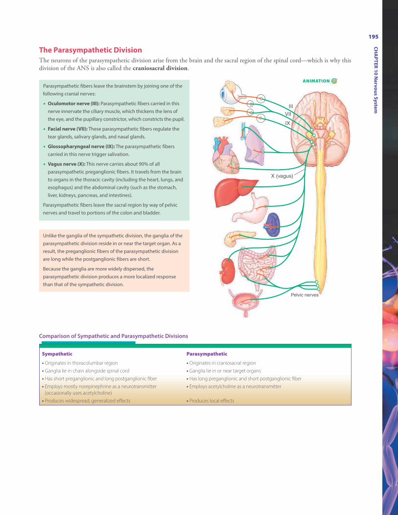
195
CH
AP
TE
R 1
0 N
erv
ou
s Sy
stem
The Parasympathetic Division
The neurons of the parasympathetic division arise from the brain and the sacral region of the spinal cord—which is why thisdivision of the ANS is also called the craniosacral division.
III
VII
X (vagus)
IX
Pelvic nerves
Parasympathetic fibers leave the brainstem by joining one of the
following cranial nerves:
• Oculomotor nerve (III): Parasympathetic fibers carried in this
nerve innervate the ciliary muscle, which thickens the lens of
the eye, and the pupillary constrictor, which constricts the pupil.
• Facial nerve (VII): These parasympathetic fibers regulate the
tear glands, salivary glands, and nasal glands.
• Glossopharyngeal nerve (IX): The parasympathetic fibers
carried in this nerve trigger salivation.
• Vagus nerve (X): This nerve carries about 90% of all
parasympathetic preganglionic fibers. It travels from the brain
to organs in the thoracic cavity (including the heart, lungs, and
esophagus) and the abdominal cavity (such as the stomach,
liver, kidneys, pancreas, and intestines).
Parasympathetic fibers leave the sacral region by way of pelvic
nerves and travel to portions of the colon and bladder.
Unlike the ganglia of the sympathetic division, the ganglia of the
parasympathetic division reside in or near the target organ. As a
result, the preganglionic fibers of the parasympathetic division
are long while the postganglionic fibers are short.
Because the ganglia are more widely dispersed, the
parasympathetic division produces a more localized response
than that of the sympathetic division.
Comparison of Sympathetic and Parasympathetic Divisions
Sympathetic Parasympathetic
• Originates in thoracolumbar region • Originates in craniosacral region
• Ganglia lie in chain alongside spinal cord • Ganglia lie in or near target organs
• Has short preganglionic and long postganglionic fiber • Has long preganglionic and short postganglionic fiber
• Employs mostly norepinephrine as a neurotransmitter
(occasionally uses acetylcholine)
• Employs acetylcholine as a neurotransmitter
• Produces widespread, generalized effects • Produces local effects
ANIMATION

The two divisions of the ANS tend to exert opposite effects. For example, the sympathetic division increases the heart rate whilethe parasympathetic division slows it down. However, that’s not always the case. For example, the sympathetic division triggersdilation of blood vessels leading to skeletal muscles, but it also constricts blood vessels leading to most organs.
Two factors determine the effect of each division: the neurotransmitter released and the type of receptor on the target cells.
Neurotransmitters
The ANS employs two different neurotransmitters: acetylcholine (ACh) and norepinephrine (NE). Fibers that secreteacetylcholine are called cholinergic fibers. Fibers that secrete NE (sometimes called noradrenaline) are called adrenergicfibers.
Cholinergic Fibers
• Include the preganglionic fibers of both the sympathetic and
parasympathetic divisions
• Include the postganglionic fibers of the parasympathetic
division
• Secrete acetylcholine
Adrenergic Fibers
• Include most (but not all) of the postganglionic fibers of the
sympathetic division
• Secrete norepinephrine
FAST FACTRecent research has shown that neurotransmittersother than norepinephrine and acetylcholine canbind to autonomic receptors—thus opening thedoor for the development of new medications.
The Body AT WORKThe effects of the parasympathetic division tend to be short lived, while the effects of the sympathetic division last
much longer. The difference in the length of these responses is linked to the speed with which the neurotransmitter is
broken down after its release.
For example, once ACh is released into the synaptic cleft, it’s quickly broken down by the enzyme
acetylcholinesterase (AChE). As a result, the parasympathetic division exerts only short-term effects.
In contrast, NE—the neurotransmitter released only by postganglionic sympathetic fibers—may be disposed of
by one of three methods, none of which are as rapid as the breakdown of ACh. Some NE is reabsorbed by the nerve
fiber (where it is either reused or broken down by the enzyme monoamine oxidase [MAO]); some of it is also absorbed
by the surrounding tissue (where it is broken down by another enzyme). In addition, some of the NE may be absorbed
by the bloodstream and carried throughout the body, further prolonging the effects of the sympathetic division.
That Makes SenseRemember: In all pathways, preganglionic fibers are
cholinergic and secrete acetylcholine. Most sympathetic
postganglionic fibers are adrenergic. (See the diagram at
the end of the chapter for a clear example of this complex
process.)
!P
AR
T III R
eg
ula
tion
an
d In
teg
ratio
n o
f the
Bo
dy
196
Effects of the ANS on Target Organs

Nicotinic Receptors
• These receptors occur within the ganglia of the ANS, in the
adrenal medulla, and in the neuromuscular junction.
• All cells with nicotinic receptors are excited by ACh.
Muscarinic Receptors
• These receptors occur on the glands, smooth muscle, and cardiac
muscle cells of the organs innervated by cholinergic fibers.
• Cells with muscarinic receptors exhibit a variable response to
ACh: some are excited while others are inhibited. (This variable
response occurs because several subtypes of muscarinic
receptors exist.)
• This variable response allows ACh to stimulate intestinal
smooth muscle while inhibiting cardiac muscle.
Adrenergic Receptors
There are also two basic types of adrenergic receptors: alpha-(a-)adrenergic receptors and beta-(b-)adrenergicreceptors. The following principles are true most of the time:
l Cells with a-adrenergic receptors are excited by NE.l Cells with b-adrenergic receptors are inhibited by NE.
To illustrate, the binding of NE to a-adrenergic receptors in blood vessels causes the blood vessels to constrict. Arteriesthat supply blood to the heart and skeletal muscles contain b-adrenergic receptors. The binding of NE to these receptorscauses the vessels to dilate.
(Be aware, however, that both alpha and beta receptorscontain subtypes—alpha-1 [a1], alpha-2 [a2], beta-1 [b1], andbeta-2 [b2]—which can cause exceptions to these principles.)
FAST FACTA group of drugs called beta blockers bind tobeta receptors, effectively “blocking”norepinephrine and epinephrine from bindingto those same receptors. (Remember that thesympathetic nervous system stimulates theadrenal medulla to secrete epinephrine, ahormone that prolongs the sympatheticresponse.) Blocking these hormones effectivelylessens the sympathetic response, making thedrugs useful for treating such disorders as highblood pressure and irregular heartbeats.
197
CH
AP
TE
R 1
0 N
erv
ou
s Sy
stem
Receptors
After being released, the neurotransmitters bind to receptors on the effector cells of the target organs. In brief, ACh binds tocholinergic receptors and NE binds to adrenergic receptors. However, within these two categories of receptors are severalsubtypes of receptors; it’s these different types of receptors that determine the effect produced by a neurotransmitter.
To illustrate the importance of the type of receptor, consider the fact that the sympathetic division secretes NE toconstrict the blood vessels of the skin and also to dilate the blood vessels of skeletal muscles. Likewise, the parasympatheticdivision secretes ACh to contract the bladder wall and also to relax the internal urethral sphincter. These opposite effectsoccur then, not because of the neurotransmitter, but because of the receptor.
Cholinergic Receptors
Acetylcholine may bind to one of two different types of receptors:

PA
RT
III Re
gu
latio
n a
nd
Inte
gra
tion
of th
e B
od
y
198
� � � � � ! " # � $fibers of
preganglionic neuronssecrete
% � � &% � � ' ( ) * + , - � $ � . � � $ / 0 1 0 2 , - / +- ) 2 - + , 3 4 ) 3 5 ( - ) ( 1) 0 6 / - ) + 7
8 - + , 2 - + , 3 4 ) 3 5 ( - ) ( 1) 0 6 / - ) + 4 / 0 9 : " ! ! " # � $4 ) * + 0 1 / 0 , 0 ; < 7 = 1 1 4 + ( - ) 4 5 5 > ?2 - + , 3 4 ) 3 5 ( - ) ( 1 ) 0 6 / - ) +4 / 0 $ � � � � ! " # � $ 4 ) *+ 0 1 / 0 , 0 % � � 7; < ' ( ) * + , - 9 � @ � 9 A 4 ) *B ! . 9 C 9 : " ! ! " # � $ " ! $ ! @ . � " D & E F G ' ( ) * + , -H I D $ 9 " � � $ / 0 1 0 2 , - / + 7J 4 / 3 0 ,1 0 5 5J 4 / 3 0 ,1 0 5 5
� � � � � ! " # � $2 - + , 3 4 ) 3 5 ( - ) ( 1) 0 6 / - ) + + 0 1 / 0 , 0 % � � &
� � � � � ! " # � $fibers of
preganglionic neuronssecrete
% � � &% � � ' ( ) * + , -
� $ � . � � $/ 0 1 0 2 , - / + - )2 - + , 3 4 ) 3 5 ( - ) ( 1 ) 0 6 / - ) + 7
E F G ' ( ) * + , -H I D $ 9 " � � $ / 0 1 0 2 , - / + 7J 4 / 3 0 ,1 0 5 5
Parasympathetic Division
Neurotransmitters and Receptors
The following diagrams summarize the different neurotransmitters and receptors of the two divisions of the autonomicnervous system.
Sympathetic Division
ANIMATION

199
The Body AT WORKMost organs receive nerve fibers from both the
sympathetic and parasympathetic divisions. This
is called dual innervation. Typically, the effects of
the two divisions oppose one other. For example,
the sympathetic division increases heart rate,
while the parasympathetic division slows it down;
also, the sympathetic division dilates pupils, while
the parasympathetic division constricts them.
Sometimes, though, the effects of the two
divisions complement each other to produce a
desired effect. For example, parasympathetic
division stimulates salivary glands to produce a
thin secretion containing enzymes while the
sympathetic division stimulates the production of
thick mucus; both of these substances are needed
to make saliva.
FAST FACTSympathetic fibers cause both blood vesselconstriction and dilation. They do this by alteringtheir frequency of firing. An increase in firing causesblood vessels to constrict; a decrease in firing resultsin dilation. This allows the sympathetic division toregulate blood pressure and blood flow without anyhelp from parasympathetic fibers.
Life lesson: Medications and the nervous systemA number of drugs work by enhancing or inhibiting the effects of neurotransmitters. Following are some of themajor classifications of these types of drugs:
• Sympathomimetics: These drugs enhance sympathetic action by either promoting the release of epinephrineor stimulating adrenergic receptors. A common example is phenylephrine, a drug found in many coldremedies. This drug stimulates a1 receptors, which dilates the bronchial tubes (to ease breathing) andconstricts blood vessels in the nose (to ease nasal congestion).
• Sympatholytics: These drugs block the effects of epinephrine and norepinephrine, primarily by inhibitingadrenergic receptors. This class of drug is often used to treat high blood pressure: blocking a-adrenergic receptors causes the blood vessels to dilate and blood pressure to fall.
• Parasympathomimetics: These drugs—also called cholinergics—stimulate or mimic the parasympathetic division. They do this by either stimulating the nicotinic or muscarinic receptors or by inhibitingacetylcholinesterase (the enzyme that breaks down ACh). One such drug is physostigmine, which is used totreat glaucoma (increased pressure in the eye) and delayed gastric emptying.
• Parasympatholytics: Also called anticholinergics, these drugs inhibit ACh release or block its receptors to reduce the activity of the parasympathetic division. Because one of its effects is to dilate the bronchiole tubesin the lungs, this class of drug may be used to treat asthma.
Life lesson: Changes with agingThe type and degree of age-related changes in the brain and nervous system varies from one individual to thenext. Some experience significant nerve degeneration while others have few changes. In general, most peopleexperience slight slowing of thought and memory as the speed of nerve conduction slows. Other possible changesinclude reduced reflexes or sensation, leading to problems with safety.
Dementia and severe memory loss are not a normal part of aging. When they occur, they often signalunderlying disease. Illnesses not related to the brain, however, can cause changes in thinking and behavior. Forexample, an infection or unregulated blood glucose levels can cause severe confusion.
Some evidence suggests that both physical and mental exercise (such as reading, doing crossword puzzles, andengaging in stimulating conversations) can help keep thinking sharp.
CH
AP
TE
R 1
0 N
erv
ou
s Sy
stem

PA
RT
III Re
gu
latio
n a
nd
Inte
gra
tion
of th
e B
od
y
200
Action potential: Nerve impulse
Adrenergic fibers: Nerve fibers that secrete norepinephrine
Afferent neuron: Sensory nerve thatcarries impulses toward the centralnervous system
Alpha-adrenergic receptors: Receptorsto norepinephrine that generally produce an excitatory response
Autonomic nervous system:
Subdivision of the nervous system responsible for regulating activitiesthat maintain homeostasis; also calledvisceral motor system
Axon: Fiber extending from a neuron’scell body that conducts impulses
Beta-adrenergic receptors: Receptors tonorepinephrine that generally producean inhibitory response
Brainstem: Stem-like portion of thebrain that connects the cerebral hemispheres to the spinal cord; consists of the midbrain, pons, andmedulla oblongata
Central nervous system: Portion of thenervous system consisting of the brainand spinal cord
Cerebellum: Portion of the brain responsible for coordination ofvoluntary movement and balance
Cerebral cortex: The surface of thecerebrum consisting of a thin layer ofgray matter
Cerebrospinal fluid: Clear, colorlessfluid that fills the brain’s ventriclesand central canal and also bathes theoutside of the brain and spinal cord
Cerebrum: Largest portion of the brain
Cholinergic fibers: Nerve fibers that secrete acetylcholine
Cranial nerves: Nerves of the peripheralnervous system that arise directlyfrom the brain (instead of from thespinal cord)
Dermatome: A specific area of the skininnervated by a spinal nerve
Efferent neuron: Motor nerve that carries impulses away from the centralnervous system
Epidural space: Small space betweenthe outer covering of the spinal cordand the vertebrae
Frontal lobe: Portion of the cerebrumresponsible for decision making, reasoning, memory, and voluntarymovements
Gray matter: Nervous tissue containingmostly cell bodies of motor neuronsand interneurons
Hypothalamus: The bottom half of thediencephalon of the brain, which is responsible for many vital body functions
Limbic system: Complex set of structures in the brain responsible for emotion and learning
Medulla oblongata: Attaches brain tospinal cord; contains centers that govern heart rate, blood pressure, andbreathing
Meninges: Layer of fibrous connectivetissue covering the brain and spinalcord
Mixed nerve: Nerve that contains bothsensory and motor fibers
Muscarinic receptors: Receptors toacetylcholine on target cells that exhibit a variable response
Myelin: Fatty sheath encasing theaxons of many neurons; formed bySchwann cells in the peripheral nervous system and oligodendrocytesin the central nervous system
Nerve: A bundle of neurons thattransmits impulses between the brainand spinal cord and the rest of thebody
Neuroglia: Cells in the nervous systemthat support neurons but do not conduct impulses
Neurons: Nervous system cells thatconduct impulses
Nicotinic receptors: Receptors toacetylcholine on target cells that produce an excitatory response
Occipital lobe: Portion of the cerebrumresponsible for analyzing and interpreting visual information
Parasympathetic division: Division ofthe autonomic nervous system thatprimarily has a calming effect; alsocalled craniosacral division
Parietal lobe: Portion of the cerebrumconcerned with bodily sensations andproprioception
Peripheral nervous system: Portion ofthe nervous system consisting of thenetwork of nerves throughout thebody
Plexus: A network of nerves
Polarization: The electrical state of acell membrane that has an excess ofpositive ions on one side of the membrane and an excess of negativeions on the other
Reflex arc: A neural circuit that bypasses regions of the brain whereconscious decisions are made
Somatic reflex: Reflex involving contraction of a skeletal muscle afterbeing stimulated by a somatic motorneuron
Sympathetic division: Division of theautonomic nervous system responsiblefor “fight or flight” response; alsocalled thoracolumbar division
Review of Key Terms

Synapse: Space between the junctionof two neurons in a neural pathway
Temporal lobe: Portion of the brain responsible for hearing, smell, and visual recognition
Tracts: Bundles of axons in the spinal cord that serve as routes ofcommunication to and from the brain
Visceral reflexes: Reflex of the autonomic nervous system affectingorgans
White matter: Nervous tissue containing bundles of axons that carry impulses from one part of thenervous system to another
Own the InformationTo make the information in this chapter part of your
working memory, take some time to reflect on what you’ve
learned. On a separate sheet of paper, write down
everything you recall from the chapter. After you’re done,
log on to the DavisPlus website, and check out the Study
Group podcast and Study Group Questions for the chapter.
Key Topics for Chapter 10:• Divisions of the nervous system
• Types of cells found in the nervous system
• Neuron structure
• The repair of nerve fibers
• Impulse conduction in both myelinated and unmyelinated
nerve fibers
• Synapse structure and function
• The structure of the spinal cord
• Spinal tracts
• Spinal nerves
• Somatic reflexes
• Divisions of the brain
• Structure and function of the meninges
• Creation and circulation of cerebrospinal fluid
• Divisions of the brain, including the brainstem,
cerebellum, diencephalon, and cerebrum
• The limbic system
• Sensory and motor functions of the cerebral cortex
• Regions of the brain responsible for language and special
senses
• Types of brain waves as related to different stages of sleep
• Brain regions involved in memory
• The relationship between the right and left hemispheres
• Cranial nerves
• Visceral reflexes
• The structure and function of the autonomic nervous
system
• The structure and function of the sympathetic and
parasympathetic divisions of the autonomic nervous
system
• The neurotransmitters and receptors employed by the
autonomic nervous system
• The effects of the sympathetic and parasympathetic
nervous systems on target organs
201
CH
AP
TE
R 1
0 N
erv
ou
s Sy
stem

PA
RT
III Re
gu
latio
n a
nd
Inte
gra
tion
of th
e B
od
y
202
Answers: Chapter 101. Correct answer: b. Neuroglia are nervous system
cells that protect neurons. Microglia are a type ofneuroglia that perform phagocytosis and engulfmicroorganisms and debris. Ependymal cells linethe spinal cord and cavities in the brain andsecrete cerebrospinal fluid.
2. Correct answer: c. The brain requires a large supplyof blood to function. The blood supply to thebrain is not separate from the blood supplied tothe rest of the body. Cerebrospinal fluid follows itsown circulatory route and is eventually reabsorbedinto the venous system; it has nothing to do withthe blood-brain barrier.
3. Correct answer: c. Efferent neurons are motor neurons; these relay messages from the brain tomuscles and gland cells. Interneurons are foundonly in the CNS; they connect incoming sensorypathways with outgoing motor pathways.Schwann cells wrap around the axons of many peripheral nerves to form the myelin sheath.
4. Correct answer: d. Resting potential is the phase inwhich the neuron is resting but has the potentialto react. Action potential is when the neuron becomes active and conducts an impulse along anaxon. Repolarization is when the nerve cell restores its electrical balance in preparation for anew stimulus. While a neuron won’t necessarilyreact to an impulse during action potential and repolarization, the most correct answer is the refractory period, because, although the membraneis polarized, the cell won’t react to a new impulseuntil sodium and potassium have been restored totheir rightful sides of the membrane.
5. Correct answer: a. Spinal nerves carry both sensoryand motor fibers, making them capable of transmitting impulses both to and from the CNS.Spinal nerves regulate voluntary function, not autonomic function; they are not part of the autonomic nervous system.
6. Correct answer: c. The phrenic nerve is part of thecervical plexus and innervates the diaphragm. Theaxillary nerve is part of the brachial plexus andleads into the arm. The femoral nerve is derivedfrom the lumbar plexus and supplies the thigh andleg.
7. Correct answer: c. CSF nourishes the brain byproviding glucose and protein and removingmetabolic wastes. An adult brain contains about140 ml of CSF; the brain monitors levels ofcarbon dioxide; CSF continually circulatesthrough the central nervous system.
8. Correct answer: b. The pons contains tracts thatconvey signals to and from different parts of thebrain; it’s also the site where several cranial nervesoriginate. The midbrain contains centers for auditory and visual reflexes as well as neurons essential for motor control. The cerebellum plays akey role in motor functions as well as in sensoryand emotional functions.
9. Correct answer: d. The thalamus is the gatewayfor nearly all sensory impulses. The limbic system is the seat of emotion and learning. Thecerebral cortex is the gray matter covering thesurface of the brain; it handles the cerebrum’sfunctions, which include thought, reasoning,judgment, and memory.
202
Test Your Knowledge1. The impulse-conducting cells of
the nervous system are called:a. neuroglia.b. neurons.c. microglia.d. ependymal cells.
2. What is the blood-brain barrier?a. It’s a membrane surrounding
the brain that keeps bloodfrom penetrating brain tissue.
b. It’s a membrane surroundingthe brain that keeps the bloodin the brain separate from theblood in the rest of the body.
c. It’s a semi-permeable membranethat permits small moleculeslike oxygen and carbon dioxideto diffuse across to the brainbut blocks larger molecules.
d. It’s a semi-permeable membranethat prevents cerebrospinalfluid from leaving the brainand entering the bloodstream.
3. Which neurons detect sensationssuch as touch or heat and thenrelay information about the stimuli to the central nervous system?a. Efferent neuronsb. Interneuronsc. Afferent neuronsd. Schwann cells
4. A nerve cell in which phase cannot respond to a new stimulus?a. Resting potentialb. Action potentialc. Repolarizationd. Refractory period
5. Which statement regardingspinal nerves is true?a. Spinal nerves are mixed nerves,
containing both sensory andmotor fibers, making it capableof transmitting impulses intwo directions.
b. Spinal nerves are primarilymotor nerves, responsible for transmitting impulses toskeletal muscles.
c. Spinal nerves are primarilysensory nerves, responsible fortransmitting sensations fromthe muscles to the brain andspinal cord.
d. Spinal nerves are a chief component of the autonomicnervous system and are responsible for controlling autonomic functions.
6. Which key nerve is part of thesacral plexus?a. Phrenic nerveb. Axillary nervec. Sciatic nerved. Femoral nerve
7. Which statement about cerebrospinal fluid (CSF) is true?a. An adult’s brain contains about
500 ml of CSF.b. The brain monitors the level
of oxygen in CSF.c. CSF furnishes the brain with
glucose and protein.d. CSF remains relatively
stagnant within the cerebralcavity.
8. Which portion of the brain contains centers responsible forsuch vital functions as heart rate,breathing, and blood pressure?a. Ponsb. Medulla oblongatac. Midbraind. Cerebellum
9. Which brain structure influencesnearly every organ and exertscontrol over the autonomic nervous system and pituitarygland?a. Thalamusb. Limbic systemc. Cerebral cortexd. Hypothalamus

10. An injury to which part of thebrain may result in a severe personality disorder and cause socially inappropriate behavior?a. Frontal lobeb. Parietal lobec. Temporal lobed. Occipital lobe
11. Which area of the brain receivesimpulses of heat, cold, and touchfrom receptors all over the bodyand is, therefore, known as theprimary somatic sensory area?a. Precentral gyrusb. Wernicke’s areac. Broca’s aread. Postcentral gyrus
12. Which cranial nerve suppliesmost of the organs in the thoracic and abdominal cavitiesas well as those in the head andneck?a. Glossopharyngealb. Hypoglossalc. Vagusd. Vestibulocochlear
13. Brain swelling from a head injurycan compress this nerve and interfere with the ability of thepupils to react to light.a. Oculomotorb. Olfactoryc. Opticd. Facial
14. Which of the following is a characteristic of the somaticnervous system?a. It innervates glands, smooth
muscle, and cardiac muscle.b. It consists of two nerve fibers
that synapse at a ganglion be-fore reaching a target organ.
c. It secretes both acetylcholineand norepinephrine.
d. It operates under voluntarycontrol.
15. Which of the following is one ofthe actions of the parasympatheticdivision of the autonomic nervous system?a. Increases heart rateb. Constricts bronchial tubesc. Stimulates sweat glandsd. Inhibits intestinal motility
16. Which of the following statements correctly describescholinergic fibers?a. Cholinergic fibers secrete
norepinephrine.b. Cholinergic fibers include
the postganglionic fibers of the sympathetic and parasympathetic divisions.
c. Cholinergic fibers secrete acetylcholine.
d. Cholinergic fibers include thepreganglionic fibers of theparasympathetic division only.
17. The effect produced by a neurotransmitter (such aswhether it constricts or dilatesblood vessels) is ultimately determined by:a. the amount of neurotransmitter
released.b. the type of neurotransmitter
released.c. the type of receptor.d. the number of receptors.
10. Correct answer: a. The parietal lobe handles theawareness of one’s body parts; an injury here maycause someone to ignore objects on one side of thebody, even his own arm or leg. The temporal lobegoverns hearing and memory; an injury here mayimpair the ability to identify familiar objects. Theoccipital lobe is concerned with analyzing and interpreting visual information; an injury herewould affect the ability to see.
11. Correct answer: d. Neurons in the precentral gyrussend impulses through the motor tracts in thebrainstem and spinal cord to skeletal muscles.Wernicke’s area (in the left temporal lobe) andBroca’s area (in the left frontal lobe) are both concerned with language: Wernicke’s area formulates the words into phrases that complywith learned grammatical rules; Broca’s area plansthe muscle movements required to form the wordsand sends the appropriate impulses to the primarymotor cortex.
12. Correct answer: c. The glossopharyngeal nerve governs tongue movements, swallowing, and gagging. The hypoglossal nerve controls tonguemovements. The vestibulocochlear nerve is concerned with hearing and balance.
13. Correct answer: a. The olfactory nerve governs thesense of smell. The optic nerve is concerned withvision (not pupillary response). The facial nerve isconcerned with taste as well as facial expression.
14. Correct answer: d. All the other statements arecharacteristics of the autonomic nervous system.
15. Correct answer: b. All of the other actions listedcan be attributed to the sympathetic division.
16. Correct answer: c. Adrenergic fibers secrete norepinephrine. Cholinergic fibers include thepreganglionic fibers of both the sympathetic andparasympathetic divisions as well as the postganglionic fibers of the parasympathetic division.
17. Correct answer: c. The amount of neurotransmitterwould influence the strength of a response, butnot the type of response. The effect produced doesvary according to the type of neurotransmitter; however, the same neurotransmitter bound to a different receptor will have a different response.Therefore, it’s the receptor, not the neurotransmitter,that ultimately determines the response. If the receptors are blocked (thus decreasing the numberavailable), then the strength of the response wouldbe diminished.
Go to http://davisplus.fadavis.com Keyword:Thompson to see all of the resources availablewith this chapter.
203
CH
AP
TE
R 1
0 N
erv
ou
s Sy
stem

CHAPTER OUTLINESensory Receptors
The General Senses
The Special Senses
LEARNING OUTCOMES1. Distinguish between sensory receptors and
sense organs.
2. List three kinds of information obtained by
sensory receptors.
3. Name five classifications of sensory receptors.
4. Identify two categories of pain receptors.
5. Summarize the main pain pathway.
6. Name the five special senses.
7. Specify the location of taste receptors and
describe how the sensation of taste occurs.
8. Identify the location of smell receptors and
describe how the sensation of smell occurs.
9. Describe the anatomy of the inner, middle, and
outer ear.
10. Explain how hearing occurs.
11. Discuss how the vestibule and semicircular
canals allow the brain to interpret body
movement and position.
12. Describe the anatomy of the eye and its
accessory structures.
13. Describe the structure and function of the
retina.
14. Discuss how vision occurs.
15. Explain how the size of the pupil and curvature
of the lens affect vision.
16. Describe the location and purpose of rods and
cones.

11
chapter SENSE ORGANSAlthough human sense organs detect a vast array of sensations, the
environment is full of physical and chemical energy beyond our
comprehension.
Sense organs allow us to taste our food, see a beautiful sunset, hear music, and feel the touch of another person. But sensesdo more than contribute to our enjoyment of life. For one thing, our senses protect us from danger: being able to see or heardanger, and to feel pain, allows us to avoid injury. Even more importantly, experiencing sensation is vital to our mental well-being. In fact, in experiments in which subjects were placed in isolation tanks that deprived them of all sensation, thesubjects soon experienced hallucinations, extreme anxiety, delusions, and panic.
The five main senses are sight, hearing, taste, smell, and touch; however, the body’s sensory experiences involve morethan those few. In fact, many of the body’s sensory processes take place without our awareness. For example, the sensorysystem is integral in maintaining such vital functions as blood pressure, body temperature, and balance.
Scattered throughout the body are millions of sensory receptors. Some of these receptors combine with muscle and tissue toform sense organs (such as the eyes, ears, and nose). Most, though, are specialized nerve cells or nerve endings that detectphysical or chemical events outside the cell membrane.
FAST FACTOne type of energy beyond the scope of human senses iselectric fields. However, some fish, such as sharks, haveelectroreceptive organs that allow them to detect weakelectrical impulses emitted by the gills of their prey.
When the impulse arrives at the
brain, we may experience a
sensation, such as a sight or sound.
The Body AT WORKNormally, the brain filters out most of the
stimuli detected by receptors, keeping them
below our level of awareness. That’s because
many of the stimuli relate to body processes
that operate subconsciously. Also, the filtering
process protects us from being overwhelmed
by an overabundance of stimuli.
Sensory Receptors
A sensation begins
when a receptor
detects a stimulus.
If the stimulus is strong enough, the
receptor causes a sensory neuron to send
an impulse to the brain and spinal cord.

PA
RT
III Re
gu
latio
n a
nd
Inte
gra
tion
of th
e B
od
y
206
Sensory receptors transmit information about the type, the location, and the intensity of each sensation.
l Type: Each sensory receptor responds to a different stimulus; for example, cold receptors respond only to cold while lightreceptors respond only to light. This allows the nervous system to differentiate between the various sensations.
l Location: Each sensory neuron responds to stimuli in a certain area, called a receptive field. Therefore, when a particularneuron carries a stimulus to the brain, the brain knows where the stimulus originated. This is known as sensory projection. Sensitive areas of the body, such as the tips of the fingers, contain a dense population of receptors. Becauseeach neuron covers a tiny territory, the brain can pinpoint the location of a stimulus. In less sensitive areas, such as theback, a single neuron covers a much larger territory. Consequently, the brain can only identify that a stimulus occurred inthat area; it can’t pinpoint the spot with accuracy.
l Intensity: The stronger a particular stimulus, the more nerve fibers fire. This allows the brain to interpret the intensity ofa sensation.
Classification of Receptors
Receptors are classified according to the type of stimuli they detect.
The Body AT WORKWhen a stimulus is
continuous, the firing
frequency of the nerve
begins to slow, causing the
sensation to diminish. This
is known as adaptation. As
an example, think of
entering a cold body of
water. When you first step
in, the water may seem very
cold. After a few minutes,
your senses adapt and it
doesn’t feel as cold.
The Body AT WORKAnother type of specialized
receptor is the
proprioceptor. Found in
skeletal muscle, joints, and
tendons, proprioceptors
provide information about
body movement, muscle
stretch, and the general
orientation of the body. For
example, proprioceptors
allow you to orient your
body in space and to know
the position of your body
parts (such as your arm or
leg) without looking.
Chemoreceptors
These receptors react to various chemicals, including
odors and tastes, as well as the concentration of
various chemicals (such as glucose or carbon dioxide)
in the body.
Mechanoreceptors
These receptors respond to factors—such as pressure,
stretch, or vibration—that change the position of a
receptor.
Thermoreceptors
These receptors are activated by a change in
temperature.
Nociceptors
These are pain receptors that respond to tissue
damage from trauma as well as from heat, chemicals,
pressure, or a lack of oxygen.
Photoreceptors
Found only in the eyes, these receptors respond to
light.

207
CH
AP
TE
R 1
1 S
en
se O
rga
ns
The general senses include pain as well as the sensations of pressure, touch, stretch, and temperature. Receptors for thegeneral senses are widely distributed in the skin, muscles, tendons, joints, and viscera.
Pain
Nociceptors, or pain receptors, consist of free nerve endings that carry painimpulses to the brain. They’re especially abundant in the skin and mucousmembranes and also found in almost every organ. (They’re not, however, found inthe brain.) They fall into one of two categories:
l Fast pain fibers: Abundant in the skin and mucous membranes, these fibers produce a sharp, localized, stabbing-type pain at the time of injury. This is the typeof pain you experience when you stub your toe or slam your finger in a door.
l Slow pain fibers: These fibers are congregated on deep body organs and structures and produce a dull, aching pain. For example, pain sensations from abowel obstruction or appendicitis would be carried along these fibers.
J G 4 5 4 K 6 +L 2 ( ) 4 5 1 - / *
M / 4 ( ) + , 0 KL 2 ( ) - , G 4 5 4 K ( 1 , / 4 1 , N - / + 4 53 4 ) 3 5 ( - )O 0 , ( 1 6 5 4 /P - / K 4 , ( - )
L 2 ( ) 4 5 1 - / *
Pain can travel by a number of different routes (making the true source of pain often difficult to identify). The followingillustration portrays the main pain pathway for most areas of the body.
1 Injured tissues release
several chemicals that
stimulate the nociceptors
and trigger pain. (Pain
receptors may also be
stimulated from a lack of
oxygen or when tissues are
stretched.)
2 A neuron conducts a pain
signal to the dorsal horn
of the spinal cord.
4 At the same time, the
spinoreticular tract
carries pain signals to the
reticular formation of the
brainstem.
5 The thalamus relays the signal from the
spinothalamic tract to the postcentral
gyrus of the cerebrum. At that point, the
person becomes aware of the pain.
6 The impulse from the
spinoreticular tract bypasses
the thalamus and travels to the
hypothalamus and limbic system.
These areas trigger emotional and
behavioral responses to pain,
such as fear and nausea.
3 At the dorsal horn,
the spinothalamic
tract relays the pain signal
to the thalamus.
The General Senses
FAST FACTWhile nociceptors don’t occur inthe brain, they do occur in themeninges. Headaches, therefore,don’t result from pain in thebrain but from the surroundingtissues.
ANIMATION
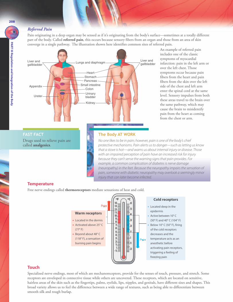
PA
RT
III Re
gu
latio
n a
nd
Inte
gra
tion
of th
e B
od
y
208
Touch
Specialized nerve endings, most of which are mechanoreceptors, provide for the senses of touch, pressure, and stretch. Somereceptors are enveloped in connective tissue while others are uncovered. These receptors, which are located on sensitive,hairless areas of the skin such as the fingertips, palms, eyelids, lips, nipples, and genitals, have different sizes and shapes. Thisbroad variety allows us to feel the difference between a wide range of textures, such as being able to differentiate betweensmooth silk and rough burlap.
Temperature
Free nerve endings called thermoreceptors mediate sensations of heat and cold.
Liver andgallbladder
Liver andgallbladder
Appendix
Ureter
Lungs and diaphragm
Heart
Stomach
Pancreas
Small intestine
Colon
Kidney
Urinarybladder
Q 4 ( )Q 4 ( )
The Body AT WORKNo one likes to be in pain; however, pain is one of the body’s chief
protective mechanisms. Pain alerts us to danger—such as letting us know
that a stove is hot—and warns us about internal injury or disease. Those
with an impaired perception of pain have an increased risk for injury
because they can’t sense the warning signs that pain provides. For
example, a common complication of diabetes is nerve damage
(neuropathy) in the feet. Because the neuropathy impairs the sensation of
pain, someone with diabetic neuropathy may overlook a seemingly minor
injury that can later become infected.
Warm receptors
• Located in the dermis
• Activated above 25° C
(77° F)
• Beyond about 48° C
(118° F), a sensation of
burning pain begins
An example of referred painincludes one of the classicsymptoms of myocardialinfarction: pain in the left arm orover the left chest. Thosesymptoms occur because painfibers from the heart and painfibers from the skin over the leftside of the chest and left armenter the spinal cord at the samelevel. Sensory impulses from boththese areas travel to the brain overthe same pathway, which maycause the brain to misidentifypain from the heart as comingfrom the chest or arm.
Referred Pain
Pain originating in a deep organ may be sensed as if it’s originating from the body’s surface—sometimes at a totally differentpart of the body. Called referred pain, this occurs because sensory fibers from an organ and those from an area of skinconverge in a single pathway. The illustration shown here identifies common sites of referred pain.
Cold receptors
• Located deep in the
epidermis
• Active between 10° C
(50° F) and 40° C (104° F)
• Below 10° C (50° F), firing
of the cold receptors
decreases and the
temperature acts as an
anesthetic before
activating pain receptors,
triggering a feeling of
freezing pain
FAST FACTDrugs used to relieve pain arecalled analgesics.

Epiglottis
Lingual tonsil
Papillae
Tongue epithelium
Supporting cell
Gustatory cell
209
CH
AP
TE
R 1
1 S
en
se O
rga
ns
The special senses—which include taste, smell, hearing, equilibrium, and vision—involve receptors that are either groupedtogether or clustered in specialized organs.
Taste buds
reside along
and between
the papillae.Taste buds look similar
to an orange, with each
segment containing 25
to 50 chemoreceptors
called gustatory cells
or taste cells.
A hair-like tip projects into an
opening called the taste pore,
which is bathed in saliva.
Chemicals dissolved in saliva
stimulate the gustatory cells,
which, in turn, stimulate certain
cranial nerves.
Taste buds send gustatory impulses
directly to the brain by one of three cranial
nerves (the facial nerve [VII], the
glossopharyngeal nerve [IX], and the vagus
nerve [X]). The cranial nerve used depends
on the location of the taste bud stimulated.
• Vallate papillae are large
papillae found at the rear of
the tongue; although few in
number, they contain up to
half of all taste buds.
• Foliate papillae form ridges
at the sides of the tongue.
• Filiform papillae are
thread-like papillae that
contain no taste buds; they
play a role in helping us
distinguish the texture of
food.
• Fungiform papillae are
especially concentrated at
the tip and sides of the
tongue.
The Body AT WORKThe four primary tastes are salty,
sweet, sour, and bitter. Researchers
have suggested a new primary
taste called umami. A Japanese
slang word meaning “delicious,”
umami refers to a “meaty” taste
resulting from amino acids.
Despite previously held views,
the tongue is not divided into
different regions according to
taste. All tastes can be detected in
all areas of the tongue that
contain taste buds; however,
some areas are more sensitive to
certain tastes. For example, the tip
of the tongue is most sensitive to
sweet tastes while the taste buds
at the rear of the tongue are most
sensitive to bitter tastes.
The Special Senses
FAST FACTA child has about 10,000taste buds. The numberdeclines with age, and elderlypeople may have fewer than5000 taste buds.
Taste
The sense of taste, or gustation, results when chemicals come in contact with taste buds. Most taste buds are locatedaround protrusions on the tongue called papillae, although a few reside in the lining of the mouth and soft palate. Thetongue contains four types of papillae:

R S T U V W X Y Z [ \ Y ] \
R S T U V W X Y Z ^ _ S ^` S X a \ Y _ S _ bc W d a X e f ^ X [ \g U b U S \ h e W d \ S e _ aR S T U V W X Y Z Y \ V \ h W X Y V \ S S b
PA
RT
III Re
gu
latio
n a
nd
Inte
gra
tion
of th
e B
od
y
210
Smell
Lining the roof of the nasal cavity is a small area of epithelium that contains receptor cells for olfaction (the sense of smell).The receptor cells—which are essentially neurons—leave the nasal cavity through pores in the ethmoid bone; they thengather together to form cranial nerve I (the olfactory nerve).
1 Incoming odor molecules
bind to cilia projecting
from the ends of the olfactory
receptor cells.
4 The signals continue on to
the primary olfactory
cortex in the brain.
3 The fibers synapse with
other neurons in olfactory
bulbs, a pair of structures
residing just underneath the
brain’s frontal lobe. Intermediate
neurons called glomeruli
partially process the impulses.
The Body AT WORKAfter a stimulus reaches the primary olfactory cortex, it may
continue on to a number of other locations in the cerebrum and
brainstem—including the hippocampus and amygdala. This
explains why certain odors can evoke a memory, an emotional
response, or even a physical response—such as vomiting.
Also noteworthy is that certain odors (such as those produced
by the chemicals ammonia, chlorine, and menthol) stimulate
nociceptors on the trigeminal nerve instead of olfactory cells. The
strong, unpleasant response that results from smelling these
chemicals is what makes “smelling salts” effective in reviving
someone who is unconscious.
FAST FACTOlfactory receptors are quicklyfatigued; they will stop sensing eventhe strongest smells after a short time.
2 This stimulates an impulse
along nerve fibers leaving
the nasal cavity through pores
in the ethmoid bone.
FAST FACTThe senses of smell and taste areclosely related. In fact, people who losetheir sense of smell typically complainthat food has lost much of its taste.

R ] U S i e [ f X i
211
CH
AP
TE
R 1
1 S
en
se O
rga
ns
Hearing
The ears provide the sense of hearing; they’re also essential for balance (equilibrium). The ear has three sections: the outerear, the middle ear, and the inner ear.
Outer Ear
The outer, or external, ear consists of the auricle (or pinna) and the auditory canal.R b b e V S \ b jk W U h \ bl [ V _ bm U S S \ _ b k \ a e V e Y V _ S U Y V U [ U S bn \ b W e ^ _ S U Y [ \ Y ] \o X V d S \ U Y [ \ Y ] \o X V d S \ Uc _ b W U V d e U [ W _ ^ \R _ W \ Y \ U Y m e f f S \ \ U Y l [ [ \ Y \ U Y
p X _ [ f i e [ f X iThe auricle (pinna) is the visible
part of the ear. Shaped by
cartilage, this part of the ear
funnels sound into the auditory
canal.
Auditory ossicles: The three smallest bones in
the body connect the eardrum to the inner ear;
they are named for their shape:
• Malleus (hammer)
• Incus (anvil)
• Stapes (stirrup)
The stapes fits within the oval window of the
vestibule, which is where the inner ear begins.
The auditory or eustachian tube
is a passageway from the middle
ear to the nasopharynx. Its
purpose is to equalize pressure on
both sides of the tympanic
membrane. Unfortunately, it can
also allow infection to spread
from the throat to the middle ear.
Middle Ear
The middle ear consists of these structures:
The auditory canal leads
through the temporal bone to
the eardrum. (The opening of the
auditory canal to the outside of
the body is called the external
acoustic meatus.) Glands lining
the canal produce secretions that
mix with dead skin cells to form
cerumen (ear wax). Cerumen
waterproofs the canal and also
traps dirt and bacteria. The
cerumen usually dries and then,
propelled by jaw movements
during eating and talking, works
its way out of the ear.
Tympanic membrane (or eardrum): This
membranous structure separates the outer ear
from the middle ear; it vibrates freely in
response to sound waves.
Life lesson: Middle ear infectionsMiddle ear infections, called otitis media, occur commonly in children. The reasonis simple: the size and position of the eustachian tube. In children, the eustachiantube is fairly short and horizontal. This short, straight passageway to thenasopharynx allows drainage from the nose to flow easily into the middle ear,spreading infection. As a child grows, the eustachian tube lengthens and slantsdownward, making the drainage of secretions into the ear, and resultantinfection, less likely.

q U b e S U Y a \ a ^ Y U [ \k _ h h X Y W e [ r V \ S S bs U e Y b t \ V W X Y e U Sa \ a ^ Y U [ \
u e ^ \ Y b X TV X V d S \ U Y [ \ Y ] \
v \ Y e S Z a h d o X V d S \ U Y f _ V Ww i e W d \ [ f X S Z a h d xR ] U S i e [ f X i
n \ b W e ^ _ S U Y [ \ Y ] \o X V d S \ U Y [ \ Y ] \
p X _ [ f i e [ f X i
PA
RT
III Re
gu
latio
n a
nd
Inte
gra
tion
of th
e B
od
y
212
Inner Ear
A complicated system of passageways within the temporal bonecontains the inner ear, which explains why this part of the ear iscalled the bony labyrinth. The membranous labyrinth lines theinside of the bony labyrinth. Fluid called perilymph cushions thespace between the two labyrinths, while another fluid, calledendolymph, occupies the inside of the membranous labyrinth.
Three separate structures form the bony labyrinth:
The Body AT WORKThe number of sound waves that occur
during a specific time frame, called
frequency, determines the pitch of a
sound. What we can hear depends on
how loud a sound is (the volume) as
well as its pitch. Human ears can
respond to sounds having a pitch
between 20 Hz (vibrations per second)
and 16,000 Hz. Many animals, though,
can hear sounds the human ear can’t
detect. For example, cats can hear
sounds up to 60,000 Hz, and bats can
detect frequencies as high as 120,000 Hz.
Vestibule: This structure,
which marks the entrance
to the labyrinths, contains
organs necessary for the
sense of balance.
Cochlea: This snail-like
structure contains the
structures for hearing.
Resting on the floor (called the
basilar membrane) of this duct
is the organ of Corti, the
hearing sense organ.
The organ of Corti consists of
a layer of epithelium
(composed of sensory and
supporting cells). Thousands
of hair cells project from this
epithelial layer and are
topped with a gelatin-like
membrane called the
tectorial membrane. Nerve
fibers extending from the
base of the hairs eventually
form the cochlear nerve
(cranial nerve VIII).
The spirals of the cochlea
are divided into three
compartments. The
middle compartment is a
triangular duct (called
the cochlear duct) filled
with endolymph; the
outer two compartments
are filled with perilymph.
Semicircular canals:
These structures are
crucial for the
maintenance of
equilibrium and balance.

k W U h \ bo X V d S \ U Y f _ V WR ] U S i e [ f X i o X V d S \ U Y [ \ Y ] \
t \ V W X Y e U Sa \ a ^ Y U [ \s U e Y V \ S S b X [X Y r U [ X T o X Y W eq U b e S U Y a \ a ^ Y U [ \c _ b W U V d e U [ W _ ^ \
l [ V _ bm U S S \ _ b213
CH
AP
TE
R 1
1 S
en
se O
rga
ns
How Hearing Occurs
Hearing occurs when sound creates vibrations in the air, known as sound waves.
5 The ripples continue
through the perilymph
and dissipate by striking the
round window.
1 Sound waves
enter the ear
and travel down the
external auditory
canal. The waves
strike the tympanic
membrane, causing
it to vibrate.
2 The vibration spreads through the
malleus, the incus, and the stapes.
3 The movement of the stapes against
the oval window shakes the perilymph
on either side of the cochlear duct.
4 The ripples in the perilymph are transmitted through the roof
of the cochlear duct to the organ of Corti. Movement here
stimulates the hairs of the organ of Corti to send nerve impulses
along the cochlear nerve. The impulses reach the auditory cortex
in the brain’s temporal lobe, where it’s interpreted as sound.
Life lesson: Hearing lossHearing loss can be divided into two main categories:
1. Conductive hearing loss. Anything that interferes with the transmission of vibrations to the inner earwill result in a hearing loss. For example, fluid in the middle ear (as a result of an infection, cold, orallergies), impacted cerumen, or a foreign body will all block the transmission of vibrations. Whenthe underlying cause is treated, hearing returns to normal. A more serious type of conductivehearing loss is otosclerosis, a condition in which the auditory ossicles fuse together. Because theossicles can’t move, the vibrations stop when they reach the middle ear. Surgical intervention cansometimes restore hearing.
2. Sensorineural (nerve) hearing loss. This type of hearing loss most often results from the death of haircells in the organ of Corti, usually a result of frequent exposure to sustained loud noise (such as thatexperienced by factory workers and musicians). Once the hairs are damaged, they never grow back,making this type of hearing loss permanent.
ANIMATION

y z { | } ~ �� � } } � ~ �
PA
RT
III Re
gu
latio
n a
nd
Inte
gra
tion
of th
e B
od
y
214
Balance
The vestibule and semicircular canals of the inner ear play a key role in the process of balance.
Three fluid-filled semicircular canals
lie at right angles to one another. This
arrangement allows each canal to be
stimulated by a different movement
of the head.
At the end of each canal is a bulb-like
area called an ampulla. Within each
ampulla is a mound of hair cells
topped by a gelatinous cone-shaped
cap called the cupula. The lightweight
cupula floats in the endolymph that
fills the semicircular canals.� � � � � �� � � � � � � � � � � � � � � �� � � � � � � � � � �
Inside the vestibule are two sense
organs: the utricle and saccule. A
patch of hair cells lies inside both
these organs. The tips of the hair
cells are covered by a gelatin-like
material; embedded throughout
the gelatin material are heavy
mineral crystals called otoliths.� � � � � � � �� � � � � � � � �¡ � � ¢ � £ ¤ � ¢ ¥ � ¦ � § � ¢ �¨ � � ¢ © � � �The Body AT WORKThe semicircular canals and the vestibule monitor different aspects of balance. The semicircular canals are primarily
concerned with the speed and direction of head movements (dynamic equilibrium). In contrast, the utricle and saccule
share responsibility for detecting the position of the head when the body is stationary and also for the sense of
acceleration when moving in a straight line (such as when riding in a car). This is called static equilibrium.
` Y U ] e W U W e X [ U S T X Y V \When the head rotates, the endolymph inside the ampulla
lags behind. When the movement stops, the endolymph
swirls past the cupula, bending it in the process. This pulls
on the hair cells, stimulating nearby nerve receptors to
send a signal to the brain via the vestibular nerve. The
brain interprets the information and triggers the responses
necessary for maintaining balance when the head or body
is suddenly moved.
Inside the vestibule, the otoliths remain level on the hair
cells when the head is level, causing minimal stimulation.
When the head tilts (or when the entire body moves
forward), the membrane and the otoliths shift, stimulating
the hair cells. This, in turn, stimulates nearby receptors of
the vestibular nerve to conduct impulses to the brain,
which produces a sense of the head’s position.

ª U V Y e a U Sb U Vk _ h \ Y e X YS U V Y e a U SV U [ U Sl [ T \ Y e X YS U V Y e a U SV U [ U S« _ V W b
ª U W \ Y U S V U [ W d _ b ª X i \ Y \ Z \ S e f¬ h h \ Y\ Z \ S e f m \ f e U S V U [ W d _ b
215
CH
AP
TE
R 1
1 S
en
se O
rga
ns
Vision
The most complex of the senses, vision depends on the eye to use light to convert stored energy into nerve impulses; thesenerve impulses travel to the brain, where they are interpreted as sight.
Accessory Structures of the Eye
Lacrimal ApparatusThe lacrimal apparatus consists of the lacrimal glandand a series of ducts (which are also called tear ducts).
Eyebrow: Perhaps the most significant role of eyebrows is to enhance
facial expressions, aiding in nonverbal communication. They also help
keep perspiration out of the eye and shield the eye from glare.
Eyelids (palpebrae): Formed
primarily by the orbicularis oculi
muscle covered with skin, the
upper and lower eyelids protect
the eye from foreign bodies and
block light when closed to allow
for sleeping. Periodic blinking
helps moisten the eyes with tears
and wash out debris.
Palpebral fissure: This is the opening
between the lids.
Tarsal glands: These glands, which lie along the
thickened area at the edge of the eye (called the tarsal
plate), secrete oil to slow the evaporation of tears and
help form a barrier seal when the eyes are closed.
Eyelashes: These hairs along the edges of the
eyelids help keep debris out of the eye. Touching
the eyelashes stimulates the blink reflex.
Conjunctiva: The conjunctiva is a
transparent mucous membrane that lines
the inner surface of the eyelid and covers
the anterior surface of the eyeball (except
for the cornea). It secretes a thin mucous
film to help keep the eyeball moist. It is
very vascular, which becomes apparent
when eyes are “bloodshot,” a result of
dilated vessels in the conjunctiva.
Lacrimal gland: This small gland secretes
tears that flow onto the surface of the
conjunctiva. Tears clean and moisten the
eye’s surface and also deliver oxygen and
nutrients to the conjunctiva. Furthermore,
tears contain a bacterial enzyme called
lysozyme that helps prevent infection.
Lacrimal punctum: This tiny
pore at the end of each
lacrimal canal drains tears into
the lacrimal canals and
nasolacrimal duct. Nasolacrimal duct: This passageway
carries tears into the nasal cavity (which
explains why crying or watery eyes can
cause a runny nose).
FAST FACTThe study of the eye and thetreatment of its diseases is calledophthalmology.

k _ h \ Y e X Y X ^ S e _ \k _ h \ Y e X Y Y \ V W _ bm \ f e U S Y \ V W _ bª U W \ Y U S Y \ V W _ bl [ T \ Y e X Y Y \ V W _ bl [ T \ Y e X Y X ^ S e _ \
PA
RT
III Re
gu
latio
n a
nd
Inte
gra
tion
of th
e B
od
y
216
Extrinsic Eye MusclesSix muscles attach to the bony walls of the orbit and to the surface of the eyeball. This group of muscles—called extrinsicmuscles because they reside outside the eyeball—moves the eye. Other muscles—called intrinsic muscles—arise from withinthe eyeball. Intrinsic muscles, which will be discussed later in this chapter, regulate the size of the pupil and change theshape of the lens.
The superior oblique muscle is innervated by the trochlear nerve (cranial nerve IV) and the lateral rectus by the abducensnerve (cranial nerve VI). All the other extrinsic muscles are innervated by the oculomotor nerve (cranial nerve III).
The Body AT WORKHuman vision is restricted to a band of
wavelengths ranging from 400 to 700 nm.
Ultraviolet wavelengths lie just above this
spectrum, while infrared wavelengths lie just
below it. Some animals, though, can detect
ultraviolet light, making colors visible that
humans are blind to. As an example, a specific
species of bird (called the blue tit) distinguish male
from female by brilliant ultraviolet feathers, the
color of which we can’t detect. Also, certain
snakes, including rattlesnakes, have a second set
of “eyes” that sense infrared radiation, allowing
them to spot prey in their vicinity.
The Body AT WORKBoth eyes work in tandem:
when one eye moves, so
does the other eye. As a
result, when one eye needs
to remain still because of an
injury or following surgery,
it’s necessary to patch both
eyes.
Four recti (or
“straight”)
muscles move
the eye up,
down, medially,
and laterally.
The superior
and inferior
oblique
muscles help
you rotate your
eyes to the side
and downward;
they also allow
you to “roll”
your eyes.
FAST FACTOver half the information in theconscious mind enters throughthe eyes.

n \ [ _ S \® Y W \ Y e X S \
Eye Anatomy
The eye is a sphere about 3/4 to 1 inch (19 to 25 mm) in diameter. It lies within the bony orbit of the skull and is partially surroundedby a layer of fat. Three layers of tissue form the surface of the eye, while the inside is divided into two fluid-filled chambers.
Tissue Layers
The eye is formed by three layers of tissue
l A fibrous outer layer (consisting of the sclera and cornea)l A vascular middle layer (consisting of the choroid, ciliary body, and iris)l A neural inner layer (consisting of the retina, optic nerve, and blood vessels)
217
CH
AP
TE
R 1
1 S
en
se O
rga
ns
The sclera—formed from dense connective tissue— is the outermost layer
of the eye. Most of the sclera is white and opaque; it forms what is called
“the white of the eye.” Blood vessels and nerves run throughout the sclera.
The cornea is a transparent extension of the sclera
in the anterior part of the eye. It sits over the iris
(the colored portion of the eye) and admits light
into the eye. It contains no blood vessels.
The iris is a ring of colored muscle; it
works to adjust the diameter of the
pupil (the central opening of the iris) to
control the amount of light entering
the eye.
The ciliary body is a thickened extension of the
choroid that forms a collar around the lens. It
also secretes a fluid called aqueous humor.
The choroid is a highly vascular layer of tissue
that supplies oxygen and nutrients to the retina
and sclera.
The retina is a thin layer of light-sensitive
cells.
Exiting from the posterior
portion of the eyeball is
the optic nerve (cranial
nerve II), which transmits
signals to the brain.
The center point of the retina, as seen through an
ophthalmoscope, is a patch of cells called the macula lutea.
Inside the macula lutea is a depression called the fovea
centralis. Most of the cones are concentrated here,
making this the area that produces the sharpest vision.
(Rods are absent from the fovea centralis; they’re mostly
clustered along the outside edges of the retina.)
Medial to the macula lutea is the optic disc. Nerve fibers
leave the retina at this point, converging to become the
optic nerve. This is also the spot where blood vessels enter
and leave the eye.
Fibrous Outer Layer
Vascular Middle Layer
Neural Inner Layer
The Retina
Lining the posterior two-thirds of the eye is the retina.Inside the retina are photoreceptors called rods and conesthat are stimulated by light rays to produce an electrical or chemical signal. The retina can be viewed by using ahand-held instrument called an ophthalmoscope to lookthrough the pupil to the back of the eye.

® [ W \ Y e X Y V d U a ^ \ Yo U [ U S X T k V d S \ a av X b W \ Y e X Y V d U a ^ \ Y
PA
RT
III Re
gu
latio
n a
nd
Inte
gra
tion
of th
e B
od
y
218
Chambers and Fluids
The eye is actually a fluid-filled sphere divided into two main cavities.
The space between the lens and the cornea is
the anterior cavity. This cavity is further divided
into an anterior chamber (anterior to the iris)
and a posterior chamber (posterior to the iris
but anterior to the lens). A clear, watery fluid
called aqueous humor fills the anterior cavity.
The lens is a transparent
disc of tissue just behind
the pupil, between the
anterior and posterior
cavities. The lens
changes shape for near
and far vision.
The posterior cavity is the larger cavity
lying posterior to the lens and anterior
cavity. It is filled with a jelly-like substance
called vitreous humor. This semi-solid
material helps keep the eyeball from
collapsing.
The ciliary body
secretes aqueous
humor that fills the
anterior cavity. The
fluid flows from the
posterior chamber,
through the pupil,
and into the
anterior chamber
of the anterior
cavity. It then
drains into a blood
vessel called the
canal of Schlemm.
The Body AT WORKThe optic disc contains no photoreceptors; consequently, it
produces a blind spot in the visual field of each eye. To identify
your own blind spot, draw a large dot on the left side of a 3” x 5”
card and an “X” on the right. Cover your right eye and hold the
card about a foot from your face. Focus on the “X” as you move
the card slightly forward or to the right and left. At some point,
the dot will disappear as it falls on the blind spot of your left eye.
« U a U r \ W XY \ W e [ U U [ fX h W e V [ \ Y ] \ v Y \ b b _ Y \e [ b e f \ \ Z \Life lesson: GlaucomaNormally, the production of aqueous humor by the ciliarybody equals the rate of absorption through the canal ofSchlemm. If the canal of Schlemm becomes obstructed,aqueous humor accumulates first in the anterior chamber and then in the posterior chamber, causing pressure withinthe entire anterior cavity to build. Rising pressure in theanterior cavity forces the lens backward, driving the vitreousbody against the choroid, which, in turn, obstructs blood flowto the retina and optic nerve. Left untreated, cells in the retinadie, the optic nerve atrophies, and blindness may result.
The condition of increased intraocular pressure is calledglaucoma. Although glaucoma can be easily diagnosedduring a routine eye exam, it remains a leading cause ofblindness. One reason is that early symptoms are vague (suchas flashes of light). Later symptoms include a narrowed fieldof vision and colored halos around artificial lights. Whileglaucoma can be treated with drugs or surgery, any resultingvision loss is permanent.
That Makes SenseRemember: The eye consists of three
spaces: the anterior chamber, the
posterior chamber, and the posterior
cavity. The anterior and posterior
chambers, together, are known as the
anterior cavity.
!
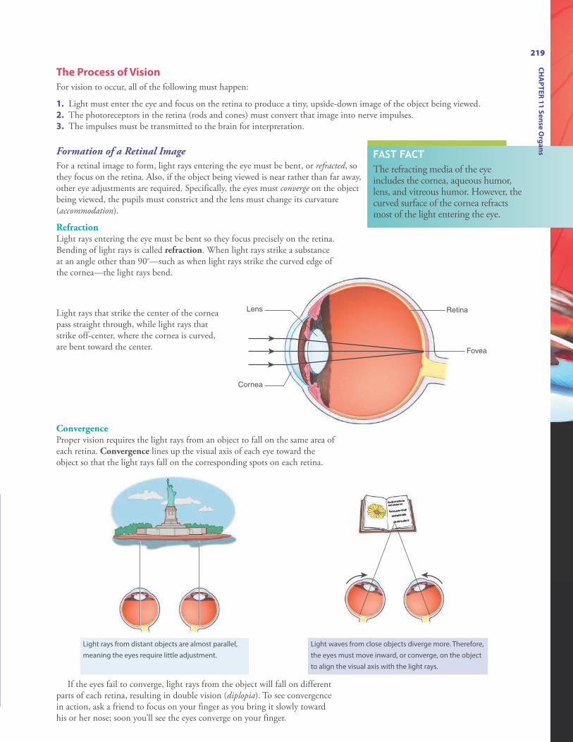
Lens
Cornea
Retina
Fovea
219
Light rays that strike the center of the corneapass straight through, while light rays thatstrike off-center, where the cornea is curved,are bent toward the center.
Light rays from distant objects are almost parallel,
meaning the eyes require little adjustment.
Light waves from close objects diverge more. Therefore,
the eyes must move inward, or converge, on the object
to align the visual axis with the light rays.
ConvergenceProper vision requires the light rays from an object to fall on the same area ofeach retina. Convergence lines up the visual axis of each eye toward theobject so that the light rays fall on the corresponding spots on each retina.
If the eyes fail to converge, light rays from the object will fall on differentparts of each retina, resulting in double vision (diplopia). To see convergencein action, ask a friend to focus on your finger as you bring it slowly towardhis or her nose; soon you’ll see the eyes converge on your finger.
The Process of Vision
For vision to occur, all of the following must happen:
1. Light must enter the eye and focus on the retina to produce a tiny, upside-down image of the object being viewed.2. The photoreceptors in the retina (rods and cones) must convert that image into nerve impulses. 3. The impulses must be transmitted to the brain for interpretation.
Formation of a Retinal Image
For a retinal image to form, light rays entering the eye must be bent, or refracted, sothey focus on the retina. Also, if the object being viewed is near rather than far away,other eye adjustments are required. Specifically, the eyes must converge on the objectbeing viewed, the pupils must constrict and the lens must change its curvature(accommodation).
RefractionLight rays entering the eye must be bent so they focus precisely on the retina.Bending of light rays is called refraction. When light rays strike a substanceat an angle other than 90°—such as when light rays strike the curved edge ofthe cornea—the light rays bend.
FAST FACTThe refracting media of the eye includes the cornea, aqueous humor,lens, and vitreous humor. However, thecurved surface of the cornea refractsmost of the light entering the eye.
CH
AP
TE
R 1
1 S
en
se O
rga
ns

Life lesson: Common visual defectsMany people experience less than perfect vision, typically caused by errors in refraction. Special lenses (eyeglasses)can be used to correct these errors. The lenses bend light rays before they reach the cornea so that, by the time therays reach the back of the eye, they focus on the retina to produce clear vision. The following figures illustratenormal vision as well as common refractive errors.
PA
RT
III Re
gu
latio
n a
nd
Inte
gra
tion
of th
e B
od
y
220
Presbyopia: With age, the lens loses flexibility—interfering with its ability tochange shape—and the focusing muscles in the eye weaken. As a result, lightfocuses behind the retina, creating difficulty focusing on objects up close (suchas when reading). Presbyopia usually begins between the ages of 40 and 50years.
Astigmatism: Astigmatism results from an uneven or asymmetrical curvatureof the cornea, causing light to be focused unevenly. People with astigmatismhave difficulty viewing fine detail in objects up close as well as at a distance.This refractive defect often accompanies myopia or hyperopia.
Hyperopia (farsightedness): When light rays focus at a point behind theretina, objects up close appear blurry. Called hyperopia, this condition occurs ifthe eyeball is too short or the cornea is flatter than normal.
Myopia (nearsightedness): When light rays focus in front of the retina insteadof directly on it, distant objects appear blurry while those up close are clear.This condition, called myopia, occurs if the eyeball is too long or the cornea hasmore curvature than normal. Myopia is the most common refractive visiondefect.
Emmetropia: When light rays focus on the retina without the need of acorrective lens, normal vision results. This is called emmetropia.
FAST FACTThe sharpness of visualperception is called visualacuity.
Life lesson: CataractsAnother common cause of visual disturbances, especiallyamong the elderly, is cataracts. A cataract is clouding ofthe lens, making vision cloudy or blurry. Other commoncomplaints include glare, colors appearing faded, andpoor night vision. Cataracts commonly occur as a part ofaging, although other risk factors include diabetesmellitus, smoking, and prolonged exposure to sunlight.Treatment includes surgery to remove the lens with thecataract and insertion of an artificial lens.

v _ h e S S U Y Zf e S U W X Ya _ b V S \ b v _ h e Sv _ h e S S U Y ZV X [ b W Y e V W X Ya _ b V S \ b
The pupillary constrictor muscle
encircles the pupil. When
stimulated by the parasympathetic
nervous system, the muscle
constricts, narrowing the pupil to
admit less light.
The pupillary dilator looks like the
spokes of a wheel. When stimulated
by the sympathetic nervous system,
this muscle contracts, pulling the
inside edge of the iris outward. This
widens the pupil and admits more
light.
221
CH
AP
TE
R 1
1 S
en
se O
rga
ns
Constriction of the PupilThe center of the cornea—just like the center of any lens—can focus lightrays better than the periphery. To reduce blurriness when focusing on anearby object, the pupil constricts to screen out peripheral light rays. Theconstriction and dilation of the pupil depends upon muscles inside the iris;these are called the intrinsic eye muscles.
Accommodation of the LensWhile the cornea refracts most of the light rays entering theeye, the lens fine-tunes the rays for sharper focus.Specifically, the curvature of the lens changes to allow theeye to focus on a near object, a process calledaccommodation.
The Body AT WORKPupils also constrict automatically
when exposed to bright light. This is
called the photopupillary reflex. In
fact, both pupils constrict even if
only one eye is illuminated, which is
why this is sometimes called the
consensual light reflex.
The nearly parallel light rays from distant objects require little
refraction. Consequently, the ciliary muscle encircling the lens relaxes
and the lens flattens and thins.
The more divergent light rays from a nearby object require more
refraction. To help focus the light rays, the ciliary muscle
surrounding the lens contracts. This narrows the lens, causing it to
bulge into a convex shape and thicken, giving it more focusing
power.
ª \ [ b W d e V ¯ \ [ bo e S e U Y Z a _ b V S \V X [ W Y U V W \ fo e S e U Y Z a _ b V S \Y \ S U ° \ f ª \ [ b W d e [ b
FAST FACTContinually using the eyes for close-upwork stresses the ciliary muscles, resultingin eyestrain. Taking periodic breaks to focuson a distant object will help lessen thestrain.
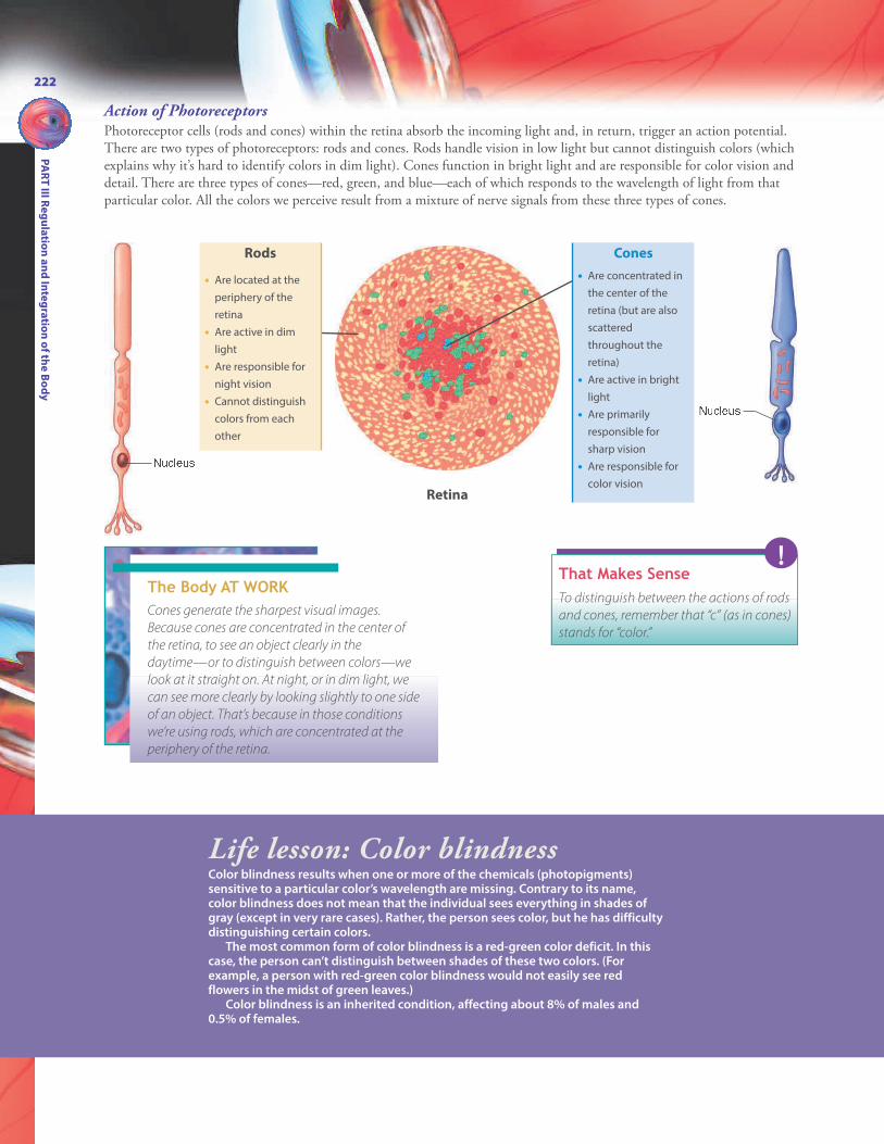
PA
RT
III Re
gu
latio
n a
nd
Inte
gra
tion
of th
e B
od
y
222
Action of PhotoreceptorsPhotoreceptor cells (rods and cones) within the retina absorb the incoming light and, in return, trigger an action potential.There are two types of photoreceptors: rods and cones. Rods handle vision in low light but cannot distinguish colors (whichexplains why it’s hard to identify colors in dim light). Cones function in bright light and are responsible for color vision anddetail. There are three types of cones—red, green, and blue—each of which responds to the wavelength of light from thatparticular color. All the colors we perceive result from a mixture of nerve signals from these three types of cones.
g _ V S \ _ bThat Makes SenseTo distinguish between the actions of rods
and cones, remember that “c” (as in cones)
stands for “color.”
!The Body AT WORKCones generate the sharpest visual images.
Because cones are concentrated in the center of
the retina, to see an object clearly in the
daytime—or to distinguish between colors—we
look at it straight on. At night, or in dim light, we
can see more clearly by looking slightly to one side
of an object. That’s because in those conditions
we’re using rods, which are concentrated at the
periphery of the retina.
Cones
• Are concentrated in
the center of the
retina (but are also
scattered
throughout the
retina)
• Are active in bright
light
• Are primarily
responsible for
sharp vision
• Are responsible for
color vision
Rods
• Are located at the
periphery of the
retina
• Are active in dim
light
• Are responsible for
night vision
• Cannot distinguish
colors from each
other
g _ V S \ _ bRetina
Life lesson: Color blindnessColor blindness results when one or more of the chemicals (photopigments)sensitive to a particular color’s wavelength are missing. Contrary to its name,color blindness does not mean that the individual sees everything in shades ofgray (except in very rare cases). Rather, the person sees color, but he has difficultydistinguishing certain colors.
The most common form of color blindness is a red-green color deficit. In thiscase, the person can’t distinguish between shades of these two colors. (Forexample, a person with red-green color blindness would not easily see redflowers in the midst of green leaves.)
Color blindness is an inherited condition, affecting about 8% of males and0.5% of females.

223
CH
AP
TE
R 1
1 S
en
se O
rga
ns
Transmission of Nervous Impulses
The final step in the visual process involves relaying the nerve impulses to the brain, wherethe images are interpreted as sight.
ª \ T W \ Z \ p e r d W \ Z \p e r d W ] e b _ U S T e \ S fª \ T W ] e b _ U S T e \ S f
That Makes SenseIt may be confusing to think that images from the right
visual field are handled by the left cerebral hemisphere, and
that images from the left visual field are handled by the
right cerebral hemisphere. It makes more sense, however,
when you remember that the right brain controls motor
responses on the left side of the body and vice versa.
Therefore, each side of the brain needs to “see” what’s
happening on the side of the body for which it initiates
movement.
!
The Body AT WORKThe optic chiasm is located
just anterior to the pituitary
gland. That’s why an
enlargement of the
pituitary gland, such as
from a tumor, can affect
vision.
Nerve impulses generated by
the rods and cones leave the
eye via the optic nerve.
Half the nerve fibers cross over
to the opposite side of the brain
at the optic chiasm. Specifically,
fibers from the nasal side cross
over while the fibers on the
temporal side remain on the
same side.
The impulses travel to the
primary visual cortex in the
occipital lobe for interpretation.
FAST FACTA lesion in the brain’s occipitallobe can cause blindness, even ifthe eyes are functioning properly.

Review of Key TermsAdaptation: The diminishing of asensation that occurs after continuousexposure to a stimulus
Analgesic: Drug used to relieve pain
Aqueous humor: Clear, watery fluidthat fills the anterior cavity of the eye
Bony labyrinth: Complicated system ofpassageways within the temporal bonethat contains the inner ear
Chemoreceptors: Receptors that reactto various chemicals, including odors,tastes, and the concentration ofvarious chemicals in the body
Choroid: Highly vascular layer oftissue in the eye that supplies oxygenand nutrients to the sclera and retina
Ciliary body: Thickened extension ofthe choroid that secretes aqueoushumor
Cochlea: Snail-like structure in innerear that contains the structures forhearing
Cones: Photoreceptors in the retinaresponsible for color vision
Conjunctiva: Transparent mucousmembrane that lines the eyelids andcovers the anterior surface of theeyeball (except for the cornea)
Cornea: Transparent extension ofsclera that sits over the iris and admitslight into the eye
Eustachian tube: Passageway frommiddle ear to nasopharynx
Fovea centralis: Depressed area in themiddle of the macula lutea thatcontains only cones; it is the area ofsharpest vision
Gustation: Sense of taste
Lacrimal gland: Small gland thatsecretes tears
Macula lutea: Center point of theretina
Mechanoreceptors: Receptors thatrespond to factors that change theposition of a receptor
Membranous labyrinth: Lines theinside of the bony labyrinth
Nociceptors: Pain receptors thatrespond to tissue damage fromtrauma as well as from heat,chemicals, pressure, or a lack ofoxygen
Olfaction: Sense of smell
Optic chiasm: Point in the brain wherehalf the optic nerve fibers cross to theopposite side of the brain
Optic disc: Spot where nerve fibersleave the retina, converging to becomethe optic nerve
Organ of Corti: Hearing sense organ
Papillae: Protrusions on the tongue onwhich taste buds are located
Photoreceptors: Receptors in the eyesthat respond to light
Referred pain: Pain originating in adeep organ that is sensed as if it’soriginating from the body’s surface
Refraction: Bending of light rays sothey focus on the retina
Retina: Innermost layer of the eye thatcontains receptors for vision
Rods: Photoreceptors in the retinaactive in dim light
Sclera: Outermost layer of the eye;formed from dense connective tissue
Thermoreceptors: Receptors activatedby a change in temperature
Tympanic membrane: Membraneseparating outer ear from middle earthat vibrates in response to soundwaves; eardrum
Vestibule: Structure of inner ear thatcontains organs necessary for sense ofbalance
Vitreous humor: Jelly-like substancethat fills the posterior cavity of the eye
PA
RT
III Re
gu
latio
n a
nd
Inte
gra
tion
of th
e B
od
y
224

Own the InformationTo make the information in this chapter part of your
working memory, take some time to reflect on what you’ve
learned. On a separate sheet of paper, write down
everything you recall from the chapter. After you’re done,
log on to the DavisPlus website, and check out the Study
Group podcast and Study Group Questions for the chapter.
Key Topics for Chapter 11:
• Types and locations of sensory receptors
• Classifications of sensory receptors
• Types and locations of pain receptors
• The main pain pathway
• Characteristics and locations of receptors for temperature
and touch
• The location of receptors for taste and the chemical and
neural process for experiencing taste
• The location of receptors for smell and the process for
experiencing smell
• Structures of the outer, middle, and inner ear
• How hearing occurs
• The role of the vestibule and semicircular canals in the
process of balance
• Accessory structures of the eye
• Anatomy of the eye, including the retina and chambers
of the eye
• The process of vision
• The action of photoreceptors
225
CH
AP
TE
R 1
1 S
en
se O
rga
ns

Answers: Chapter 111. Correct answer: c. Chemoreceptors react to
chemicals; mechanoreceptors respond to factors(such as pressure, stretch, or vibration) that changethe position of a receptor; thermoreceptorsrespond to changes in temperature.
2. Correct answer: d. Injured receptors do releaseseveral different chemicals, but this has nothing todo with identifying the source of pain. Deep bodyorgans and structures contain slow pain fibers,which produce dull, aching pain. Pain signals thattravel to the thalamus proceed to the postcentralgyrus, making the individual aware of pain, whilesignals that travel to the limbic system triggeremotional responses to pain.
3. Correct answer: a. Olfaction is the sense of smell.Equilibrium is the sense of balance. Convergenceis when the visual axis of each eye lines upproperly to the incoming light rays.
4. Correct answer: c. The vestibule contains organsnecessary for the sense of balance. The semicircularcanals are crucial for the maintenance of balanceand equilibrium. The auricle, or pinna, is thevisible part of the external ear.
5. Correct answer: a. All of the other conditionswould interfere with the transmission of vibrationsto the inner ear, resulting in a conductive hearingloss.
226
PA
RT
III Re
gu
latio
n a
nd
Inte
gra
tion
of th
e B
od
y
Test Your Knowledge1. Nociceptors are receptors that
respond to:a. various chemicals inside and
outside the body.b. pressure, stretch, or vibration.c. pain from tissue damage.d. changes in temperature.
2. The true source of pain can bedifficult to identify because:a. injured tissues release several
different chemicals that stimulate the nociceptors andtrigger pain.
b. deep body organs and structures don’t contain painfibers.
c. some pain signals travel to thethalamus while others travel tothe limbic system.
d. sensory impulses from different areas of the bodyoften travel to the brain overthe same pathway.
3. The sense of taste is called:a. gustation.b. olfaction.c. equilibrium.d. convergence.
4. Which inner ear structure contains the structures for hearing?a. Vestibuleb. Semicircular canalsc. Cochlead. Auricle
5. Which condition would cause asensorineural hearing loss?a. Death of hair cells in the organ
of Cortib. Fluid in the middle earc. Fusion of the auditory ossiclesd. Impacted cerumen

Go to http://davisplus.fadavis.com Keyword:Thompson to see all of the resources availablewith this chapter.
6. Vibration of the tympanic membrane triggers vibration inthe:a. eustachian tube.b. auditory ossicles.c. bony labyrinth.d. perilymph.
7. Which is a normal function oftears?a. Aid in the refraction of light
raysb. Deliver oxygen and nutrients
to the conjunctivac. Transmit light rays to the
cornead. Fill the anterior cavity
8. The outermost layer of the eye is the:a. sclera.b. ciliary body.
c. choroid.d. retina.
9. What condition results when therate of absorption of aqueoushumor through the canal ofSchlemm is less than its production?a. Cataractsb. Astigmatismc. Hyperopiad. Glaucoma
10. Rods are:a. active in dim light.b. concentrated in the center of
the retina.c. responsible for color vision.d. primarily responsible for sharp
vision.
6. Correct answer: b. The eustachian tube is apassageway from the middle ear to thenasopharynx; it does not respond to vibrations inthe tympanic membrane. The bony labyrinth isthe part of the temporal bone containing thestructures of the inner ear. Perilymph moves inresponse to vibrations transmitted by the auditoryossicles through the oval window.
7. Correct answer: b. Tears do not aid in therefraction or transmission of light rays. Aqueoushumor fills the anterior cavity.
8. Correct answer: a. The ciliary body and choroidare parts of the middle vascular layer. The retina ispart of the inner neural layer.
9. Correct answer: d. Cataracts result from cloudingof the lens. Astigmatism results from an uneven orasymmetrical curvature of the cornea. Hyperopia,or farsightedness, results when light rays focus at apoint behind the retina.
10. Correct answer: a. All of the other descriptionspertain to cones.
227
CH
AP
TE
R 1
1 S
en
se O
rga
ns

CHAPTER OUTLINEOverview of the Endocrine System
Hormones
The Pituitary Gland and Hypothalamus
Pineal Gland
Thymus
Thyroid Gland
Parathyroid Glands
Adrenal Glands
Pancreas
Gonads
Other Endocrine Cells and Chemicals
LEARNING OUTCOMES1. Define hormone.
2. List the organs of the endocrine system.
3. Differentiate between the actions of the
endocrine system and the nervous system.
4. Compare and contrast the chemical makeup
and actions of steroid and nonsteroid
hormones.
5. Describe the anatomical relationship between
the pituitary gland and hypothalamus.
6. Identify the hormones, as well as the actions of
the hormones, secreted by both the pituitary
gland and the hypothalamus
7. Explain how pituitary secretions are controlled
by the nervous system as well as by negative
feedback.
8. Describe the structure and location of the
major endocrine glands.
9. Identify the hormones secreted by the major
endocrine glands and describe their functions.

12chapter ENDOCRINE SYSTEM
Pituitary
Hypothalamus
Thyroid
Thymus
Adrenals
Pancreas
Parathyroids
Pineal
Testes(male)
Ovaries (female)
The endocrine system produces a multitude of hormones with effects
as wide ranging as influencing mood to controlling reproduction.
The endocrine system consists of a variety of glands andspecialized cells throughout the body. These glands and cellssecrete chemicals (called hormones) that influence almostevery cell and organ in the body. Hormones determine,among other things, when you feel hungry or full, whetheryou’re fat or thin, how you handle stress, and even how yousleep.
The endocrine and nervous systems often work hand-in-hand to promote communication between cells, allowing themto coordinate and integrate their activities. Although theyshare a common goal—homeostasis—their methods foraccomplishing this vary. For example, the nervous system useslightning-fast electrical signals to communicate, making it theideal choice to oversee such time-sensitive processes asbreathing and movement. In contrast, the endocrine systemcommunicates through slower-acting hormones, placing it incharge of body processes that happen more slowly, such as cellgrowth.
FAST FACTThe study of the endocrinesystem and the diagnosis andtreatment of its disorders iscalled endocrinology.

PA
RT
III Re
gu
latio
n a
nd
Inte
gra
tion
of th
e B
od
y
230
Comparison of Endocrine and Nervous Systems
The actions of the endocrine and nervous systems complement one another to ensurethat the body maintains homeostasis. Their methods of action differ, though, asdetailed below.
Endocrine cells
Targetcells
HormoneBloodstream
Neuron
Target cells
Neuro-transmitter
That Makes SenseTo differentiate between endocrine and exocrine glands,
remember: EXocrine glands have EXit routes (ducts) to an
environment EXternal to themselves. ENDocrine glands
secrete hormones withIN the body that END up in the
bloodstream.
!
The endocrine system consists of the glands shown in the figure on the previous page as well as clusters of hormone-secreting cells in various organs, including the brain, heart, and small intestine.
FAST FACTEven after a stimulus ceases, theeffects of the endocrine systemcan persist for days or evenweeks.
The Body AT WORKEndocrine glands are ductless glands: they secrete hormones directly
into the bloodstream. (In contrast, exocrine glands secrete hormones
into ducts, which, in turn, lead to a surface in some other location,
such as the body's surface—as in sweat glands—or the digestive
tract—as in digestive enzymes.) Once in the bloodstream, each
hormone travels throughout the body. In the process, the cells of
many different organs are exposed to a particular hormone; however,
only cells having receptors for that hormone (called target cells) will
respond. In other words, a hormone acts only on cells with receptors
specific to that hormone; this is called specificity.
Overview of the Endocrine System
Endocrine system
• Employs hormones to relay messages
• Distributes hormones throughout the body via the bloodstream
• Responds slowly to stimuli (seconds to days)
• Exerts long-lasting effects
• Adapts slowly to continual stimulation
Nervous system
• Employs neurotransmitters to relay messages
• Secretes neurotransmitters into tiny space of a synapse
• Responds to stimuli quickly (milliseconds)
• Exerts short-lived effects
• Adapts quickly to continual stimulation

231
CH
AP
TE
R 1
2 E
nd
ocrin
e S
yste
m
Nucleus
Blood vessel
Secondmessenger
Enzyme
Reaction
Hormones can be classified as steroid or nonsteroid. Steroid hormones are synthesized from cholesterol; they include maleand female sex hormones as well as aldosterone (secreted by the adrenal cortex). Nonsteroid, or protein-based, hormones aresynthesized from amino acids. They can be further divided into protein hormones (such as insulin), peptide hormones (suchas antidiuretic hormone), or amino acid derivative hormones (such as epinephrine and norepinephrine). Regardless ofclassification, hormones travel through the bloodstream to stimulate specific target cells. The target cells may lie anywhere inthe body: close to the gland or far from it.
Once a hormone reaches the target cell, it binds with a receptor to trigger changes within the cell. How this occurs,though, depends upon whether the hormone is a steroid or protein-based hormone.
Protein-based hormones can’t penetrate the cell wall.
Consequently, they bind to receptors on the cell
surface. The binding of the hormone activates a
second messenger system: a cascade of processes that
results in the production of a second messenger.
The second messenger activates specific enzymes.
The enzymes influence cellular reactions, producing
the cell’s response to the hormone.
Steroid hormones, which are derived from cholesterol
(a lipid), pass easily through a cell’s membrane.
Once inside the cell, they bind to receptors in the
nucleus associated with the DNA. This triggers the
synthesis of new proteins, which then alters
metabolism in the cell.
The Body AT WORKThe more receptors a target cell has, the more sensitive it
will be to a hormone. Likewise, the fewer the receptors,
the less sensitive it will be. Cells constantly create new
receptors to replace older ones. Furthermore, cells can
add to their receptors, making them more sensitive to a
hormone. As an example, during the final stage of
pregnancy, cells in the uterus increase their receptors for
oxytocin, the hormone responsible for triggering uterine
contractions during childbirth.
Hormones
FAST FACTThe body employs a number ofsecond messenger molecules;however, the most prominentmay be cyclic adenosinemonophosphate, or cAMP.

Hypophysealportalsystem
Hypothalamus
Anterior pituitary
Posterior pituitary
Optic chiasm
Pituitary gland
The tiny, pea-sized pituitary gland sits
just underneath the hypothalamus. It
lies cradled in the sella turcica, a
cavity within the sphenoid bone.
A stalk called the infundibulum
connects the hypothalamus and
pituitary.
Despite its small size, the pituitary gland is
actually two distinct glands: the anterior
pituitary, or adenohypophysis, and the
posterior pituitary, or neurohypophysis.
These two glands are made of different tissue,
excited by different types of stimuli, and secrete
different hormones.Anterior Pituitary
The anterior pituitary—the larger of the two pituitary glands—consists ofglandular tissue. It synthesizes and secretes a number of very importanthormones, all under the direction of the hypothalamus.
Neurons within the hypothalamus synthesize
various hormones. Some, called releasing
hormones, stimulate the anterior pituitary to
secrete its hormones. Others, called inhibiting
hormones, suppress hormone secretion by
the anterior pituitary.
The neurons of the hypothalamus release their
hormones into a system of blood vessels called
the hypophyseal portal system.
The blood travels straight to the anterior
pituitary, where the hormones from the
hypothalamus act on target cells in the
anterior pituitary.
This stimulates the anterior pituitary to
release, or to suppress the release of, certain
hormones into the general circulation.
The Pituitary Gland and Hypothalamus
PA
RT
III Re
gu
latio
n a
nd
Inte
gra
tion
of th
e B
od
y
232
The pituitary gland—especially in conjunction with the nearby hypothalamus—exerts more influence on body processesthan any other endocrine gland.
ANIMATION
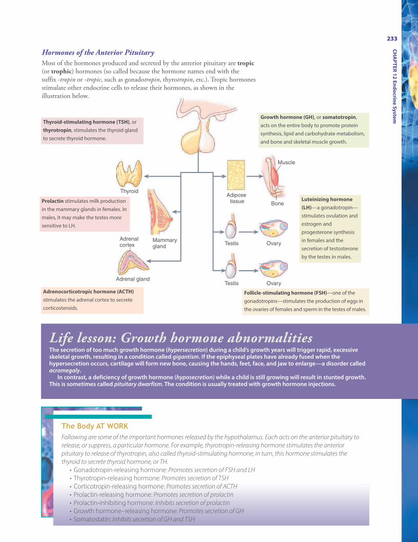
Thyroid
Mammary gland
Adrenal cortex
Adrenal gland
Adipose tissue Bone
Testis
OvaryTestis
Ovary
Muscle
Follicle-stimulating hormone (FSH)—one of the
gonadotropins—stimulates the production of eggs in
the ovaries of females and sperm in the testes of males.
Luteinizing hormone
(LH)—a gonadotropin—
stimulates ovulation and
estrogen and
progesterone synthesis
in females and the
secretion of testosterone
by the testes in males.
Growth hormone (GH), or somatotropin,
acts on the entire body to promote protein
synthesis, lipid and carbohydrate metabolism,
and bone and skeletal muscle growth.
Thyroid-stimulating hormone (TSH), or
thyrotropin, stimulates the thyroid gland
to secrete thyroid hormone.
Prolactin stimulates milk production
in the mammary glands in females. In
males, it may make the testes more
sensitive to LH.
Adrenocorticotropic hormone (ACTH)
stimulates the adrenal cortex to secrete
corticosteroids.
The Body AT WORKFollowing are some of the important hormones released by the hypothalamus. Each acts on the anterior pituitary to
release, or suppress, a particular hormone. For example, thyrotropin-releasing hormone stimulates the anterior
pituitary to release of thyrotropin, also called thyroid-stimulating hormone; in turn, this hormone stimulates the
thyroid to secrete thyroid hormone, or TH.
• Gonadotropin-releasing hormone: Promotes secretion of FSH and LH
• Thyrotropin-releasing hormone: Promotes secretion of TSH
• Corticotropin-releasing hormone: Promotes secretion of ACTH
• Prolactin-releasing hormone: Promotes secretion of prolactin
• Prolactin-inhibiting hormone: Inhibits secretion of prolactin
• Growth hormone–releasing hormone: Promotes secretion of GH
• Somatostatin: Inhibits secretion of GH and TSH
233
CH
AP
TE
R 1
2 E
nd
ocrin
e S
yste
m
Hormones of the Anterior Pituitary
Most of the hormones produced and secreted by the anterior pituitary are tropic(or trophic) hormones (so called because the hormone names end with the suffix -tropin or -tropic, such as gonadotropin, thyrotropin, etc.). Tropic hormonesstimulate other endocrine cells to release their hormones, as shown in theillustration below.
Life lesson: Growth hormone abnormalitiesThe secretion of too much growth hormone (hypersecretion) during a child’s growth years will trigger rapid, excessiveskeletal growth, resulting in a condition called gigantism. If the epiphyseal plates have already fused when thehypersecretion occurs, cartilage will form new bone, causing the hands, feet, face, and jaw to enlarge—a disorder calledacromegaly.
In contrast, a deficiency of growth hormone (hyposecretion) while a child is still growing will result in stunted growth.This is sometimes called pituitary dwarfism. The condition is usually treated with growth hormone injections.
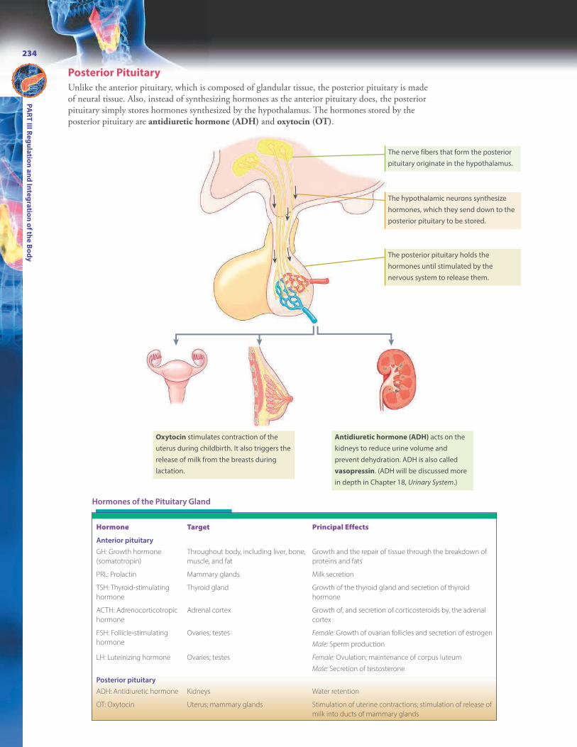
Oxytocin stimulates contraction of the
uterus during childbirth. It also triggers the
release of milk from the breasts during
lactation.
Antidiuretic hormone (ADH) acts on the
kidneys to reduce urine volume and
prevent dehydration. ADH is also called
vasopressin. (ADH will be discussed more
in depth in Chapter 18, Urinary System.)
The nerve fibers that form the posterior
pituitary originate in the hypothalamus.
The hypothalamic neurons synthesize
hormones, which they send down to the
posterior pituitary to be stored.
The posterior pituitary holds the
hormones until stimulated by the
nervous system to release them.
PA
RT
III Re
gu
latio
n a
nd
Inte
gra
tion
of th
e B
od
y
234
Posterior Pituitary
Unlike the anterior pituitary, which is composed of glandular tissue, the posterior pituitary is madeof neural tissue. Also, instead of synthesizing hormones as the anterior pituitary does, the posteriorpituitary simply stores hormones synthesized by the hypothalamus. The hormones stored by theposterior pituitary are antidiuretic hormone (ADH) and oxytocin (OT).
Hormone Target Principal Effects
Anterior pituitary
GH: Growth hormone
(somatotropin)
Throughout body, including liver, bone,
muscle, and fat
Growth and the repair of tissue through the breakdown of
proteins and fats
PRL: Prolactin Mammary glands Milk secretion
TSH: Thyroid-stimulating
hormone
Thyroid gland Growth of the thyroid gland and secretion of thyroid
hormone
ACTH: Adrenocorticotropic
hormone
Adrenal cortex Growth of, and secretion of corticosteroids by, the adrenal
cortex
FSH: Follicle-stimulating
hormone
Ovaries; testes Female: Growth of ovarian follicles and secretion of estrogen
Male: Sperm production
LH: Luteinizing hormone Ovaries; testes Female: Ovulation; maintenance of corpus luteum
Male: Secretion of testosterone
Posterior pituitary
ADH: Antidiuretic hormone Kidneys Water retention
OT: Oxytocin Uterus; mammary glands Stimulation of uterine contractions; stimulation of release of
milk into ducts of mammary glands
Hormones of the Pituitary Gland

235
CH
AP
TE
R 1
2 E
nd
ocrin
e S
yste
m
Control of Pituitary Secretions
The pituitary gland does not release a steady flow of hormones. Rather, it releases hormones inphases or pulses. For example, growth hormone is mainly secreted at night, whereas LH surges inthe middle of the menstrual cycle. The central nervous system plays a role in controlling hormonesecretion. So, too, do the target organs themselves through the process of negative feedback.
Negative Feedback
TRH stimulates the anterior
pituitary to release thyroid-
stimulating hormone (TSH).
Cold stimulates the
hypothalamus to release
thyrotropin-releasing
hormone (TRH).
At the same time, the
increased thyroid
hormone inhibits release
of TSH by the pituitary.
TSH stimulates the
thyroid to release
thyroid hormones.
Thyroid hormones
stimulate metabolism,
increasing warmth.
Control by the Central Nervous System
The brain constantly monitors conditions both inside and outside the body. Inturn, it triggers the release of hormones as needed. For example, when it’s cold, ittriggers the release of TSH to promote body heat. After a hearty meal, it stimulatesthe secretion of GH to break down fats for energy and use amino acids to buildtissue.
Control by Negative Feedback
When the pituitary stimulates another endocrine gland to secrete its hormone, thathormone is then fed back to the pituitary, telling it to stop further release of thetropic hormone. This process is called negative feedback. Negative feedback loopsare used extensively to regulate hormones in the hypothalamic-pituitary axis. Thefollowing figure illustrates the negative feedback process as it relates to theregulation of body temperature.
FAST FACTThe use of negative feedback tocontrol hormone secretion,combined with the fact thathormones have a limited lifespan, causes most hormonesto be secreted in “pulses.”
ANIMATION

Hypothalamus
Pituitary
gland
Larynx
Thyroid gland
Trachea
Lung
Heart
The pineal gland produces melatonin, a hormone that rises at night, when sunlight is absent, and falls during the day. Highmelatonin levels trigger sleepiness, making it a key factor in the sleep-wake cycle. Although yet to be proven, scientistsspeculate that the pineal gland may also regulate the timing of puberty.
In children, the thymus gland is large. Beginning in puberty,though, the gland starts to shrink. By the time one reaches oldage, the gland consists of mostly fat and fibrous tissue.
The thymus secretes thymosin and thymopoietin, twohormones having a role in the development of the immunesystem. Although it secretes hormones, making it a member ofthe endocrine system, the actions of the hormones make thethymus part of the immune system. (For more information onthe thymus gland, see Chapter 16, Lymphatic & ImmuneSystems.)
FAST FACTA number of people take supplementalmelatonin orally to combat jet lag or othersleep disorders. While the supplements aregenerally regarded as safe for short-term use,taking too much can increase one’s risk forblood clots and seizures.
Tucked in the center of the brain,
on the roof of the brain’s third
ventricle, is the tiny, cone-shaped
pineal gland.
The thymus lies in the mediastinum just
beneath the sternum.
Life lesson: Seasonal affective disorderLow levels of melatonin have been linked to mood disorders, particularly seasonal affective disorder (SAD).Occurring during winter months, when exposure to sunlight is limited, people with SAD complain of sleepiness,depression, irritability, and carbohydrate cravings. Exposure to special high-intensity lights for several hours eachday often relieves symptoms.
Pineal Gland
PA
RT
III Re
gu
latio
n a
nd
Inte
gra
tion
of th
e B
od
y
236
Thymus

Larynx
Isthmus
Trachea
Thyroid
colloid
The largest endocrine gland, the thyroid, consists of two large lobes connected by a narrow band oftissue called the isthmus.
Thyroid tissue is made of tiny sacs called thyroid follicles.
Each follicle is filled with a thick fluid called thyroid colloid.
The cells lining the sacs secrete the two main thyroid hormones:
T3 (triiodothyronine) and T4 (thyroxine). Unlike other glands,
the thyroid gland can store the hormones for later use.
Cells between the thyroid follicles, called parafollicular cells,
secrete another hormone called calcitonin. Secreted in
response to rising blood calcium levels, calcitonin triggers
the deposition of calcium in bone, thus promoting bone
formation. The effects of calcitonin are particularly important
in children.
The thyroid gland resides in the neck, just
below the trachea, where it is wrapped
around the anterior and lateral portions of
the trachea.
The Body AT WORKAs previously described, TSH (released from the anterior
pituitary) stimulates the release of thyroid hormone (TH) from
the thyroid gland. The overall effect of TH is to increase the
body’s metabolic rate, which, in turn, increases heat
production. (This explains why TH is released when the body is
exposed to cold.) Just a few of the other effects of TH are
increased rate and strength of heart contractions, increased
respiratory rate, and increased appetite. TH is also crucial for
growth and development: it promotes the development of
bone; the nervous system; and skin, hair, nails, and teeth.
237
CH
AP
TE
R 1
2 E
nd
ocrin
e S
yste
m
Thyroid Gland
FAST FACTThe abbreviations T3 and T4refer to the number of iodineatoms in each hormone: T3has three iodine atoms whileT4 has four.

PA
RT
III Re
gu
latio
n a
nd
Inte
gra
tion
of th
e B
od
y
238
PTH is the main hormone the body uses to maintain normal levels of calcium in the blood. Normal nerve and musclefunction, blood clotting, cell membrane permeability, and the function of certain enzymes all depend on adequate levels ofcalcium.
Thyroidgland
Posterior view
Life lesson: Thyroid disordersA number of significant disorders develop from hyper- or hyposecretion of thyroid hormone. If a child isborn without a thyroid gland, the lack of thyroid hormone (TH) leads to cretinism: a condition characterizedby retarded growth and sexual development, a low metabolic rate, and mental retardation. If thehyposecretion develops later in life, a lowered metabolic rate causes weight gain, hair loss, and fatigue. Ifhypothyroidism is severe or prolonged, a condition called myxedema may result, which involves swellingand firmness of the skin.
Hypersecretion of TH stimulates the body’s metabolism, resulting in a condition called Graves’ disease.Thought to be due to an autoimmune disorder, Graves’ disease causes unexplained weight loss, increasedheart rate, nervousness, and exophthalmos—a protrusion of the eyeballs due to swelling of tissue behindthe eye.
An enlarged thyroid gland due to a dietary deficiency of iodine is called simple goiter. Iodine is the basisfor TH. Without enough iodine, the production of TH falls. Consequently, the anterior pituitary never receivesa negative feedback message to stop producing TSH. Not only that, because TH levels are low, the anteriorpituitary tries to stimulate the thyroid even more by secreting greater levels of TSH. Instead of triggering theproduction of TH (which the thyroid gland can’t do without enough iodine), the TSH stimulates growth ofthyroid tissue, leading to thyroid enlargement and goiter.
Located on the posterior surface of the thyroid are four parathyroid glands. These glands secrete parathyroid hormone(PTH) in response to low blood levels of calcium.
The parathyroid glands are embedded in the
posterior lobes of the thyroid. Most people
have four parathyroid glands, but the number
of glands, as well as their locations, can vary.
FAST FACTThe margin of safety for bloodcalcium levels is so narrow that adrop in blood calcium levels assmall as 1% causes the secretionof PTH to double.
Parathyroid Glands

239
CH
AP
TE
R 1
2 E
nd
ocrin
e S
yste
m
Calcium Homeostasis
Calcitonin (secreted by the thyroid) has antagonistic effects on PTH. Theinteraction of these two hormones helps the body achieve calcium homeostasis.
Blood Ca2+
excess
Thyroid
releases
calcitonin
Ca2+ moves
from blood
to bone
Blood Ca2+
levels
decrease
Normal blood
calcium levels
Blood Ca2+
deficiency
Parathyroid
releases PTH
Ca2+ moves from
bones, kidneys,
and intestines
to blood
Blood Ca2+
levels
increase
Regulation of blood
calcium levels
In an effort to achieve calcium homeostasis, PTH exerts its influence on the bones, kidneys, and intestines:
PTH inhibits new bone formation while stimulating
the breakdown of old bone, causing calcium (and
phosphate) to move out of bone and into the blood.
PTH encourages the kidneys to reabsorb calcium—
blocking its excretion into the urine—while
promoting the secretion of phosphate. PTH also
prompts the kidneys to activate vitamin D,
necessary for intestinal absorption of calcium.
After its activation by the kidneys, vitamin D allows
the intestines to absorb calcium from food; the
calcium is transported through intestinal cells and
into the blood.
FAST FACTNinety-nine percent of thebody’s calcium is found inthe bones.
ANIMATION

PA
RT
III Re
gu
latio
n a
nd
Inte
gra
tion
of th
e B
od
y
240
FAST FACTTetany from hypocalcemiatypically occurs following theaccidental removal of theparathyroid glands duringthyroid surgery.
Hormone Target Principal Effects
Thyroid
Triiodothyronine (T3) and
thyroxine (T4)
Most tissues Increases rate of metabolism
Calcitonin Bone Increases deposition of Ca2+ in bones,
lowering blood Ca2+ levels
Parathyroid
Parathyroid hormone (PTH) Bone, kidneys Increases blood Ca2+ levels by increasing
removal of Ca2+ from bone, reducing
urinary excretion of Ca2+, and increasing
absorption of Ca2+ by the intestines
Hormones of the Thyroid and Parathyroid Glands
Life lesson: Calcium abnormalitiesCalcium ions normally contribute to the difference in polarity across the cellmembrane (with the outer surface of the membrane having a positive charge and theinterior having a negative). When calcium is in short supply, the charge differencelessens, and sodium ions can more easily enter the cell. As was discussed in the lastchapter, it’s the inflow of sodium that excites nerve and muscle cells.
So, in a calcium deficiency (hypocalcemia), nerve and muscle cells become moreexcitable. The excessive excitation leads tomuscle tremors, spasms, or tetany—thesustained contraction of a muscle. In fact, acardinal sign of hypocalcemia is a tetany ofthe hands and feet called carpopedal spasm.As shown here, the muscles of the handscontract tightly, unable to relax. (Similarcontractions occur in the feet.) If calciumlevels drop further, the muscles of the larynxcan spasm enough to stop airflow.
When the blood contains too manycalcium ions (hypercalcemia), the excess
calcium ions bind to the cell’s surface. Fewer sodium ions flow in and the cell becomesless responsive, resulting in such symptoms as muscle weakness, sluggish reflexes,and, in severe cases, cardiac arrest.

Capsule of connective tissue
Adrenal medulla
Adrenal
gland
Kidney
241
CH
AP
TE
R 1
2 E
nd
ocrin
e S
yste
m
Classes of Hormones Secreted by Adrenal Cortex
The hormones secreted by the adrenal cortex fall into one of the following three classes.
The adrenal glands perch on the top of each kidney. Instead of being one gland, though, each adrenal gland is actually twodistinct glands. The inner portion—called the adrenal medulla—consists of modified neurons and functions as part of thesympathetic nervous system. The outer portion—called the adrenal cortex—is glandular tissue and secretes steroidhormones called corticosteroids.
Adrenal Medulla
The adrenal medulla contains modified
neurons (called chromaffin cells) that act as
part of the sympathetic nervous system.
These cells secrete catecholamines
(specifically, epinephrine and
norepinephrine) in response to stimulation.
Catecholamines:
• Prepare the body for physical activity by
increasing heart rate and blood pressure,
stimulating circulation to the muscles, and
dilating the bronchioles; to maximize
blood flow to the areas needed for physical
activity, they also inhibit digestion and
urinary production.
• Boost glucose levels (a source of fuel) by
breaking down glycogen into glucose
(glycogenolysis) and converting fatty
acids and amino acids into glucose
(gluconeogenesis).
Adrenal Cortex
The adrenal cortex consists of three
layers of glandular tissue, which are
shown below. Each layer secretes a
different corticosteroid.
Mineralocorticoids
• The principal
mineralocorticoid is
aldosterone.
• Aldosterone acts on the
kidneys to promote
Na+ retention and K+
excretion.
• In turn, it also causes water
retention.
(For more information on
aldosterone, see Chapter 19,
Fluid, Electrolyte, & Acid-Base
Balance.)
Glucocorticoids
• The principal glucocorticoid is
cortisol.
• Glucocorticoids help the body adapt to
stress and repair damaged tissue by
stimulating the breakdown of fat and
protein, converting fat and protein to
glucose, and releasing fatty acids and
glucose into the blood.
• They have an anti-inflammatory effect.
• They also suppress the immune system
if secreted over a long term.
• Glucocorticoids are essential for
maintaining a normal blood pressure.
Sex Steroids
• Sex steroids include a weak form of androgen that is
converted to the more potent androgen testosterone.
• The testes produce much more testosterone, making this
an unimportant source of testosterone in men.
• Androgens stimulate development of pubic and axillary
hair and sustain sex drive (libido) in both sexes.
• The sex steroids also include small amounts of
estrogen.
• Because the amount is small, it has little importance
during reproductive years.
• However, it is the only source of estrogen after
menopause.
Zona glomerulosa (the
outermost layer): Secretes
mineralocorticoids
Zona fasciculata (the
middle layer): Secretes
glucocorticoids
Zona reticularis (the
innermost layer): Secretes
sex steroids
Adrenal Glands
FAST FACTGlucocorticoids affect nearlyevery cell in the body.

PA
RT
III Re
gu
latio
n a
nd
Inte
gra
tion
of th
e B
od
y
242
Life lesson: Adrenal disordersHypersecretion of cortisol from the adrenal gland results in a collection ofsymptoms called Cushing syndrome. Classic symptoms include a “moon face” (arounded face) and a “buffalo hump” (a hump between the shoulders resultingfrom a redistribution of body fat). Because excess cortisol also disruptscarbohydrate and protein metabolism, other symptoms include hyperglycemia,muscle weakness, and edema. The breakdown of protein causes muscle wastingand loss of bone mass, while the retention of sodium and water leads tohypertension.
The hypersecretion of adrenal androgens often accompanies Cushingsyndrome. This results in premature puberty in children and the development ofmasculine characteristics (deepening voice, increased body hair, and thedevelopment of facial hair) in women.
Hyposecretion of mineralocorticoids and glucocorticoids occurs in a disordercalled Addison’s disease, also called adrenal cortical insufficiency. Symptomsinclude a loss of fluid and electrolytes, weakness, muscle wasting, dehydration,and weight loss. Without treatment, adrenal insufficiency is life threatening.
Hormone Target Principal Effects
Adrenal medulla
Epinephrine Most tissues Enhances the effects of the sympathetic
nervous system
Norepinephrine Most tissues Enhances the effects of the sympathetic
nervous system
Adrenal cortex
Aldosterone Kidney Promotes Na+ retention and K+ excretion,
which leads to water retention
Cortisol Most tissues Stimulates the breakdown of fat and protein
and the conversion of fat and protein to
glucose; enhances tissue repair; anti-
inflammatory; in large amounts, inhibits the
immune system
Adrenal androgens Sex organs Promotes growth of pubic and axillary hair;
sex drive
Adrenal estrogens Sex organs Physiologically insignificant
Hormones of the Adrenal Glands
FAST FACTProlonged stress results in increasedblood levels of cortisol, which, in turn,leads to immune system dysfunction.One reason this occurs is that highconcentrations of cortisol causelymphatic tissues to atrophy and thenumbers of white blood cells todecline.
Life lesson: Changes with AgingWith age, some target tissues become less sensitive to theircontrolling hormone. The amount of hormone secreted maychange, and the hormone may be metabolized more slowly.Following are some common effects of age on the endocrinesystem:
• The thyroid gland becomes more nodular, leading to adecrease in secretion of thyroid hormones and a decrease inmetabolism.
• Changes in levels of parathyroid hormone may contribute toosteoporosis.
• Cells become less sensitive to insulin after age 50, whichleads to a gradual rise in fasting blood glucose levels.
• Secretion of growth hormone decreases, resulting indecreased muscle mass and an increase in fat storage.

243
CH
AP
TE
R 1
2 E
nd
ocrin
e S
yste
m
Liver
Stomach
Spleen
Small intestine
Colon
Beta cell
Alpha cell
Delta cell
Bile duct
The pancreas is somewhat unique in that it contains both endocrine and exocrine tissues. The vast majority of the pancreasacts as an exocrine gland, but a small percentage serves an important endocrine function. (For more information on the roleof the pancreas as an exocrine gland, see Chapter 20, Digestive System.)
The pancreas lies just behind the stomach,
with its head tucked in the curve of the
beginning of the small intestine (duodenum)
and its tail reaching to the spleen.
Exocrine cells, called acini, secrete
digestive enzymes into ducts that
drain into the small intestine.
Interspersed with the exocrine cells are
clusters of endocrine cells; these cells are
called pancreatic islets or the islets of
Langerhans. The pancreatic islets contain
several different types of cells, the main
ones being alpha cells, beta cells, and delta
cells.
Alpha Cells
Alpha cells secrete the hormone
glucagon. Between meals, when blood
glucose levels fall, glucagon stimulates
liver cells to convert glycogen into glucose
and also to convert fatty acids and amino
acids into glucose (gluconeogenesis). The
resulting glucose is released into the
bloodstream, causing blood glucose levels
to rise.
Beta Cells
Beta cells secrete the hormone insulin.
After eating, the levels of glucose and
amino acids in the blood rise. Insulin
stimulates cells to absorb both of these
nutrients, causing blood glucose levels to
fall.
Delta Cells
Delta cells secrete somatostatin, a
hormone that works within the pancreas to
regulate the other endocrine cells.
Specifically, it inhibits the release of both
glucagon and insulin. It also inhibits the
release of growth hormone.
The Body AT WORKThe body must constantly
work to keep blood glucose
levels steady. If levels fall too
low, cells won’t have
enough fuel. On the other
hand, too much glucose
will trigger disorders such as
autoimmune disease or
pancreatitis.
Pancreas
FAST FACTThe pancreas contains1 to 2 millionpancreatic islets, andyet, they constituteonly about 2% of allpancreatic tissue.

PA
RT
III Re
gu
latio
n a
nd
Inte
gra
tion
of th
e B
od
y
244
Regulation of Blood Glucose
Two of the major pancreatic hormones—insulin and glucagon—have oppositeeffects on levels of blood glucose, which helps maintain blood glucose levels withina normal range.
After eating, blood glucose levels rise as glucose flows
from the digestive tract into the bloodstream.
When blood glucose levels drop below a certain point
(such as after skipping a meal), the alpha cells of the
pancreas release glucagon into the blood.
Glucagon stimulates the liver to break down stored
glycogen into glucose, which it then releases into the
bloodstream. This causes blood glucose levels to rise.
High glucose levels stimulate the beta cells of the
pancreas to secrete insulin.
Insulin triggers two reactions:
• It stimulates the cells to take up more glucose.
• It causes the liver to take up glucose and store
it as glycogen.
The combined result is that glucose levels return to normal levels.
Glucose
– +
Glucose
– +
Glucose
– +
Glucose
– +
1
2
3
4
5
Glucose
Glucose
Insulin
Glucagon
Glucose
Glucagon
Glucose
Glucose
Glycogen
ANIMATION

245
CH
AP
TE
R 1
2 E
nd
ocrin
e S
yste
m
Life lesson: Diabetes mellitusOne of the most common endocrine disorders, the incidence of diabetes in the United Statescontinues to rise. According to the American Diabetes Association, over 29 millionAmericans, or 9.3% of the population, have diabetes. Of those, thousands experience seriouscomplications: 810 people die, 230 undergo an amputation of a lower limb, 120 begin kidneydialysis or receive a kidney transplant, and 55 go blind each day. What’s more, it’s estimatedthat about 6 million of the people with diabetes don’t even know they have the disease.
Diabetes results from an inadequate amount of insulin or from a diminished number ofnormal insulin receptors. Either way, without enough insulin, or without enough insulinreceptors, glucose can’t enter cells. As a result, cells are deprived of their main energy source,and glucose builds up in the blood. This produces high levels of blood glucose(hyperglycemia)—one of the cardinal signs of diabetes.
High levels of blood glucose trigger a number of physiological changes that produce theclassic signs of this disease. For example, the kidneys normally filter blood plasma andconvert it to urine; as it does, it removes glucose and returns it to the bloodstream. Inhyperglycemia, the high levels of glucose overwhelm the kidneys, and excess glucose “spillsover” into the urine (glycosuria).
To flush out this extra load of glucose, the kidneys produce more urine (polyuria). In turn,this dehydrates the body, triggering excessive thirst (polydipsia). In addition to drinkingexcessive quantities of fluids, people with untreated diabetes also experience continuoushunger (polyphagia). That’s because, even though the blood is carrying an overabundantsupply of glucose, the cells are starving because the glucose can’t enter.
If left untreated, the body begins to burn protein and fat for energy. Besides causingfatigue and weight loss, this abnormal metabolism produces an acidic byproduct calledketone bodies. As ketones accumulate, blood pH drops, causing acidosis. Unchecked, this willprogress to diabetic ketoacidosis, causing symptoms such as nausea, vomiting, fruity odor ofthe breath, and possibly coma and death.
Diabetes also damages blood vessels (resulting in heart attacks, strokes, decreasedcirculation in the extremities, and even blindness from damaged blood vessels in the retina)as well as nerves (resulting in numbness and tingling). Kidney disease is another commoncomplication.
FAST FACTAdults with diabetes haveheart disease death rates two tofour times higher than adultswithout diabetes.
Type 1 Diabetes Characteristics Type 2 Diabetes Characteristics
Average age at onset Before age 30 Usually after age 40
Rate of onset Rapid Gradual
Percent of all diabetics 10% 90%
Cause Deficiency of insulin resulting from the
destruction of beta cells of the pancreatic
islets
Loss of insulin receptors on target cells,
leading to insulin resistance
Contributing factors Uncertain; may be caused by autoimmune
disease
Heredity combined with excess body weight
and sedentary lifestyle; also more prevalent in
Native Americans, Hispanics, and African
Americans
Treatment Daily insulin injections Lifestyle changes may control disease; if not,
oral diabetic medications or insulin injections
may be used
Type 1 diabetes was formerly called
juvenile-onset diabetes or insulin-dependent
diabetes mellitus (IDDM)
Type 2 diabetes was formerly called
maturity-onset diabetes or non –insulin-
dependent diabetes mellitus (NIDDM)
another common complication.
There are two main types of diabetes: type 1 and type 2. The characteristicsof each type are outlined in the table below.
Characteristics of Type 1 and Type 2 Diabetes Mellitus
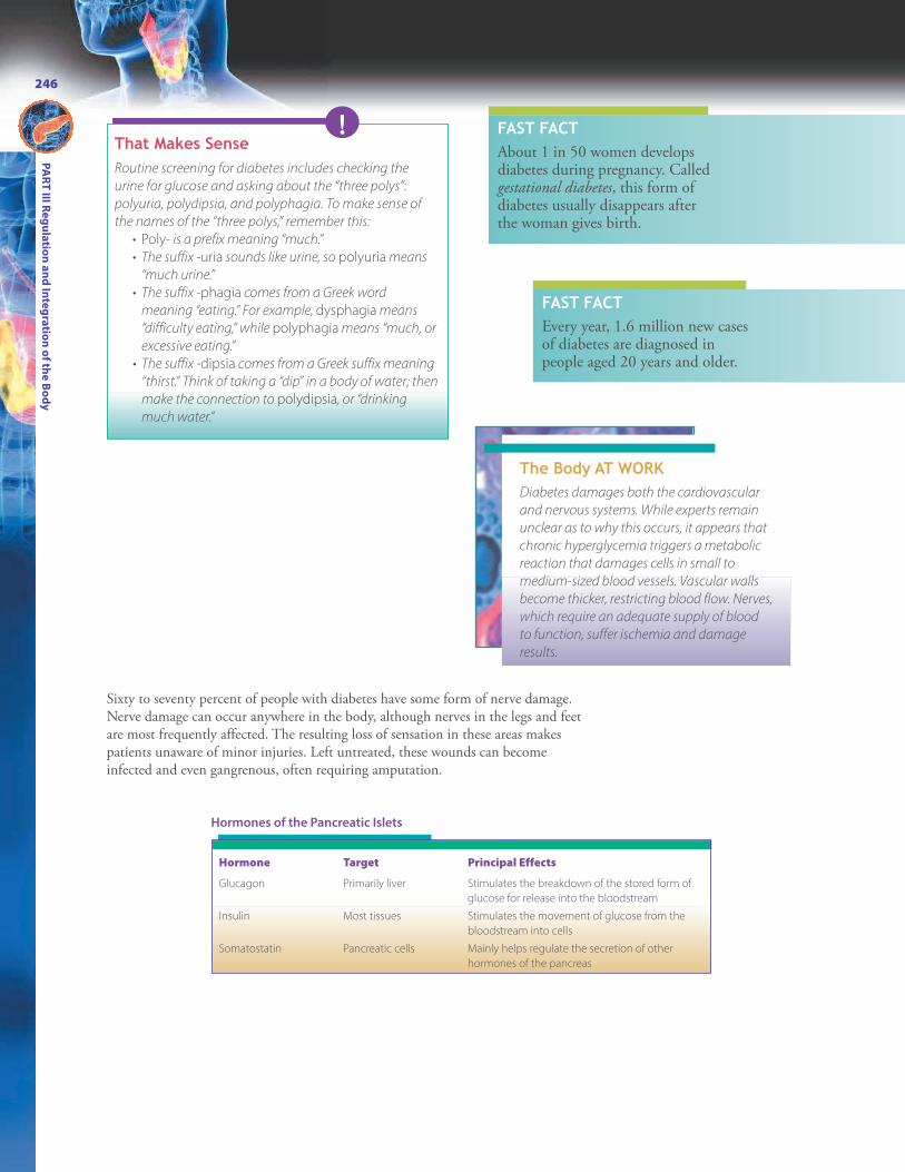
PA
RT
III Re
gu
latio
n a
nd
Inte
gra
tion
of th
e B
od
y
246
FAST FACTEvery year, 1.6 million new casesof diabetes are diagnosed inpeople aged 20 years and older.
That Makes SenseRoutine screening for diabetes includes checking the
urine for glucose and asking about the “three polys”:
polyuria, polydipsia, and polyphagia. To make sense of
the names of the “three polys,” remember this:
• Poly- is a prefix meaning “much.”
• The suffix -uria sounds like urine, so polyuria means
“much urine.”
• The suffix -phagia comes from a Greek word
meaning “eating.” For example, dysphagia means
“difficulty eating,” while polyphagia means “much, or
excessive eating.”
• The suffix -dipsia comes from a Greek suffix meaning
“thirst.” Think of taking a “dip” in a body of water; then
make the connection to polydipsia, or “drinking
much water.”
!
Hormone Target Principal Effects
Glucagon Primarily liver Stimulates the breakdown of the stored form of
glucose for release into the bloodstream
Insulin Most tissues Stimulates the movement of glucose from the
bloodstream into cells
Somatostatin Pancreatic cells Mainly helps regulate the secretion of other
hormones of the pancreas
Hormones of the Pancreatic Islets
FAST FACTAbout 1 in 50 women developsdiabetes during pregnancy. Calledgestational diabetes, this form ofdiabetes usually disappears afterthe woman gives birth.
Sixty to seventy percent of people with diabetes have some form of nerve damage.Nerve damage can occur anywhere in the body, although nerves in the legs and feetare most frequently affected. The resulting loss of sensation in these areas makespatients unaware of minor injuries. Left untreated, these wounds can becomeinfected and even gangrenous, often requiring amputation.
The Body AT WORKDiabetes damages both the cardiovascular
and nervous systems. While experts remain
unclear as to why this occurs, it appears that
chronic hyperglycemia triggers a metabolic
reaction that damages cells in small to
medium-sized blood vessels. Vascular walls
become thicker, restricting blood flow. Nerves,
which require an adequate supply of blood
to function, suffer ischemia and damage
results.

247
CH
AP
TE
R 1
2 E
nd
ocrin
e S
yste
m
Ovary
Testis
Gonads—the testes in males and the ovaries in females—are the primary sex organs. They produce sex hormones, whichstimulate the production of sperm (in males) and eggs (in females). They also influence the development of secondary sexcharacteristics during puberty. (For further information, see Chapter 23, Reproductive Systems.)
The cells of the ovarian follicle secrete estrogen. Estrogen promotes the
development of female characteristics (such as breast development) and also
contributes to the development of the reproductive system.
After ovulation, the corpus luteum (the tissue left behind after a rupture of a
follicle during ovulation) secretes progesterone. Progesterone, in combination
with estrogen, helps maintain the uterine lining during pregnancy.
Specialized cells within the testes secrete
testosterone. Testosterone triggers the
development of male sexual characteristics;
it also sustains sperm production.
FAST FACTNonsteroidal anti-inflammatory drugs (NSAIDS),such as ibuprofen, relieve pain and fever byblocking the synthesis of certain prostaglandins.
Not all hormones are secreted from specific, well-defined glands. In fact, organs such as the heart, liver, kidneys, stomach,small intestines, and placenta contain clusters of cells that secret hormones. Just a few examples:
l When stretched by rising blood pressure, heart muscle cells secrete atrial natriuretic peptide (ANP), a hormone thattriggers changes to lower blood pressure.
l The placenta secretes estrogen, progesterone, and other hormones that help maintain a woman’s pregnancy.l The stomach secretes several hormones that help regulate the digestive process.
Also realize that hormones and neurotransmitters are not the only chemical messengers in the body. Certain cells in thebody secrete other chemicals that help regulate function. Perhaps the most significant of these is prostaglandins.
Prostaglandins
Made by cells throughout the body, prostaglandins are lipid molecules that, like hormones,integrate the activities of other cells. Unlike hormones—which are released into the bloodstream sothey can influence the activities of widely separated organs—prostaglandins are released within thetissue where they are produced. As a result, instead of influencing activities in distant organs,prostaglandins influence the activities of neighboring cells.
The body manufactures a multitude of different prostaglandins. The various prostaglandins playmany roles. For example, some prostaglandins promote pain and fever, while others act asvasodilators and vasoconstrictors. They also are involved in inflammation, blood clotting, uterinecontraction, reproduction, and digestion.
Gonads
Other Endocrine Cells and Chemicals

Review of Key TermsAdenohypophysis: Anterior pituitarygland
Adrenal cortex: Outer portion of theadrenal gland that secretescorticosteroids
Adrenal gland: Gland perched on topof the kidney; consists of two distinctglands (the adrenal medulla and theadrenal cortex)
Adrenal medulla: Inner portion ofadrenal gland that functions as part ofthe sympathetic nervous system
Catecholamines: Epinephrine andnorepinephrine; secreted by theadrenal medulla
Corticosteroids: Steroid hormonessecreted by the adrenal cortex
Endocrine gland: Ductless glands thatsecrete hormones directly into thebloodstream
Gonads: Primary sex organs, which arethe testes in males and ovaries infemales
Graves’ disease: Disorder resultingfrom hypersecretion of thyroidhormone
Hormones: Chemicals secreted byglands and specialized cells of theendocrine system
Neurohypophysis: Posterior pituitarygland
Pancreas: Secretes digestive enzymes(exocrine function) as well ashormones used to regulate bloodglucose levels (endocrine function)
Parathyroid glands: Glands embeddedon the posterior surface of the thyroidgland that secrete a hormone used toregulate blood calcium levels
Pineal gland: Produces the hormonemelatonin, which increases at nightand decreases during the day
Pituitary gland: Small gland attachedto the lower surface of thehypothalamus that secretes a numberof hormones that regulate manybodily processes; consists of ananterior and posterior lobe
Target cells: Cells having receptors fora particular hormone
Tetany: Sustained muscle contraction;may result from hypocalcemia
Thymus: Secretes hormones having arole in the development of theimmune system
Thyroid gland: Gland in the neck thatsecretes hormones that affect thebody’s metabolic rate
PA
RT
III Re
gu
latio
n a
nd
Inte
gra
tion
of th
e B
od
y
248
Own the InformationTo make the information in this chapter part of your
working memory, take some time to reflect on what you’ve
learned. On a separate sheet of paper, write down
everything you recall from the chapter. After you’re done,
log on to the DavisPlus website, and check out the Study
Group podcast and Study Group Questions for the chapter.
Key Topics for Chapter 12:
• Function of the endocrine system
• Comparison of the endocrine and nervous systems
• The classification and actions of hormones
• The relationship of the pituitary gland and hypothalamus
• Hormones of the hypothalamus and pituitary gland
• Control of pituitary secretions by the central nervous
system and negative feedback
• The location, structure, and function of the pineal gland,
thymus, thyroid gland, parathyroid glands, adrenal
glands, pancreas, and gonads
• Other endocrine tissues and chemicals

Test Your Knowledge1. Which is a characteristic of
exocrine glands?a. They secrete hormones into
the bloodstream.b. Ducts carry their secretions to
the body’s surface.c. They exert a long-lasting effect.d. They respond slowly to
stimuli.
2. Which endocrine gland influences more body processesthan any other endocrine gland?a. Thyroidb. Pituitaryc. Adrenal cortexd. Limbic system
3. A key difference between the anterior and posterior pituitary isthat the posterior pituitary:a. consists of glandular tissue.b. secretes hormones under the
direction of the hypothalamus.c. receives hormones from the
hypothalamus via the hypophyseal portal system.
d. stores hormones released fromthe hypothalamus.
4. Negative feedback is a process bywhich:a. a hormone is fed back to the
pituitary, telling it to stop further release of that hormone.
b. the deficiency of a certain hormone stimulates a gland torelease more of that hormone.
c. the central nervous systemmonitors the conditions insideand outside of the body and, inturn, triggers the release of hormones as needed.
d. hormone levels fall precipitously, triggering shock.
5. Cold temperatures are most likelyto stimulate the release of whichhormone by the anterior pituitary?a. Growth hormoneb. ACTHc. Thyroid-stimulating hormoned. Prolactin
6. Following thyroid surgery, a patient is most at risk for whichdisorder?a. Graves’ diseaseb. Goiterc. Hypercalcemiad. Hypocalcemia
7. To help the body cope withstress, the adrenal cortex secreteswhich class of hormones?a. Mineralocorticoidsb. Catecholaminesc. Glucocorticoidsd. Sex steroids
8. Which gland has more exocrinethan endocrine tissue?a. Thyroid glandb. Adrenal glandc. Pancreasd. Thymus
9. The chief role of insulin is to:a. stimulate cells to take up more
glucose.b. stimulate the liver to break
down stored glycogen into glucose.
c. stimulate the pancreas to release glucagon into theblood.
d. trigger the conversion of fattyacids and amino acids into glucose.
10. A hypersecretion of growth hormone during adulthoodcauses:a. gigantism.b. pituitary dwarfism.c. acromegaly.d. myxedema.
Answers: Chapter 121. Correct answer: b. Exocrine glands have ducts that
carry their secretions to the body’s surface, whileendocrine glands are ductless glands that secretetheir hormones into the bloodstream. The othercharacteristics are true of endocrine glands.
2. Correct answer: b. Although the thyroid andadrenal cortex affect important body processes, thepituitary influences a number of organs andprocesses. The limbic system is not part of theendocrine system.
3. Correct answer: d. All of the other statements arecharacteristics of the anterior pituitary.
4. Correct answer: a. None of the other answers is adefinition of negative feedback.
5. Correct answer: c. Thyroid-stimulating hormonestimulates the secretion of thyroid hormone by thethyroid gland, which, in turn, would boostmetabolism and heat production. Growthhormone promotes growth and tissue repair.ACTH stimulates the secretion of corticosteroidsby the adrenal cortex. Prolactin stimulates milksecretion by the mammary glands.
6. Correct answer: d. Accidental removal of theparathyroid glands during thyroid surgery triggershypocalcemia. Graves’ disease is the result ofhypersecretion of thyroid hormone. Goiter is anenlarged thyroid gland due to a dietary deficiencyof iodine. Hypercalcemia would result fromexcessive secretion of parathyroid hormone.
7. Correct answer: c. The principal glucocorticoid iscortisol, which helps the body adapt to stress. Theprincipal mineralocorticoid is aldosterone, whichaids in water retention by the kidneys.Catecholamines are secreted by the adrenalmedulla, not the adrenal cortex. Sex steroidsstimulate the development of secondary sexcharacteristics.
8. Correct answer: c. The other glands contain onlyendocrine tissue.
9. Correct answer: a. Glucagon stimulates the liver tobreak down stored glycogen into glucose. Fallingblood glucose levels stimulate the pancreas torelease glucagon into the blood. Glucagon triggersthe conversion of fatty acids and amino acids intoglucose.
10. Correct answer: c. Hypersecretion of growthhormone during a child’s growth years causesgigantism. Hyposecretion of growth hormoneduring a child’s growth years causes pituitarydwarfism. Myxedema is a condition involvingswelling and firmness of the skin as a result ofprolonged or severe hypothyroidism.
249
CH
AP
TE
R 1
2 E
nd
ocrin
e S
yste
m
Go to http://davisplus.fadavis.com Keyword:Thompson to see all of the resources availablewith this chapter.