Embed Size (px)
Citation preview

Anatomy & Physiology of Mechanical Digestion
ANATOMY & PHYSIOLOGY 13-14

Avian Digestive Tract

Ruminates

Monogastric Digestive System

Digestive System Divisions
• Alimentary Canal (Gastrointestinal Tract)– These are all the structures that food passes
through during digestion– One, long, tube open at both ends– Starting with the mouth, ending with the anus
• Accessory Organs– These are the structures that aid in digestion
of food, but in which no food passes– Liver, gallbladder, pancreas, salivary glands
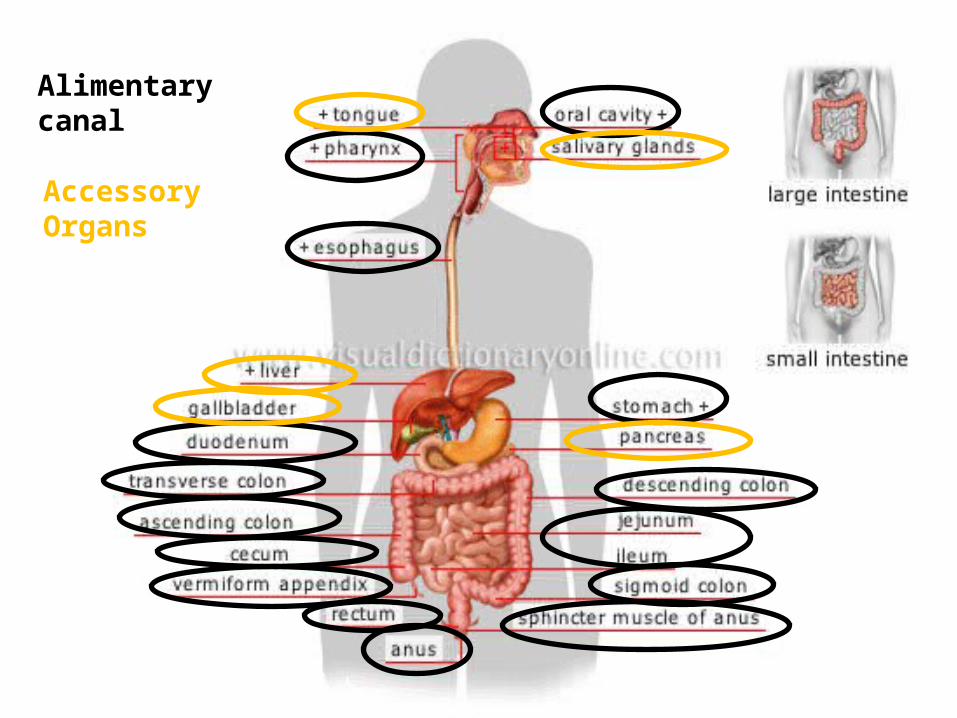
Alimentary canal
Accessory Organs

CONNECTIONS

Anatomy of the Oral Cavity
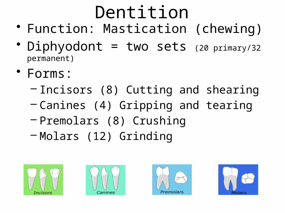
Dentition• Function: Mastication (chewing)• Diphyodont = two sets (20 primary/32 permanent)
• Forms: – Incisors (8) Cutting and shearing – Canines (4) Gripping and tearing– Premolars (8) Crushing– Molars (12) Grinding

Palates
• • Palatum Durum (hard palate): Upper, anterior roof of the mouth, covered with rugae (folds) to allow backwards movement of food
• Palatum Velum (i.e. “Soft Palate”): Upper, posterior roof of the mouth. Closes off access to nasopharynx during swallowing

Anatomy of Tongue
• Tongue: Muscle that mixes food with saliva, pushes food into the oropharynx, and contains gustatory papillae (taste receptors)
• Lingual frenulum: Fold of mucous membrane that attaches the tongue to the floor of the mouth

Tonsils
• Lymphoid tissue = first line of immune defense for aerodigestive tract
• Largest pre-puberty; atrophy after puberty

Deglutition and Pharyngeal-Esophageal Anatomy
• Deglutition = swallowing• Food enters into the
oropharynx, then passes into the laryngopharynx
• Closure of the epiglottis allows passage of food into the esophagus and prevents aspiration (particulates in lungs)

Peristalsis
• Alternating contractions between circular and longitudinal muscle of the pharynx and esophagus
• Physically separates food into small spheres (bolus) and moves it through the esophagus

Hiatal Hernia

STOMACH
• Initial site of protein hydrolysis/digestion
• Primary site of mechanical digestion via rumination
• Absorption of water and alcohol

Dr. William Beaumont and Alexis St. Martin (August 1825)


Cardiac Sphincter(antrum cardiacum)
• Food enters the stomach through this muscle via the esophagus
• Accidental opening of this structure may lead to Gastro-Esophageal Reflux Disorder (GERD)

Fundus
• Means “bottom” in Latin but is the left anterior curvature of the stomach
• Stores food for appx. 1 hour
• Digestive gases collect here

Anatomy of Stomach Body
• Greater and lesser curvature
• Gastric canal – can hold appx. 1 gallon of food
• Rugae (increase surface area)

Pyloric Antrum & Sphincter
• Muscular terminus of stomach
• Involved in rumination of food
• Food exits the gastric canal via passage through the pyloric sphincter

DUODENUM
• First section of small intestine
• Drastic rise in pH due to addition of bile salts
• Receives secretions of pancreas
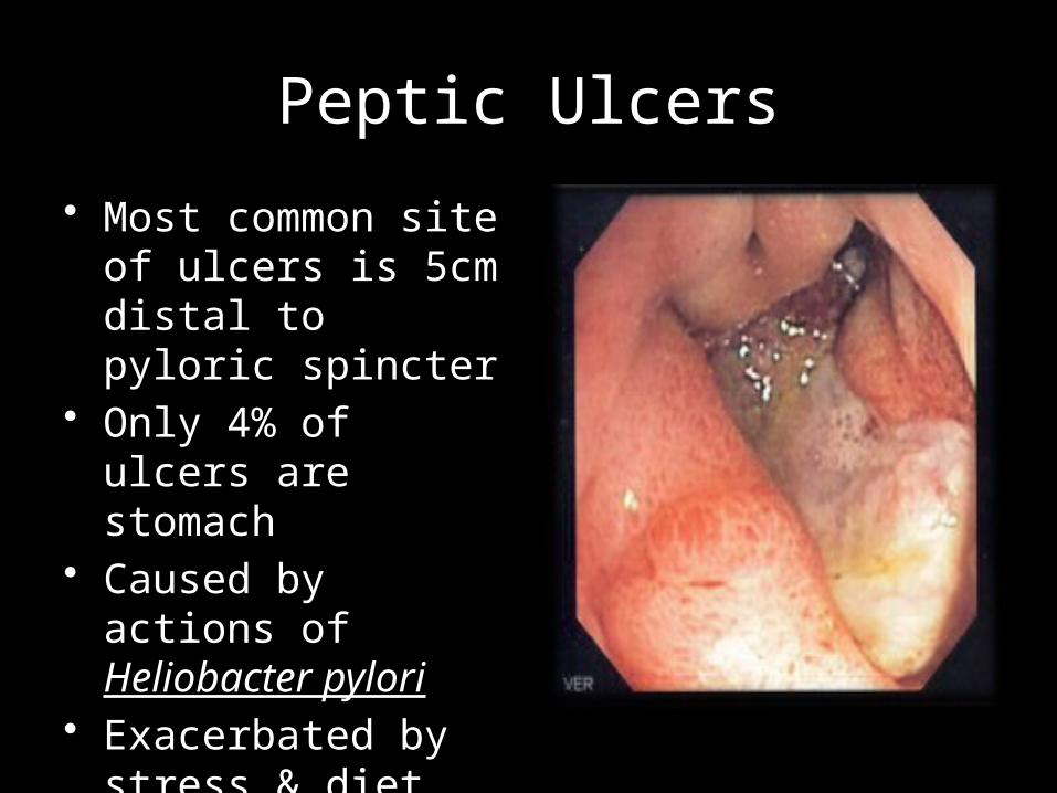
Peptic Ulcers
• Most common site of ulcers is 5cm distal to pyloric spincter
• Only 4% of ulcers are stomach
• Caused by actions of Heliobacter pylori
• Exacerbated by stress & diet


SMALL INTESTINE
• Jejunum (8ft)• Ileum (12ft)• Site of absorption and
chemical digestion

VILLI
• Small (1mm) projections of small intestine that drastically increase surface area of small intestine
• Cells lining villi die and are consumed!
• Contain capillaries for transport of material
• Contain lacteals to transport fats
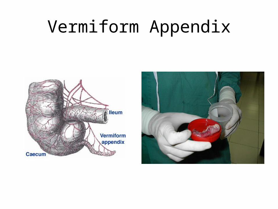
Vermiform Appendix

COLON

Colonic Form and Function
• 1.5m divided into– Ascending– Transverse– Descending– Sigmoid
• Involved in conduction of solid waste
• Reabsorption of water via standing gradient osmosis

Anal Sphincter
• Muscular bands that control expulsion of feces
• Regulated via recto-anal inhibitory reflex (RAIR)
• Flatuaria is a loss of control over RAIR