Embed Size (px)
Citation preview

Gut, 1981, 22, 534-541
Ultrastructure of endocrine-like cells in laminapropria of human gastric mucosa*J STACHURA, W J KRAUSE, AND K J IVEYtFrom the Veterans Administration Medical Center, Long Beach, California; University of California Irvine,Irvine, California; Department of Anatomy, University of Missouri School of Medicine, Columbia,Missouri; and the Institute ofPathology, Copernicus Medical Academy, Krakow, Polantd
SUMMARY Endocrine cells of gastric and gut mucosa are commonly thought to be present onlywithin mucosal glands. In a previous report, we described argyrophilic cells in the lamina propriain 40% of surgical gastric specimens, using light microscopy. All these patients had chronic gastritis.Argyrophilia, however, is a non-specific reaction which could occur in other than endocrine cells.The present study was undertaken to describe the ultrastructure of argyrophil cells in the laminapropria. In five patients with chronic gastritis, endoscopic biopsies were taken from the fundic,intermediate, and pyloric areas of the stomach. Single and/or clustered argyophil cells were seen
by light microscopy in the lamina propria of the intermediate and pyloric are.as. On electron-microscopy, these cells had the following characteristics of endocrine-like cells: they were
characterised by numerous electron dense granules in the cytoplasm, 100-300 nm in diameter; thecytoplasm contained poorly-developed rough endoplasmic reticulum and well-developed smoothendoplasmic reticulum with occasional vesicles. Immunostaining gave negative results for variousgastrointestinal hormones. These ultrastructural characteristics of lamina propria cells are similarto endocrine cells of the APUD series. We conclude that endocrine-like cells occur in the laminapropria of the human stomach in the presence of chronic gastritis.
Endocrine cells of gastrointestinal mucosa areusually found within mucosal glands.' Lightmicroscopic examination has suggested thepresence of endocrine cells in the lamina propriaof intermediate and pyloric mucosa of patho-logical human gastric mucosa.2 Argyrophiliccells, singly or in groups, were found in the laminapropria of 22 of 56 surgical gastric specimens ofpatients operated on for gastric or duodenalulcer or gastric cancer. All subjects with argyro-philic cells in the lamina propria had chronicgastritis.2 The argyrophilic reaction, however, isnon-specific, and thus it could only be hypothe-sised that these argyrophilic cells were endocrinecells.2 This study presents ultrastructuralevidence for endocrine-like morphological char-acteristics of these argyrophilic cells in thegastric lamina propria of man.*This work was supported in part by the CRC NIH Grant No.RR 00287-12 and Biomedical Grant No. RR 07053, and by theMedical Research Service of the Veterans Administration.tAddress for all correspondence and reprint requests: Dr K J Ivey,Gastroenterology Section (IIIG), Veterans Administration MedicalCenter, 5901 East Seventh Street, Long Beach, California 90822, USA.
Received for publication 9 January 1981
Methods
Biopsies of five subjects (four female, one male,aged 25-47 years) were investigated by light andelectron microscopy. Biopsies were chosen from60 patients endoscoped during a three-monthperiod for investigation of gastric or duodenalulcer disease or dyspepsia. A duplicate biopsy wastaken from pyloric (about 2 cm from pylorus),intermediate (7-9 cm from pylorus along greatercurva,ture), and body (mid-greater curvature)areas in each patient. One biopsy was routinelyexamined histologically, the other was collected inEpon blocks. Biopsies from five of these 60subjects showing argyrophil positive laminaproprial cells in the pyloric and/or intermediatearea were chosen for ultrastructural investiga-tions.
Tissue for histological examination was fixed in10% neutral formalin and embedded in paraffin.Paraffin sections were stained with haematoxylinand eosin (H and E). Periodic acid-Schiff (PAS)reaction, with Grimelius method for argyrophilia,3and the Masson-Fontana4 argentaffin reaction
534
on March 28, 2020 by guest. P
rotected by copyright.http://gut.bm
j.com/
Gut: first published as 10.1136/gut.22.7.534 on 1 July 1981. D
ownloaded from

Ultrastructure ofendocrine-like cells in lamina propria ofhuman gastric mucosa
Table Immunohistochemical investigations ofendocrine-like cells in lamina propria by PAPmethods5
Antiserum to Dilution Sourceused
Gastrin I and II 1:500 CalbiochemGlucagon 1:1000 CalbiochemSomatostatin 1:300 A Arimura. Tulane Univ.
School of Med, New Orleans,Louisiana, USA
VIP 1:2500 S 1 Said, Depts of InternalMed. and Pharmacol., Univ.Texas, Dallas, USA
Secretin 1:500 CalbiochemPancreatic polypeptide 1:500 Lilly Res Labs, Indianapolis,(bovine) Indiana, USASubstance P 1:500 R H Ho, Dept of Anatomy,
Ohio State Univ, Columbus,USA
were performed.Tissue for ultrastructural examination was
fixed in 2% glutaraldehyde in phosphate buffer,pH 7.4 at 4°C for two hours. Tissues were thenwashed in phosphate buffer, pH 74, and post-fixed in 2% osmium tetroxide for two hours,dehydrated in a graded series of ethyl alcohol,and embedded in Epon 812 resin. Silver-grey thinsections were stained with uranyl acetate andlead citrate and examined on a Philips 300 elec-tron microscope operated at 80 KV.
Immunostaining for gut hormones was per-formed according to Sternberger's PAP method5using formalin-fixed paraffin sections. Antiseraused, dilution and source are shown in the Table.
Results
LIGHT MICROSCOPYChronic gastritis was present in the biopsies ofthe pyloric and intermediate areas in all fivesubjects showing argyrophil positive lamina pro-prial cells. Focal intestinal metaplasia was pre-sent in two of these subjects. Focal superfici'algastritis occurred in the fundic area.Based on the Grimelius argyrophil reaction,
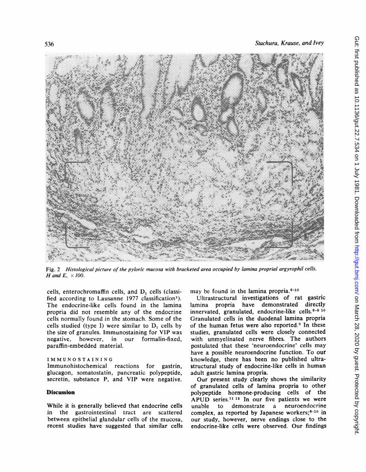
we found numerous positive staining endocrinecells in the gastric pyloric and fundic glands ashas been well described by others. In addition,single and/or groups of argyrophil cells werefound in the lamina propria as well, in thepyloric and intermediate zones (Fig. 1). The posi-tion of these cells in the lamina propria of themucosa is shown in Fig. 2. The Masson-Fontanaargentaffin reaction was negative.
ELECTRONMICROSCOPYUltrastructural examination showed these argyro-phil cells in the lamina propria to be endocrine-
1P... WFig. 1 Groups of argyrophil cells (large arrow)localised in the lamina propria. Single argyrophil cellsalso are observed within glandular epithelium (smallarrow). x 260.
like cells (Figs 3 and 4). They were characterisedby round and oval electron dense granules 100-200 nm in diameter in some cells (type I, Fig. 5)and 150-300 nm in other cells (type II, Fig. 6).The nuclei of these cells were oval and slightlyirregular in shape, with well-developed, smoothendoplasmic reticulum, with occasional vacuoleformation. Mitochondria were not very numer-ous. Lysosomal bodies containing electrondense material were also noticed. Cell boundariesusually were poorly defined, as they were inter-mingled with collagen fibres. Occasionally, theendocrine-like cells formed small clusters of twoto five cells (Figs 7 and 8). Some cells werefound close to the basal lamina of the gastricglands; others, however, were situated deep inthe connective tissue of the lamina propria, faraway from the glandular cells. We observednerve fibres close to some endocrine-like cells.Other endocrine-like cells were situated adja-cent to small blood vessels (Fig. 8).A moderate inflammatory infiltrate with
numerous plasma cells was observed within thelamina propria, as were granulated mast cells.Parietal and mucous cells present in the gastricglands were morphologically normal. Endocrinecells in the normal distribution within gastricglands were primarily enterochromaffin-like
535
on March 28, 2020 by guest. P
rotected by copyright.http://gut.bm
j.com/
Gut: first published as 10.1136/gut.22.7.534 on 1 July 1981. D
ownloaded from
Stachura, Krause, and Ivey
*wk
s \ff* as wtTh \E
Fig. 2 Histological picture of the pyloric mucosa with bracketed area occupied by lamina proprial argyrophil cells.Hand E, x 100.
cells, enterochromaffin cells, and D, cells (classi-fied according to Lausanne 1977 classification').The endocrine-like cells found in the laminapropria did not resemble any of the endocrinecells normally found in the stomach. Some of thecells studied (type I) were similar to D, cells bythe size of granules. Immunostaining for VIP wasnegative, however, in our formalin-fixed,paraffin-embedded material.
IMMUNOSTAINI NG
Immunohistochemical reactions for gastrin,glucagon, somatostatin, pancreatic polypeptide,secretin, substance P, and VIP were negative.
Discussion
While it is generally believed that endocrine cellsin the gastrointestinal tract are scatteredbetween epithelial glandular cells of the mucosa,recent studies have suggested that similar cells
may be found in the lamina propria."-10Ultrastructural investigations of rat gastric
lamina propria have demonstrated directlyinnervated, granulated, endocrine-like cells.6-8 10Granulated cells in the duodenal lamina propriaof the human fetus were also reported.9 In thesestudies, granulated cells were closely connectedwith unmyelinated nerve fibres. The authorspostulated that these 'neuroendocrine' cells mayhave a possible neuroendocrine function. To ourknowledge, there has been no published ultra-structural study of endocrine-like cells in humanadult gastric lamina propria.Our present study clearly shows the similarity
of granulated cells of lamina propria to otherpolypeptide hormone-producing cells of theAPUD series.1' 12 In our five patients we wereunable to demonstrate a neuroendocrinecomplex, as reported by Japanese workers;8-10 inour study, however, nerve endings close to theendocrine-like cells were observed. Our findings
536
on March 28, 2020 by guest. P
rotected by copyright.http://gut.bm
j.com/
Gut: first published as 10.1136/gut.22.7.534 on 1 July 1981. D
ownloaded from

Ultrastructure ofendocrine-like cells in lamina propria ofhuman gastric mucosa
A ,c
Fig. 3 Endocrine-like cell (E) situated in the connective tissue of lamina propria. Note the secretory granules(arrow). x 7000. i'~~~~~~~~~~~ _Fig. 4 Increased magnification of the cell shown in Fig. 3. Note the irregular distribution and variation in size ofsecretory granules (arrows). A fibroblast nucleus is seen at the upper right. x 10 000.
537
on March 28, 2020 by guest. P
rotected by copyright.http://gut.bm
j.com/
Gut: first published as 10.1136/gut.22.7.534 on 1 July 1981. D
ownloaded from

Stachura, Krause, and Ivey
Fig. 5 Details of the endocrine-like cell (type I). Note secretory granules (G) of 100-200 nm in diameter andendoplasmic reticulum (arrows). x 17 500.
were also different from those of Solcia whodescribed a nodular hyperplasia of pyloricendocrine cells closely applied to pyloric glands.13The close relationship between neural and
endocrine tissues has been widely recognised andled Pearse"l 12 to hypothesise the origin of theAPUD series in neurally programmed ectodermalcells. This hypothesis is supported by commonpeptides found in both neural tissue and in gutendocrine cells.'4-'9 An alternative viewpoint tothis hypothesis is that there is a common endo-dermal origin of all gut epithelial cells, includingendocrine cells.20-22 From our study, we are un-able to provide any evidence as to the origin ofthese endocrine-like cells. The function of laminaproprial endocrine-like cells remains unknown.Our efforts to identify some of the known guthormones by immunohistochemistry were
negative.Endocrine-like cells in the lamina propria
could be due to migration of endocrine cells fromwithin the mucosal glands. Or, these cells mayhave been present in the lamina propria de novoand have been stimulated to proliferate in patho-logical conditions such as chronic gastritis. Byanalogy, carcinoids are considered to developfrom gut endocrine cells. These tumours, how-ever, typically are found in the lamina propria orthe submucosa of the gut (and not in theglandular epithelium).4 23 24 At some stage,therefore, endocrine cells which can proliferateinto carcinoid tumours must be present in thelamina propria. Our present study providesevidence that endocrine-like cells can occur in thelamina propria, at least in pathological conditionsof the stomach such as chronic gastritis.
538
on March 28, 2020 by guest. P
rotected by copyright.http://gut.bm
j.com/
Gut: first published as 10.1136/gut.22.7.534 on 1 July 1981. D
ownloaded from

Ultrastructure ofendocrine-like cells in lamina propria ofhuman gastric mucosa
Fig. 6 Secretory granules of a different (type II) endocrine-like cell observed in the lamina propria. Larger(150-300 nm) granules are seen. Note the microfilaments (arrow). x 17 500.
.....
Fig. 7 A group of endocrine-like cells in the lamina propria. Note poorly defined cell boundaries. A verticalscore mark is noticeable. x 7500.
539
on March 28, 2020 by guest. P
rotected by copyright.http://gut.bm
j.com/
Gut: first published as 10.1136/gut.22.7.534 on 1 July 1981. D
ownloaded from

540 Stachura, Krause, and Ivey
~ ~ ~ ~ ~ ~ ~ ~ ~ ~ ~ ~ ~ ~ ~ ~ ~ ~ ~ ~ ~ ~ ~ ~ ~ ~~~ ~..
so-
M~ ~
Fig. 8 Two endocrine-like cells (E) situated in the lamina propria close to the blood vessel (V). x 8000.
References
'Solcia E, Polak JM, Pearse AGE, et al. Classificationof gastroentero-pancreatic endocrine cells. In:Bloom SR, ed. Gut hormones. Edinburgh: ChurchillLivingstone, 1978: 40-8.
2Stachura J, Urban A, Bigaj M, et al. Histochemicaland ultrastructural observation of endocrine cells inpathological gastric mucosa. Folia HistochemCytochem (Krakow) 1978; 16:287-98.
3Grimelius L. A silver nitrate stain for a2 cells inhuman pancreatic islets. Acta Soc Med Upsal 1968;73:243-70.4Masson P. Carcinoids (argentaffin-cell tumours) andnerve hyperplasia of the appendicular mucosa. AmJ Pathol 1928; 4:181-211.
5Sternberger LA. Immunocytochemistry. EnglewoodCliffs NJ: Prentice-Hall. 1974.
.Fujita T, Kobayashi S. Structure and function ofgut endocrine cells. Int Rev Cytol 1977; suppl 6:187-233.7Matsuo Y, Seki A. The coordination of gastro-intestinal hormones and the autonomic nerves. AinJ Gastroenterol 1978; 69:21-50.8Matsuo Y, Seki A, Fekuda S. Neuroendocrine cellsin the lamina propria of rat stomach. In: Fujita T,ed. Endocrine gut and pancreas. Amsterdam:Elsevier, 1976: 159-65.9Osaka M, Kobayashi S. Duodenal basal-granulatedcells in the human foetus, with special reference totheir relationship to nervous elements. In: Fujita T,ed. Endocrine gut and pancreas. Amsterdam:Elsevier, 1976: 133-44.
10Matsuo Y, Seki A. Integration of hormonal andneural control of gastro-intestinal secretion. A.sianMed J 1976; 19:15-56.
1Pearse AGE, Polak JM. The diffuse neuroendocrinesystem and the APUD concept. In: Bloom SR, ed.Gut hormones. Edinburgh: Churchill Livingstone,1978: 33-9.
12Pearse AGE, Takor T. Embryology of the diffuseneuroendocrine system and its relationship to thecommon peptides. Fed Proc 1979; 38:2288-94.
13Solcia E, Capella C, Varsalle G, et al. Endocrinecells of the gastric mucosa. Int Rev Cytol 1975;42:223-86.
14Larsson LI. Gastrointestinal cells producing endo-crine, neurocrine and paracrine messengers. ClinGastroenterol 1980; 9:485-516.
'5Sundler F, Hakanson R, Leander S. Peptidergicnervous system in the gut. Clin Gastroenterol 1980;9:517-44.
16Zimmermann EG. Peptides in the brain and gut:Introductory remarks. Fed Proc 1979; 38:2286-7.
17Sundler F, Alumets J, Hakanson R. Peptides in thegut with the dual distribution in nerves and endo-crine cells. In: Bloom SR, ed. Gut hormones.Edinburgh: Churchill Livingstone, 1978: 406-13.
18Fuxe K, Andersson K, Hokfelt T, et al. Localisationand possible function of peptidergic neurons andtheir interactions with the central catecholamineneurons, and the central actions of gut hormones.Fed Proc 1979; 38:2333-2340.
'9Strause E, Yalow RS. Gastrointestinal peptides inthe brain. Fed Proc 1979; 38:2320-4.
20Cheng H, Leblond CP. Origin, differentiation andrenewal of the four main epithelial cells types inthe mouse small intestine. V. Unitarian theory ofthe origin of the four epithelial cell types. Am JAnat 1974; 141:537-62.
21Pictet RL, Rall LB, Phelps P, et al. The neuralcrest and the origin of the insulin-producing and
on March 28, 2020 by guest. P
rotected by copyright.http://gut.bm
j.com/
Gut: first published as 10.1136/gut.22.7.534 on 1 July 1981. D
ownloaded from

Ultrastructure ofendocrine-like cells in lamina propria ofhuman gastric mucosa 541
other gastrointestinal hormone-producing cells.Science 1976; 191:191-2.
22Le Douarin NM. The embryological origin of theendocrine cells associated with the digestive tract:Experimental analysis based on the use of a stablecell marking technique. In: Bloom SR, ed. Guthormones. Edinburgh: Churchill Livingstone, 1978:49-56.
23Tischler AS, Dichter MA, Biales B, et al. Neuro-endocrine neoplasms and their cells of origin.N Engl J Med 1977; 296:919-25.
24Soga J. Neoplasm of GEP endocrine cells: thepresent day concept of carcinoids. In: Fujita T, ed.Endocrine gut and pancreas. Amsterdam: Elsevier,1976: 387-94.
on March 28, 2020 by guest. P
rotected by copyright.http://gut.bm
j.com/
Gut: first published as 10.1136/gut.22.7.534 on 1 July 1981. D
ownloaded from