Embed Size (px)
Citation preview

1120-6721/301-05$02.50/0
H. GONG, H. HAYASHIDA, T. KITAOKA, T. AMEMIYA
European Journal of Ophthalmology / Vol. 11 no. 3, 2001 / pp. 301-305
Ultrastructural study of primary lacrimal adenocarcinoma
INTRODUCTION
Adenocarcinoma of the lacrimal gland is a rare ma-lignancy with a distinctive histologic pattern. It is com-moner in males and has been reported to metastasizeearly, with a low survival rate (1, 2). The ultrastructureof the normal lacrimal gland and of some of its epithe-lial tumors has been well described (3-5) but the finestructure of adenocarcinoma of the lacrimal gland hasnot been clearly defined. This report presents the histopatho-logic features and ultrastructural findings of a case ofadenocarcinoma of the lacrimal gland.
PATIENT AND METHODS
Case report
A 59-year-old Japanese man was referred to theDepartment of Ophthalmology because of a one-monthhistory of a right upper lid mass. He had a history of
cerebrovascular accident nine years ago and his fam-ily history was non-contributory. Visual acuity was 0.4on the right and 0.9 with + 1.0 D spherical lens on theleft. Hertel exophthalmometer was 20 mm, OD and16 mm, OS. The upper lid was slightly full but no dis-crete mass was palpated. The rest of the physical ex-amination was unremarkable. Computed tomography(CT) of the orbit showed an isolated dense mass lat-eral to the right eye with bony destruction of the lacrimalgland fossa. CT scans delected no primary lesion oth-er than the right lacrimal gland tumor.
The tumor was completely removed with its capsuleby a conjunctival approach under general anesthesia.The tumor subsequently recurred, and the patient diedof an unknown cause one year after operation.
Laboratory methods
Specimens were obtained in the operating room andimmediately cut into two parts. One part, for lightmicroscopic examination, was placed immediately
PURPOSE. Primary adenocarcinoma of the lacrimal gland is a rare malignant tumor of the or-bit. Up to now, there has been no presentation of its ultrastructural features. The histopatho-logical findings and fine structures of one case of adenocarcinoma of the lacrimal gland aredescribed in the present work.METHODS. The patient was a 59-year-old Japanese man with proptosis that had persistedfor one month. A tumor was extirpated, and the tissues were prepared for light and elec-tron microscopic examination.RESULTS. Electron microscopic examination demonstrated that the tumor cells had well-de-veloped microvilli and lumens. These ultrastructure features are similar to those seen inadenocarcinomas at other sites.CONCLUSIONS. These observations suggest that the accurate diagnosis of rare malignant ade-nocarcinoma depends not only on routine techniques such as light microscopy of hema-toxylin-eosin and PAS-diastase stained slides, but also on electron microscopic findings.(Eur J Ophthalmol 2001; 11: 301-5)
KEY WORDS. Adenocarcinoma, Lacrimal gland, Electron microscope
Accepted: April 23, 2001
© by Wichtig Editore, 2001
Department of Ophthalmology, Nagasaki University School of Medicine, Nagasaki - Japan
Case report
SHORT COMMUNICATION
302
Ultrastructural study of primary lacrimal adenocarcinoma
in 10% neutral formalin and embedded in paraffin.The sections were stained with hematoxylin eosin(H&E) and PAS. The other part, for electron microscopy,was cut into small pieces and fixed in 4% glutaraldehydein 0.05 M cacodylate buffer for one hour, then post-fixed in 1% osmium tetroxide in veronal acetate bufferfor one hour after an overnight washing with 0.05M cacodylate buffer containing 0.44 M sucrose andembedded in Luveak 812. Ultrathin sections werecut with a Porter-Blum MT 2 microtome and exam-ined with Hitachi H-300 and JEOL JEM-1210 elec-tron microscopes.
RESULTS
Gross findings
The tumor mass measured 26 x 21 mm and had afirm cut surface.
Light microscopic findings
The tumor was surrounded by a membrane of con-nective tissue and infiltrated by inflammatory cells.There were a few lumen-like spaces containing alcian
Fig. 1 - Light microscopic appearance of an adenocarcinomaof the lacrimal gland. The lumens of acinous structures areseen. The nuclei are exceptionally large, and mitotic figurescan be observed (Original magnification 200 x).
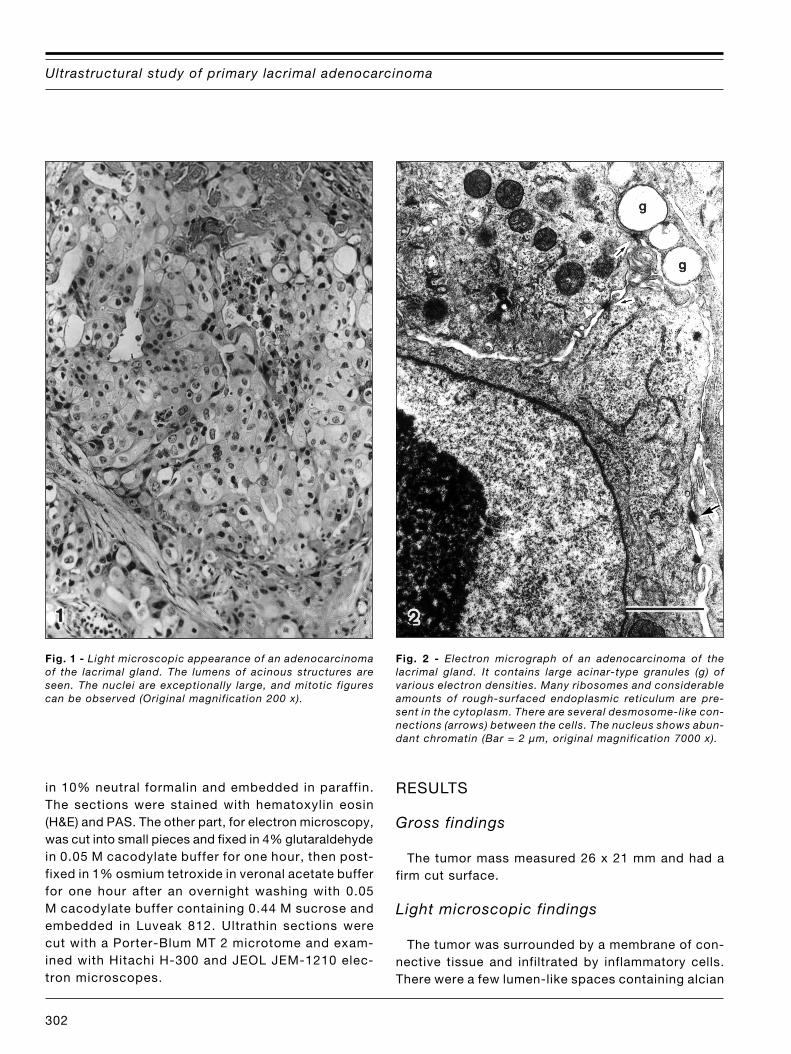
Fig. 2 - Electron micrograph of an adenocarcinoma of thelacrimal gland. It contains large acinar-type granules (g) ofvarious electron densities. Many ribosomes and considerableamounts of rough-surfaced endoplasmic reticulum are pre-sent in the cytoplasm. There are several desmosome-like con-nections (arrows) between the cells. The nucleus shows abun-dant chromatin (Bar = 2 µm, original magnification 7000 x).

303
Gong et al
blue staining materials. Cytological examinationshowed neoplastic cells containing large hyperchro-matic nuclei; some were arranged in an adenoid pat-tern near the normal lacrimal gland (Fig. 1).
Electron microscopic findings
The tumor cells were of two types, dark and light,with various degrees of differentiation. In highly dif-ferentiated tumor cells, the cytoplasm contained many
organelles: mitochondria, rough-surfaced endoplas-mic reticulum and ribosomes (Fig. 2). The nuclei werelarge and irregular. The nucleoli were large, and thechromatin of the nucleus was conspicuous. There weredesmosome-like connections between the cells (Fig.2). The tumor cells formed luminal structures (Fig. 3),with many microvilli on their edges, and the lumenshad low electron-dense secretion granules (Fig. 3).Dark and light cells were seen (Figs. 3-4), and manymultivesicular bodies (Fig. 4).
Fig. 3 - Electron micrograph of an adenocarcinoma of thelacrimal gland. The nest contains light cells (lc) and darkcells (dc). In the former cytoplasmic organelles are abun-dant, but in the latter cytoplasm is sparse. In a lumen struc-ture (l), the apical parts of the cells have numerous microvill i(arrows), and there are mucus granules within the lumen.Numerous mucus granules of various sizes and density arealso seen in the cytoplasm (g) (Bar = 6 µm, original magni-fication 2000 x).
Fig. 4 - A tumor cell nest consists of light and dark cells. Adark cell contains numerous multivesicular bodies (arrows). Lightcells have many ribosomes, mitochondria and rough-surfaced en-doplasmic reticulum (Bar = 3 µm, original magnification 5000 x).

304
Ultrastructural study of primary lacrimal adenocarcinoma
DISCUSSION
The classification of epithelial neoplasms of the lacrimalgland is roughly comparable to that of the salivary gland(6), both these tumors having similar histopathologicfeatures (7, 8). However, some of the neoplasms com-monly found in salivary glands do not occur inlacrimal glands (8, 9).
The most common lacrimal gland epithelial neoplasmis pleomorphic adenoma (51%) (10). The remainingtumors are malignant, the most common being ade-noid cystic carcinoma (30%). Adenocarcinoma is thebasic malignant neoplasm of all glandular structures(2, 11, 12), showing tumor-forming histologic patternswith a mucus-producing tendency particularly in thedifferentiated forms. It can occur de novo in 7% ofpatients with lacrimal gland tumors (5, 9, 10, 13), andis more common in males than females. Metastasisoccurs earlier and the survival rate is low comparedto adenoid cystic carcinoma (10, 14).
Lacrimal adenocarcinomas show lumen formation(2, 5), whereas a pseudoluminal pattern with tubular-like matrix material is more usual in adenoid cysticcarcinoma (3, 5, 15). The mucin content of adeno-carcinoma can be demonstrated with mucicarmine andalcian blue stains; these features are absent in theundifferentiated type (16). The undifferentiated typeof adenocarcinoma does not show these features. Theproliferating cells of an adenocarcinoma are pleomorphic,mitotically active, irregularly layered in the tubular struc-tures, and arranged in sheets or cords in extralumi-nal areas (2).
The diagnosis of adenocarcinoma, in general, de-pends on four routine techniques: light microscopyof H&E stained tissues, mucicarmine staining, PASstaining, and electron microscopy (16), but the fre-quency of their use varies and their diagnostic sen-sitivity may vary (17-19). Since adenocarcinomas ofthe lacrimal gland are very malignant, accurate diag-nosis is important from both the therapeutic and theprognostic standpoint (1, 2, 5).
In the present study we did not find typical lumenswith PAS-stained mucoid material, which were not-ed in light microscopic examinations. Only gaping lu-mens between tumor cells were seen typical of ade-nocarcinoma of the lung. Under electron microscopythe three most common epithelial tumors of the lacrimalgland (benign mixed tumor, malignant mixed tumor,
and adenoid cystic carcinoma) have significant ul-trastructural differences (3). In benign mixed tumor,the inner cuboidal or columnar cells lining the tubu-lar structures have characteristics similar to those ofa normal lacrimal gland. Malignant mixed tumors haveabnormal features: large indented nuclei, scanty cy-toplasm, lipid inclusions, and abundant deposits ofglycogen. In adenoid cystic carcinoma, the tumor cellscontain ductal-type granules, which may resemble basalductal cells with bundles, and the cystoid spaces ofthe tumor are composed of peripheral multilaminarbasement membrane material and central thin fibrils.
Our study highlights the typical fine structural al-terations in the tumor cells. There was an increase inmitochondria, free ribosomes and rough-surfaced en-doplasmic reticulum. The Golgi apparatus reflectedincreased mucus production in the tumor cells. In mosttumor cells, there was a marked increase of multi-vesicular bodies, suggesting heightened metabolic ac-tivity. Variations in the morphology of mucus gran-ules and surface microvilli are also seen in other ade-nocarcinomas (11-12, 20-21). The most important ul-trastructural features of our case were lumens withsecretion granules and microvilli on the apical por-tion of the tumor cells. These findings are diagnosticof adenocarcinoma (16, 22). The nuclear changes arecommon in human malignancies.
Adenocarcinoma of a lacrimal gland arising de novo is rarely malignant and must be differentiatedfrom adenoid cystic carcinoma in the lacrimal gland.There are of five types of adenoid cystic carcinoma:basaloid (solid), cribriform (Swiss cheese), scleros-ing, comedocarcinoma (basaloid units with central necro-sis) and tubular (manifesting true duct formation). Inthe present case light and electron microscopic ex-amination revealed no basaloid masses, duct forma-tion, necrosis in the tumor or cribriform pattern. Therewere also no pleomorphic adenoma-like findings suchas hyalinized connective tissues, myxoid changes, orfocal squamous metaplasia.
In pleomorphic adenomas the inner cells lining thetubules are usually similar to the duct cells of normallacrimal glands (23), and the outermost cells have manytonofilaments. In this patient the pattern was rathersimple, with none of the features of adenoid cysticcarcinoma or pleomorphic adenoma. The inner cellslining the tubules had many secreting granules, roughendoplasmic reticulum, ribosomes, microvilli, and desmo-

305
Gong et al
some-like intercellular attachments despite the pres-ence of many dark cells. To our knowledge, there havebeen no previous reports on the ultrastructural fea-tures and fine structures of this tumor.
Reprints requests to:Huaqing Gong, MD, PhDDepartment of Ophthalmology,Nagasaki University School of Medicine,Nagasaki 852-8501, [email protected]
REFERENCES
1. Zimmerman LE, Sanders TE, Ackerrnan LV. Epithelialtumors of the lacrimal gland: Prognostic and therapeuticsignificance of histologic types. Int Ophthalmol Clin 1962;2: 337-67.
2. Henderson JW. Primary epithelial neoplasms. Orbital tu-mors. 3rd ed. Raven Press Litd: New York, 1994; 323-42.
3. Iwamoto T, Jakobiec FA. A comparative ultrastructur-al study of the normal lacrimal gland and its epithelialtumors. Hum Pathol 1982; 13: 236-62.
4. Konrad EA, Thiel HJ. Adenocarcinoma of the lacrimalgland with sebaceous differentiation: A clinical studyusing light and electron microscopy. Graefes Arch ClinExp Ophthalmol 1983; 221: 81-5.
5. Wright JE, Rose GE, Garner A. Primary malianant neo-plasms of the lacrimal gland. Br J Ophthalmol 1992;76: 401-7.
6. Foote-Jr FW, Frazell EL. Tumors of the major salivaryglands. Cancer 1953; 6: 1065-123.
7. Godtfredsen E. Pathology of mucous and salivary glandtumors in the lacrimal gland and the relation to extra-orbital mucous and salivary gland tumors. Br J Oph-thalmol 1948; 32: 171-9.
8. Forrest AW. Pathologic criteria for effective manage-ment of epithelial lacrimal gland tumors. Am J Oph-thalmol 1971; 71: 178-92.
9. Zimmerman LE. New concepts regarding certain orbitaland lacrimal gland tumors. Ocular and adnexal tumors.St Louis: CV Mosby, 1964; 395-428.
10. Font RL, Gamel JW. Epithelial tumors of the lacrimalgland: an analysis of 265 cases. Ocular and adnexaltumors. Birmingham: Aesculapius Publishing, 1978; 787-805.
11. Koga A, Momii S, Eguchi M, Makino T. Ultrastructureof well-differentiated adenocarcinoma of the gallblad-der. Ultrastruct Pathol 1991; 15: 41-8.
12. Kudo R, Sagae S, Hayakawa O, Ito E, Horimoto E,Hashimoto M. Morphology of adenocarcinoma in situand microinvasive adenocarcinoma of the uterinecervix: A cytologic and ultrastructural study. Acta Cy-tol 1991; 35: 109-16.
13. Ni C, Kuo PK, Dryja TP. Histopathological classifica-tion of 272 primary epithelial tumors of the lacrimal gland.Chin Med J 1992; 105: 481-5.
14. Evans HL, Batsakis JS. Polymorphous low-grade ade-nocarcinoma of minor salivary glands: A study of 14cases of a distinctive neoplasm. Cancer 1984; 53: 935-42.
15. Vangveeravong S, Katz SE, Rootman J, White V. Tu-mors arising in the palpebral lobe of the lacrimal gland.Ophthalmology 1996: 103: 1606-18.
16. McGregor DH, Dixon AY, McGregor DK. Adenocarci-noma of the lung: A comparative diagnostic study us-ing light and electron microscopy. Hum Pathol 1988;19: 910-3.
17. Churg A. The fine structure of large cell undifferenti-ated carcinoma of the lung: Evidence for its relation tosquamous cell carcinoma and adenocarcinoma. HumPathol 1978; 9: 143-56.
18. Auerbach O, Frasen JM, Parks VR, Carter HW. A com-parison of World Health Organization (VVHO) classifi-cation of lung tumors by light and electron microscopy.Cancer 1982; 50: 2079-88.
19. Nash G. The diagnosis of lung cancer in the 80's: Willroutine light microscopy suffice? Hum Pathol 1983; 4:1021-3.
20. Goldman H, Ming SC. Fine structure of intestinal meta-plasia and adenocarcinoma of the human stomach. LabInvest 1968; 18: 203-10.
21. Elema JD, Keuning HM. The use of electron mi-croscopy for the diagnosis of cancer in bronchial biop-sies. Hum Pathol 1988; 19: 304-8.
22. Kaneko C, Niimi H, Shinzato M, Shamoto M. Compar-ative studies of the same adenocarcinoma cells,macrophages, and mesothelial cells by light mi-croscopy, scanning electron microscopy, and trans-mission electron microscopy. Diagn Cytopathol 1994;11: 333-42.
23. Erlandson RA, Corden-Cardo C, Higins TJ. Histogen-esis of benign pleomorphic adenoma (mixed tumor) ofthe major salivary glands. An ultrastructural and im-munohistochemical study. Am J Surg Pathol 1984: 8:803-20.