Embed Size (px)
Citation preview
Photochemistry and Photobiology, 1997, 65(3): 535-537
Technical Note
Two-dimensional Imaging of Ultraweak Photon Emission from Germinating Soybean Seedlings with a Highly Sensitive CCD Camera
Masaki Kobayashi”, B. Devarajl, Masashi Usal, Yukina Tanno’, Motohiro Takedal and Humio lnaba1.* ’Biophotonics Information Laboratories, Yamagata, Japan and 2Tohoku Institute of Technology, Sendai, Japan
Received 23 September 1996; accepted 10 December 1996
ABSTRACT
A novel application of a highly sensitive charge-coupled device (CCD) camera for imaging of spontaneous ultra- weak photon emission from living organisms (biophoton emission) is described. The performance of the CCD camera for low-level light imaging is theoretically com- pared to a conventionally used two-dimensional photon counting tube, and we deduce that in the wavelength re- gion above 700 nm and for measurement periods over 2000 s, the CCD camera is advantageous over the two- dimensional photon counting tube. Experimental results on the two-dimensional biophoton imagery of germinat- ing soybean seedlings also suggest that the wide-range spectral sensitivity of the CCD camera is effective for low-level light imaging from living organisms that have a predominant emission spectrum in the red and near-IR wavelength region.
INTRODUCTION
Extremely weak light, spontaneously emitted from living or- ganisms without any external excitation or stimulation, is commonly referred to as “biophoton emission” ( I ) . The in- tensity of biophoton emission is estimated to be on the order of less than -10- l 6 W/cm* (or -10’ photods cm2 at around 600 nm) on the sample surface (2). The process of biophoton emission is generally considered to originate from internal biochemical reactions associated with oxidative metabolic processes that occur regularly within a living body and hence is closely related to living states. However, in many cases, specific light emitters or details of emission mechanisms are not clearly understood due to the complexity of living sys- tems and appropriate low-level light detection systems that cover the entire spectrum of biophoton emission. A number of phenomena suggest that there is a relationship between
*To whom correspondence should be addressed at: Biophotonics Information Laboratories, Yamagata Advanced Technology Re- search and Development Center, 2-2-1 Matsuei, Yamagata 990, Japan. Fax: 81-236-47-3108; e-mail: [email protected].
0 1997 American Society for Photohinlngy 003 1-8655/97 $5.00+0.00
biophoton emission and physiological andor pathological conditions.
In the case of growing soybean seedlings, it has been re- ported by the first measurement of two-dimensional emission pattern that photon emission around the area of active mi- tosis is relatively strong (3) and emission intensity correlates with growth rate of the root (4). suggesting the relationship between biophoton emission and cellular activities. Rela- tively strong light emission is also observed from mechani- cally wounded regions of soybean seedlings and the emis- sion mechanism is ascribed to oxidative reactions of the per- oxidase-H,O, system. It has also been suggested that ultra- weak light emission occurs upon injury, suggesting that light emission is a reflection of the defense mechanisms of the plant to injury or infection (5.6). The detection and charac- terization of biophoton emission would therefore provide useful insight to the various metabolic processes involved and could also be used in the near future as a noninvasive diagnostic technique through the gathering of unique optical information.
We reported earlier the various properties of biophoton emission phenomena based on spatial distribution analysis of ultraweak biophoton emission using a highly sensitive two-dimensional imaging system (2,3,7,8). In this paper, we report the first biophoton emission images of a germinating seedling obtained using a highly sensitive charge-coupled device (CCD)t camera. We also compare the performance of the CCD camera with conventional low-level light detec- tors and demonstrate the potential usefulness of the CCD camera for biophoton imaging.
The conventional imaging systems used for low-level light imaging uses a two-dimensional photon counting tube (2D PMT) that has excellent sensitivity. However, the quantum efficiency of the photocathode is very low, approximately 20% at peak wavelength of 400 nm and less than 5% in the red or near-IR regions, and hence is not useful for a wide variety of biophoton emission measurements. The minimum detectable optical power at each pixel of a 2D PMT with a signal-to-noise ratio of unity can be defined as
tAhhreviarions: CCD, charge-coupled device; RMS, root mean square; 2D PMT, two-dimensional photon counting tube.
535

536 Masaki Kobayashi et a/.
Figure 1. (a) Ultraweak biophoton emission image of germinating soy- bean under intact conditions obtained by a CCD camera. Integration time is 30 min. Emission intensity is indicat- ed by the gray scale. Randomly dis- tributed white spots represent cosmic ray-induced background noise. (b) Photograph of the same sample under weak light illumination.
where Ndl is the number of dark counts per unit time at a single element of 2D PMT, T is the measurement time, h is Planck's constant, c is the velocity of light, -ql is the quantum efficiency of the PMT photocathode, X is the wavelength and S is the area of pixel. In the case of a CCD camera, the minimum detectable power is defined as
where Nd2 is the number of electrons contributing to the dark current per unit time at a single element of the CCD, q2 is the quantum efficiency of the CCD and N, represents the electron root mean square (RMS) of readout noise of am- plifier circuit per pixel. From the above equations it can be deduced that if the CCD is operated at extremely low dark current and longer exposure times to reduce the contribution of the readout noise, the signal-to-noise ratio of the CCD camera would be superior to the conventional 2D PMT un- der similar conditions of wavelength and measurement time.
MATERIALS AND METHODS Imaging apparatus. A liquid nitrogen-cooled, highly sensitive CCD camera system (ATCZOOC, Photometrics, AZ, USA) that includes a back-illuminated, thinned type Si-CCD (TK1024AB2-G I , Tektro- nix, OR, USA) is used for imaging of biophoton emission in our measurements. The CCD format is 1024 X 1024 pixels for a full frame with each pixel size being 24 X 24 pm. Dark current of the device is 0.225 e-/h pixel (=6.25 X e /s pixel) at -120°C in MPP (multipinned phase) mode and the readout noise is 3.3 e - RMS/pixel. In the experiments, the CCD camera was operated in 2 X 2 binning mode, then spatial resolution was 48 X 48 pm. Quan- tum efficiency of the system is 73% at 700 nm. A lens system (Nikkor 50 mm F1.2S. Nikon, Tokyo, Japan) was used in tandem to collect the emitted ultraweak light. Samples were placed on a mechanical stage in a light-tight sample chamber and focused by adjusting the distance between the sample and lens system.
Plant material. Samples were comprised of etiolated soybean seedlings (Glycine max) that are dark adapted for 3 4 days at 25°C after germination. The length of the root system was approximately 3 0 4 0 mm during the growth period. Seeds were washed thoroughly in distilled water prior to planting and were not sterilized to prevent oxidative damage to samples. The seedlings had no visible physical injury or infection, and sample treatment was canied out under weak light illumination to prevent the influence of delayed fluorescence. Samples were placed in a sample chamber made of synthetic resin with ultrapure water. Background photon emission was not observed from the sample chamber.
Mechanical injury to the cotyledon was done with a knife, form- ing a cross mark 2-3 mm in depth and 10 mm long.
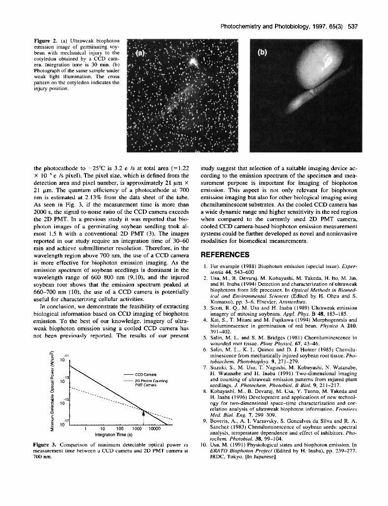
RESULTS AND DISCUSSION The biophoton image obtained under intact conditions is dis- played in Fig. l a with a sample photograph under weak light illumination shown in Fig. lb. Spatial distribution and pat- tern of biophoton emission intensity in the hypocotyl area is clearly observed. The hypocotyl area that indicates strong emission is considered to be active in cell growth and res- piration; hence, increased emission intensity suggests that biophotons carry information on cellular metabolic activities (3). The biophoton image of an etiolated soybean seedling with mechanical injury to the cotyledon is displayed in Fig. 2a with a sample photograph under weak light illumination in Fig. 2b. Strong emission is observed at the injured posi- tion of the cotyledon and at the noninjured hypocotyl area. The mechanism of light emission from the wounded region (5,7) suggests the contribution of endogenous H,O,-perox- idase system and it possibly reflects the defensive reactions of the seedlings to injury or infection. Similar light emission phenomena were also observed on wounding of the hypo- cotyl or radicle region of germinating seedlings. In the case of radicle excision, it was reported (8) that there is an in- crease in light emission also at the uninjured hypocotyl re- gion, which is distant from the radicle. This phenomenon was observed a few hours after excision and was speculated as the defensive response against injury such as respiratory activity or peroxidase synthesis (8). The observed CCD im- ages in Fig. l a and Fig. 2a also have higher spatial resolution than 2D PMT images (3,7), suggesting the potential useful- ness of the CCD camera for specific localization of the emis- sion intensity for understanding the details of activated areas and characterizing the morphological information associated with the physiological and/or pathological state of the plant.
In Fig. 3, the calculated minimum detectable optical pow- er of a CCD camera is compared to that of a 2D PMT as a function of measurement time at a wavelength of 700 nm. The performance of the 2D PMT camera is obtained exper- imentally using a Photon Imaging Acquisition System (PIAS. Hamamatsu Photonics, Japan) that has a 15 mm di- ameter multialkaline photocathode and a 512 X 512 pixel resolved position-sensitive detector. Dark current on cooling
Photochemistry and Photobiology, 1997, 65(3) 537
Figure 2. (a) Ultraweak biophoton emission image of germinating soy- bean with mechanical injury to the cotyledon obtained by a CCD cam- era. Integration time is 30 min. (b) Photograph of the same sample under weak light illumination. The cross pattern on the cotyledon indicates the injury position.
the photocathode to -25°C is 3.2 e-/s at total area ( = I .22 X e-/s pixel). The pixel size, which is defined from the detection area and pixel number, is approximately 21 pm X 21 pm. The quantum efficiency of a photocathode at 700 nm is estimated at 2.13% from the data sheet of the tube. As seen in Fig. 3, if the measurement time is more than 2000 s, the signal-to-noise ratio of the CCD camera exceeds the 2D PMT. In a previous study it was reported that bio- photon images of a germinating soybean seedling took al- most 1.5 h with a conventional 2D PMT (3). The images reported in our study require an integration time of 30-60 min and achieve submillimeter resolution. Therefore, in the wavelength region above 700 nm, the use of a CCD camera is more effective for biophoton emission imaging. As the emission spectrum of soybean seedlings is dominant in the wavelength range of 600-800 nm (9,10), and the injured soybean root shows that the emission spectrum peaked at 660-700 nm (lo), the use of a CCD camera is potentially useful for characterizing cellular activities.
In conclusion, we demonstrate the feasibility of extracting biological information based on CCD imaging of biophoton emission. To the best of our knowledge, imagery of ultra- weak biophoton emission using a cooled CCD camera has not been previously reported. The results of our present
L
n - CCD Camera
2D Photon Counting PMT Camera
-13 10
-1 5 10
-17 i n .-
1 l o 100 1000 10000 Integration Time (s)
Figure 3. Comparison of minimum detectable optical power vs measurement time between a CCD camera and 2D PMT camera at 700 nm.
study suggest that selection of a suitable imaging device ac- cording to the emission spectrum of the specimen and mea- surement purpose is important for imaging of biophoton emission. This aspect is not only relevant for biophoton emission imaging but also for other biological imaging using chemiluminescent substrates. As the cooled CCD camera has a wide dynamic range and higher sensitivity in the red region when compared to the currently used 2D PMT camera, cooled CCD camera-based biophoton emission measurement systems could be further developed as novel and noninvasive modalities for biomedical measurements.
REFERENCES 1. For example (1988) Biophoton emission (special issue). Exper-
ientia 44, 543-600 2. Usa, M., B. Devaraj, M. Kobayashi, M. Takeda, H. Ito, M. Jin,
and H. Inaba (1994) Detection and characterization of ultraweak biophotons from life processes. In Optical Methods in Biomed- ical and Environmental Sciences (Edited by H. Ohzu and S. Komatsu), pp. 3-6. Elsevier, Amsterdam.
3. Scott, R. Q., M. Usa and H. Inaba (1989) Ultraweak emission imagery of mitosing soybeans. Appl. Phys. B 48, 183-185.
4. Kai, S., T. Mitani and M. Fujikawa (1994) Morphogenesis and bioluminescence in germination of red bean. Physica A 210, 391-402.
5 . Salin, M. L. and S. M. Bridges (1981) Chemiluminescence in wounded root tissue. Plant Phvsiol. 67, 43-46.
6. Salin, M. L., K. L. Quince and D. J. Hunter (1985) Chemilu- minescence from mechanically injured soybean root tissue. Pho- tobiochem. Phorobiophys. 9, 27 1-279.
7. Suzuki, S., M. Usa. T. Nagoshi, M. Kobayashi, N. Watanabe, H. Watanabe and H. Inaba (1991) Two-dimensional imaging and counting of ultraweak emission patterns from injured plant seedlings. J. Photochem. Photobiol. B Biol. 9, 21 1-217.
8. Kobayashi, M., B. Devaraj, M. Usa. Y. Tanno, M. Takeda and H. Inaba (1996) Development and applications of new technol- ogy for two-dimensional space-time characterization and cor- relation analysis of ultraweak biophoton information. Frontiers Med. Biol. Eng. 7 , 299-309.
9. Boveris, A., A. 1. Varsavsky, S. Goncalves da Silva and R. A. Sanchez ( 1983) Chemiluminescence of soybean seeds: spectral analysis, temperature dependence and effect of inhibitors. Pho- tochem. Photobiol. 38, 99-1 04.
10. Usa, M. (1991) Physiological states and biophoton emission. In ERATO Biophoton Project (Edited by H. Inaba), pp. 239-277. JRDC, Tokyo. [In Japanese]