Embed Size (px)
Citation preview

JARQ 24, 1-14 (1990)
Structure and Function of the Haustorium in Germinating Coconut Palm Seed
Yukio SUGIMUMA and Taka MURAKAMI*
Abstract During the germination of coconut seed, a haustorium was formed from the distal portion of the embryo. Compared with various parts of tissues within a haustorium, the surface tissues were markedly different in (1) bearing undulating structure which closely attached with the degradating endosperm, (2) possessing starch grains and oil droplets in developing haustorium, (3) accumulating relatively high amounts of sucrose and starch, and (4) being the presence of considerably higher activities of phosphoglucomutase and phosphoglucose isomerase. In addition, vascular bundles were situated near the outer surface. In view of this tissue specific features, it is likely that the surface layer consisting of epithelium and adjacent cells plays a key role in absorption of oil reserves released from degraded endosperm and conversion into sugars. Possible interactions between haustorium and endosperm are discussed.
Discipline: Crop production Additional key words: Cocos nucifera L., endosperm, histochemistry, sugar metabolism
Introduction
Plant seeds can be grouped into (I) starchy, (2) proteid and (3) oil types on the basis of main components stored in the seeds. In regard to the mechanisms of metabolizing reserve materials within the seeds during the period of their germinat ion, a number of reports have been published, dealing with such different types of seeds.
Coconut palms (Cocos nucijera L.) produce oilbearing seeds which are commercially important as a source of vegetable oil. The palm manifests its special features in the process of germination and seedling development. The seeds contain a small size of embryo and a copious amount of endosperm. Th·e distal portion of the embryo increases in size to form a haustorium which remains within the seed and expands extensively as the endosperm disappears . This expansion is carried on until the haustorium completely fills the seed. Based on such structural changes taking place within the seed, it is considered that the haustorium may play a key role during the
period of germination and initial seedl ing growth2•8>. Since coconut seeds are large in size, they provide
a convenient system in investigating embryoendosperm interactions during the stage of germination and subsequent growth. The authors' previous study relating to germination of coconut palm covered the following aspects: ( I) structure of the haustorium and the endosperm, (2) function of the haustorium, (3) carbohydrate metabolism in the haustorium in relation to the seedling growth, and (4) regulatory function of the endogenous gibberellins in the haustorium 12>. The present paper reviews the results of the current studies with special emphasis on(!) process of the haustorium development associated with germination, (2) cytological and histochemical changes in the haustorium at its different developmental stages, and (3) quantitative patterns of sugars at various parts of the haustorium.
Process of germination
A matured embryo is cylindrical in shape, being approximately 10 mm in length. The location of
Tochigi Research Laboraiories, Kao Corporation {lchikai-machi, Haga, Tochigi, 321-34 Japan) • Department of Applied Physiology, National Insticute of Agrobiological Resources (Tsukuba, Ibaraki, 305 Japan)

2
embryo is in the endosperm just below the germ pore, which shows a dark circular spot on the endocarp (Plate 1- 1). In a longitudinal section, the plumule and radicle can be distinguished within the proximal end of the embryo (Plate 1-2). The plumule in section shows a central mcristematic zone surrounded by the scaly-leaf primordia, which in turn are enclosed by the coleoptile (Plate 1-3). II is situated at a certain angle to the central axis of the embryo. A small slit is evident above the coleopti le (Plate 1-4). The radicle is situated opposite to the plumule and within the apical mass of mcristematic cells oriented towards the suspensory region (Plate 2-1). The cells containing yellowish brown materials, likely tannin, distribute at the region tapered (Plate 2-2). The proximal part of the embryo is separated by a small constriction from the cotyledon which will develop into the haustorium (Plate l-2). These morphological features well confirm the earlier observationsz· 13•11>.
Three cell-types in the cotyledon consisting of parenchyma, procambial and prorodermal, can be distinguished by their shapes, sizes and positions. The parenchyma cells have ilri isodiametric shape. The protodermal cells are tabular in outline and form a very distinct layer around the surface of the cotyledon. The procambial cells are narrow and elongate in the long axis of the embryo. Within the cotyledon, a bundle of procambial strands is developed (Plate l-2, 3, 4).
On germination, the embryo simultaneously develops in two directions as follows: (1) from proximal end of the embryo, the apical part forces its way out through the germ pore and the plumule and radicle rhen grows outside the endocarp, and (2) from distal end of the embryo, rhe cotyledon expands 10 form a pear-shaped haustorium inside tJ1e central cavity of the seed (Plate 2- 3). As the initial growth of seedling begins gradually, the haustorium substantially increase its size (Plate 2-4), whi le the surrounding endosperm is digested and replaced by developing haustorium. Chi ld showed quantitarive changes in haustorium and endosperm at different intervals after seed bedding2>.
At1atomy of the haustorium
With the purpose of identifying possible interac
tions between haustorium and endosperm, micro-
JARQ 24(1) 1990
scopic investigations on tissues of the developing haustorium were undertaken . Once the haustorium development takes place, the outer surface which is closely attached with the endosperm expands substantially. On the auached surface, a countless, minute and protuberant structure is specifically
formed (Plate 2-5). The undulating structure is pale-yellow. Pieces of degradation products from the endosperm cover the surface and lodge in the troughs induced by invaginations (Plate 3-1, 2). The outermost layer of haustorium, epithelium, consists of rectangular cells (Plate 3- 2). Cells of the interior parenchyma increase gradually in size during the haustorium development. The degree of the increase depends on the location of the relevant cells. Those near the outer surface change least in size and shape, while those near the cemer expand with an irregular shape. The central tissue in the matured haustorium consists of loosely-connected amorphous cells with large intercellular spaces among them (Plate 3-3, 4). It seems that the substantial increase of haustorium in size, which keeps it in continual physical contact with the degrading endosperm, is mainly
caused by the expansion of intercellular space. The vascular bundles extend from shoot apex to the distal tip of the haustorium and run in parallel to the haustorium surface (Plate 3- 5). The distance from the surface 10 the bundles increases gradually as the surrounding parenchyma cells enlarge.
Histochemistry of the haustorium
Cellular distributions of starch grains and oil droplets were subjected to histochemical analyses based on the sections of various sizes of the haustorium.
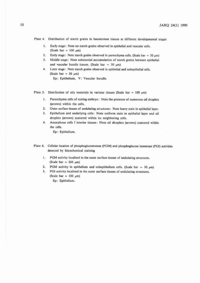
Starch grains : At the early stage of the haustorium development, starch grains accumulate in the entire parenchyma tissues, while no starch grains are present in the epithelial and vascular tissues (Plate 4- 1, 2). This distribution pattern changes in the following stages, where the haustorium develops actively: the epithelium and neighboring cells located in outer t issues have a large amount of starch grains (Plate 4-3), in co111ras1, in the imerior tissues, the number of the grain-containing cells decreases gradually towards the cemral tissue. Most of the starch grains disappear in the spongy-like cells in the central tissue at the later stages of haustorium

3
Plate 1.

4 JARQ 24(1) 1990
Pla1e 2.

5
Plate 3.

6
Plate I. Morphology of resting embryo and i ts apical meristem
I. Half-cut seed showing position of embryo. (Scale bar = 10 mm)
2. Longitudinal section of resting emt>ryo.
Square indicates the apical part containing plumule and radicle. (Scale bar = I mm)
3. Apical meristem surrounded by the scaly-leaf primordia. (Scale bar = 100 1,m)
4. Slit in proximal end of embryo. (Scale bar = 100 µm)
Am: Apical meristem of plumule, C: Constriction, Ee: Endocarp,
Es: Endosperm, Op: Germ pore, Pc: Procambial strand,
Pd : Protodermal, SI: Slit, Sp: Suspensor.
Plate 2. Radiclc meristcm in resting embryo and structural change.s of haustorium
I. Radicle meristem towards the suspensional region. (Scale bar = 100 µm)
2. Tip region of suspensor where cells having yellowish brown materials (arrows)
are distributed. (Scale bar ·= I 00 µ,m)
3. Half-cut seed at early stage of germination. (Scale bar = 10 mm)
4. The sequence of haustorium expansion occurred from early (I) to later (7) stages
of the development. (Scale bar = 30 mm)
5. Longitudinal view of undulating structures formed in immediately adjacent area
to the degradating endosperm. (Scale bar = 200 µm)
Es: Endosperm, H: Haustorium, Pd: Protodermal,
Ra: Radicle meristem, R: Root, Sp: Suspensor.
JARQ 24(1) 1990
Plate 3. Location of degradation products from endosperm and cellular structure of haustorium
I. Surface of undulating structure covered by degradation products from endosperm.
(Scale bar = IO µm)
2. Longitudinal section of undulating structure showing degradation products (arrow)
entered in a trough. (Scale bar = 50 µm)
3. Interior parenchyma cells at initial st:age of haustorium development.
(Scale bar = I 00 µm)
4. Interior parenchyma eel.ls at later stage of haustorium development.
(Scale bar = 200 µm)
5. Longitudinal and transverse view of haustorium showing the arrangement of vas
cular bundles (arrows). (Scale bar = 30 mm)
Arrows a and b indicate approximate level for transverse views (a) and (b).

7
Plate 4.

8 JARQ 24(1) 1990
Plate 5.

9
. .
Plate 6.
10 JARQ 24(1) 1990
Plate 4. Distribution of starch grains in haustorium tissues at different developmental stages
I . Early stage: Note no starch grains observed in epithelial and vascular cells .
(Scale bar = I 00 µm)
2. Early stage: Note starch grains observed in parenchyma cells. (Scale bar = SO µm)
3. Middle stage: Note substantial accumulation of starch grains between epithelial
and vascular bundle tissues. (Scale bar = SO µm)
4. Later stage: Note starch grains observed in epithelial and subepithelial cells.
(Scale bar = 30 µm)
Ep: Epithelium, V: Vascular bundle .
Plate S. Distribution of oily materials in various tissues (Scale bar = I 00 µm)
I . Parenchyma cells of resting embryo: Note the presence of numerous oil droplets
(arrows) within the cells.
2 . Outer surface tissues of undulating structures: Note heavy stain in epithelial layer.
3. Epithelium and underlying cells: Note uniform stain in epithelial layer and oil
droplets (arrows) scattered within it.s neighboring cells.
4. Amorphous cells f interior tissues: Note oil diroplets (arrows) scattered within
the cells.
Ep: Epithelium.
Plate 6. Cellular location of phosphoglucomutase (PGM) and phosphoglucose isomerase (PGI) activities
detected by histochemical staining
I. PGM activity localized in the outer su.rface tissues of undulating structures. (Scale bar = 200 µm)
2. PGM activity in epithelium and sut>epithelium cells. (Scale bar = SO ,,m)
3. PG! activity localized in the outer surface tissues of undu lating structures.
(Scale bar = I 00 µm)
Ep: Epithelium.

development, whereas some amount of the grains is stored in the outer tissues including epithelium throughout the developmental stages (Plate 4-4).
Oil droplets: Cells of cotyledon in the resting enbryo have a number of oil droplets (Plate 5-1). A substantial amount of oily materials drived from cmdosperm reserves exists on the surface of undulating structure (Plate 5-2). Oils as stained with Sudan III are contained in not only epithelium but also its underlying cells (Plate 5-3). This pattern found in the outer surface layer is consistent regardless of the growth stages of haustorium. Oil droplets scattered in the interior tissues (Plate 5- 4) decrease during the haustorium development, and eventually disappear completely. This site-specific localization is similar to that of the starch grains.
The in situ visualization of enzymic proteins is a powerful tool to determine the actual locations of enzymes at a cellular level. Both phosphoglucomu-tase (PGM) and phosphoglucose isomerase (PGI) activities were surveyed by a histochemical assay10•11 ·12l. Distinct activities of these enzymes are confined only in the epithelial and subepithelial layers (Plate 6-1, 2, 3). No or little activities are present in other parts of the cells. This indicates that the epithelial and subepithelial cells are of metabolically active state in terms of sugar conversion.
Sugar met.abol.ism of the developing haustorium
It is known that haustoria contain reducing sugars. In connection with the haustorium growth, a quantitative pattern of major sugars such as glucose, fructose, sucrose and starch was analyzed on the basis of enzymatic measurements9
•12
•18>. The
change in sugar content was monitored to identify its relationship with the haustorium increase in size accompanied by the germination development.
The resting embryo before haustorium development stores sucrose at concentration of 9 l 07o of the total sugar content extracted. Once the haustoriunn development begins to take place, the amount of sucrose decreases rapidly, being followed by a gradual increase. After reaching a plateau, the sucrose content decreases again during the later stage of development (Fig. I) . Such a pattern of changes can be understood as an outcome of the varying rates of synthesis and consumption: sucrose synthesized is utilized for supplying an energy required by the
100
~
.i: 80 .!!9 " ,:
~ o!: 60 .. .§. s i: 40 .. i: 8
~ 20
"'
0 )!!
11
40 60
Haustorium growth (diameler, mm)
lli : Resting embryo • : Glucose o : Fructose "' : Sucrose • : S1arch
Fig. 1. Quantitative changes in sugars during haustorium development
growing seedling, thereby the sucrose concentration in haustorium may decrease at the later stage.
A temporary increase in starch content takes place at the early stage, suggesting that overproduced sucrose be converted into starch as a stored matter (Fig. 1). This quantitative change in starch corresponds well to the histochemical observations explained in the previous section of this paper. The patterns of change in glucose and fructose contents are almost the same: their concentrations maintain a steady level of 9-16 µg/mg fresh weight throughout the haustorium growth stage with an exception in the initial stage (Fig. I) . It is most likely that these monosaccharides may be produced by the cleavage of sucrose. This cleavage may be caused by sucrose reaction observed by Balasubramaniam et aJ.1>.
In order to identify accumulation sites of the sugars, the haustoria were separated in different stages into seven parts as illustrated in Fig. 2. At the surface and its neighboring tissues, a relatively high amount of sucrose and starch accumulates as major sugars. In contrast , the amount of both

12
B1
Fig. 2. Longitudinal sketch of haus1orium 10 be divided into various ponions for sugar analyses
Haustoriurn Diameter : 52mm
-.... .s::. .21 Q)
3: .s::. <I) Q) ... -0)
E ...... 0)
.3--C Q) .... C 0 (.) ... t1l 0) :,
(/)
40
20
20
60
40
20
40
20
JARQ 24(1) 1990
glucose and fructose is considerably higher in the central tissues than !hose in outer surrace (Fig. 3). These distribution patterns of the four different sugar species are inherent characteristics, independent of size of the haustoria used.
General discussions and conclusions
A sequent breakdown of coconut endosperm was described at a cellular level in our earlier report 121 : the process of endosperm breakdown is always confined 10 a thin zone directly adjacent to invaginated surface of the haustorium and continues at a rate commensurate with haustorium development. This morphological evidence may indicate that the haustorium plays a regulating role of endosperm breakdown. The cell wall hydrolases are very likely to be closely associated with the endosperm break-
74mm 88mm
Q) (/) 0 (.) :,
a Q) (/)
0 .... (.) :, ... u.
Q) <I) 0 ... (.) :, (/)
.s::. (.) ... t1l -(/)
0 LL....L...C::i=ic:::::1.....L_L_L..J_J_....C:t:::::L..J._L...L_l,_t_t:~-.L.JL..l._L_J
Part of
Haustorium
1 2 3 4 3 2 1 1 2 3 4 3 2 1 -A B A B A
Fig. 3. Site-specific variation or glucose, fructose, sucrose and starch con1cms at different s tages of haus1oriu111 developmem
Pans 1-4 and haus1orium A, B refer 10 Fig. 2.
B
down. There might be three possibilities in regard 10 the original location of cell wall hydrolases. Enzymes are: (I) synthesized by haustorium and secreted into the endosperm; (2) synthesized de novo
in the endosperm as a result of an inductive signal coming from the haustorium; or (3) stored in the endosperm and activated or released by a regulatory signal from the haustorium. Using date palm, DeMason et al. 6> suggested that date haustorium in some way activate and cause the release of the endo-0-mannase which is stored in the endosperm cells. However, the possibility that the enzyme was secreted from the haustorium in some inactive form could not be ruled out under their conditions.
The radio-labelled fatly acids and triacylglycerols are absorbed and metabolized by oil palm haustori -um where the glyoxylate bypass enzymes are Jocated15
•161
• The epithelium has an important role in germinating rice seed for the hydrolytic digestion of starch reserves in the endosperm••>. Taking these evidences into account, possible functions of the epithelium in the strucLUre invaginated imo the endosperm are: (I) t.o secrete cell wall hydrolases and/or signal factors involved for endosperm break down; (2) to absorb oily reserves released from degrading endosperm cells; and (3) LO modify oi ly reserves incorporated and to transfer resultan L
metabolites into adjacent cells. In view of the posi tion of the vascular tissues near the epithelium, it is suggested that sucrose which builds up in the epithelium and the adjacent cells be transported LO
upward parts via the vascular tissues for seedling growth. These functions may be maintained until the photosynthetic machinery begins to operate. If this is the case, site-specific location of the oil droplets and the four sugar species as stated earlier can be well justified. The high activities of PGM and PG! are present only in the epithelial cells, indicating that these cells are different in sugar metabolism from the other types of cells in the haustorium.
Based on the common feature observed in date palm3- 7>. oil palm 15• 16l and coconut palm2•12>, the haustorium is mainly an absorptive and storage organ which provides the seedling with products of endosperm hydrolysis before the seedling can afford itself by photosynthesis. Further investigation is needed to elucidate functions of the hauslOrium io more details, in panicular those of the epithelial
13
layer invaginated into endosperm, by biochemical
approaches.
The authors would like to thank Professors K. lnada, Tottori University, and Y. Ota, Tokyo University of Agriculture, for their cont inuous encouragement, and also to Drs. M. Saito, K. Okamoto, S. lloh and K. Otsuji of Kao Corporation for their valuable suggest ions and helpful
discussions.
References
J) 8alasubramaniam, K. el al. (1973): Biochemical changes during germina1ion of 1he coconut (Cocos 1111cijera). A11n. Bot .• 37, 439-445.
2) Child, R. (1964): Coconu1s. Longmans, Green and Co., Lid., London, 65-<>9.
3) DeMason, D. A. & Thomson, W.W. (1981): S1rucrnre and ul1ramucture of the co1ylcdon of da1e palm (Phoenix dactylijera L.). Bot. Caz .• 142, 320-328.
4) OeMason, 0. A., S<:xton, R. & Orantreid, J . S. (1984): Slruc1ure, composition and physiological slate of the endosperm of Phoenix dactyfijera L., An 11. Bot .. S2, ?1-80.
5) DeMason, D. A. (1984) : Grow1h parameters in the cotyledon of da1e seedlings. Bot. Caz., 145, t76-183.
6) DeMason, D. A. e1 al. ( 1985): S1ruccure and biochemis1ry of endosperm breakdown In date palm (Phoenix dactylijera L.) seeds. Protoplasma, 126, 159-167.
7) OcMason, 0. A. (1985): His1ochcmical and ultras1ruc-1ural changes in the haustorium of date tphoe11ix dac1ylifer11 L.). Protoplasma, 126, 168-177.
8) Fremond, Y., Ziller, R. & Lamo1hc, M. N. (1966): The coconut palm. Iniernational Po1ash lns1i1u1e. Berne and Swi1zerland, 36-38.
9) Jones , M. G. K. ct al. (1977): Enzymic assay of 10- 7
10 IO- " moles of sucrose in plan1 tissues. Plant Physiol. , 60, 379-383.
10) Murakami, T . (1983): The histochemical distribu1lon and activity of phosphoglucomu1ase in tissue cells of sugar beet petiole. Jpn. J. Crop Sci., 52, 177-182 [In Japanese with English summary].
11) Murakami, T. (1985): The his1ochemieal detection of phosphoglueose isomerase in tissue cells of sugar beet petiole. Jp11. J. Crop Sci. , 54, 283-284 )In Japanese with English summary).
t2) Murakami, T. & Sugimura, Y. (1987) : S1ruc1urc and function of the haustorium in coconu1 palm during germina1ion. Bull. Nat. Inst. Agrobiol. Resour., 3, 11-57 [In Japanese wi1h English summary].
13) Nuri1a-Toruan (1978): Pcrtumbuhan dan perkembangan embrio kelapa (Cocos m,cijem L.) dalam kullur aseptik . Menara Perkebwwn, 46 , 213-216.

14
14) Okamoto, K. & Aka , . , .. ~. T. (1979): Enzymic mechanisms of starch hrc:tkdown in germinating rice seeds. 7 . Amylase formation in the epithelium. Plan/ Physiol., 63, 336-340.
15) Oo, K. C. & Stumpf, P. K. (1983): Some cnzymic activities in the germinating oil palm (Elaeis g11i11ee11-sis) seedling. Pla11t Physiol., 73, 1028-1032.
16) Oo, K. C. & Stumpf, P . K. (1985): The metabolism of the germinating oil palm (Elaeis gui11ee11sis) seedling.
JARQ 24(1) 1990
In vivo studies. Plant Physiol., 78, 1033-1037. 17) Sclvaratnam, E. M. (1952): Embryo of the coconut.
Nature, 169, 714-715. 18) Takebe, M. & Murakami, T. (1984): The enzymatic
determination of sugars in sugar beet plants with hcxokinase and glucose-6-phosphate dehydrogenase. Jpn. J. Crop Sci. S3, 113-114 (In Japanesel .
(Received ror publication, March 15, 1989)