Embed Size (px)
Citation preview

123
TUMOR and STAGING DATA cont. Extensive material in this section comes with permission from the AJCC Cancer Staging Manual, Sixth Edition,
Springer-Verlag, New York 2002. See the Permissions page.
AJCC TNM Staging System At this time, it is the COC's plan to continue collection of "manual" TNM staging even after Collaborative Staging goes into effect. The MCR now plans to NOT collect these fields for diagnoses made beginning in 2004. The AJCC staging fields are all required for pre-2004 diagnoses for the MCR. Cases diagnosed beginning in 2003 are TNM-staged using the AJCC Cancer Staging Manual, Sixth Edition (with updates). The codes used to record the staging information can be found in the FORDS Manual and this MCR Manual. Both the staging manual and a coding manual must be used to record AJCC staging fields. Both clinical and pathologic staging fields are collected by the MCR. If you have enough information to specifically stage a case clinically and pathologically, then both stages should be specifically reported. The FORDS Manual, on its p. 23, and the AJCC Cancer Staging Manual, Sixth Edition note that both clinical and pathologic staging should be recorded. Use the codes for "unknown" and "not applicable" to complete the staging fields whenever appropriate. Some physicians seem to feel that a case with an unknown stage (for example, only an incisional biopsy of the primary tumor has been done) "cannot be staged"; everyone should be aware that a case with a correct "unknown" or "not applicable" stage assignment has been staged and can be recorded with appropriate codes (that do not appear in the Cancer Staging Manual). The FORDS Manual specifies that some TNM fields should be left empty when a physician did not supply the staging information. None of the TNM fields may be left empty for the MCR -- the MCR MUST receive the staging information for pre-2004 diagnoses. The MCR is not concerned with whom staged the case, as long as the information is correct and is coded correctly. (This is why we don't collect the "Staged By" fields.) If the coded staging information we're receiving from you in the AJCC fields is known to be incorrect or questionable, please explain the situation in a Staging Narrative; the MCR will want to correct such stages on our data system. Those wishing to select cases in which staging was assigned only by physicians can use the "Staged By" fields to identify such records. Cases in which a registrar has had to supply the staging will not meet the COC's physician staging requirements but will satisfy the MCR's reporting requirements.

124
TUMOR and STAGING DATA cont. Extensive material in this section comes with permission from the AJCC Cancer Staging Manual, Sixth Edition,
Springer-Verlag, New York 2002. See the Permissions page. Not all types of cancer are AJCC-stageable. Use the primary site codes listed at the beginning of each chapter in the Cancer Staging Manual. The lists of histologic types in each chapter are only a guide to indicate the cancer types which can be AJCC-staged using that staging scheme. Any cancer types listed as exclusions at the beginning of each chapter are to NOT be AJCC-staged with that scheme. Some chapters are specifically limited to certain cancer types only (such as skin melanomas). The lymphoma staging scheme applies to histologic types 9590-9729 and does not limit its applicable primary sites. Examples: The beginning of the esophagus chapter (p. 91) indicates that all of primary site
C15._ is included. The section titled "Primary Site" also discusses how to handle tumors arising in the esophagogastric junction (C16.0). Page 91 specifies that sarcomas are excluded. Page 94 (under Histopathologic Type) says, "The classification applies to all carcinomas." Page 95 lists specific histologic types, including adenosarcoma. The code for papillary carcinoma in situ (8050/2) is not listed here, but obviously it could be staged with this chapter.
Pediatric cancers do not have special TNM coding in the AJCC Cancer Staging Manual, Sixth Ed. They would ordinarily be considered unstageable in this system. If a physician has chosen to stage a pediatric case using TNM (clinically or pathologically), then this staging may be coded and unknown codes should be used for any unspecified fields.
Examples: A child has soft tissue sarcoma and the TNM staging has been left unstaged. Both clinical and pathologic staging would be coded as unstageable -- T88N88M88, Stage Grouping 88.
A child has soft tissue sarcoma and a physician clinically stages it using the sarcoma chapter in the AJCC Cancer Staging Manual, Sixth Ed. The requirements for pathologic staging are not reached. Code the clinical TNM and Stage Grouping to record the physician's stage, and use unknown codes for the pathologic fields -- TX_NX_MX_, Stage Grouping 99.
If the primary site is not definitely known, AJCC staging of the cancer should be based on "reasonable clinical certainty" of a primary site identification. If there is not "reasonable clinical certainty" indicating one primary site, then the AJCC staging should be "not applicable" (as for an unknown primary site).
Examples: A scan finds brain metastases. The physician states that the primary site is probably lung. Use the AJCC scheme for lung primaries to stage this.
A patient has liver metastases and the primary site may be colon or lung. If the primary site is not clearly identified, this case should be AJCC-staged T88N88M88, Stage Grouping 88.
A patient has two primaries that are both carcinomas. Metastatic carcinoma is also found in the liver and its source cannot be determined. Stage both primaries as metastatic unless later information identifies which primary spread to the liver.

125
TUMOR and STAGING DATA cont. Extensive material in this section comes with permission from the AJCC Cancer Staging Manual, Sixth Edition,
Springer-Verlag, New York 2002. See the Permissions page. Lymph nodes are not often surgically removed for known in-situ tumors. The AJCC classification is therefore usually "pTis cN0 cM0, cStage Grouping 0" because there is usually only clinical evaluation of nodal and distant disease (see page 6 in the AJCC's Cancer Staging Manual, Sixth Edition). Timing rule: For both Clinical and Pathologic staging information, use only information as described below obtained through the completion of the most definitive first-course surgery or within four months of diagnosis, whichever is longer. Patients who do not have first-course surgery are staged with the information available four months after diagnosis. If the cancer extent has progressed before surgery takes place, then the information obtained from the surgery does not reflect the "at-diagnosis" stage; if you have waited for surgery to take place and disease progression has been noted instead (for example, a PSA level rises), the staging should be based on the information available before the progression. If staging information has been affected by pre-surgical treatment, then again the information obtained from surgery will not reflect the "at-diagnosis" stage; use the appropriate Descriptor field to denote that the corresponding stage uses information obtained after neoadjuvant therapy (yTNM). The Clinical AJCC classification (cTNM) is based on information and evidence obtained before treatment and is important for planning initial treatment. It is especially important for sites which are accessible for clinical examination, including the cervix, oral cavity and larynx. Physical examination, imaging, endoscopy, biopsy, surgical exploration and other relevant pre-treatment findings are the basis of clinical staging. Evaluate the clinical stage of disease using all information available before the first treatment. In compliance with AJCC rules, do not change the clinical staging based on information obtained later after treatment begins -- the clinical stage should always reflect the initial clinical impression of the disease extent before any treatment. The clinical stage and pathologic stage should not necessarily agree. If a decision is made to not treat a patient, the "time period" for gathering clinical staging information ends at that decision. The Pathologic AJCC classification (pTNM) is based on clinical information obtained before treatment supplemented by additional evidence from surgery and pathologic examination of resected specimens. It is a combination of all findings through the most definitive surgery (for example, metastases only found after definitive surgery are not included in the AJCC staging). The pathologic stage provides the best data to estimate prognosis and evaluate results. Pathologic assessment of the primary tumor requires a resection or biopsy adequate to evaluate the highest pT category. Pathologic regional lymph node assessment requires the surgical removal of enough nodes to confirm the absence of regional lymph node metastasis and evaluate the highest pN category. There is no minimum number of nodes that must be examined -- even one sentinel lymph node may be sufficient for some cancers.

126
TUMOR and STAGING DATA cont. Extensive material in this section comes with permission from the AJCC Cancer Staging Manual, Sixth Edition,
Springer-Verlag, New York 2002. See the Permissions page. There are also retreatment (rTNM) classifications for recurrent disease. These employ the regular TNM categories but the staging evaluation is made at the time of the recurrence retreatment. Because the "r" prefix is not recorded in any data field, it is impossible to tell rTNM staging information from regular cTNM and pTNM information, so please do NOT record rTNM staging in the cTNM and pTNM fields collected by the MCR. We expect recurrences reportable to the MCR to have "unknown" or "not applicable" stages. If you must record the recurrent stage in these fields, be sure to tell us in a Staging Narrative field that the stage recorded is from the time of recurrent diagnosis. Do not confuse the lower-case "r" prefix with the "R" categories for residual tumor. There are also autopsy classifications (aTNM) for staging information derived for autopsy-only cases (Class 5). The "a" prefix is not recorded in any data field, but these stages are comparable to regular TNM information and the MCR will accept aTNM staging information recorded in the standard collected Clinical and Pathologic TNM fields. If a medical record contains ambiguous terms describing disease extent, try to clarify this with a physician and use the following lists as guidelines within the context of AJCC staging information (not all forms of a word or phrase can be shown here):
Terms Indicating Tumor Terms NOT Indicating Tumor Involvement or Extension for AJCC Involvement or Extension for AJCC
adherent approaching apparent equivocal compatible with possible consistent with questionable encroaching upon suggests fixation/fixed very close to induration into onto out onto probable suspect suspicious to
Examples: "muscle probably involved" -- Muscle is involved. "tumor approaches muscle" -- Muscle is not involved.

127
TUMOR and STAGING DATA cont. Extensive material in this section comes with permission from the AJCC Cancer Staging Manual, Sixth Edition,
Springer-Verlag, New York 2002. See the Permissions page. The MCR collects 2 characters in each TNM field and one character in each Descriptor field. The MCR does not collect various other supplementary prefixes, suffixes and staging extensions used in the AJCC system that are not captured in the coded fields:
aTNM when the stage is determined from autopsy findings (the MCR collects only previously unsuspected cases found incidentally through autopsy);
F0, F1 for the fibrosis score for liver cancer; LX, L0 and L1 for lymphatic invasion; rTNM when recurrences are staged after a disease-free interval; RX, R0, R1 and R2 for residual tumor following treatment; "U" and "L" for upper and lower regional neck lymph nodes for head and neck sites; VX, V0, V1 and V2 for venous invasion.
If these prefixes, suffixes or extensions are recorded at your facility, please include the information in one of the Staging Narratives. For example, for a MCR-reportable recurrence, if you code a rTNM stage, the MCR will not realize that the TNM stage we are seeing dates from the time of recurrence unless you tell us so in a narrative. Clinical Descriptor NAACCR Version 11.1 field "TNM Clin Descriptor", Item 980, column 581 This field records a code describing additional detail about the type of staging information recorded in the clinical TNM fields. Some of these codes reflect suffixes, prefixes or subscripts that may be added onto TNM elements or stage groupings. When none of the coded situations below applies to a particular case, a zero (0) is recorded. The field may not be left empty. The codes follow:
Descriptor category Code None; no special Descriptor (1-5) applies. 0 "E" Stage Grouping for extranodal lymphomas 1 "S" Stage Grouping for lymphomas involving the spleen 2 "m" T Element; multiple tumors in the primary site (at diagnosis) counted as one primary case
3
"E+S" Stage Grouping for extranodal lymphomas involving the spleen 5 A TNM prefix/suffix/subscript probably applies, but you don't know which; unknown Descriptor category.
9
Note: Codes 4 and 6 involve staging done after therapy. These do not apply to clinical staging.

128
TUMOR and STAGING DATA cont. Extensive material in this section comes with permission from the AJCC Cancer Staging Manual, Sixth Edition,
Springer-Verlag, New York 2002. See the Permissions page. Clinical T NAACCR Version 11.1 field "TNM Clin T", Item 940, columns 573-574 Under the TNM system, the T Element is used to describe the primary tumor's size and/or extension. Always refer to the AJCC Cancer Staging Manual, Sixth Edition for detailed site-specific/histology-specific coding rules. The clinical T classification (cT) is based on information and evidence obtained before treatment. It is especially important for sites that are accessible for clinical examination, including cervix, oral cavity, and larynx. The physical examination, imaging, endoscopy, incisional biopsy, surgical exploration, and other relevant findings are the basis of clinical staging. Evaluate the clinical stage of disease using all information available before the first cancer-directed treatment. When there are multiple simultaneous tumors being reported as one primary, the T Element for only the largest individual tumor or the individual tumor with the highest T category is coded. The tumor multiplicity is recorded in the Clinical Descriptor field. The EOD -- Tumor Size field will reflect the size of the largest tumor. You may include tumor multiplicity information in the Staging Narratives or Narrative Primary Site fields. The number of tumors is important in determining the T Element for some cancer types. Stage simultaneous bilateral independent tumors in paired organs (two primaries) separately.
Examples: There are two simultaneous duct carcinomas in the upper outer quadrant of the right breast -- one with diameter 0.4 cm, the other with diameter 0.8 cm. The case is reported with T1B because this corresponds to the size of the larger lesion. The Staging Narratives should include the fact that there were two tumors, along with their sizes.
Two primary tumors -- one sized 1.1 cm, the other 2.1 cm -- in the same lobe of the liver, without any vascular invasion. The coded T Element is T2_. Since this could also describe a single tumor with vascular invasion, use a Staging Narrative to specify the specific situation that was coded.
A patient is diagnosed in May with a 1-cm duct carcinoma of the right breast and a 0.5-cm lobular carcinoma of the left breast. Stage each primary separately (T1B for the right, T1A for the left).
The following general definitions are used throughout the T Element classification:
TX - primary tumor cannot be assessed or is unknown T0 - no evidence of a primary tumor Tis - carcinoma in situ (a pathologic T category) T1, T2, T3, T4 - describe increasing size and/or local extent of the primary tumor

129
TUMOR and STAGING DATA cont. Extensive material in this section comes with permission from the AJCC Cancer Staging Manual, Sixth Edition,
Springer-Verlag, New York 2002. See the Permissions page. Use X_ when the site or histologic type has an AJCC staging scheme but there is not enough information to assign a T value.
Example: A patient has a fine needle biopsy of a breast mass. The cytology identifies infiltrating duct carcinoma. The patient is lost to follow-up. There is not enough information for clinical staging. Record cTX_NX_MX_.
TX_ is also coded for certain lung cancers (occult) when a primary tumor mass cannot be evaluated. Code T88 is not included in AJCC staging. The addition of this code enables registries to distinguish unstaged cases in which the site or histologic type has no AJCC staging scheme from cases that could not be staged because the information was incomplete. Use T88 when the site or histologic type does not have an AJCC staging scheme (or does not have a scheme for classifying the T Element).
Examples: Leukemia, trachea, brain primary -- There are no staging schemes in the AJCC Cancer Staging Manual, Sixth Edition for these cancers. Record T88N88M88.
The pathology report identifies a breast mass as sarcoma. The breast staging scheme in the AJCC Cancer Staging Manual, Sixth Edition does not apply to sarcomas. Record T88N88M88.
Lymphomas have AJCC Stage Grouping schemes, but not TNM Elements. Record T88N88M88 and the appropriate Stage Grouping code.
Choose the lower (less advanced) T category when there is uncertainty in which category to assign. For example, in the larynx supraglottis squamous cell carcinoma scheme, the T4a category specifies that the tumor invades through the thyroid cartilage, while the T3 category includes minor thyroid cartilage erosion; so if a supraglottis tumor invades deeply into but not through thyroid cartilage, it would be classified T3 because it does not meet the T4a requirements. The MCR collects 2 characters in this field. If the value is only one character, enter it on the left and leave the second space blank. The following table shows how each T category should be coded (both cT and pT categories are included in this table).

130
TUMOR and STAGING DATA cont. Extensive material in this section comes with permission from the AJCC Cancer Staging Manual, Sixth Edition,
Springer-Verlag, New York 2002. See the Permissions page.
T Category Code T Category Code T Category Code T Category Code TX* X_ T1mic 1M T2 2_ T4 4_
T0 0_ T1 1_ T2a 2A T4a 4A
Ta A_ T1a 1A T2b 2B T4b 4B
Tis IS T1a1 A1 T2c 2C T4c 4C
Tispu SU T1a2 A2 T3 3_ T4d 4D
Tispd SD T1b 1B T3a 3A T not applicable** 88
T1b1 B1 T3b 3B T1b2 B2 T3c 3C
T1c 1C
* This cancer has a Sixth Edition AJCC T classification scheme, but there is not enough information to specify the T; occult lung cancers (primary tumor can't be evaluated).
** There is no Sixth Edition AJCC cT classification for this cancer. Clinical N NAACCR Version 11.1 field "TNM Clin N", Item 950, columns 575-576 The N Element identifies the absence or presence of regional lymph node metastases. Always refer to the AJCC Cancer Staging Manual, Sixth Edition for appropriate site-specific and histology-specific coding rules. The following general definitions are used throughout the TNM classification:
NX - regional lymph nodes cannot be assessed or status unknown N0 - nodes were assessed; no evidence of regional lymph node metastasis N1, N2, N3 - indicate increasing involvement of regional lymph nodes
Classify a primary tumor that directly extends into lymph nodes in the N Element as lymph node metastasis (rather than in the T Element as continuous extension of the primary tumor). For colorectal cancers, smoothly contoured metastatic nodules resembling lymph nodes in surrounding fat tissue should be counted in the N Element even if no actual lymph node tissue is found in them; but irregularly contoured nodules in nearby fat should be counted in the extent of the T Element. Metastasis in any lymph node not specified as regional in the appropriate AJCC staging scheme should be considered distant and classified in the M Element.

131
TUMOR and STAGING DATA cont. Extensive material in this section comes with permission from the AJCC Cancer Staging Manual, Sixth Edition,
Springer-Verlag, New York 2002. See the Permissions page. Use code NX_ when the site or histologic type has an AJCC staging scheme but there is not enough information to assign an N Element code.
Example: A testicular mass is biopsied. The biopsy identifies an embryonal carcinoma. The patient is lost to follow-up. The requirements for clinical N staging of testicular cancers have not been met. Code cNX_.
Code N88 is not included in AJCC staging, but this code helps distinguish unstaged cases with no AJCC staging scheme from cases with a staging scheme that could not be staged. Use N88 when the site/histologic type does not have an AJCC N staging scheme.
Examples: Leukemia, pituitary gland, ill-defined digestive primary site -- These do not have staging schemes in the AJCC Cancer Staging Manual, Sixth Edition. Record T88N88M88.
The pathology report identifies a gastric sarcoma. The stomach staging scheme in the AJCC Cancer Staging Manual, Sixth Edition does not apply to sarcomas. Record T88N88M88.
Gestational trophoblastic tumors do not have N categories. Record N88. Choose the lower (less advanced) N category when there is any uncertainty. The MCR collects 2 characters in this field. If there is only one character, enter it on the left and leave the second space blank.
N Category Code N Category Code NX* X_ N2 2_ N0 0_ N2a 2A N1 1_ N2b 2B N1a 1A N2c 2C N1b 1B N3 3_
N3a 3A N3b 3B N3c 3C N not applicable** 88
* This cancer has a Sixth Edition AJCC N classification scheme, but there is not enough
information to specify the N.
** There is no Sixth Edition AJCC cN classification for this cancer.

132
TUMOR and STAGING DATA cont. Extensive material in this section comes with permission from the AJCC Cancer Staging Manual, Sixth Edition,
Springer-Verlag, New York 2002. See the Permissions page. Clinical M NAACCR Version 11.1 field "TNM Clin M", Item 960, columns 577-578 The M Element records the presence or absence of distant metastases (including spread to non-regional lymph nodes). Always refer to the AJCC Cancer Staging Manual, Sixth Edition for appropriate site-specific and histology-specific coding rules. Metastasis in any lymph node not specified as regional in the appropriate AJCC staging scheme should be considered distant and classified in the M Element. The following general definitions are used throughout the TNM classification:
MX - presence of distant metastasis cannot be assessed or is unknown M0 - no known distant metastasis M1 - distant metastasis present
Use MX_ when the site or histologic type has an AJCC staging scheme but there is not enough information to code an M Element.
Example: A patient has a fine needle biopsy of a breast mass. Cytology identifies infiltrating duct carcinoma. The patient is lost to follow-up. There is not enough information for clinical staging. Record TX_NX_MX_.
Code M88 is not included in AJCC staging, but its use helps registries distinguish unstaged cases in which the site/histology has no AJCC staging scheme from cases that could not be staged because of incomplete information. Use M88 when the site or histologic type does not have an AJCC staging scheme.
Examples: Leukemia, parathyroid, cerebrum -- There are no staging schemes in the AJCC Cancer Staging Manual, Sixth Edition for these cancers. Record T88N88M88.
The medical record identifies a breast mass as sarcoma. The breast staging scheme in the AJCC Cancer Staging Manual, Sixth Edition does not apply to sarcomas. Record T88N88M88.
The MCR collects 2 characters in this field. The MCR does not collect the additional notations "PUL", "OSS", "HEP", etc. (see page 7 in the Cancer Staging Manual, Sixth Edition) to denote the site(s) of distant metastasis. Please include any known site(s) of distant metastasis in a Staging Narrative field. Choose the lower (less advanced) M category when there is any uncertainty in which category to assign.

133
TUMOR and STAGING DATA cont. Extensive material in this section comes with permission from the AJCC Cancer Staging Manual, Sixth Edition,
Springer-Verlag, New York 2002. See the Permissions page. Codes for Clinical M follow:
M Category Code MX* X_ M0 0_ M1 1_ M1a 1A M1b 1B M1c 1C M not applicable** 88
* This cancer has a Sixth Edition AJCC M classification scheme, but there is not enough information to specify the M Element.
** There is no Sixth Edition AJCC cM classification for this cancer. Clinical Stage Grouping
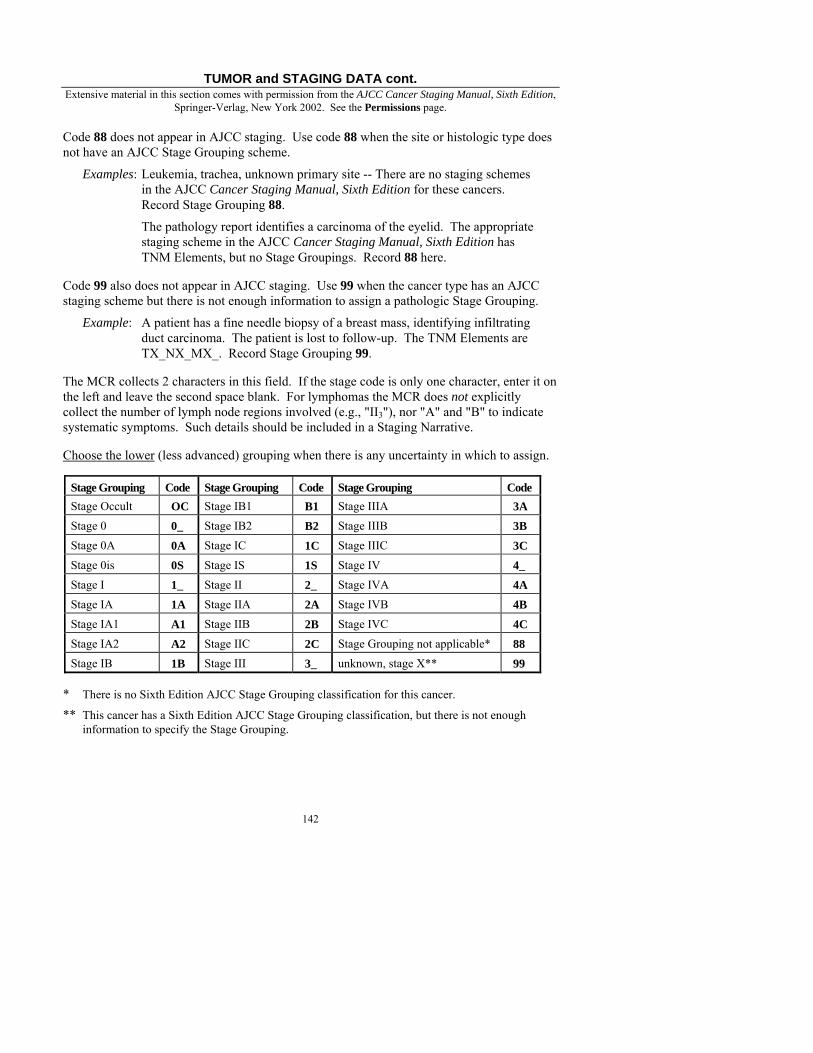
NAACCR Version 11.1 field "TNM Clin Stage Group", Item 970, columns 579-580 The Stage Grouping indicates the anatomic extent of disease and groups cases which are expected to have similar prognoses. The Clinical Stage Grouping is important for selecting and evaluating the primary therapy. The TNM Stage Grouping is usually based on the previously coded TNM Elements. Lymphomas have only Stage Groupings in the TNM system (no TNM Elements). Many of the opthalmic cancers have TNM Elements but no Stage Groupings. Tumor Size, histopathologic Grade, Age at Diagnosis, risk factors, or serum tumor marker data are needed to determine the Stage Grouping for some cancer types. (When appropriate, relevant risk factor information can be included in a Staging Narrative.) Always refer to the AJCC Cancer Staging Manual, Sixth Edition for appropriate site-specific coding rules. Code 88 does not appear in AJCC staging. Use code 88 when the site or histologic type does not have an AJCC Stage Grouping scheme.
Examples: Leukemia, central nervous system, adrenal gland, unknown primary -- There are no staging schemes in the AJCC Cancer Staging Manual, Sixth Edition for these cancers. Record Stage Grouping 88.
Carcinoma of the eyelid -- The appropriate staging scheme in the AJCC Cancer Staging Manual, Sixth Edition has TNM Elements, but no Stage Groupings. Record Stage Grouping 88.

134
TUMOR and STAGING DATA cont. Extensive material in this section comes with permission from the AJCC Cancer Staging Manual, Sixth Edition,
Springer-Verlag, New York 2002. See the Permissions page. Code 99 also does not appear in AJCC staging. Use code 99 when the site or histologic type has an AJCC staging scheme but there is not enough information to assign a Stage Grouping.
Example: A patient has a fine needle biopsy of a breast mass. The cytology identifies infiltrating duct carcinoma. The patient is lost to follow-up. The AJCC TNM elements are TX_NX_MX_. Record Stage Grouping 99.
The MCR collects 2 characters in this field. If the code is only one digit, enter it on the left and leave the second space blank. For lymphomas the MCR does not explicitly collect the number of lymph node regions involved (e.g., "II3"), nor "A" and "B" to indicate systematic symptoms. Such details should be included in a Staging Narrative. Choose the lower (less advanced) category when there is any uncertainty in which to assign.
Stage Grouping Code Stage Grouping Code Stage Grouping Code Stage Occult OC Stage IB1 B1 Stage IIIA 3A Stage 0 0_ Stage IB2 B2 Stage IIIB 3B Stage 0A 0A Stage IC 1C Stage IIIC 3C Stage 0is 0S Stage IS 1S Stage IV 4_ Stage I 1_ Stage II 2_ Stage IVA 4A Stage IA 1A Stage IIA 2A Stage IVB 4B Stage IA1 A1 Stage IIB 2B Stage IVC 4C Stage IA2 A2 Stage IIC 2C Stage Grouping not applicable* 88 Stage IB 1B Stage III 3_ unknown, stage X** 99
* There is no Sixth Edition AJCC Stage Grouping classification for this cancer.
** This cancer has a Sixth Edition AJCC Stage Grouping classification, but there is not enough information to specify the Stage Grouping.
Pathologic Descriptor NAACCR Version 11.1 field "TNM Path Descriptor", Item 920, column 571 This field records a code describing additional detail about the type of staging information recorded in the pTNM fields. Some of these codes reflect suffixes, prefixes or subscripts that may be added onto TNM elements or stage groupings. When none of the coded situations below applies to a particular case, a zero (0) is recorded. The field may not be left empty.

135
TUMOR and STAGING DATA cont. Extensive material in this section comes with permission from the AJCC Cancer Staging Manual, Sixth Edition,
Springer-Verlag, New York 2002. See the Permissions page. The codes for Pathologic Descriptor follow:
Descriptor category Code None; no special Descriptor applies. 0 "E" Stage Grouping for extranodal lymphomas 1 "S" Stage Grouping for lymphomas involving the spleen 2 "m" T Element; multiple tumors in the primary site (at diagnosis) counted as one primary case
3
"y" prefix; staged after the start of pre-surgical (neoadjuvant) therapy* 4 "E+S" Stage Grouping for extranodal lymphomas involving the spleen 5 "m" and "y"; multiple tumors in the primary site and staging done after the start of neoadjuvant* therapy
6
A TNM prefix/suffix/subscript probably applies, but you don't know which; unknown Descriptor category.
9
* Radiation therapy, chemotherapy, hormone therapy, immunotherapy or endocrine procedures may
have affected the extent of disease before the pathologic staging was done. Pathologic T NAACCR Version 11.1 field "TNM Path T", Item 880, columns 563-564 The Pathologic T Element (pT) describes the primary tumor's size and/or extension. Refer to the AJCC Cancer Staging Manual, Sixth Ed. for site-specific/histology-specific coding rules. Pathologic classification is based on information obtained before treatment and supplemented by additional evidence from surgery and pathologic examination of resected specimens. It is a combination of all findings through the most definitive surgery done. The pathologic stage provides the most precise data to estimate prognosis and calculate end results. Pathologic assessment of the primary tumor generally requires a resection of the primary tumor or biopsy specimen adequate to evaluate the highest pT category. When there are multiple simultaneous tumors being reported as one primary, the T Element for only the largest individual tumor or the individual tumor with the highest T category is coded. The tumor multiplicity is captured in the Pathologic Descriptor field. The EOD--Tumor Size field will reflect the size of the largest tumor. You may include specific tumor multiplicity information in a Staging Narrative or Narrative Primary Site fields. The number of tumors is important in determining the T Element for some cancer types. Simultaneous independent bilateral tumors in paired organs are staged separately (multiple primaries).

136
TUMOR and STAGING DATA cont. Extensive material in this section comes with permission from the AJCC Cancer Staging Manual, Sixth Edition,
Springer-Verlag, New York 2002. See the Permissions page.
Examples: There are two simultaneous duct carcinomas in the upper outer quadrant of the right breast -- one with diameter 0.4 cm, the other with diameter 0.8 cm. The case is reported with T1B because this corresponds to the size of the larger lesion. The Staging Narratives should include the fact that there were two tumors, along with their sizes.
There are two primary tumors -- sized 1.1 cm and 2.1 cm -- in the same lobe of the liver, without any vascular invasion. The coded T Element is T2_. As this could also describe a single tumor with vascular invasion, use a Staging Narrative to specify the specific situation that was coded.
A patient is diagnosed in May with a 1-cm duct carcinoma of the right breast and a 0.5-cm lobular carcinoma of the left breast. Stage each primary separately (T1B for the right, T1A for the left).
Many chapters in the AJCC staging system specifically include a classification for carcinomas in situ as "Tis". If there is an accepted histologic classification for carcinoma in situ as determined by a pathologist, you may use "pTis" even if the Cancer Staging Manual, Sixth Edition does not include this category for the given primary site. The following general definitions are used throughout the TNM classification:
TX - primary tumor cannot be assessed or is unknown. T0 - no evidence of a primary tumor Tis - carcinoma in situ* T1, T2, T3, T4 - describe increasing size* and/or local extent of the primary tumor
* Note: For AJCC staging schemes in which a specific tumor size plays an important role in assigning the T Element category (such as breast carcinomas), there is sometimes confusion about how to stage an in-situ case that has a recorded tumor size. All lesions that are completely in situ (no invasive component) are assigned pTis regardless of the tumor size. A large in-situ tumor does not have the same prognosis as an invasive cancer with the same tumor size. pT1_, pT2_, etc. are assigned to invasive cancers of increasing size and/or extent. For a tumor with both in situ and invasive components, only the invasive component's size should be used for assigning the T Element. Use code X_ when the site or histologic type has an AJCC staging scheme but there is not enough information to assign a T value.
Example: A biopsy of a breast mass identifies infiltrating duct carcinoma. The patient is lost to follow-up. The AJCC staging scheme requires excision of the primary tumor with macroscopically clean margins for pathologic staging. Record pTX_.

137
TUMOR and STAGING DATA cont. Extensive material in this section comes with permission from the AJCC Cancer Staging Manual, Sixth Edition,
Springer-Verlag, New York 2002. See the Permissions page. Code T88 is not included in AJCC staging. This code enables the MCR to distinguish cases in which the site or histologic type has no AJCC staging scheme from cases that could not be staged because the information was incomplete. Use T88 when the site or histologic type does not have an AJCC staging scheme.
Examples: Leukemia, brain primary -- These have no staging schemes in the AJCC Cancer Staging Manual, Sixth Ed. Record T88N88M88.
The pathology report identifies a breast mass as sarcoma. The breast staging scheme in the AJCC Cancer Staging Manual, Sixth Ed. does not apply to sarcomas. Record T88N88M88.
Lymphomas have AJCC Stage Groupings, but no TNM Elements. Record T88N88M88.
Choose the lower (less advanced) T category when there is uncertainty in which category to assign. For example, in the larynx supraglottis squamous cell carcinoma scheme, the T4a category specifies that the tumor invades through the thyroid cartilage, while the T3 category includes minor thyroid cartilage erosion; so if a supraglottis tumor invades deeply into but not through thyroid cartilage, it would be classified T3 because it does not meet the T4a requirements. The MCR collects 2 characters in this field. For only one character, enter it on the left and leave the second space blank. The following table shows the code for each T category.
T Category Code T Category Code T Category Code T Category Code TX* X_ T1mic 1M T2 2_ T4 4_
T0 0_ T1 1_ T2a 2A T4a 4A
Ta A_ T1a 1A T2b 2B T4b 4B
Tis IS T1a1 A1 T2c 2C T4c 4C
Tispu SU T1a2 A2 T3 3_ T4d 4D
Tispd SD T1b 1B T3a 3A T not applicable** 88
T1b1 B1 T3b 3B T1b2 B2 T3c 3C
T1c 1C
* This cancer has a Sixth Edition AJCC T classification scheme, but there is not enough information to specify the T; occult lung cancers (primary tumor mass not present or not evaluable).
** There is no Sixth Edition AJCC pT classification for this cancer.

138
TUMOR and STAGING DATA cont. Extensive material in this section comes with permission from the AJCC Cancer Staging Manual, Sixth Edition,
Springer-Verlag, New York 2002. See the Permissions page. Pathologic N NAACCR Version 11.1 field "TNM Path N", Item 890, columns 565-566 Pathologic N (pN) identifies the absence or presence of regional lymph node metastases. Always refer to the AJCC Cancer Staging Manual, Sixth Edition for appropriate site-specific and histology-specific coding rules. The following general definitions are used throughout the TNM classification:
NX - regional lymph nodes cannot be assessed or status unknown N0 - nodes were assessed and there was no pathologic evidence of regional lymph
node metastasis N1, N2, N3 - indicate increasing involvement of regional lymph nodes
A lymph node may appear pathologically negative for cancer (when the specimen is examined under the microscope) and yet harbor a small number of cells which are cancerous. These may be micrometastases (> 0.2 mm and < 2.0 mm in largest dimension) or isolated tumor cells (a single cancerous cell or a tiny group of them < 0.2 mm in largest dimension). The presence of these cells may be detected by ordinary staining or by special immunohistochemical or molecular tests. Flow cytometry and DNA analysis may also be used to detect individual cells that are "suggestive" for being cancerous. A node as a whole may be pathologically negative even if isolated tumor cells are detected. If there are multiple isolated tumor cells and/or micrometastases in a node, use the size of the largest cancer deposit to determine the N category. Immunohistochemical and molecular test results may now be recorded in the 2-character pN codes (added in 2004, although the MCR no longer collects TNM fields beginning with 2004 diagnoses). If the primary tumor extends directly into a lymph node, classify this in the N Element as a lymph node metastasis (rather than in the T Element). For colorectal cancers, smoothly contoured metastatic nodules resembling lymph nodes in surrounding fat tissue should be counted in the N Element even if no actual lymph node tissue is found in them; but irregularly contoured nodules in nearby fat should be counted in the extent of the T Element. Metastasis in any lymph node not specified as regional in the appropriate AJCC staging scheme should be considered distant metastasis and classified in the M Element. Use code NX_ when the site or histologic type has an AJCC staging scheme but there is not enough information to assign an N Element code.
Example: A patient has a biopsy of a testicular mass. The biopsy identifies an embryonal carcinoma. The patient is lost to follow-up. This type of case has an AJCC staging scheme, but no assessment of regional lymph node involvement was made. Record NX_.

139
TUMOR and STAGING DATA cont. Extensive material in this section comes with permission from the AJCC Cancer Staging Manual, Sixth Edition,
Springer-Verlag, New York 2002. See the Permissions page. Code 88 does not appear in AJCC staging. Its use enables registries to distinguish cases unstaged because of insufficient information from those unstaged because they have no AJCC staging scheme. Use code 88 when the site/histology does not have an AJCC staging scheme.
Examples: Adrenal gland, unknown primary site -- These have no staging schemes in the AJCC Cancer Staging Manual, Sixth Ed. Record T88N88M88.
The pathology report identifies a breast mass as sarcoma. The breast staging scheme in the AJCC Cancer Staging Manual, Sixth Ed. does not apply to sarcomas. Record T88N88M88.
Gestational trophoblastic tumors do not have N categories. Record N88. Choose the lower (less advanced) N category when there is uncertainty. The MCR collects 2 characters. If there is only one character, enter it on the left and leave the second space blank.
N Category Code N Category Code NX* X_ N2 2_ N0** 0_ N2a 2A N0(i-) I- N2b 2B N0(i+) I+ N2c 2C N0(mol-) M- N3 3_ N0(mol+) M+ N3a 3A N1 1_ N3b 3B N1a 1A N3c 3C N1b 1B N not applicable*** 88
N1c 1C N1mi 1M
* This cancer has a Sixth Edition AJCC N classification scheme, but there is not enough information to specify the N.
** Code 0_ included N0(i-), N0(i+), N0(mol-) and N0(mol+) before separate codes were added for these categories in 2004.
*** There is no Sixth Edition AJCC pN classification for this cancer.
Pathologic M NAACCR Version 11.1 field "TNM Path M", Item 900, columns 567-568 The M Element describes the presence or absence of distant metastases (including non-regional lymph nodes). Refer to the AJCC Cancer Staging Manual, Sixth Ed. for site- and histology-specific coding rules.

140
TUMOR and STAGING DATA cont. Extensive material in this section comes with permission from the AJCC Cancer Staging Manual, Sixth Edition,
Springer-Verlag, New York 2002. See the Permissions page. Metastasis in any lymph node not specified as regional in the appropriate AJCC staging scheme should be considered distant and classified in the M Element. The following general definitions are used throughout the TNM classification:
MX - presence of distant metastasis cannot be assessed or is unknown M0 - no known distant metastasis M1 - distant metastasis present
For the evaluation of distant lymph nodes, as with the pN Element, a distant node may be pathologically negative for cancer (when the specimen is examined under the microscope) and yet harbor isolated tumor cells detected by immunohistochemical or molecular tests. Flow cytometry and DNA analysis may also be used to detect individual cells that are "suggestive" for being cancerous. As long as the node as a whole is pathologically negative, it is coded M0_ even if isolated tumor cells or micrometastases are detected. In general, if a distant metastasis has been microscopically confirmed, this alone may satisfy the requirements for a pM category (such as pM1_ or pM1A) and the Pathologic Stage Group may be assigned. Use MX_ when the site or histologic type has an AJCC staging scheme but there is not enough information to specify an M Element.
Example: A patient's breast mass biopsy finds infiltrating duct carcinoma. The patient is lost to follow-up. Breast carcinomas have an AJCC staging scheme, but the status of distant metastasis has not been evaluated. Record TX_NX_MX_.
Code 88 does not appear in AJCC staging. This code allows registries to distinguish unstaged cases in which the site/histology has no AJCC staging scheme from cases that could not be staged because of incomplete information. Use M88 when the site or Histologic Type does not have an AJCC Staging scheme.
Examples: Leukemia, central nervous system, an ill-defined pelvic site -- These have no staging schemes in the AJCC Cancer Staging Manual, Sixth Ed. Record T88N88M88.
Pathology identifies gastric sarcoma. The stomach staging scheme in the AJCC Cancer Staging Manual, Sixth Ed. does not apply to sarcomas. Record T88N88M88.
The MCR collects 2 characters in this field. The MCR does not collect the additional notations "PUL", "OSS", "HEP", etc. (see p. 7 in the Cancer Staging Manual, Sixth Ed.) to denote the site(s) of distant metastasis. Please include any known site(s) of distant metastasis in a Staging Narrative.

141
TUMOR and STAGING DATA cont. Extensive material in this section comes with permission from the AJCC Cancer Staging Manual, Sixth Edition,
Springer-Verlag, New York 2002. See the Permissions page. Choose the lower (less advanced) category when there is any uncertainty in which to assign.
M Category Code MX* X_ M0 0_ M1 1_ M1a 1A M1b 1B M1c 1C M not applicable** 88
* This cancer has a Sixth Edition AJCC M classification scheme, but there is not enough information to specify the M.
** There is no Sixth Edition AJCC pM classification for this cancer. Pathologic Stage Grouping
NAACCR Version 11.1 field "TNM Path Stage Group", Item 910, columns 569-570 The Stage Grouping describes the anatomic extent of disease. Different cases which fall into the same Stage Grouping are expected to have similar prognoses. The Pathologic Stage Grouping can be used as a guide for the need of adjuvant therapy, for reporting end results, and for the estimation of prognosis. In order to assign a Pathologic Stage Grouping it is not always necessary to have three specific Pathologic TNM Elements. If sufficient tissue has been removed for pathologic examination to evaluate the highest T and N categories (that is, pT and pN are specified), you may use the specific pM or cM to assign a Pathologic Stage Grouping. In general, if a distant metastasis has been microscopically confirmed, this alone may satisfy the requirements for a pathologic M category (such as pM1_ or pM1A) and the Pathologic Stage Grouping may be assigned. The TNM Stage Grouping is usually based on the coded TNM Elements. Lymphomas have only Stage Groupings in the TNM system (no TNM Elements). Many of the opthalmic cancers have TNM Elements but no Stage Groupings. Tumor Size, histopathologic Grade, Age at Diagnosis, risk factors, or serum tumor marker data are needed to determine the Stage Grouping for some cancer types. (When relevant, risk factor information should be included in a Staging Narrative.) Always refer to the AJCC Cancer Staging Manual, Sixth Ed. for appropriate site-specific and histology-specific coding rules.
142
TUMOR and STAGING DATA cont. Extensive material in this section comes with permission from the AJCC Cancer Staging Manual, Sixth Edition,
Springer-Verlag, New York 2002. See the Permissions page. Code 88 does not appear in AJCC staging. Use code 88 when the site or histologic type does not have an AJCC Stage Grouping scheme.
Examples: Leukemia, trachea, unknown primary site -- There are no staging schemes in the AJCC Cancer Staging Manual, Sixth Edition for these cancers. Record Stage Grouping 88.
The pathology report identifies a carcinoma of the eyelid. The appropriate staging scheme in the AJCC Cancer Staging Manual, Sixth Edition has TNM Elements, but no Stage Groupings. Record 88 here.
Code 99 also does not appear in AJCC staging. Use 99 when the cancer type has an AJCC staging scheme but there is not enough information to assign a pathologic Stage Grouping.
Example: A patient has a fine needle biopsy of a breast mass, identifying infiltrating duct carcinoma. The patient is lost to follow-up. The TNM Elements are TX_NX_MX_. Record Stage Grouping 99.
The MCR collects 2 characters in this field. If the stage code is only one character, enter it on the left and leave the second space blank. For lymphomas the MCR does not explicitly collect the number of lymph node regions involved (e.g., "II3"), nor "A" and "B" to indicate systematic symptoms. Such details should be included in a Staging Narrative. Choose the lower (less advanced) grouping when there is any uncertainty in which to assign.
Stage Grouping Code Stage Grouping Code Stage Grouping Code Stage Occult OC Stage IB1 B1 Stage IIIA 3A Stage 0 0_ Stage IB2 B2 Stage IIIB 3B Stage 0A 0A Stage IC 1C Stage IIIC 3C Stage 0is 0S Stage IS 1S Stage IV 4_ Stage I 1_ Stage II 2_ Stage IVA 4A Stage IA 1A Stage IIA 2A Stage IVB 4B Stage IA1 A1 Stage IIB 2B Stage IVC 4C Stage IA2 A2 Stage IIC 2C Stage Grouping not applicable* 88 Stage IB 1B Stage III 3_ unknown, stage X** 99
* There is no Sixth Edition AJCC Stage Grouping classification for this cancer.
** This cancer has a Sixth Edition AJCC Stage Grouping classification, but there is not enough information to specify the Stage Grouping.

143
TUMOR and STAGING DATA cont. Extensive material in this section comes with permission from the AJCC Cancer Staging Manual, Sixth Edition,
Springer-Verlag, New York 2002. See the Permissions page. TNM Edition Number NAACCR Version 11.1 Item 1060, column 593-594
Like other TNM fields, this field should be filled for the MCR for pre-2004 diagnoses only. This field identifies the edition of the AJCC Manual for Staging of Cancer that was used to stage the case. Staging criteria and timing inclusion rules differ between editions. This code allows analysis of cases grouped by edition number. The staging manual that is appropriate for the year of diagnosis of a case should be used; although this field may be automatically filled by your software system, please code the book that was actually used to stage the case when this is known, even if it is not the appropriate edition to have used for the given year of diagnosis (if you can manually enter this code on your system). The MCR uses this field to determine which book we must use to interpret the coded stage information.
AJCC Staging Edition Code not staged (case has an AJCC staging scheme,
but staging was not done) 00
First Edition (for cases diagnosed before 1984) 01 Second Edition (for cases diagnosed 1984-1988) 02 Third Edition (for cases diagnosed 1989-1992) 03 Fourth Edition (for cases diagnosed 1993-1997) 04 Fifth Edition (for cases diagnosed 1998-2002) 05 Sixth Edition (for cases diagnosed 2003-?) 06 not applicable (case does not have an AJCC
staging scheme in the edition used) 88
unknown edition (case was AJCC-staged, but the edition used is unknown) 99
If the year of diagnosis is unknown to you, stage the case as if it had been diagnosed in your facility's Date of First Contact year. SEER Summary Stage 1977 NAACCR Version 11.1 Item 760, column 529 Refer to the Third Edition of this manual for coding diagnoses made before 2001. This field should be left empty for diagnoses made beginning in 2001; it must be filled for diagnoses made before 2001.

144
TUMOR and STAGING DATA cont. SEER Summary Stage 2000 NAACCR Version 11.1 Item 759, column 528 This field is required for all diagnoses made between January 1, 2001 and December 31, 2003. Beginning with diagnoses made in 2004, this field is replaced by the Summary Stage 2000 field derived from the Collaborative Staging fields. Thus, this field must be filled for diagnoses made between 2001 and 2003; it should be left empty for diagnoses made before 2001 and for those made after 2003. SEER Summary Staging groups cases into broad categories (such as localized, regional and distant). Note: Before Collaborative Staging takes effect, the COC only requires Summary Staging for cases which are not TNM-staged. The MCR requires both Summary Staging and TNM staging for all reportable cases diagnosed between 2001 and 2003 (use "unknown" and "not applicable" codes as necessary). All cases diagnosed between 2001 and 2003 must be staged using the red SEER Summary Staging Manual 2000 (published 2001, with updates). All pre-2001 diagnoses must be staged using the green Summary Staging Guide (1977, with updates from SEER Extent of Disease coding changes). The two books have different staging schemes and rules -- be sure to use the correct book to stage each case based on its diagnosis year. If your data system shows both fields "SEER Summary Stage 1977" and "SEER Summary Stage 2000" on-screen, be sure that the correct field gets filled in based on each case's diagnosis year; if your system shows only one Summary Stage field, be sure to use the correct book to stage each case based on its diagnosis year; when the case is exported your system will put that single field into the appropriate place in the NAACCR layout based on each case's diagnosis year. If the year of diagnosis is unknown to you, stage the case as if it had been diagnosed in your facility's Date of First Contact year. General Guidelines Rules governing Summary Staging appear in the SEER Summary Staging Manual 2000's first chapter (pages 2-15). The entire set of Guidelines on p. 10 is very important to keep in mind as you apply any specific staging scheme. Some of the Guidelines are paraphrased here: • Instructions in a specific staging scheme take precedence over the General Guidelines on
p. 10.
• Summary staging is based on a combination of clinical, operative and pathologic assessments. Clinical evaluations may include important staging information, such as skin involvement, missing from operative and path reports; but if some part of the clinical assessment is disproved by operative or pathologic findings, use the op/path findings. If information from an operative and path report conflict, priority goes to the pathologic assessment. An autopsy report should be given the same priority as a pathology report.

145
TUMOR and STAGING DATA cont. • When you have AJCC TNM staging but no direct Summary Stage information, assign the
Summary Stage 2000 code that is most equivalent to what the TNM staging reflects. If the medical record conflicts with a physician's TNM stage, the information in the record takes precedence; try to consult with the physician to see if s/he has information not available in the record, or if the record was incomplete at the time of the TNM staging.
• Include all information available within the following timeframe: through completion of first-course-of-treatment* surgeries or
within four months of diagnosis in the absence of disease progression, whichever is longer.
This applies to all cancers, including prostate. Do not stage too soon (before all the information you need is in the medical record). You must still report to the MCR in a timely manner, however; so when surgery is delayed for a case, you may need to send the case to us with an unknown Summary Stage; then be sure to call us (617-624-5680 or 617-624-5653) when the staging is complete so that we can update it.
* Note: This refers to the SEER definition of "first course of treatment" which may include a much longer timeframe than the COC definition under certain circumstances only. Applying the COC definition here should not make a difference in most cases. See the section "Unresolved Issues" in the NAACCR Standards for Cancer Registries, Volume II: Data Standards and Data Dictionary (any recent edition) for a concise comparison of the COC and SEER definitions of "first course of treatment".
• Exclude any metastasis known to have developed after the diagnosis was established.
• Information obtained after the start of treatment (radiation, chemotherapy, etc.) may be used unless it falls outside the timeframe described in the "surgery/four-months" Guideline. But if pre-surgical treatment affected the extent of disease found at surgery, then use the pre-treatment information for staging. (For example, a lung cancer case is clinically regionalized to lymph nodes and the patient receives chemotherapy before surgery; the surgery finds no positive nodes; the Summary Stage should reflect the clinically diagnosed node involvement that existed at diagnosis. So just as disease progression should not be included in Summary Staging, disease improvement resulting from treatment should also not be included. This is the MCR's interpretation of the Summary Stage rules.)
• Certain staging schemes are used for certain histologic types regardless of primary site: Kaposi Sarcoma of All Sites (page 274); Hodgkin and Non-Hodgkin Lymphomas of All Sites, excluding Mycosis Fungoides
and Sezary Disease of Skin, Vulva, Penis, Scrotum (page 278); Hematopoietic, Reticuloendothelial, Immunoproliferative, and Myeloproliferative
Neoplasms (page 280).
If a case has one of these histologies, ignore the primary site when choosing your staging scheme (e.g., a stomach lymphoma should be staged using the lymphoma scheme, even though the stomach scheme does not specify that it excludes lymphomas.)

146
TUMOR and STAGING DATA cont. Most schemes apply to all other histologies for the given primary sites, except as noted. See the lists on pages 285-287 of the staging manual.
Some schemes are limited to certain sites and histologies in combination: Melanoma of Skin, Vulva, Penis, Scrotum (page 173) and Conjunctiva (page 252); Mycosis Fungoides and Sezary Disease of Skin, Vulva, Penis, Scrotum (page 176); Melanoma of Cornea, Retina, Choroid, Ciliary Body, Eyeball, and Overlapping and
Other Eye (page 256); Retinoblastoma (page 258). Ambiguous terms may be used in the medical record to describe tumor involvement. The following lists should be used as a guide when assigning Summary Stage 2000. (Not all forms of the words can be shown here.)
Involved: Consider the following terms to be indicative of cancer involvement: • adherent • apparent(ly) • appear(s) to • comparable with • compatible with • consistent with • contiguous with • continuous with • encroaching upon* • extension to, into, onto or out onto • features of • fixation to another structure** • fixed** • impending perforation of • impinging upon • impose/imposing on • incipient invasion
• induration*** • infringe(s)/infringing • into* • intrude(s) • invasion to, into, onto or out onto • matted (indicates involvement for lymph
nodes only) • most likely • onto* • overstep(s) • presumed • probable • protruding into (unless encapsulated) • suspect(ed) • suspicious • to* • up to
* indicates cancer involvement whether found in a clinical, operative or pathologic description ** indicates that the other structure or tissue is involved *** used to describe surrounding fibrous or connective tissue adjacent to the tumor; interpreted as
extension of the malignant growth

147
TUMOR and STAGING DATA cont. Not Involved: Consider the following terms to be indicative of cancer non-involvement:
• abuts • approaching • approximates • attached • cannot be excluded • cannot be ruled out • effaces/effacing/effacement • encases/encasing • encompass(es) • entrapped • equivocal
• extension to without invasion • extension to without involvement of • kiss(es)/kissing • matted (except for lymph nodes) • possible • questionable • reaching out • rule(s) out • suggests • very close to • worrisome
The special use of some terms in the Summary Staging Manual 2000 are discussed on page 14 therein. When used in other places, these terms may have different meanings. "adjacent organ(s)": Anatomic structures with specific physiologic functions other than (or in addition to) support and storage that are located next to the primary site organ. "adjacent structure(s)": connective tissues large enough to have been given a specific name (for example, brachial artery, broad ligament) "adjacent tissue(s), NOS": This term may appear in the staging schemes for ill-defined and non-specific sites. The term is used to mean "the unnamed tissue(s) that immediately surround an organ or structure containing a primary cancer". The tumor has invaded past the outer border (capsule, serosa, other edge) of the primary organ into the organ's supportive structures but has not invaded larger structures and adjacent organs. "connective tissue(s)": These do not generally have specific names. They include adipose tissue, aponeuroses, blood vessels, bursa, fascia, fatty tissues, fibrous tissues, ganglia, ligaments, lymphatic channels, muscle, nerves (spinal, sympathetic, peripheral), skeletal muscle, subcutaneous tissue, synovia, tendons, tendon sheaths, unidentified vessels and veins. Blood, cartilage and bone are not "connective tissues" in the Summary Staging manual. "cortex", "cortical": the external or outer surface layer of an organ "marrow", "medulla", "medullary": the interior central portion of an organ "parenchyma": the functional portion of an organ, as distinguished from its framework or stroma; the place where most malignancies arise "stroma": the cells and tissues that support, store nutrients, and maintain viability within an organ; consists of connective tissue, vessels and nerves; provides the framework of an organ

148
TUMOR and STAGING DATA cont. In Situ (Code 0) A diagnosis of "in situ" must be based on microscopic examination of tissue or cells. An in-situ tumor has all the characteristics of malignancy except invasion (i.e., the basement membrane has not been penetrated). A tumor that displays any degree of invasion is not classified as in situ (it is at least localized). For example, if a report states "carcinoma in situ of the cervix showing microinvasion of one area", then the tumor is not in situ. A primary tumor may involve more than one site (e.g., cervix and vagina, labial mucosa and gingiva) and still be in situ if it does not show any invasion. If a tumor is Summary Staged as in situ, its Behavior Code (see pages 97-98) is 2. Organs and tissues that have no epithelial layer and basement membrane cannot be Summary Staged as in situ; only carcinomas and melanomas may be staged in situ. For carcinomas and melanomas, if all reports are negative for disease spread and the pathologist states that the cancer is noninvasive or noninfiltrating, code as 0. Certain terms indicate an in situ Summary Stage:
confined to epithelium
intracystic intraductal intraepidermal intraepithelial
intrasquamous involvement up to but
not including the basement membrane
no penetration below the basement membrane
no stromal invasion noninfiltrating noninvasive preinvasive Stage 0
Localized (Code 1) A localized tumor invades beyond the basement membrane, but is still confined entirely to the organ of origin. For most sites a localized tumor may be widely invasive or have spread within the organ, as long as it does not extend beyond its outer limits and there is no evidence of metastasis to other parts of the body. If all reports are negative for disease spread and the pathologist states that the cancer is invasive or infiltrating, use code 1. Inaccessible Sites - Clinical diagnosis alone may be insufficient for staging a tumor "localized" when the primary site and regional nodes are inaccessible, such as with the esophagus or lung. Without confirmation from surgery/autopsy, it is usually preferable to use code 9 ("unstageable"); but, if a physician stages the case "localized", or if clinical reports (like CT scans) provide enough information to rule out further disease spread, code 1 may be used; if surgery was done, study the operative report for evidence of direct extension or metastasis; if surgery and radiology have produced no such evidence, assign code 1. Vessel / Lymphatic Involvement -- Invasion of blood vessels, lymphatics and/or nerves within the primary site is localized, unless there is evidence of disease outside the site. Microinvasive -- This term, used by pathologists to describe the earliest invasive stage, has precise meaning for cancer of certain sites. Microinvasive cancers are staged as "localized".

149
TUMOR and STAGING DATA cont. Regional (Codes 2, 3, 4, 5) A tumor at the "regional" stage has grown beyond the limits of the organ of origin -- into adjacent organs or tissues by direct extension, and/or to regional lymph nodes by metastasis. Cancer becomes regional when there is the potential for spread by more than one lymphatic or vascular supply route. If in situ, localized and distant stage categories have been ruled out, then the stage may be assumed to be regional. Neoplasms appearing to be in the "regional" stage must be evaluated very carefully to make sure they have not spread any further.
Example: A malignant tumor of the stomach or gallbladder often passes through the wall of the primary organ into surrounding tissues. Before coding as regional, make certain that imaging scans do not reveal metastasis to lung or bone and that surgery did not reveal metastases to non-regional tissues. Check timely progress notes and discharge summary for any mention of metastases.
Regional, by Direct Extension or Contiguous Spread Only (Code 2) -- Sometimes a cancer spreads to surrounding organs or tissue with no involvement of regional lymph nodes. The cancer invades through the wall of the organ of origin into surrounding organs and/or adjacent tissues. Before assigning code 2 to such a case, make sure that tissue adjacent to the original organ is actually involved. The terms "penetrating", "extension" and "metastases" are sometimes used to describe spreading within an organ, such as the large intestine or bladder, in which case the stage might still be "localized" (code 1). The Summary Staging Manual lists organs and structures considered to be regional for each site. Regional, to Lymph Nodes Only (Code 3) -- If a cancer continues to grow after the onset of local invasion, regional lymph nodes draining the area usually become involved. The cancer invades the walls of lymphatics and may travel to and grow in nearby nodes. Enter code 3 if nodal involvement is indicated and there is no other evidence of extension beyond the organ of origin. For carcinomas, if there are lymph nodes involved, then the stage is at least regional. Words like "local" and "metastasis" appearing in medical records sometimes cause confusion in staging. Failure to recognize the names of regional lymph nodes might lead to incorrect staging. The Summary Staging Manual and the AJCC's Cancer Staging Manual contain helpful information about the names of regional and distant nodes.
Examples: "Carcinoma of the stomach with involvement of local lymph nodes" should, lacking further evidence, be considered "regional" and coded 3.
Statements like "carcinoma of the breast with axillary lymph node metastasis" and "carcinoma of the stomach with metastasis to perigastric nodes" indicate metastasis to regional nodes and should be assigned code 3.

150
TUMOR and STAGING DATA cont. Regional nodes are listed for each Summary Stage scheme. Consider the farthest specific node chain involved. Any nodes that are removed along with the resected primary site specimen that are not specifically identified should be considered "regional lymph nodes, NOS". If a specific node chain is named but is not listed in the staging scheme, first determine if the recorded name is synonymous with a listed regional chain (see page 284 in the Summary Staging Manual); otherwise, assume that these are distant lymph nodes (7). Unless stated to be contralateral or bilateral, assume that lymph nodes mentioned are ipsilateral (homolateral). For lymphomas, any mention of lymph nodes indicates involvement. For solid tumors, the terms "fixed", "matted" and "mass in the mediastinum, retroperitoneum, and/or mesentery" without specific information as to the types of tissue involved are considered to indicate lymph node involvement. The terms "palpable", "enlarged", "visible swelling", "shotty" and "lymphadenopathy" are to be ignored except for lung primaries; for lung primaries, these terms are interpreted as regional lymph node involvement. Bilateral lymph node metastases do not necessarily indicate distant spread. For primaries on the body's midline (e.g., esophagus), bilateral node involvement is regional disease.
Regional, Direct Extension and Lymph Nodes (Code 4) -- Use code 4 when a tumor has metastasized to regional lymph nodes and also has spread to regional tissue via direct extension, but there is no evidence of metastasis to a distant site or distant nodes.
Regional, NOS (Code 5) -- If available information states only that a cancer has spread regionally, use Summary Stage code 5. This indicates that you cannot determine if the spread is to regional nodes only, by direct extension of the tumor only, or both. Some staging schemes have this as the only Regional code available because "direct extension" and "regional lymph node" categories don't apply (for example, brain cancers and lymphomas). Distant Site(s) and/or Distant Node(s) Involved (Code 7) Distant metastases are tumor cells that have broken away from the primary tumor, traveled to other parts of the body, and have begun to grow there. This may be called "diffuse", "disseminated", "metastatic", "remote" or "secondary" disease. In most cases there is no continuous trail of tumor cells between the primary and distant sites. Cancer cells may travel from the primary site and grow distantly by several routes: by direct tumor extension of the primary tumor through adjacent tissues into a non-regional
organ; by travel in lymph channels beyond the first (regional) drainage area to distant nodes; invasion of blood vessels within the primary site, allowing hematogenous (blood-borne)
disease spread through blood vessels in distant sites; by implantation or seeding through fluid within a body cavity.
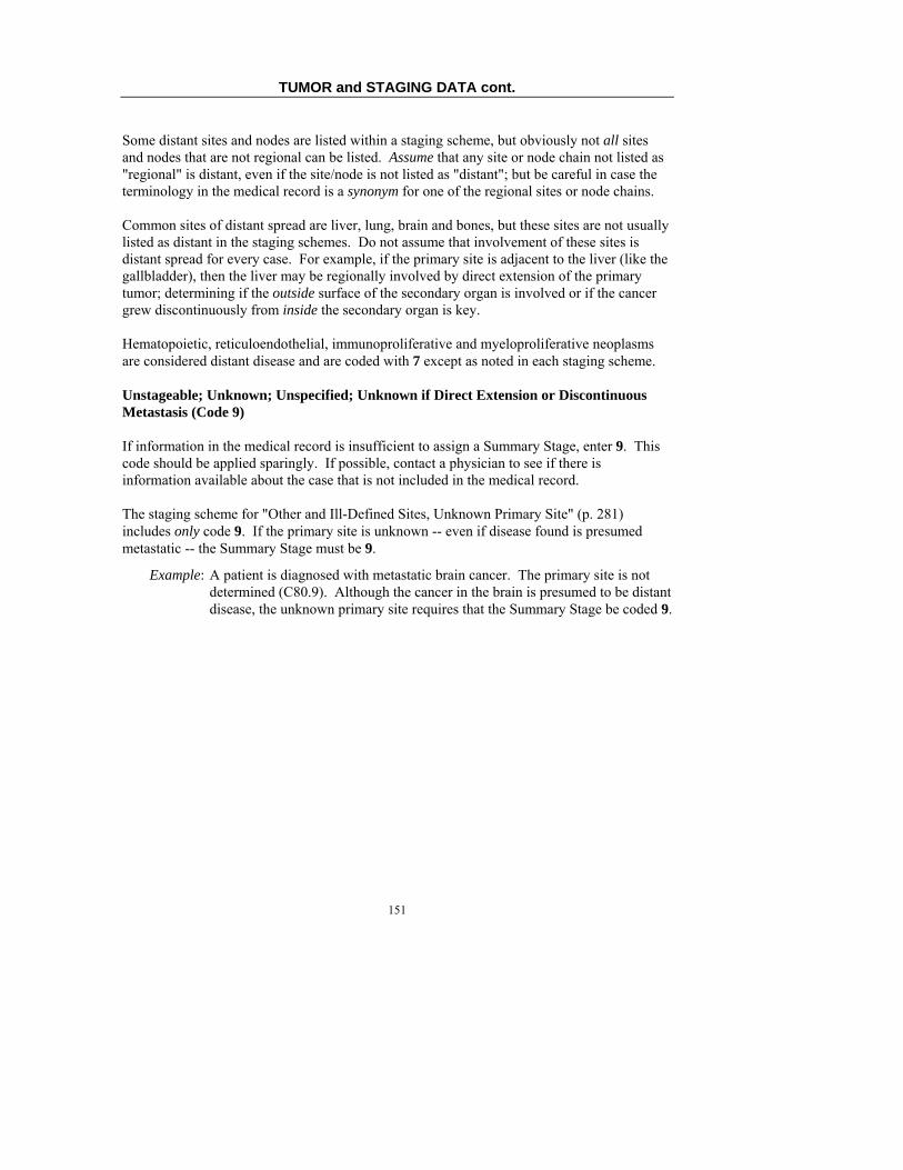
151
TUMOR and STAGING DATA cont. Some distant sites and nodes are listed within a staging scheme, but obviously not all sites and nodes that are not regional can be listed. Assume that any site or node chain not listed as "regional" is distant, even if the site/node is not listed as "distant"; but be careful in case the terminology in the medical record is a synonym for one of the regional sites or node chains. Common sites of distant spread are liver, lung, brain and bones, but these sites are not usually listed as distant in the staging schemes. Do not assume that involvement of these sites is distant spread for every case. For example, if the primary site is adjacent to the liver (like the gallbladder), then the liver may be regionally involved by direct extension of the primary tumor; determining if the outside surface of the secondary organ is involved or if the cancer grew discontinuously from inside the secondary organ is key. Hematopoietic, reticuloendothelial, immunoproliferative and myeloproliferative neoplasms are considered distant disease and are coded with 7 except as noted in each staging scheme. Unstageable; Unknown; Unspecified; Unknown if Direct Extension or Discontinuous Metastasis (Code 9) If information in the medical record is insufficient to assign a Summary Stage, enter 9. This code should be applied sparingly. If possible, contact a physician to see if there is information available about the case that is not included in the medical record. The staging scheme for "Other and Ill-Defined Sites, Unknown Primary Site" (p. 281) includes only code 9. If the primary site is unknown -- even if disease found is presumed metastatic -- the Summary Stage must be 9.
Example: A patient is diagnosed with metastatic brain cancer. The primary site is not determined (C80.9). Although the cancer in the brain is presumed to be distant disease, the unknown primary site requires that the Summary Stage be coded 9.

152
TUMOR and STAGING DATA cont. Use only the codes shown in the SEER Summary Staging Manual 2000 for a specific staging scheme. The general code categories follow:
Extent of Disease Code in situ 0 localized only 1 regional, by direct extension only 2 regional, to lymph nodes only 3 regional (both 2 and 3) 4 regional, NOS 5 distant site(s)/node(s) involved 7 unstageable, unknown, or unspecified; unknown if extension or metastasis; death certificate-only case 9
Not all of these codes apply to every staging scheme. For example, in the malignant Brain and Cerebral Meninges scheme (page 266), only codes 1, 5, 7 and 9 are applicable. Regional Nodes Examined NAACCR Version 11.1 Item 830, columns 541-542 NOTE: This field becomes involved in the Collaborative Staging system for diagnoses made beginning in 2004. The MCR will continue to collect this field for ALL diagnosis years. The instructions below apply for diagnoses made before 2004. For diagnoses made beginning in 2004, and for any case abstracted after your software is updated for 2004 Collaborative Staging, follow the Collaborative Staging Manual and Coding Instructions for this field. This field describes the total number of regional lymph nodes examined by a pathologist (including regional lymph nodes removed by lymph node biopsy and lymph node aspiration). Include nodes considered "regional" and used in the pN Element according to the AJCC Cancer Staging Manual, Sixth Ed. (The MCR will re-code this field in our offices if the particular nodes coded are not regional in the SEER system.) Code all regional lymph nodes removed during the First Course of Therapy (see pages 169-170 for the definition of First Course of Therapy). If nodes were removed at different times during First Course of Treatment, be sure to include all of them here. Do not include nodes removed just to establish recurrence or disease progression (as these would be removed after First-Course Therapy). Do not include regional nodes that are only clinically evaluated as being involved by disease.

153
TUMOR and STAGING DATA cont. Ignore the effects of neoadjuvant therapy (Radiation, Chemotherapy, etc., done before surgery) on regional nodes. Record the total number of regional nodes examined during First Course of Therapy, even if the patient had pre-surgery therapy. Use code 00 when no regional nodes were removed. Use code 95 when a lymph node aspiration was performed and the cytology or histology was positive for malignant cells, but no nodes were actually removed. Use code 99 if information about regional lymph node examination is completely unknown, and for sites and histologies for which regional lymph node removal is not applicable*:
cerebral meninges primary site (C70.0);
brain primary site (C71._);
ill-defined primary site (C76._);
lymphomas with lymph node primary site** (C77._ and 9590-9729);
unknown primary site (C80.9);
multiple myeloma (9732);
Letterer-Siwe disease (9754);
hematopoietic, reticuloendothelial, immunoproliferative and myeloproliferative diseases (primary sites C42.0, C42.1, C42.3, C42.4 or histologies 9750, 9760-9764, 9800-9820, 9826, 9831-9920, 9931-9964, 9980-9989).
* This list changes slightly under Collaborative Staging [with the addition of placenta (C58.9) and other central nervous system (C72._) primary sites, and the lymphoma range excludes 9700 and 9701].
** The FORDS Manual specifies this coding rule and accepts that a specific number of lymph nodes may be recorded here for lymphomas with a primary site other than C77._. SEER (who has the major responsibility for defining this field before Collaborative Staging takes effect) instructs that 99 should be recorded for all lymphomas regardless of primary site. The MCR will collect this field according to the COC's rules and will change the codes as necessary at the MCR to adhere to SEER rules.

154
TUMOR and STAGING DATA cont. The codes for Regional Nodes Examined follow:
No regional lymph nodes were examined. 00
One regional lymph node was examined. 01
Two regional lymph nodes were examined. 02
...exact number of regional lymph nodes examined… ...
Ninety or more regional lymph nodes were examined. 90 No regional lymph nodes examined, but a regional node aspiration was done.
95
Regional lymph node removal documented as a sampling, and # of regional nodes unknown/not stated.
96
Regional lymph node removal documented as dissection, and # of regional nodes unknown/not stated.
97
Regional lymph nodes surgically removed, but # of nodes unknown/not stated and their removal was not documented as a "sampling" or a "dissection". Unknown number of regional lymph nodes were examined.
98
unknown if any regional nodes were examined; not applicable*; not stated; death certificate-only case
99
* For 2003 diagnoses, this means primary sites C42.0, C42.1, C42.3, C42.4, C70.0, C71._, C76._, C80.9; histologies 9732, 9754, 9750, 9760-9764, 9800-9820, 9826, 9831-9920, 9931-9964, 9980-9989; and C77._ with histologies 9590-9729. This list changes slightly under Collaborative Staging.
Regional Nodes Positive NAACCR Version 11.1 Item 820, columns 539-540 NOTE: This field becomes involved in the Collaborative Staging system beginning with 2004 diagnoses. Some of the category/code definitions changed under Collaborative Staging. The pre-Collaborative Staging codes are presented here for completeness because they still applied in 2003, but now the 2004 definitions and codes (as described in the Collaborative Staging Manual and Coding Instructions) should be used, regardless of diagnosis year. Although most of the Collaborative Staging fields only begin to apply with diagnoses made in 2004, the MCR will continue to collect this field for ALL diagnosis years.

155
TUMOR and STAGING DATA cont. I. For cases diagnosed beginning in 2004, and for pre-2004 diagnoses abstracted after Collaborative Staging software updates: Code this field according to the rules in the Collaborative Staging Manual and Instructions. II. For cases diagnosed before 2004 and abstracted before Collaborative Staging software updates (the old rules): This field describes the number of regional lymph nodes examined by a pathologist and reported as being positive for cancer involvement. Include all regional nodes removed during First Course of Treatment (see pages 169-170 for the definition of First Course of Treatment) and found to be positive by pathologic examination. Include nodes considered "regional" and used in the pN Element according to the AJCC Cancer Staging Manual, Sixth Ed. (The MCR will re-code this field when necessary to include only nodes considered regional in the SEER system.) Be sure that the number coded in this field (up to 89) does not exceed the number coded for Regional Nodes Examined. Only regional lymph nodes found to be positive by pathologic examination are counted here. If a regional lymph node is not found to be positive by pathologic examination, but the node is found to contain isolated tumor cells (< 2 mm in largest dimension or micrometastases), do NOT count the node as being positive here.
Examples: Pathology report reads "11/17 nodes examined contain metastatic squamous cell carcinoma". Enter 11 for "Regional Nodes Positive".
No regional lymph nodes were removed during first course of treatment. "Regional Nodes Examined" is 00, and "Regional Nodes Positive" is 98.
All regional lymph nodes examined are negative based on pathology. Two test positively under immunohistochemical staining or H&E. Record 00.
Ignore the effects of neoadjuvant therapy (Radiation, Chemotherapy, etc., before Surgery) on regional node disease status. Record the number of regional nodes found to be positive even if the patient had treatment before their removal.
Example: A patient is diagnosed clinically with regional lymph node involvement. She has chemotherapy, and then regional nodes are removed during surgery. None are then found to be positive by pathology. Record 00.
Use code 97 when the cytology or histology from a regional lymph node aspiration is positive for malignant cells. Use 97 when pathology reports positive regional nodes, but the exact number is not recorded. Use code 98 when no regional lymph nodes were found positive because none were ever examined (rather than 00).

156
TUMOR and STAGING DATA cont. Use code 99 if information about the regional lymph node status is unknown, or if regional lymph node removal is not applicable for the case: cerebral meninges primary site (C70.0); brain primary site (C71._); ill-defined primary site (C76._); lymphomas with lymph node primary site* (C77._ and 9590-9729); unknown primary site (C80.9); multiple myeloma (9732); Letterer-Siwe disease (9754); hematopoietic, reticuloendothelial, immunoproliferative and myeloproliferative
diseases (primary sites C42.0, C42.1, C42.3, C42.4 or histologies 9750, 9760-9764, 9800-9820, 9826, 9831-9920, 9931-9964, 9980-9989).
* The FORDS Manual specifies this rule and accepts that a specific number of lymph nodes may be recorded here for lymphomas with a primary site other than C77._. SEER (who has the major responsibility for defining this field until Collaborative Staging takes effect) instructs that 99 should be recorded for all lymphomas regardless of primary site. The MCR will collect this field according to COC rules and change the codes as necessary at the MCR to adhere to SEER rules.
For cases diagnosed before 2004 and abstracted before Collaborative Staging software updates, the codes for Regional Nodes Positive follow:
All regional nodes examined were negative. 00 one positive regional node 01 two positive regional nodes 02 ...exact number of positive regional nodes… ... ninety-six or more positive regional nodes 96 Positive regional nodes were reported, but the number was not specified. positive result from regional lymph node aspiration
97
No regional nodes were examined. 98 Regional nodes were examined, but it's unknown if they were positive or negative. not applicable*; unknown if regional nodes were examined; death certificate-only case
99
* primary sites C42.0, C42.1, C42.3, C42.4, C70.0, C71._, C76._, C80.9; histologies 9732, 9754, 9750, 9760-9764, 9800-9820, 9826, 9831-9920, 9931-9964, 9980-9989; C77._ with histologies 9590-9729

157
TUMOR and STAGING DATA cont. EOD--Extension NAACCR Version 11.1 Item 790, columns 534-535 This field is not required for the MCR, but we will collect anything that we find in this field when you submit case records. If you use SEER Extent of Disease coding at your facility, you may fill this field according to the rules in the SEER Extent of Disease, 1988: Codes and Coding Instructions, Third Ed. (1998, plus updates). The field will be read at the MCR but not edited. The MCR no longer collects this field beginning with 2004 diagnoses. EOD--Extension Prostate Pathology
NAACCR Version 11.1 field "EOD--Extension Prost Path", Item 800, columns 536-537 This field is not required for the MCR. If you use SEER Extent of Disease coding, you may fill this field according to the rules in the SEER Extent of Disease, 1988: Codes and Coding Instructions, Third Ed. (1998 plus updates). The field will be read at the MCR, but not edited. The MCR no longer collects this field beginning with 2004 diagnoses. EOD--Lymph Node Involvement
NAACCR Version 11.1 field "EOD--Lymph Node Involv", Item 810, column 538 This field is not required for the MCR. If you use SEER Extent of Disease coding at your facility you may fill in this field according to the rules in the SEER Extent of Disease, 1988: Codes and Coding Instructions, Third Ed. (1998 plus updates). The field will be read at the MCR, but edited. The MCR no longer collects this field starting with 2004 diagnoses.
Staging Narratives The following seven fields are free text fields that should include information important to understanding and interpreting exactly how the case was diagnosed, evaluated and staged. The CDC/NPCR requires central registries to collect staging narratives. Use standard abbreviations. Please be concise, but be sure to include relevant details such as the exact names of involved nodes and metastatic sites. Dates may be important to understand the order in which information about the case was accumulated. Dividing up the information among categories (clinical exams vs. pathologic assessments, for example) helps us interpret how to weigh all the information. Information related to staging is most important to us, but please include anything else that you think will be important for us to know about the case's diagnosis or staging. For diagnoses made beginning in 2004 under Collaborative Staging (CS), NAACCR has made recommendations for the types of information that should be included in each narrative field; the MCR is adopting these content suggestions because they seem to be sensible recommendations, independent of diagnosis year. page last updated July 2007
Deleted: The MCR requests that the CS "input" fields be backed up (summarized) by the Text--Staging field so that we do not have to read the other six (long) Staging Narratives unless we need further detail.

158
TUMOR and STAGING DATA cont. Because all Massachusetts facilities that diagnose and/or treat cancer are required to report to the MCR, we should eventually receive a complete account of all activities related to how each patient was diagnosed, evaluated and treated (through the beginning of first course of therapy). Your own facility is the best source of information about what went on there; the second-hand reporting of results obtained at other facilities is often less accurate than the information we should receive directly from those facilities. If you are including relevant information obtained from other facilities or physician offices, please indicate which information came from where. In case of conflicting information received from multiple facilities, this helps the MCR determine who actually did what to the patient and when. Text--Physical Exam NAACCR Version 11.1 field "Text--DX Proc--PE", Item 2520, columns 2645-2844 This narrative field records information summarized from patient history and physical examinations. Up to 200 characters are allowed. The field may be left empty for cases of Class 3 or 9. Please put the information that is most pertinent to staging up front. Enter something like "NA" or "NONE" if the medical record includes no information relevant to this text category, but please note if this information seems to be missing from the record (i.e., you would expect to find physical exam notes for this case in the record). Please do NOT record sensitive patient information that the MCR is not authorized to collect and that does not concern the central registry! For example, information on HIV or AIDS status, alcohol or other drug abuse, mental illness, venereal disease, hepatitis and family cancer history do not belong in a data item collected by the MCR. If you wish to record such information on your data system, use a field that is not collected by the MCR. If your facility or Cancer Committee insists that such confidential information must be recorded in the history/physical exam narrative, the MCR will be glad to discuss this with someone from your facility. NAACCR provides suggestions for the types of information to be recorded here (if not recorded elsewhere): • physical examination date(s) • patient age, gender, race/ethnicity • patient history related to cancer diagnoses • primary site (if known at this time) • histology (if known at this time) • tumor location (other than primary site) • approximate tumor size • palpable lymph nodes • positive clinical findings, followed by negative findings • clinical impressions related to cancer • treatment plan (if known at this time)
page last updated July 2007
Deleted: Leave the field empty
Deleted: Information captured in the coded CS fields based on the history and physical exams should be summarized in the Text--Staging field for the MCR.

159
TUMOR and STAGING DATA cont. Text--X-Ray/Scan
NAACCR Version 11.1 field "Text--DX Proc--X-ray/Scan", Item 2530, columns 2845-3094 This narrative records information summarized from imaging reports. Up to 250 characters are allowed. The field may be left empty for cases of Class 3 or 9. Please put the information that is most pertinent to staging or cancer diagnosis up front. Enter something like "NA" or "NONE" if the medical record includes no information relevant to this text category, but please note if this information seems to be missing from the record (i.e., you would expect to find imaging results in this record). Do not include information that the MCR is not authorized to collect. NAACCR provides suggestions for the types of information to be recorded here (if not recorded elsewhere): • X-ray/scan dates • primary site (if known at this time) • histology (if known at this time) • tumor location (other than primary site) • approximate tumor size • lymph node notations • positive clinical findings, followed by negative findings • extent of disease spread, metastases Text--Scopes NAACCR Version 11.1 field "Text--DX Proc--Scopes", Item 2540, columns 3095-3344 This narrative records information summarized from endoscopic examinations. Up to 250 characters are allowed. The field may be left empty for cases of Class 3 or 9. Please put information most pertinent to staging up front. Enter something like "NA" or "NONE" if the medical record includes no information relevant to this text category, but please note if this information seems to be missing from the record (i.e., you would expect to find endoscopy results in this record). Do not include information that the MCR is not authorized to collect. NAACCR provides suggestions for information to record here (if not elsewhere): • endoscopic examination dates • primary site, tumor location • histology (if known at this time) • tumor size • lymph node notations • positive clinical findings, followed by negative findings page last updated July 2007
Deleted: Leave the field empty
Deleted: Information captured in the coded CS fields based on imaging should be summarized in the Text--Staging field for the MCR.
Deleted: Leave the field empty
Deleted: Information captured in the coded CS fields based on scopes should be summarized in the Text--Staging field for the MCR.

160
TUMOR and STAGING DATA cont. Text--Lab Tests
NAACCR Version 11.1 field "Text--DX Proc--Lab Tests", Item 2550, columns 3345-3594 This narrative records information summarized from laboratory results other than cytology or histopathology. Up to 250 characters are allowed. The field may be left empty for cases of Class 3 or 9. Please put information most pertinent to staging or cancer diagnosis up front. Enter something like "NA" or "NONE" if the record includes no information relevant to this text category, but please note if this information seems to be missing from the record (i.e., you would expect to find test results for this case). Do not include information that the MCR is not authorized to collect. NAACCR gives suggestions for the types of information to record here (if not elsewhere): • types of laboratory tests/tissue specimens • positive results, followed by negative findings • tumor markers, serum and urine electrophoresis, special studies
• for breast cancer: Estrogen/Progesterone Receptor Assay (ERA, PRA), Her2/neu • for prostate cancer: Prostatic Specific Antigen (PSA) • for testicular cancer: Human Chorionic Gonadotropin (hCG), Alpha Fetoprotein
(AFP, αFP), Lactate Dehydrogenase (LDH) • other tests
• test dates Text--Operative NAACCR Version 11.1 field "Text--DX Proc--Op", Item 2560, columns 3595-3844 This field records information summarized from operative reports. Up to 250 characters are allowed. The field may be left empty for cases of Class 3 or 9. Please put information most pertinent to staging up front. Enter something like "NA" or "NONE" if the medical record includes no information relevant to this text category, but please note if surgical information seems to be missing from the record. Do not include information that the MCR is not authorized to collect. NAACCR provides suggestions for the information to record here (if not elsewhere): • dates and descriptions of biopsies and all other surgical procedures from which staging
information was derived • number of lymph nodes removed (regardless of cancer involvement) • gross size of tumor removed • residual tumor status • evidence of cancer invasion in areas surrounding the surgical sites
page last updated July 2007
Deleted: Leave the field empty
Deleted: Information captured in the coded CS fields based on lab tests should be summarized in the Text--Staging field for the MCR.
Deleted: Leave the field empty
Deleted: Information captured in the coded CS fields based on surgeries should be summarized in the Text--Staging field for the MCR.

161
TUMOR and STAGING DATA cont.
Text--Pathology NAACCR Version 11.1 field "Text--DX Proc--Path", Item 2570, columns 3845-4094 This narrative is for information summarized from cytology and histopathology reports. Up to 250 characters are allowed. The field may be left empty for cases of Class 3 or 9. Please put information most pertinent to staging or cancer diagnosis up front. Enter something like "NA" or "NONE" if the record includes no information relevant to this field, but please note if pathology information seems to be missing from the record. Do not include information that the MCR is not authorized to collect. Please note that your data system may link to an electronic pathology report if you enter just a report number here; but then the MCR only receives that number and we cannot see the report. (The report is stored on your system and it doesn't get sent to the MCR.) Information from the report must be put into this field and not just a report number. This field has become even more important because of the 2007 MP/H rules for using the Final Diagnosis from the most representative pathology report for assigning histology in most cases. NAACCR gives suggestions for what should be recorded here (if not elsewhere): • procedure dates • types of tissue specimens • specific cell type and grade, including all important modifying phrases (e.g.,
"predominantly…", "with features of…", "with foci of…", "with elements of…", etc.) • tumor size • extent of disease spread • surgical margin involvement • number of lymph nodes examined and involved • positive findings, followed by negative findings • notation if pathology is reviewing outside slides, providing a consult/second opinion, etc. • pathologist's comments on differential diagnoses, such as "…rules out", "…favors", etc. Text--Staging NAACCR Version 11.1 Item 2600, columns 4175-4474 Please no longer summarize all CS-related information in this single field for the MCR; categorize the information into the seven separate Staging Narratives as appropriate. This field might include the number of tumors in the primary site, risk factors not in the history or physical examination, or disagreements/problems with physician staging. If there is anything relevant to the staging that you could not fit into one of the other narratives, include it here (labeled, for example, as "Path continued...", or use asterisks or "…" to link two connected narratives).
page last updated July 2007
Deleted: Leave the field empty
Deleted: Information captured in coded CS fields based on pathology should be summarized in the Text--Staging field for the MCR.
Deleted: The MCR requests that information captured in the coded CS fields be summarized here so that we do not have to read the details in all six other Staging Narratives except when necessary. You may use all the Staging Narratives normally (per national data standards) if you wish, and also summarize the information needed for CS in Text--Staging.

162
TUMOR and STAGING DATA cont. Include anything relevant to the diagnosis or staging which doesn't really belong in one of the other Staging Narratives. Up to 300 characters are allowed. The field may be left empty for cases of Class 3 or 9. Enter something like "NA" or "NONE" in this field if it is not applicable to a particular case. NAACCR provides suggestions for the types of information to be recorded here: • dates of procedures that provided additional information for staging • organs/tissues found to be involved by direct tumor extension • tumor size • surgical margin status • number and sites of positive lymph nodes (regional or distant) • sites of distant metastases • relevant physician comments or other comments from the record • staging information from outside physicians or facilities
Staging Fields Collected in the Collaborative Staging Era In the Collaborative Staging System, all of the information necessary to assess a cancer case's staging (both AJCC and SEER staging) is captured in many coded fields. A computer algorithm can then be used by a facility or central registry to produce "derived" stages based on the coded information. The algorithm also uses Histologic Type ICD-O-3, Behavior Code ICD-O-3, Primary Site Code, Age at Diagnosis and Grade to derive stages. Additional information not necessary for producing stages may also be collected in Collaborative Staging fields (especially Site-Specific Factor fields). Collaborative Staging goes into effect with diagnoses beginning in 2004. The MCR will collect ALL of the Collaborative Staging coded information fields (shown here) and will NOT collect any of the "derived" stage fields. (If your data system automatically outputs the "derived" fields when exporting case reports for the MCR, we will just ignore them when uploading the data.) Specifically, the MCR does not collect from reporting facilities the fields Derived AJCC M, Derived AJCC M Descriptor, Derived AJCC N, Derived AJCC N Descriptor, Derived AJCC Stage Group, Derived AJCC T, Derived AJCC T Descriptor, Derived AJCC--Flag, Derived SS1977, Derived SS1977--Flag, Derived SS2000, and Derived SS2000--Flag. We also are not planning to collect the field CS Version Latest (added to the NAACCR Version 10.1 layout in October 2003).
page last updated July 2007
Deleted: Leave the field empty

163
TUMOR and STAGING DATA cont. For cases diagnosed beginning in 2004:
You must code these fifteen fields for the MCR: CS Tumor Size, CS Extension, CS Tumor Size/Ext Eval, CS Lymph Nodes, CS Reg Nodes Eval, Regional Nodes Examined, Regional Nodes Positive, CS Mets at DX, CS Mets Eval, and the six CS Site-Specific Factor fields. All of these fields, including Regional Nodes Examined and Regional Nodes Positive, should be coded using the detailed rules and instructions in the Collaborative Staging Manual and Coding Instructions book. The field CS Version 1st must also be submitted to the MCR, but your software will probably fill this field automatically. Do NOT code EOD -- Tumor Size and Summary Stage 2000 for cases diagnosed beginning in 2004. The "manual" AJCC staging fields will also NOT be collected by the MCR for diagnoses made beginning in 2004: TNM Edition Number; Clinical Descriptor, TNM Elements and Stage Grouping; and Pathologic Descriptor, TNM Elements and Stage Grouping.
For cases diagnosed before 2004:
It will probably be most straightforward for reporting facilities if as many cases as possible diagnosed before 2004 are completely abstracted and exported for the MCR before your software is updated for 2004 diagnoses. The fields having names starting with "CS" should not be filled for pre-2004 diagnoses. Collaborative Staging does not apply to pre-2004 diagnoses. The "manual" staging fields must be coded for the MCR: SEER Summary Stage 1977 for pre-2001 diagnoses; SEER Summary Stage 2000 for diagnoses made between January 1, 2001 and December 31, 2003; for pre-2004 diagnoses: TNM Edition Number; Clinical Descriptor, TNM Elements and Stage Grouping; Pathologic Descriptor, TNM Elements and Stage Grouping; and EOD -- Tumor Size; and Regional Nodes Examined and Regional Nodes Positive for all diagnosis years. Do not confuse the separate fields EOD -- Tumor Size and CS Tumor Size. Fill in the EOD field for pre-2004 diagnoses using the rules in this manual or the FORDS Manual-- not the rules for CS Tumor Size in the Collaborative Staging Manual and Coding Instructions.

164
TUMOR and STAGING DATA cont.
The "manual" fields Regional Nodes Examined and Regional Nodes Positive are being absorbed into Collaborative Staging, and these fields are required by the MCR for all diagnosis years. Some of the EOD code/category definitions changed for Regional Nodes Positive under Collaborative Staging. Some EOD codes had to be converted: 90 changes from meaning "90 positive nodes" to "90 or more positive nodes". 91 - 94 and 96 (which meant "91-94 and 96 positive nodes") are no longer valid codes. 95 changes from meaning "95 positive nodes" to "positive node aspiration". 97 changes from meaning "an unknown # of positive nodes or positive node
aspiration" to just "an unknown # of positive nodes".
Old codes 91 - 96 will be converted into the new code 90. Old code 97 will be converted to the new 95 when Regional Nodes Examined is coded
95 (indicating an aspiration). Old code 97 becomes new code 97 whenever Regional Nodes Examined is not coded
95 (aspiration not indicated). Your software vendor should have taken care of these conversions. After your 2004 software update, code both Regional Nodes Examined and Regional Nodes Positive in accordance with the rules in the Collaborative Staging Manual and Coding Instructions regardless of diagnosis year.

165
TUMOR and STAGING DATA cont. CS Version 1st NAACCR Version 11.1 Item 2935, columns 705-710 This field records the "version" of the Collaborative Staging instructions and codes used to fill the Collaborative Staging fields. That is, as changes are made in the future to any of the Collaborative Staging "schema", this will be tracked by creating a different version number for the new rules or codes. The field holds six digits: the first two document the main version; differences in the middle two digits represent minor changes that have occurred in the main version; and the last two digits signify insignificant changes. Your software (or the Collaborative Staging software) will almost certainly fill this field automatically with the correct version number. If you re-code a case for some reason with a newer set of Collaborative Staging rules or codes, you may need to manually update the original version number stored here. Note that this field was added to the NAACCR Version 10.1 layout in October 2003, so vendors, software users and the MCR had to make late adjustments for it. Correct codes thus far are 010000 (original 2004 CS version), 010100 (version 1.01, August 2004), 010200 (version 1.02, April 2005), and 010300 (version 1.03, September 2006 and December 2006). (A field trial version is also accepted but not expected by the MCR.) CS Tumor Size NAACCR Version 11.1 Item 2800, columns 629-631 This Collaborative Staging field replaces the EOD-Tumor Size field for diagnoses made beginning in 2004. Briefly, this field records the primary tumor's largest dimension in millimeters. Refer to the Collaborative Staging Manual and Coding Instructions for specific instructions and codes. Leave this item empty for diagnoses made before 2004. CS Extension NAACCR Version 11.1 Item 2810, columns 632-633 This Collaborative Staging field applies to diagnoses made beginning in 2004. It holds information describing the primary tumor's direct growth extension for most cancer types. Refer to the Collaborative Staging Manual and Coding Instructions for specific instructions and codes. Leave this item empty for diagnoses made before 2004. CS Tumor Size/Ext Eval NAACCR Version 11.1 Item 2820, column 634 This Collaborative Staging field applies to diagnoses made beginning in 2004. It describes the clinical or pathologic source of the primary tumor ("T") information -- CS Tumor Size and CS Extension. Refer to the Collaborative Staging Manual and Coding Instructions for specific instructions and codes. Leave this item empty for diagnoses made before 2004. page last updated July 2007
166
TUMOR and STAGING DATA cont. CS Lymph Nodes NAACCR Version 11.1 Item 2830, columns 635-636 This Collaborative Staging field applies to diagnoses made beginning in 2004. (It replaces one of the EOD fields.) It records the furthest extent of regional lymph node involvement. Refer to the Collaborative Staging Manual and Coding Instructions for specific instructions and codes. Leave this item empty for diagnoses made before 2004. CS Reg Node Eval NAACCR Version 11.1 Item 2840, column 637 This Collaborative Staging field applies to diagnoses made beginning in 2004. It describes the clinical or pathologic source of the qualitative regional lymph node ("N") information -- CS Lymph Nodes. Refer to the Collaborative Staging Manual and Coding Instructions for specific instructions and codes. Leave this item empty for diagnoses made before 2004. Note that the quantitative fields Regional Nodes Examined and Regional Nodes Positive are becoming part of Collaborative Staging, but the MCR collects these two fields for ALL diagnosis years so they are not just Collaborative Staging fields to us. These two fields are described on pages 152-156 of this manual; the rules and codes are in the Collaborative Staging Manual and Coding Instructions. CS Mets at DX NAACCR Version 11.1 Item 2850, columns 638-639 This Collaborative Staging field applies to diagnoses made beginning in 2004. It replaces an EOD field. It records the extent of discontinuous distant disease. Refer to the Collaborative Staging Manual and Coding Instructions for specific instructions and codes. Leave this field empty for diagnoses made before 2004. CS Mets Eval NAACCR Version 11.1 Item 2860, column 640 This Collaborative Staging field applies to diagnoses made beginning in 2004. It describes the clinical or pathologic source of the distant metastases ("M") information -- CS Mets at DX. Refer to the Collaborative Staging Manual and Coding Instructions for specific instructions and codes. Leave this item empty for diagnoses made before 2004.

167
TUMOR and STAGING DATA cont.
CS Site-Specific Factor 1 NAACCR Version 11.1 Item 2880, columns 641-643 This Collaborative Staging field applies to diagnoses made beginning in 2004. It records specific information for some types of cancer, and holds 888 for cancer types to which it does not apply. Refer to the Collaborative Staging Manual and Coding Instructions for specific instructions and codes. Leave this item empty for diagnoses made before 2004. CS Site-Specific Factor 2 NAACCR Version 11.1 Item 2890, columns 644-646 See CS Site-Specific Factor 1. Refer to the Collaborative Staging Manual and Coding Instructions for specific instructions and codes. Leave empty for pre-2004 diagnoses. CS Site-Specific Factor 3 NAACCR Version 11.1 Item 2900, columns 647-649 See CS Site-Specific Factor 1. Refer to the Collaborative Staging Manual and Coding Instructions for specific instructions and codes. Leave empty for pre-2004 diagnoses. CS Site-Specific Factor 4 NAACCR Version 11.1 Item 2910, columns 650-652 See CS Site-Specific Factor 1. Refer to the Collaborative Staging Manual and Coding Instructions for specific instructions and codes. Leave empty for pre-2004 diagnoses. CS Site-Specific Factor 5 NAACCR Version 11.1 Item 2920, columns 653-655 See CS Site-Specific Factor 1. Refer to the Collaborative Staging Manual and Coding Instructions for specific instructions and codes. Leave empty for pre-2004 diagnoses. CS Site-Specific Factor 6 NAACCR Version 11.1 Item 2930, columns 656-658 See CS Site-Specific Factor 1. Refer to the Collaborative Staging Manual and Coding Instructions for specific instructions and codes. Leave empty for pre-2004 diagnoses.
Note for the Derived Collaborative Staging fields (not collected by the MCR): Some fields successfully derived by the CS algorithm (Derived AJCC T, N, M, Stage Group, SS1977 and SS2000) contain "storage codes". When viewed on normal system screens or in system reports, you may see "display strings" instead of the storage codes so that these fields will be easily interpreted. If performing data analyses using the actual codes stored in these fields, you may need to use the "storage codes". Display strings and storage codes for the derived fields follow and can also be found in the Collaborative Staging Manual Part I, Appendix 2, pp. I67-I71, and in other manuals (FORDS, SEER, NAACCR data dictionaries).

168
TUMOR and STAGING DATA cont.
Derived AJCC T, N, M, Stage Group Display String
Storage Code
Display String
Storage Code
Display String
Storage Code
Display String
Storage Code
Display String
Storage Code
T0 90 N0 00 M0 00 0 00 III 50Ta 01 N0(i-) 01 M1 10 0a 01 IIIA 52Tis 05 N0(i+) 02 M1a 11 0is 02 IIIB 53Tispu 06 N0(mol-) 03 M1b 12 I 10 IIIC 54Tispd 07 N0(mol+) 04 M1c 13 IA 12 IIIS 60T1 10 N0 NOS 09 M1 NOS 19 IA1 13 IIISA 58T1mic 11 N1 10 MX 99 IA2 14 IIISB 59T1a 12 N1a 11 NA 88 IB 15 IIIE 57T1a1 13 N1b 12 IB1 16 IIIEA 55T1a2 14 N1c 13 IB2 17 IIIEB 56T1b 15 N1mi 18 IC 18 IIIES 63T1b1 16 N1 NOS 19 IS 19 IIIESA 61T1b2 17 N2 20 ISA 23 IIIESB 62T1c 18 N2a 21 ISB 24 III NOS 51T1 NOS 19 N2b 22 IE 22 IV 70T2 20 N2c 23 IEA 20 IVA 72T2a 21 N2 NOS 29 IEB 21 IVB 73T2b 22 N3 30 I NOS 11 IVC 74T2c 23 N3a 31 II 30 IV NOS 71T2 NOS 29 N3b 32 IIA 32 OCCULT 90T3 30 N3c 33 IIB 33 UNK 99T3a 31 N3 NOS 39 IIC 34 NA 88T3b 32 NX 99 IIS 40T3c 33 NA 88 IISA 38T3 NOS 39 IISB 39T4 40 IIES 43T4a 41 IIESA 41T4b 42 IIESB 42T4c 43 IIE 37T4d 44 IIEA 35T4 NOS 49 IIEB 36TX 99 II NOS 31NA 88
Derived AJCC Descriptors Derived SS1977 and SS2000 Display String and Storage
Code
Meaning, Description
Display String
Storage Code
Meaning, Description
a autopsy stage IS 0 in situ; non-invasive c clinical stage L 1 localized N not applicable RE 2 regional, direct extension only p pathologic stage RN 3 regional node(s) only
y pathologic stage after pre-surgical treatment
RE+RN 4 regional, direct extension and node(s)
_ [blank] T, N or M not derived R NOS 5 regional, NOS D 7 distant; systemic U 9 unknown; not staged NA 8 not applicable; benign or
borderline behavior

TUMOR and STAGING DATA cont. Recording Tumor Markers for CS Site-Specific Factor Fields (from materials prepared by April Fritz, approved by the CS Task Force, and distributed in April 2005; http://www.cancerstaging.org/cstage/tumormarkers.pdf) The information on pages 168A-168E is intended as a guide to help find the appropriate test results in the medical record and to identify which laboratory test/tumor marker results should be coded in the CS Site-Specific Factor fields. (See the alternate names listed after each Site-Specific Factor's standard name.) There are also some notes for informational purposes describing each factor tested. The information in this section is of limited use. The specific results of tumor marker and laboratory tests and how those results are expressed varies with the particular laboratory involved. The normal reference ranges and notes in this section are included as background information only and should NOT be used by the registrar to code categories as normal, borderline or elevated. This is especially important when test results from an unfamiliar laboratory are found in the medical record.
Whenever possible, code the physician's interpretation of test results. For example, if the physician accepts test results as being lower than normal, normal, or elevated, assign the corresponding code for the CS field no matter how the numbers may look.
In the absence of a physician's interpretation, if the reference range for the particular laboratory is listed on the test report, the registrar can use that information to assign the appropriate CS code. For example, if physician help is unavailable and the lab documentation indicates that a result of "123" is higher than normal, code this as higher than normal even if "123" would be normal or low according to other laboratories and the information appearing here.
If there is no physician help available and no reference results from the laboratory, it is not necessarily safe to code specific Site-Specific Factors using the ranges shown in this section. Your facility may review the reference information in the SEER Tumor Markers document and approve its use for coding CS fields (in the absence of physician or laboratory information), or your facility may develop its own guidelines for registrar use.
Abbreviations and units of measurement in this section: gram = g milligram = mg (one thousandth of a gram, 0.001 g) microgram = μg (one millionth of a gram, 0.000001 g, or one thousandth of a milligram, 0.001
mg; may also be recorded as "ug") nanogram = ng (one billionth of a gram, 0.000000001 g, or one thousandth of a microgram,
0.001 μg) liter = l or L milliliter = ml (one thousandth of a liter, 0.001 l; may also be recorded as "mL") International Unit = IU A nanogram per milliliter (ng/ml) is the same as a microgram per liter (μg/l). A microgram per milliliter (μg/ml) is the same as a milligram per liter (mg/l). An International Unit per liter (IU/l) is the same as a milli-International Unit per milliliter
(mIU/ml).
page added October 2005 168A

TUMOR and STAGING DATA cont. For the Colon and Rectosigmoid/Rectum schemas: Site-Specific Factor 1, Carcinoembryonic Antigen (CEA) results
Code the clinician's interpretation of the highest value before treatment, based on the reference range used by the lab. After obtaining a baseline value prior to treatment, a lower result on a subsequent test indicates a response to treatment and an increasing value indicates a possible recurrence.
normal reference range for a non-smoker: < 2.5 ng/ml (2.5 μg/l) normal reference range for a smoker: < 5 ng/ml (5 μg/l)
Notes: A result of more than 10 ng/ml (10 μg/l) is unlikely to accompany a benign disease. Results of more than 100 ng/ml (100 μg/l) probably indicate metastatic disease spread. A CEA elevation may have causes other than colorectal cancer. For the Liver and Intrahepatic Bile Ducts scheme: Site-Specific Factor 1, Alpha Fetoprotein (AFP, αFP, aFP, alpha feto-protein) results
Code the clinician's interpretation of the highest value before treatment, based on the reference range used by the lab.
normal reference range for males and non-pregnant females: < 5.4 ng/ml (5.4 μg/l)
Notes: A result of more than 500 ng/ml is unlikely to accompany a benign disease. A result of more than 1000 ng/ml is usually due to hepatocellular carcinoma. AFP is more useful in monitoring treatment response than for diagnostic purposes. It is also a marker for testicular cancer. A nanogram (ng) of AFP is approximately a milli-International Unit (mIU) of AFP. For the Melanoma of Skin/Vulva/Penis/Scrotum scheme: Site-Specific Factor 4, LDH (lactate dehydrogenase, LD, lactase dehydrogenase, lactic acid dehydrogenase, Total LDH) results
Code the clinician's interpretation of the highest value before treatment, based on the reference range used by the lab. Remember that for coding elevated levels, multiply the upper limit of the lab's stated "normal" range by 1.5 and 10, and compare with the patient's results.
normal reference range: None is presented here because this varies widely by lab and patient age, and results may be expressed in several different kinds of measurement units.
Notes: Also see the notes on the Site-Specific Factor 4 code table in this schema. Total LDH should be the test result that is coded here, but five fractions of LDH measure tissue-specific cellular damage: LD1 and LD2 are for heart, red blood cells and kidneys; LD3 is for lung; and LD4 and LD5 are for liver, skin and skeletal muscles. Elevated LDH is an indirect indication of damage to an organ, such as metastatic cancer involvement or a myocardial infarction, and it is not used to diagnose melanoma. A LDH test may be included in a liver or hepatic panel/profile, a cardiac panel, or a general metabolic panel of tests. 168B page added October 2005

TUMOR and STAGING DATA cont. For the Breast scheme: Site-Specific Factor 1, Estrogen Receptor Assay (ERA, ER, Estrogen Receptor Status, Estradiol Receptor, Estrogen Binding Protein, hormone receptor (with PRA) status
Code the pathologist's interpretation of the assay value. If values are obtained for multiple pathology specimens, use the value from the most representative tumor specimen (usually the sample containing the largest volume of tumor).
reference ranges for % of tumor cell nuclei responding to immunoperoxidase staining: < 5% negative 5 - 19% borderline (may be expressed as 1+ or +) > 20% positive (20-80% may be expressed as 2+ or ++, and >80% may be expressed
as 3+ or +++)
reference ranges when expressed as femtomoles of cytosol protein per milligram: < 3 fmol/mg negative 3 - 10 fmol/mg borderline > 10 fmol/mg positive ( > 100, highly positive)
Notes: ER positivity is a favorable prognostic factor in meningioma and breast and endometrial carcinoma. It indicates the cancer may respond well to endocrine (hormone) therapy. Combined ER and PR positivity is associated with increased responses to anti-estrogen therapies. Site-Specific Factor 2, Progesterone Receptor Assay (PRA, PR, PgR, Progesterone Receptor Status, hormone receptor (with ERA) status
Code the pathologist's interpretation of the assay value. If values are obtained for multiple pathology specimens, use the value from the most representative tumor specimen (usually the sample containing the largest volume of tumor).
reference ranges for % of tumor cell nuclei responding to immunoperoxidase staining: same as for Estrogen Receptor Assay (above)
reference ranges when expressed as femtomoles of cytosol protein per milligram: < 5 fmol/mg negative 5 - 10 fmol/mg borderline > 10 fmol/mg positive ( > 100, highly positive)
Notes: PR positivity is a favorable prognostic factor in breast and endometrial carcinoma and meningioma because this indicates the cancer may respond well to endocrine (hormone) therapy. Combined ER and PR positivity is associated with increased responses to anti-estrogen therapies.
page added October 2005 168C

TUMOR and STAGING DATA cont. For the Ovary scheme: Site-Specific Factor 1, Carbohydrate Antigen 125 (CA-125, CA125, cancer antigen 125)
Code the clinician's interpretation of the highest value before treatment, based on the reference range used by the lab.
normal reference range: < 35 units/ml
Notes: A normal result does not rule out cancer. A value of more than 35 is highly correlated with cancer. 35-65 units/ml may be considered a borderline value. A value of more than 200 is unlikely to be caused by a benign condition. CA-125 results monitor for the success of treatment and recurrence, but it is not specific to ovarian cancer alone. After obtaining a baseline value prior to treatment, a lower result on a subsequent test indicates a response to treatment, and an increasing value indicates a possible recurrence. For the Prostate scheme: Site-Specific Factor 1, Prostatic Specific Antigen Lab Value (PSA, Serum PSA, Total PSA, prostate specific antigen) (not the same as Free PSA or Precursor PSA)
Code the highest value before a diagnostic biopsy or treatment. The values used for this field and Site-Specific Factor 2 should be taken from the same lab test.
Notes: Serum PSA is the most sensitive tumor marker for monitoring prostate cancer, including response to treatment and disease progression. PSA results may be negative when prostate cancer is found on digital rectal exam, and they will not then be helpful for monitoring the disease in that patient. Site-Specific Factor 2, Prostatic Specific Antigen Interpretation
Code the clinician's interpretation of the PSA Lab Value coded in Site-Specific Factor 1.
normal reference range: Generally, up to 4.0 ng/ml is normal, but this varies by patient's race and age. The optimal range is up to 2.6 ng/ml. Normal ranges by patient's age group and race follow.
Age White Black Asian 40-49 years < 2.5 ng/ml < 2.0 ng/ml < 2.0 ng/ml 50-59 years < 3.5 ng/ml < 4.0 ng/ml < 3.0 ng/ml 60-69 years < 4.5 ng/ml < 4.5 ng/ml < 4.0 ng/ml 70-79 years < 6.5 ng/ml < 5.5 ng/ml < 5.0 ng/ml
Notes: Interpretation of the PSA value is a clinical judgment. The registrar should NOT assign a code for this field based on the normal ranges shown above. Code the physician's categorization of the result as elevated (positive), normal (negative), etc. 168D page added October 2005

TUMOR and STAGING DATA cont. For the Testis scheme: Site-Specific Factor 1, Alpha Fetoprotein (AFP, αFP, aFP, alpha feto-protein) results
Code the range containing the highest value after orchiectomy and before other treatment, based on the reference range used by the lab.
normal reference range for males and non-pregnant females: < 15 ng/ml (15 μg/l)
Notes: Results of more than 500 ng/ml indicate that a benign disease is unlikely. There is poor prognosis for testicular cancer patients with AFP levels of more than 10,000 ng/ml. AFP levels fall to under 25 ng/ml (25 μg/l) by 25-35 days after an orchiectomy. AFP and HCG results indicate specific cell types of testicular cancers -- elevated AFP values are associated with nonseminomatous malignancies and mixed cell tumors, while pure seminomas and teratomas do not secrete AFP. AFP results are more useful for monitoring response to therapy than for diagnosis. Site-Specific Factor 2, Human Chorionic Gonadotropin (HCG, bHCG, βHCG, beta HCG, beta hCG) results
Code the range containing the highest value after orchiectomy and before other treatment, based on the reference range used by the lab.
normal reference range: < 10 mIU/ml (10 IU/l) (~ 2 ng/ml)
Notes: HCG falls to undetectable levels 5-8 days after an orchiectomy. HCG and AFP results are used to identify specific cell types of testicular cancers -- HCG is secreted by some nonseminomatous germ cell tumors and mixed tumors. Site-Specific Factor 3, LDH (lactate dehydrogenase, LD, lactase dehydrogenase, lactic acid dehydrogenase, Total LDH) results
Code the clinician's interpretation of the highest value before treatment, based on the reference range used by the lab. Remember that for coding elevated levels, you need to multiply the upper limit of the lab's stated "normal" range by 1.5 and 10 and compare the patient's results with these.
normal reference range: None is presented here because this varies widely by laboratory and patient age, and results may be expressed in several different kinds of measurement units.
Notes: Total LDH should be the test result coded here, but five fractions of LDH measure tissue-specific cellular damage: LD1 and LD2 are for heart, red blood cells and kidneys; LD3 is for lung; and LD4 and LD5 are for liver, skin and skeletal muscles. Elevated LDH is an indication of possible tumor burden, such as metastatic involvement. LDH is elevated in 60% of patients with nonseminomatous germ cell tumors. A LDH test may be included in a liver or hepatic panel/profile, a cardiac panel, or a general metabolic panel of tests.
page added October 2005 168E

TUMOR and STAGING DATA cont.
(blank page)
168F