Embed Size (px)
Citation preview
A N A T O M I C S T A G E / P R O G N O S T I C G R O U P S
Stage 0 Tis N0 M0Stage IA T1* N0 M0Stage IB T0 N1mi M0
T1* N1mi M0Stage IIA T0 N1** M0
T1* N1** M0T2 N0 M0
Stage IIB T2 N1 M0T3 N0 M0
Stage IIIA T0 N2 M0T1* N2 M0T2 N2 M0T3 N1 M0T3 N2 M0
Stage IIIB T4 N0 M0T4 N1 M0T4 N2 M0
Stage IIIC Any T N3 M0Stage IV Any T Any N M1
Copy
right
2009
Am
erica
n Jo
int Co
mm
ittee
on Ca
ncer
• Pr
inted
with
perm
ission
from
the A
JCC.
1 of 2
A m e r i c a n J o i n t C o m m i t t e e o n C a n c e r
7th E D I T I O N
Primary Tumor (T) TX Primary tumor cannot be assessed T0 No evidence of primary tumor Tis Carcinoma in situ Tis (DCIS) Ductal carcinoma in situ Tis (LCIS) Lobular carcinoma in situ Tis (Paget’s) Paget’s disease of the nipple NOT associated with
invasive carcinoma and/or carcinoma in situ (DCIS and/or LCIS) in the underlying breast parenchyma. Carcinomas in the breast parenchyma associated with Paget’s disease are categorized based on the size and characteristics of the parenchymal disease, although the presence of Paget’s disease should still be noted
Distant Metastases (M) M0 No clinical or radiographic evidence of distant
metastases cM0(i+) No clinical or radiographic evidence of distant
metastases, but deposits of molecularly or microscopically detected tumor cells in circulating blood, bone marrow, or other nonregional nodal tissue that are no larger than 0.2 mm in a patient without symptoms or signs of metastases
M1 Distant detectable metastases as determined by classic clinical and radiographic means and/or histologically proven larger than 0.2 mm
Notes * T1 includes T1mi.
** T0 and T1 tumors with nodal micrometastases only are excluded from Stage IIA and are classified Stage IB.
• M0 includes M0(i+).
• The designation pM0 is not valid; any M0 should be clinical.
• If a patient presents with M1 prior to neoadjuvant systemic therapy, the stage is considered Stage IV and remains Stage IV regardless of response to neoadjuvant therapy.
• Stage designation may be changed if postsurgical imaging studies reveal the presence of distant metastases, provided that the studies are carried out within 4 months of diagnosis in the absence of disease progression and provided that the patient has not received neoadjuvant therapy.
• Postneoadjuvant therapy is designated with “yc” or “yp” prefix. Of note, no stage group is assigned if there is a complete pathologic response (CR) to neoadjuvant therapy, for example, ypT0ypN0cM0.
>10–20 mm=T1c
>5–10 mm=T1b>1–5 mm=T1a
T4aT1 T2
T3
T1 Tumor ≤ 20 mm in greatest dimension T1mi Tumor ≤ 1 mm in greatest dimension T1a Tumor > 1 mm but ≤ 5 mm in greatest dimension T1b Tumor > 5 mm but ≤ 10 mm in greatest dimension T1c Tumor > 10 mm but ≤ 20 mm in greatest dimension T2 Tumor > 20 mm but ≤ 50 mm in greatest dimension T3 Tumor > 50 mm in greatest dimension
T4 Tumor of any size with direct extension to the chest wall and/or to the skin (ulceration or skin nodules)
Note: Invasion of the dermis alone does not qualify as T4
T4a Extension to the chest wall, not including only pectoralis muscle adherence/invasion
T4b Ulceration and/or ipsilateral satellite nodules and/or edema (including peau d’orange) of the skin, which do not meet the criteria for inflammatory carcinoma
T4c Both T4a and T4b T4d Inflammatory carcinoma (see “Rules for
Classification”)
Financial support for AJCC 7th Edition Staging Posters provided by the American Cancer Society
Breast Cancer Staging
>20–50 mm
>50 mm
Direct extension to chest wall not including pectoralis muscle.
Financial support for AJCC 7th Edition Staging Posters provided by the American Cancer Society
Copy
right
2009
Am
erica
n Jo
int Co
mm
ittee
on Ca
ncer
• Pr
inted
with
perm
ission
from
the A
JCC.
2 of 2
7th E D I T I O NBreast Cancer StagingA m e r i c a n J o i n t C o m m i t t e e o n C a n c e r
Notes * “Clinically detected” is defined as detected by imaging studies (excluding lymphoscintigraphy) or
by clinical examination and having characteristics highly suspicious for malignancy or a presumed pathologic macrometastasis based on fine needle aspiration biopsy with cytologic examination. Confirmation of clinically detected metastatic disease by fine needle aspiration without excision biopsy is designated with an (f) suffix, for example, cN3a(f). Excisional biopsy of a lymph node or biopsy of a sentinel node, in the absence of assignment of a pT, is classified as a clinical N, for example, cN1. Information regarding the confirmation of the nodal status will be designated in site-specific factors as clinical, fine needle aspiration, core biopsy, or sentinel lymph node biopsy. Pathologic classification (pN) is used for excision or sentinel lymph node biopsy only in conjunction with a pathologic T assignment.
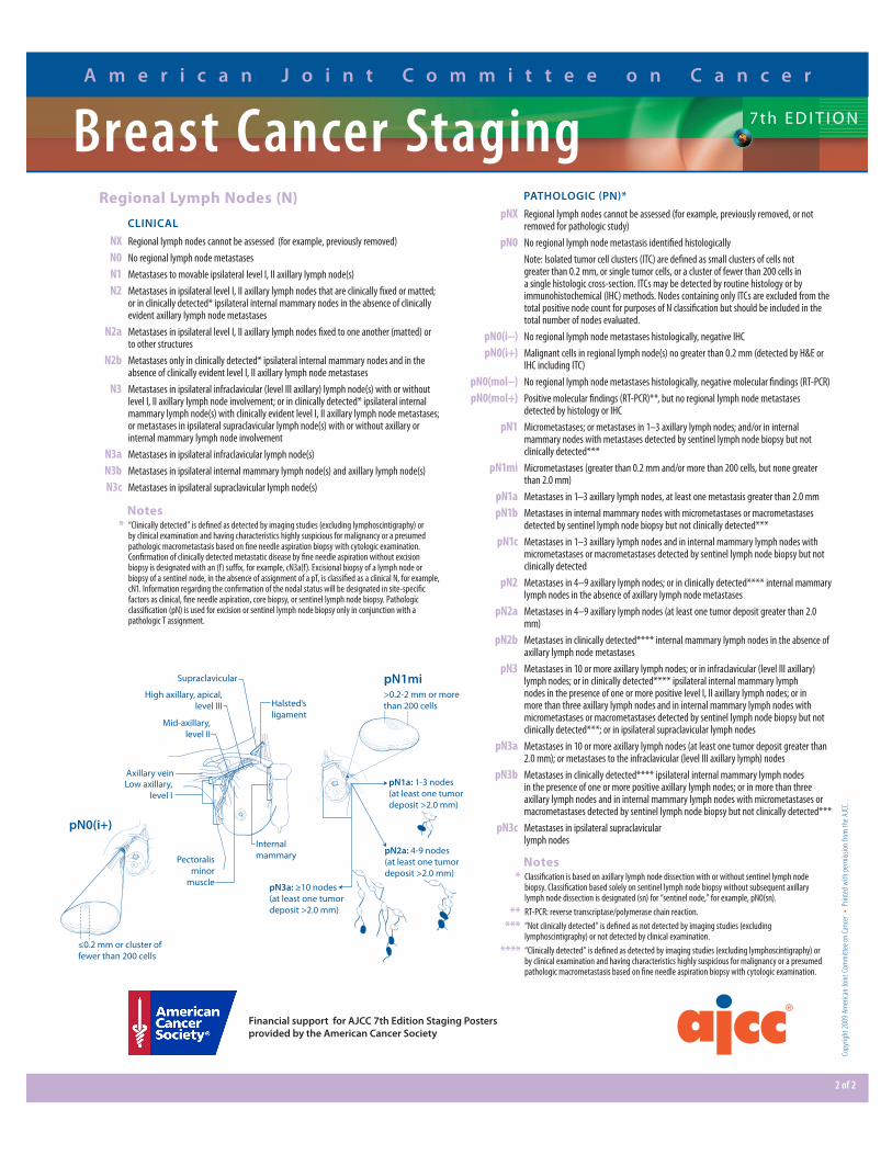
Regional Lymph Nodes (N)CLINICAL
NX Regional lymph nodes cannot be assessed (for example, previously removed) N0 No regional lymph node metastases N1 Metastases to movable ipsilateral level I, II axillary lymph node(s) N2 Metastases in ipsilateral level I, II axillary lymph nodes that are clinically fixed or matted;
or in clinically detected* ipsilateral internal mammary nodes in the absence of clinically evident axillary lymph node metastases
N2a Metastases in ipsilateral level I, II axillary lymph nodes fixed to one another (matted) or to other structures
N2b Metastases only in clinically detected* ipsilateral internal mammary nodes and in the absence of clinically evident level I, II axillary lymph node metastases
N3 Metastases in ipsilateral infraclavicular (level III axillary) lymph node(s) with or without level I, II axillary lymph node involvement; or in clinically detected* ipsilateral internal mammary lymph node(s) with clinically evident level I, II axillary lymph node metastases; or metastases in ipsilateral supraclavicular lymph node(s) with or without axillary or internal mammary lymph node involvement
N3a Metastases in ipsilateral infraclavicular lymph node(s) N3b Metastases in ipsilateral internal mammary lymph node(s) and axillary lymph node(s) N3c Metastases in ipsilateral supraclavicular lymph node(s)
PATHOLOGIC (PN)*
pNX Regional lymph nodes cannot be assessed (for example, previously removed, or not removed for pathologic study)
pN0 No regional lymph node metastasis identified histologically Note: Isolated tumor cell clusters (ITC) are defined as small clusters of cells not
greater than 0.2 mm, or single tumor cells, or a cluster of fewer than 200 cells in a single histologic cross-section. ITCs may be detected by routine histology or by immunohistochemical (IHC) methods. Nodes containing only ITCs are excluded from the total positive node count for purposes of N classification but should be included in the total number of nodes evaluated.
pN0(i−) No regional lymph node metastases histologically, negative IHC pN0(i+) Malignant cells in regional lymph node(s) no greater than 0.2 mm (detected by H&E or
IHC including ITC) pN0(mol−) No regional lymph node metastases histologically, negative molecular findings (RT-PCR) pN0(mol+) Positive molecular findings (RT-PCR)**, but no regional lymph node metastases
detected by histology or IHC pN1 Micrometastases; or metastases in 1–3 axillary lymph nodes; and/or in internal
mammary nodes with metastases detected by sentinel lymph node biopsy but not clinically detected***
pN1mi Micrometastases (greater than 0.2 mm and/or more than 200 cells, but none greater than 2.0 mm)
pN1a Metastases in 1–3 axillary lymph nodes, at least one metastasis greater than 2.0 mm pN1b Metastases in internal mammary nodes with micrometastases or macrometastases
detected by sentinel lymph node biopsy but not clinically detected*** pN1c Metastases in 1–3 axillary lymph nodes and in internal mammary lymph nodes with
micrometastases or macrometastases detected by sentinel lymph node biopsy but not clinically detected
pN2 Metastases in 4–9 axillary lymph nodes; or in clinically detected**** internal mammary lymph nodes in the absence of axillary lymph node metastases
pN2a Metastases in 4–9 axillary lymph nodes (at least one tumor deposit greater than 2.0 mm)
pN2b Metastases in clinically detected**** internal mammary lymph nodes in the absence of axillary lymph node metastases
pN3 Metastases in 10 or more axillary lymph nodes; or in infraclavicular (level III axillary) lymph nodes; or in clinically detected**** ipsilateral internal mammary lymph nodes in the presence of one or more positive level I, II axillary lymph nodes; or in more than three axillary lymph nodes and in internal mammary lymph nodes with micrometastases or macrometastases detected by sentinel lymph node biopsy but not clinically detected***; or in ipsilateral supraclavicular lymph nodes
pN3a Metastases in 10 or more axillary lymph nodes (at least one tumor deposit greater than 2.0 mm); or metastases to the infraclavicular (level III axillary lymph) nodes
pN3b Metastases in clinically detected**** ipsilateral internal mammary lymph nodes in the presence of one or more positive axillary lymph nodes; or in more than three axillary lymph nodes and in internal mammary lymph nodes with micrometastases or macrometastases detected by sentinel lymph node biopsy but not clinically detected***
pN3c Metastases in ipsilateral supraclavicular lymph nodes
Notes * Classification is based on axillary lymph node dissection with or without sentinel lymph node
biopsy. Classification based solely on sentinel lymph node biopsy without subsequent axillary lymph node dissection is designated (sn) for “sentinel node,” for example, pN0(sn).
** RT-PCR: reverse transcriptase/polymerase chain reaction.
*** “Not clinically detected” is defined as not detected by imaging studies (excluding lymphoscintigraphy) or not detected by clinical examination.
**** “Clinically detected” is defined as detected by imaging studies (excluding lymphoscintigraphy) or by clinical examination and having characteristics highly suspicious for malignancy or a presumed pathologic macrometastasis based on fine needle aspiration biopsy with cytologic examination.
Low axillary,level I
Mid-axillary,level II
High axillary, apical,level III
Supraclavicular
Pectoralisminor
muscle
Internalmammary
Axillary vein
Halsted’sligament
>0.2-2 mm or morethan 200 cells
pN1mi
pN1a: 1-3 nodes(at least one tumordeposit >2.0 mm)
pN2a: 4-9 nodes(at least one tumordeposit >2.0 mm)
pN3a: ≥10 nodes(at least one tumordeposit >2.0 mm)
≤0.2 mm or cluster offewer than 200 cells
pN0(i+)