Embed Size (px)
Citation preview

288 | Healio.com/Pediatrics PEDIATRIC ANNALS 42:7 | JULY 2013
FEATURE
Tularemia is a rare zoonosis caused by Francisella tularensis. Al-though many animals may be
infected with tularemia, human infection most commonly occurs via an insect vec-tor such as a tick or deer fly. In the US, most cases of tularemia occur in the sum-mer in the south-central states, specifically Missouri, Arkansas, and Oklahoma. There are six major tularemia clinical syndromes each with different clinical presentations: ulceroglandular tularemia (42%-75% of all tularemia cases), glandular tularemia (15%-44% of all tularemia cases), oro-pharyngeal tularemia, oculoglandular tularemia, typhodial tularemia, and pneu-monic tularemia. The diagnosis of tula-remia is typically made clinically, taking into account exposure history and clinical manifestations and confirmed by serologic testing. Aminoglycosides are the drugs of choice for the treatment of tularemia. Tu-laremia prevention is best accomplished by keeping away from dead or infected ani-mals and avoiding ticks.
ETIOLOGY AND EPIDEMIOLOGYTularemia is a zoonotic infection
caused by F. tularensis, a small, fastidi-ous, aerobic gram-negative coccobacillus. There are four distinct subspecies of F. tu-larensis; however, disease is mainly caused by F. tularensis subspecies tularensis (type A) and F. tularensis subspecies holarctica (type B).1 Type A is more virulent and is primarily found in North America.1 Type B is found throughout the Northern Hemi-sphere, mainly in Europe and Asia, and causes milder infection than type A.1 F. tu-larensis is highly contagious; only a small inoculum is needed to produce disease.1
More than 100 species of mammals have been noted to be infected with tulare-mia. This includes rabbits, hares, muskrats, prairie dogs, skunks, raccoons, rats, voles, squirrels, sheep, cattle, and cats.1 Disease transmission can occur via handling the carcass of an infected animal, via the bite of a infected animal, or via ingestion of meat from a diseased animal.
The bite of an insect vector such as a tick, deer fly, or flea can also transmit tula-remia to humans. Insects become infected when they feed on an infected animal; ticks can also become infected by transovarian passage.2 In the United States, ticks are the most common and important insect vector of tularemia.2 Tick species that transmit tularemia to humans include Amblyomma americanum (lonestar tick), Dermacentor andersoni (wood tick), and Dermacentor variabilis (dog tick).
Tularemia can also be caused by con-tact with aerosolized bacteria from mow-ing lawns, working on farms, or working
in laboratories where F. tularensis is pres-ent. The disease can also be transmitted by drinking water contaminated with F. tula-rensis; this organism can survive in water and animal carcasses for long periods. Frozen rabbit meat has remained infective for greater than 3 years.3 Person-to-person transmission of tularemia does not occur.
In the US, 90 to 154 cases of tularemia have been reported yearly to the Centers for Disease Control and Prevention (CDC) from 2001 to 2010.4 Tularemia has been reported by every state except Hawaii.4 Ar-kansas, Oklahoma, and Missouri account for approximately 50% of the cases of tula-remia reported in the US each year.5 Figure 1 (see page 289) is a CDC map detailing the locations of reported cases of tularemia from 2001 to 2010.4
Tularemia: Epidemiology, Diagnosis, and TreatmentNada S. Harik, MD
Nada S. Harik, MD, is Assistant Professor, De-
partment of Pediatrics, Division of Pediatric
Infectious Diseases, University of Arkansas for
Medical Sciences.
Address correspondence to: Nada S. Harik,
MD, Department of Pediatrics, Division of Pediat-
ric Infectious Diseases, University of Arkansas for
Medical Sciences, 1 Children’s Way, Slot 512-11,
Little Rock, AR 72202-36591; email: hariknada@
uams.edu.
Disclosure: The author has no relevant finan-
cial relationships to disclose.
doi: 10.3928/00904481-20130619-13
© S
hutte
rsto
ck

PEDIATRIC ANNALS 42:7 | JULY 2013 Healio.com/Pediatrics | 289
FEATURE
Tularemia presents most commonly in the summer, due to high tick activity, in the south-central US and peaks in the winter, the primary hunting season, in the northeastern US.1 Individuals at risk for developing infection include hunters, trap-pers, taxidermists, grounds maintenance workers, sheep herders/shearers, labora-tory workers, those with tick exposure, and those living in or traveling to areas where tularemia is endemic. The highest inci-dence of tularemia occurs in children (and in adults older than age 75 years); boys have a higher incidence of infection than girls.5 The higher incidence in boys is most likely due to their greater participation in activities such as hunting that increase exposure to tularemia. Figure 2 (see page 290) shows the age and gender of reported tularemia cases from 2001 to 2010.4
CLINICAL SYNDROMES AND MANIFESTATIONS
The incubation period of tularemia is 1 to 21 days, with an average of 2 to 5 days. There are six major tularemia clinical syn-dromes, which are classified by the portal of entry of the infection (see Table 1).
Ulceroglandular TularemiaThe most common syndrome, account-
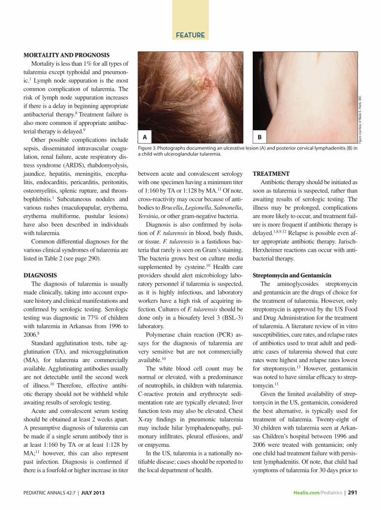
ing for between 42% and 75% of all cases of tularemia, is ulceroglandular tulare-mia.6-8 This syndrome is characterized by a painful swollen papule at the portal of en-try of the infection (skin) that becomes an ulcer. Tender lymphadenopathy is present proximal to the papule/ulcer (see Figure 3, page 291). Fever and malaise are common-ly seen with ulceroglandular tularemia.
Glandular TularemiaGlandular tularemia, representing 15%
to 44% of all cases of tularemia,6-8 presents with tender lymphadenopathy. Involved lymph nodes are most commonly axillary, inguinal, or cervical. The portal of entry with glandular tularemia is unknown but most likely is through the skin. Common additional symptoms include fever and
malaise. The most common sites of lymph node involvement in a recent review of pediatric tularemia in Arkansas were head and neck (33%), followed by inguinal ad-enitis (30%).8
In 50% of untreated cases of ulcero-glandular or glandular tularemia, lymph nodes suppurate and drain.1 Lymph node suppuration can occur even in the setting of appropriate antibacterial therapy. Glandu-lar tularemia is more common in children than adults; 44% of children compared with 16% of adults had primary glandular tularemia in a Missouri study of tularemia.7
Likewise, in a review of 30 cases of pediat-ric tularemia from 1996 through 2006 from Arkansas, the majority of children were
younger than 6 years and had ulceroglan-dular or glandular disease.8
Oropharyngeal TularemiaTraditionally representing less than
5% of cases of tularemia, infection with oropharyngeal tularemia is established through the oropharyngeal mucosa, most commonly by eating undercooked meat from an infected animal.6,7 The hallmarks of oropharyngeal tularemia are severe pharyngitis (out of proportion to pharyn-geal appearance), cervical lymphadenitis, and fever. Oral ulcers and/or an oropha-ryngeal pseudomembrane may be pres-ent. Cervical lymph nodes may suppurate and drain.
Figure 1. Reported cases of tularemia, United States, 2001 to 2010. One dot placed randomly within county of residence for each confirmed case. (From US Centers for Disease Control and Prevention4)
TABLE 1.
Common Characteristics of the Six Tularemia Clinical Syndromes
Tularemia Syndrome Characteristics Portal of Entry
Ulceroglandular Skin papule followed by ulcer, tender lymph-
adenopathy, fever
Skin
Glandular Tender lymphadenopathy, fever Unknown (likely skin)
Oropharyngeal Severe pharyngitis, cervical lymphadenitis, fever Oropharyngeal mucosa
Oculoglandular Conjunctivitis, Parinaud’s oculoglandular
syndrome
Conjunctiva
Typhodial Fever of unknown cause, sepsis, myalgia, headache Oropharyngeal mucosa or respiratory tract
Pneumonic Pneumonia, fever Respiratory tract

290 | Healio.com/Pediatrics PEDIATRIC ANNALS 42:7 | JULY 2013
FEATURE
Oculoglandular TularemiaOculoglandular tularemia was noted
to cause 4% of all cases of tularemia in Missouri from 2000 to 2007.7 Nodular conjunctivitis, conjunctival inflammation, and edema are typically seen and corneal ulcers may occur. Regional lymphadeni-tis is also seen. Oculoglandular tularemia can manifest as Parinaud’s oculoglandular syndrome (conjunctivitis and painful ip-silateral preauricular lymphadenopathy). The conjunctiva is the portal of entry for oculoglandular tularemia and infection is usually caused by direct inoculation from infected fingers.
Typhodial TularemiaThe portal of entry for typhodial tula-
remia is either the oropharyngeal mucosa by ingestion (more common in children) or the respiratory tract by inhalation (more common in adults).1 Typhoidal tularemia is a serious illness that often presents with septic shock. Fever is present without lo-calizing signs. Headaches, myalgias, pha-ryngeal pain, and diarrhea are common symptoms. Hepatomegaly and splenomeg-aly are usually seen. Given the nonspecific
symptoms, a history of tularemia exposure is often needed before this diagnosis is considered.
Pneumonic TularemiaPneumonic tularemia is uncommon in
children. In a Missouri study of 107 cases of tularemia, 4% of children compared with 39% of adults had primary pneumon-
ic tularemia.7 Type A Francisella pneu-monic tularemia has a high mortality rate and is the most severe form of tularemia. Symptoms include fever, cough, and chest pain. Pulmonary infiltrates, hilar adenopa-thy, and/or pleural effusions may be pres-ent. Via inhalation of aerosolized bacteria, the respiratory tract is the portal of entry for pneumonic tularemia.
Figure 2. Reported tularemia cases in the United States by age and gender, 2001 to 2010. (From US Centers for Disease Control and Prevention4)
80
70
60
50
40
30
20
10
00 5 10 15 20 25 30 35 40 45 50 55 60 65 70 75 80 85 90
Male Female
5 year age group
Case
s
TABLE 2.
Differential Diagnoses for the Various Clinical Syndromes of Tularemia
Ulceroglandular and Glandular Tularemia
Oropharyngeal Tularemia Oculoglandular Tularemia
Typhodial Tularemia Pneumonic Tularemia
Staphylococcus aureus
lymphadenitis
Streptococcal pharyngitis Bartonella Bacterial sepsis Typical and atypical bacterial pneumonia
Staphylococcus pyogenes
lymphadenitis
Diphtheria Sporotrichosis Malaria Tuberculosis
Tuberculosis Viral pharyngitis Tuberculosis Brucellosis Legionnaire’s disease
Non-tuberculous mycobacterium Syphilis Q fever Q fever
Bartonella Coccidioidomycosis Rickettsial diseases Fungal pneumonia
Anthrax HSV Ehrlichiosis Viral pneumonia
HIV Typhoid fever Psittacosis
Infectious mononucleosis Disseminated tuberculosis
Plague
Sporotrichosis Disseminated histoplasmosis
Lymphoma
Lymphogranuloma venereum
Lymphoma
PEDIATRIC ANNALS 42:7 | JULY 2013 Healio.com/Pediatrics | 291
FEATURE
MORTALITY AND PROGNOSISMortality is less than 1% for all types of
tularemia except typhoidal and pneumon-ic.1 Lymph node suppuration is the most common complication of tularemia. The risk of lymph node suppuration increases if there is a delay in beginning appropriate antibacterial therapy.8 Treatment failure is also more common if appropriate antibac-terial therapy is delayed.9
Other possible complications include sepsis, disseminated intravascular coagu-lation, renal failure, acute respiratory dis-tress syndrome (ARDS), rhabdomyolysis, jaundice, hepatitis, meningitis, encepha-litis, endocarditis, pericarditis, peritonitis, osteomyelitis, splenic rupture, and throm-bophlebitis.1 Subcutaneous nodules and various rashes (maculopapular, erythema, erythema multiforme, pustular lesions) have also been described in individuals with tularemia.
Common differential diagnoses for the various clinical syndromes of tularemia are listed in Table 2 (see page 290).
DIAGNOSISThe diagnosis of tularemia is usually
made clinically, taking into account expo-sure history and clinical manifestations and confirmed by serologic testing. Serologic testing was diagnostic in 77% of children with tularemia in Arkansas from 1996 to 2006.8
Standard agglutination tests, tube ag-glutination (TA), and microagglutination (MA), for tularemia are commercially available. Agglutinating antibodies usually are not detectable until the second week of illness.10 Therefore, effective antibi-otic therapy should not be withheld while awaiting results of serologic testing.
Acute and convalescent serum testing should be obtained at least 2 weeks apart. A presumptive diagnosis of tularemia can be made if a single serum antibody titer is at least 1:160 by TA or at least 1:128 by MA;11 however, this can also represent past infection. Diagnosis is confirmed if there is a fourfold or higher increase in titer
between acute and convalescent serology with one specimen having a minimum titer of 1:160 by TA or 1:128 by MA.11 Of note, cross-reactivity may occur because of anti-bodies to Brucella, Legionella, Salmonella, Yersinia, or other gram-negative bacteria.
Diagnosis is also confirmed by isola-tion of F. tularensis in blood, body fluids, or tissue. F. tularensis is a fastidious bac-teria that rarely is seen on Gram’s staining. The bacteria grows best on culture media supplemented by cysteine.10 Health care providers should alert microbiology labo-ratory personnel if tularemia is suspected, as it is highly infectious, and laboratory workers have a high risk of acquiring in-fection. Cultures of F. tularensis should be done only in a biosafety level 3 (BSL-3) laboratory.
Polymerase chain reaction (PCR) as-says for the diagnosis of tularemia are very sensitive but are not commercially available.10
The white blood cell count may be normal or elevated, with a predominance of neutrophils, in children with tularemia. C-reactive protein and erythrocyte sedi-mentation rate are typically elevated; liver function tests may also be elevated. Chest X-ray findings in pneumonic tularemia may include hilar lymphadenopathy, pul-monary infiltrates, pleural effusions, and/or empyema.
In the US, tularemia is a nationally no-tifiable disease; cases should be reported to the local department of health.
TREATMENTAntibiotic therapy should be initiated as
soon as tularemia is suspected, rather than awaiting results of serologic testing. The illness may be prolonged, complications are more likely to occur, and treatment fail-ure is more frequent if antibiotic therapy is delayed.1,8,9,12 Relapse is possible even af-ter appropriate antibiotic therapy. Jarisch-Herxheimer reactions can occur with anti-bacterial therapy.
Streptomycin and GentamicinThe aminoglycosides streptomycin
and gentamicin are the drugs of choice for the treatment of tularemia. However, only streptomycin is approved by the US Food and Drug Administration for the treatment of tularemia. A literature review of in vitro susceptibilities, cure rates, and relapse rates of antibiotics used to treat adult and pedi-atric cases of tularemia showed that cure rates were highest and relapse rates lowest for streptomycin.13 However, gentamicin was noted to have similar efficacy to strep-tomycin.13
Given the limited availability of strep-tomycin in the US, gentamicin, considered the best alternative, is typically used for treatment of tularemia. Twenty-eight of 30 children with tularemia seen at Arkan-sas Children’s hospital between 1996 and 2006 were treated with gentamicin; only one child had treatment failure with persis-tent lymphadenitis. Of note, that child had symptoms of tularemia for 30 days prior to
Figure 3. Photographs documenting an ulcerative lesion (A) and posterior cervical lymphadenitis (B) in a child with ulceroglandular tularemia.
Figu
re c
ourte
sy o
f Nad
a S.
Har
ik, M
D.
A B

292 | Healio.com/Pediatrics PEDIATRIC ANNALS 42:7 | JULY 2013
FEATURE
initiation of therapy with gentamicin, mak-ing treatment failure more likely.
The recommended pediatric dose of gentamicin for treatment of tularemia is 5 mg/kg divided every 8 or 12 hours and the typical treatment course with aminogly-cosides is 10 days. However, extension of therapy may be indicated for severe disease or for those children with prolonged symp-toms prior to diagnosis. Aminoglycoside levels should be monitored closely during therapy due to potential ototoxicity and nephrotoxicity. Once-daily gentamicin has been reported to be successful for the treat-ment of adults with glandular tularemia,14
but no data are available on the efficacy of once-daily gentamicin for the treatment of tularemia in children.
Alternative Antibiotic TherapiesAlternative antibiotic therapies for tula-
remia include doxycycline and ciprofloxa-cin. Relapse is more common in patients treated with tetracyclines (12%) than gen-tamicin (6%).13 Therefore, doxycycline is not recommended as a first-line therapy for tularemia. In addition, a longer treatment course (14 days) is recommended due to the increased relapse rate.11
Unless the benefits outweigh the risks, doxycycline should not be given to chil-dren younger than 8 years for the treatment of tularemia because of the potential for teeth staining.
Ciprofloxacin has been shown to have efficacy in the treatment of tularemia. For example, a Swedish study documented the successful treatment of 12 children with ulceroglandular tularemia with oral ciprofloxacin.15 However, most studies on fluoroquinolone efficacy have been done in Europe where F. tularensis subspecies hol-arctica (type B) predominates. F. tularensis holarctica causes far less severe disease than F. tularensis tularensis, the major caus-ative agent of tularemia in North America.16
Ciprofloxacin is not recommended for the treatment of tularemia in children younger than 18 years because of the po-tential for joint and/or cartilage injury.
Beta-lactams, clindamycin, and trime-thoprim-sulfamethoxazole are not effective for the treatment of tularemia.
PREVENTION Tularemia prevention is best accom-
plished by steering clear of infected ani-mals and insect vectors. Common sense strategies include avoiding dead or sick animals and areas that are tick-infested.
For hunters or abbatoir workers, ani-mals should not be skinned with bare hands; gloves and eye protection are indi-cated when removing animal skin. All wild game should be cooked thoroughly before eating. Patients should be counseled to not drink untreated water. When mowing the lawn, care should be taken to avoid mow-ing over any sick or dead animals.
When engaging in outdoor activities, to prevent bites from tick and deer flies, pro-tective clothing such as long pants tucked into long socks and long sleeves should be worn. Insect repellents that contain DEET (diethyltoluamide) provide protec-tion against ticks but need to be reapplied frequently. Formulations that contain 10% to 30% DEET can be used in children 2 months and older.11 Children should be checked for ticks frequently, especially in the warmer months, in tularemia-endemic areas. Ticks should be removed as soon as possible using tweezers, not fingers, by grabbing the tick as close to the skin sur-face as possible, then pulling straight up. Hands should be washed immediately after removing a tick.
As there is no evidence for person-to-person transmission of tularemia, isolation of infected individuals is not indicated.
Although tularemia is a rare zoonosis, pediatricians need to be aware of this in-fection as the diagnosis is typically made clinically based on the appropriate expo-sure history and the classic clinical mani-festations of the various tularemia syn-dromes. Pediatricians in the south-central US are ever vigilant for this infection. Pe-diatricians across the US should be on the lookout for tularemia in children who have
traveled to endemic areas, especially dur-ing summer months.
REFERENCES 1. Feigin RD, Nag PK. Tularemia. In: Feigin RD,
Cherry JD, Demmler-Harrison GJ, Kaplan SL (eds). Textbook of Pediatric Infectious Dis-eases. 6th ed. Philadelphia, PA: WB Saunders; 2009:1725-1734.
2. Hopla CE. The ecology of tularemia. Adv Vet Sci Comp Med. 1974;18:25-53.
3. Chin JE (ed). Control of Communicable Diseas-es Manual. 17th ed. Washington DC: American Public Health Association; 2000.
4. Centers for Disease Control and Prevention. Re-ported tularemia cases by state, United States, 2001 – 2010 and reported tularemia cases by age and sex, United States, 2001 – 2010. Available at: www.cdc.gov/tularemia/statistics/. Accessed June 10, 2013.
5. Centers for Disease Control and Prevention. Tu-laremia—United States, 1990-2000. Morb Mor-tal Wkly Rep. 2002;51:181-184.
6. Levy PD, Chiang WK. Update on emerging infections: news from the Centers for Dis-ease Control and Prevention. Ann Emerg Med. 2002;40:356-360.
7. Centers for Disease Control and Prevention. Tularemia-Missouri, 2000-2007. Morb Mortal Wkly Rep. 2009;58:744-748.
8. Snowden J, Stovall S. Tularemia: retrospective review of 10 years’ experience in Arkansas. Clin Pediatr. 2011;50:64-68.
9. Kaya A, Deveci K, Uysal IO, et al. Tularemia in children: evaluation of clinical, laboratory and therapeutic features of 27 tularemia cases. Turk J Pediatr. 2012;54:105-112.
10. Tärnvik A, Chu MC. New approaches to diagno-sis and therapy of tularemia. Ann NY Acad Sci. 2007;1105:378-404.
11. Committee on Infectious Diseases. Tularemia. In: Pickering LK, Baker CJ, Kimberlin DW, Long SS (eds). Red Book: 2012 Report of the Committee on Infectious Disease. 29th ed. Elk Grove Village, IL: American Academy of Pedi-atrics; 2012:768-769.
12. Penn RL, Kinasewitz GT. Factors associated with a poor outcome in tularemia. Arch Intern Med. 1987;147:265-268.
13. Enderlin G, Morales L, Jacobs RF, et al. Strep-tomycin and alternative agents for the treatment of tularemia: review of the literature. Clin Infect Dis. 1994;19:42-47.
14. Hassoun A, Spera R, Dunkel J. Tularemia and once-daily gentamicin. Antimicrob. Agents Che-mother. 2006;50:824.
15. Johansson A, Berglund L, Gothefors L, et al. Ciprofloxacin for treatment of tularemia in chil-dren. Pediatr Infect Dis J. 2000;19:449-453.
16. Pechous RD, McCarthy TR, Zahrt TC. Working toward the future: insights into Francisella tula-rensis pathogenesis and vaccine development. Microbiol Mol Biol Rev. 2009;73:684-711.