Embed Size (px)
Citation preview

Vol.:(0123456789)1 3
Knee Surgery, Sports Traumatology, Arthroscopy https://doi.org/10.1007/s00167-018-5251-5
HIP
Treatment strategies for ischiofemoral impingement: a systematic review
Naoki Nakano1 · Haitham Shoman1 · Vikas Khanduja1
Received: 23 February 2018 / Accepted: 22 October 2018 © The Author(s) 2018
AbstractPurpose There has been relatively little information about the treatment for ischiofemoral impingement (IFI) because of its rarity as well as the uncertainty of diagnosis. The aim of this study was to provide the reader with the available treatment strategies and their related outcomes for IFI based on the best available evidence, whilst highlighting classically accepted ways of treatment as well as relatively new surgical and non-surgical techniques.Methods A systematic review of the literature from Medline, Embase, AMED, Cochrane and Google Scholar was undertaken since inception to December 2017 following the PRISMA guidelines. Clinical outcome studies, prospective/retrospective case series and case reports that described the treatment outcome for IFI were included. Animal or cadaveric studies, trial protocols, diagnostic studies without any description of treatments, technical notes without any results, and review articles were excluded.Results This systematic review found 17 relevant papers. No comparative studies were included in the final records for quali-tative assessment, which means all the studies were case series and case reports. Eight studies (47.1%) utilised non-surgical treatment including injection and prolotherapy, followed by endoscopic surgery (5 studies, 29.4%) then open surgery (4 studies, 23.5%). Mean age of the participants was 41 years (11–72 years). The mean follow-up was 8.4 months distributed from 2 weeks to 2.3 years. No complications or adverse effects were found from the systematic review.Conclusion Several treatment strategies have been reported for IFI, and most of them have good short- to medium-term outcomes with a low rate of complications. However, there are no comparative studies to assess the superiority of one tech-nique over another, thus further research with randomised controlled trials is required in this arena. This study explores the wide variety and categories of different treatments used for IFI to guide physicians and shed light on what can be done for this challenging cohort of patients.Level of evidence III.
Keywords Ischiofemoral impingement · Quadratus femoris · Endoscopy · Extra-articular impingement · Systematic review · Hip
Introduction
Ischiofemoral impingement (IFI) is an uncommon cause of pain and snapping in the hip, buttock, and groin. The pathol-ogy occurs because of a reduction of space between the lesser trochanter (LT) and the lateral border of the ischium,
which leads to entrapment of the quadratus femoris (QF) muscle [37]. IFI was first described by Johnson in 1977 in three patients who had undergone an osteotomy of the hip or a hip replacement previously [18]. However, despite it being described almost 40 years ago, it is still frequently misdi-agnosed or neglected because of its rarity, and the absence of specific clinical findings and diagnostic tests [14, 26]. Recently, several studies on the radiological features of IFI and distance between the ischium and the LT, i.e. ischiofem-oral distance‚ have been published [14, 20, 31]. We have also recently reported on the normal ischiofemoral distance (measured as the smallest distance between the lateral cortex of ischial tuberosity and the medial cortex of the LT) and its
* Vikas Khanduja [email protected]
1 Department of Trauma and Orthopaedic Surgery - Young Adult Hip Service, Addenbrooke’s-Cambridge University Hospitals NHS Foundation Trust, Hills Road, Box 37, Cambridge CB2 0QQ, UK
Knee Surgery, Sports Traumatology, Arthroscopy
1 3
variations using the computed tomography (CT) data of 298 normal hips and found that the mean ischiofemoral distance was 18.6 mm in females and 23 mm in males and that it increased by 1.06 mm for each 1 mm of offset and dropped by 0.09 mm with each year of age [14]. In addition, it was reported that narrowed ischiofemoral distance was associ-ated with abnormal magnetic resonance imaging (MRI) sig-nal intensity in the QF muscle [20, 31]. Furthermore, some studies report that the QF muscle signal changes on MRI or symptoms of IFI could be observed in patients without ischiofemoral distance narrowing (e.g. due to tumour [30] or exostosis [35, 38]), keeping the pathogenesis of IFI uncer-tain [20, 28].
There is relatively little information available on the best management strategy for patients with IFI. This is mainly because of the uncertainty of diagnosis and the fact that conservative treatment such as physiotherapy or activity modification is undertaken as a first step in the manage-ment of most cases with IFI [2, 26]. Surgical treatment is reserved for patients in whom conservative treatment fails. Until recently, excision of the LT with an open approach had been recommended as a normal operative technique for IFI with a narrowed ischiofemoral distance [2], however, with the improvement in arthroscopic techniques and devices, some authors report on the entire LT being accessed and resected endoscopically [10, 19, 40].
Currently, there is a lack of evidence in the literature that provides hip surgeons with evidence-based recommenda-tions on the management of IFI, and no systematic review has been published in this arena thus far. The aim of this study, therefore, was to provide the reader with the available treatment strategies and their related outcomes for IFI based on the best available evidence, whilst highlighting classically accepted ways of treatment as well as relatively new surgical and non-surgical techniques. The objective of this systematic review would be to look at patients from both genders with no demographic restriction, who had any treatment for IFI to treat and alleviate buttock and posterior hip pain with or without distal neuropathic pain radiation and by including the studies reporting on IFI treatment and this would provide the current treatment strategies in practice.
Materials and methods
Search strategy
The PICOS tool was adopted to formulate the research question and modified since no comparators were sought in this study [24]. The study included randomised trials, cohort studies, case controls, and case studies as the study designs of interest. The protocol of this systematic review was developed and has been registered in the International
Prospective Register of Systematic Reviews (PROSPERO 2017 CRD42017084855) [17].
Two reviewers searched the online databases (Medline, Embase, AMED, Cochrane, and Google Scholar) for the literature describing the outcomes of treatments for IFI. The Preferred Reporting Items for Systematic Reviews and Meta-Analyses (PRISMA) guidelines were used for design-ing this study [25]. Database search was conducted on 31st, December 2017 and retrieved articles from the databases since inception to the search date. The electronic search cita-tion algorithm used was: [ischiofemoral (Title/Abstract) OR ischiofemoral (Title/Abstract)]. The search also included the yet to be printed search results and grey literature. Results were pooled and exported to Mendeley reference manager software (Elsevier, Amsterdam, The Netherlands) and dupli-cates were removed electronically and manually. The two reviewers independently reviewed all the titles and abstracts. The remaining search results were divided equally between the two reviewers and reviewed in duplicate applying the inclusion and exclusion criteria. Any discrepancies at the title and abstract stage as well as the full-text stage were resolved by consensus between the two reviewers and the third more senior author. This process led to 100% agree-ment between the authors.
Study selection (inclusion and exclusion criteria)
Levels 1, 2, 3, 4, and 5 evidence (according to the Oxford Centre for Evidence-Based Medicine [29]) English language studies were eligible for inclusion in the systematic review. We excluded duplicate subject publications within separate unique studies. Both electronically published articles and print journals were included for this review. Clinical out-come studies, prospective case series, retrospective case series and case reports that described the outcomes of treat-ments for IFI were included. Procedures regardless of open surgery, endoscopic surgery or non-surgical treatment were included. Studies on animal models and basic science stud-ies (e.g. cadaveric studies) were excluded. Studies describing trial protocols without any results, diagnostic studies without any description of treatments, technical notes without any results, and review articles were also excluded. The detailed inclusion and exclusion criteria are shown in Table 1.
Data extraction and analysis
Both the reviewers independently extracted the relevant study data from the final pool of included articles and recorded this data on a spreadsheet designed a priori in Microsoft Excel 2013 (Microsoft Corporation, Redmond, Washington, USA). Participant-specific demographics extracted from each study included the number of cases, gender distribution, mean age with range (years), mean

Knee Surgery, Sports Traumatology, Arthroscopy
1 3
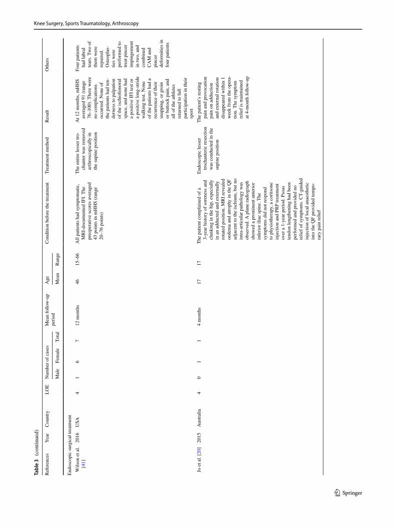
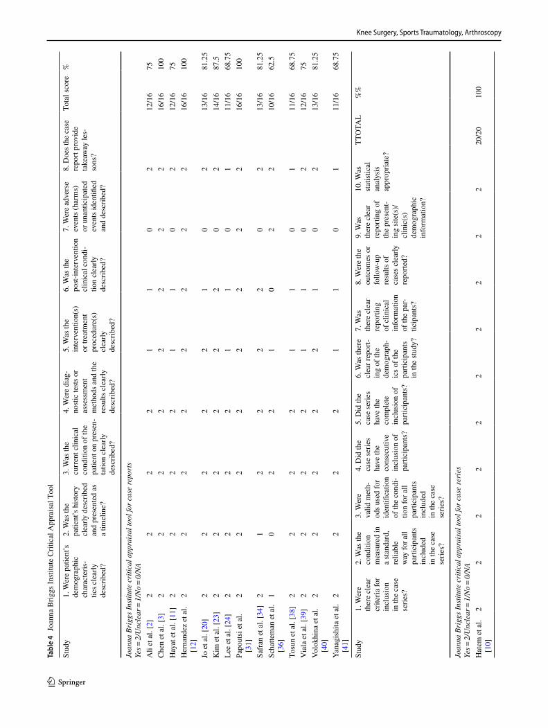
length of follow-up, physical, clinical or radiological con-dition before the treatment, treatment strategy used in the study, final outcome, and specific comments (if any). Study-specific demographics extracted from each study included the level of evidence according to the simplified evidence level table from the Centre for Evidence-Based Medicine, Oxford, country where the study was conducted and the year of publication. Studies were then analysed and assessed using the Joanna Briggs Institute Critical Appraisal Check-list (JBICAC) for case reports and case series [17]. A scor-ing system was implemented based on the findings from the studies. JBICAC scores the answers to its questions as yes, no, unclear or not applicable. We then allocated numbers to each answer where domains answering with yes gets 2 points, unclear gets 1 and no gets 0. A scoring of 16 and 20 indicated the maximum points of case report and case series, respectively.
Statistical analyses
The extracted data were then analysed using Microsoft Excel 2013. Statistical analyses in this study focused on descriptive statistics by calculating the mean values for ages and follow-up times providing an overview summary statistic of ages and follow-up times.
Results
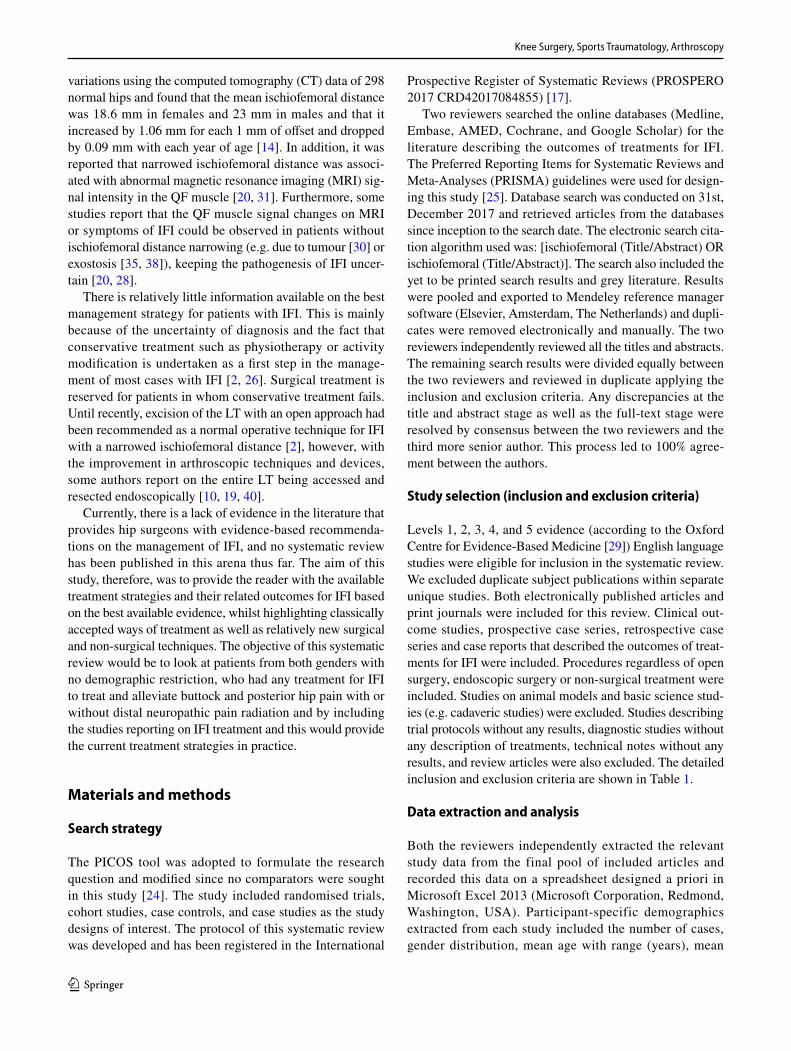
The initial search found a total of 381 studies from all the databases. The search process led to 100% agreement among three authors. Duplicates found were 165 articles and were removed. A total of 216 articles were then identi-fied for title screening. One hundred and thirty-four arti-cles were excluded based on the inclusion criteria leaving 82 articles for abstract screening. Fifty-seven articles were then excluded and 25 were included for full-text review.
Eventually, 17 studies met all the inclusion criteria and were eligible for critical appraisal, quality assessment and included in the study. Of the participants, 15 (35.7%) were males and 27 (64.3%) were females (data availability: 100%). Mean age of the participants was 41 years (range 11–72 years). The mean follow-up period was 8.4 months distributed from 2 weeks to 2.3 years after the treatment. Study demographics are shown in Table 2. No complica-tion or adverse event was found from the systematic review. The flow chart of the literature search algorithm is shown in Fig. 1. The oldest study included in this review was pub-lished in 2011. All the studies included in this systematic review were level 4 studies, which means there were no com-parative studies found. Due to lack of homogeneity in stud-ies, a meta-analysis was deemed unsuitable for this study.
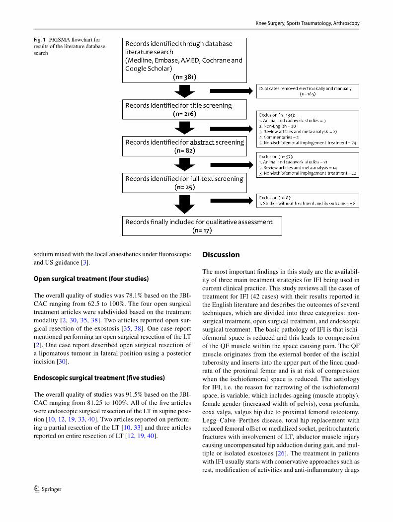
Three major treatment strategies were found from this systematic review. Eight studies (47.1%) utilised non-sur-gical treatment including injection and prolotherapy, fol-lowed by endoscopic surgery (five studies, 29.4%) and then open surgery (four studies, 23.5%). Data extracted from all the studies are shown in Table 3. The outcomes of quality assessment using JBICAC are shown in Tables 4 and 5.
Non‑surgical treatment (eight studies)
The overall quality of the eight articles was 80% based on the JBICAC ranging from 68.75 to 100% [3, 11, 21–23, 37, 39, 41]. Four articles reported using conservative treatment (e.g. activity restriction, intake of non-steroidal anti-inflam-matory drugs (NSAIDs) or physiotherapy) [11, 23, 37, 41]. Three articles reported the outcome following ultrasound (US) guided injection [21, 22, 39]. One article mentioned deploying prolotherapy with polydeoxyribonucleotide
Table 1 Inclusion and exclusion criteria applied to articles identified in the literature
Inclusion criteria1. All levels of evidence2. Written in the English language3. Studies on humans4. Studies reporting the outcome of treatment for ischiofemoral
impingementExclusion criteria1. Studies describing trial protocols without any results2. Animal studies3. Basic science studies (e.g. cadaveric studies)4. Diagnostic studies without any description of treatments5. Technical notes without any results6. Reviews, systematic reviews
Table 2 Demographics of the study
Parameter
Studies analysed 17 studiesLevels of evidence: 4 17 studies (100%)
Case series 3 studies (17.6%) Case report 14 studies (82.4%)
Participants (cases) Male 15 (35.7%) Female 27 (64.3%)
Mean follow-up time (range) 8.4 months (2 weeks–2.3 years)
Mean participant age (range) 41.0 (11–72) yearsApproach of treatment Non-surgical treatment 8 studies (47.1%) Open surgery 4 studies (23.5%)
Endoscopic surgery 5 studies (29.4%)
Knee Surgery, Sports Traumatology, Arthroscopy
1 3
sodium mixed with the local anaesthetics under fluoroscopic and US guidance [3].
Open surgical treatment (four studies)
The overall quality of studies was 78.1% based on the JBI-CAC ranging from 62.5 to 100%. The four open surgical treatment articles were subdivided based on the treatment modality [2, 30, 35, 38]. Two articles reported open sur-gical resection of the exostosis [35, 38]. One case report mentioned performing an open surgical resection of the LT [2]. One case report described open surgical resection of a lipomatous tumour in lateral position using a posterior incision [30].
Endoscopic surgical treatment (five studies)
The overall quality of studies was 91.5% based on the JBI-CAC ranging from 81.25 to 100%. All of the five articles were endoscopic surgical resection of the LT in supine posi-tion [10, 12, 19, 33, 40]. Two articles reported on perform-ing a partial resection of the LT [10, 33] and three articles reported on entire resection of LT [12, 19, 40].
Discussion
The most important findings in this study are the availabil-ity of three main treatment strategies for IFI being used in current clinical practice. This study reviews all the cases of treatment for IFI (42 cases) with their results reported in the English literature and describes the outcomes of several techniques, which are divided into three categories: non-surgical treatment, open surgical treatment, and endoscopic surgical treatment. The basic pathology of IFI is that ischi-ofemoral space is reduced and this leads to compression of the QF muscle within the space causing pain. The QF muscle originates from the external border of the ischial tuberosity and inserts into the upper part of the linea quad-rata of the proximal femur and is at risk of compression when the ischiofemoral space is reduced. The aetiology for IFI, i.e. the reason for narrowing of the ischiofemoral space, is variable, which includes ageing (muscle atrophy), female gender (increased width of pelvis), coxa profunda, coxa valga, valgus hip due to proximal femoral osteotomy, Legg–Calve–Perthes disease, total hip replacement with reduced femoral offset or medialized socket, peritrochanteric fractures with involvement of LT, abductor muscle injury causing uncompensated hip adduction during gait, and mul-tiple or isolated exostoses [26]. The treatment in patients with IFI usually starts with conservative approaches such as rest, modification of activities and anti-inflammatory drugs
Fig. 1 PRISMA flowchart for results of the literature database search

Knee Surgery, Sports Traumatology, Arthroscopy
1 3
Tabl
e 3
Det
ails
of 1
7 stu
dies
incl
uded
in th
e sy
stem
atic
revi
ew
Refe
renc
esYe
arC
ount
ryLO
EN
umbe
r of c
ases
Mea
n fo
llow
-up
perio
dA
geC
ondi
tion
befo
re th
e tre
atm
ent
Trea
tmen
t met
hod
Resu
ltO
ther
s
Mal
eFe
mal
eTo
tal
Mea
nR
ange
Con
serv
ativ
e tre
atm
ent
Kim
et a
l. [2
2]20
16So
uth
Kor
ea4
86
142
wee
ks53
.433
–72
Patie
nts h
ad lo
wer
but
tock
pai
n w
hich
loca
lised
at a
poi
nt
halfw
ay b
etw
een
the
late
ral
prom
inen
ce o
f the
gre
ater
tro
chan
ter a
nd th
e is
chia
l tu
bero
sity
cor
resp
ondi
ng to
th
e lo
catio
n of
the
QF
belly
. M
ean
VAS
befo
re in
ject
ion
was
6.7
(ran
ge 3
–10)
Und
er u
ltras
ound
gu
idan
ce, 8
mL
of
0.25
% li
doca
ine
was
in
ject
ed in
to th
e Q
F. T
he n
umbe
r of
QF
inje
ctio
n w
as:
1, fi
ve p
atie
nts;
2,
seve
n pa
tient
s; 3
, on
e pa
tient
; 4, o
ne
patie
nt (T
he a
vera
ge
freq
uenc
y of
inje
c-tio
n w
as 2
.5 ti
mes
)
Two
wee
ks a
fter t
he
last
inje
ctio
n, 1
0 of
14
pat
ient
s exp
ress
ed
thei
r sat
isfa
ctio
n as
ex
celle
nt o
r goo
d (3
pa
tient
s exp
ress
ed it
as
fair
and
1 pa
tient
ex
pres
sed
it as
poo
r).
Ther
e w
ere
no c
om-
plic
atio
ns o
bser
ved.
A
t the
fina
l fol
low
-up
, mea
n VA
S w
as
3.4
(ran
ge, 0
–8)
Che
n et
al.
[3]
2017
USA
40
11
5 m
onth
s34
3411
-mon
th h
istor
y of
left
poste
-rio
r glu
teal
pai
n. It
initi
ally
pr
esen
ted
as a
dul
l ach
e in
th
e bu
ttock
and
ham
strin
g ar
ea, e
xace
rbat
ed a
fter 3
0 m
in
of ru
nnin
g or
cyc
ling,
rate
d at
NPR
of 5
/10
but c
hang
ed
to 7
/10
with
act
ivity
. The
pa
tient
den
ied
the
histo
ry o
f pr
ior t
raum
a. P
rior t
reat
-m
ents
incl
ude
phys
ical
th
erap
y, c
hiro
prac
tic, a
s wel
l as
fluo
rosc
opic
ally
gui
ded
corti
coste
roid
inje
ctio
n of
the
isch
ial b
ursa
, sac
roili
ac jo
int,
intra
-arti
cula
r hip
join
t and
pi
rifor
mis
mus
cle,
all
with
out
impr
ovem
ent
Afte
r loc
al a
naes
thes
ia,
100
units
inco
botu
li-nu
m to
xin
A re
con-
stitu
ted
in 2
.5 m
L of
ster
ile sa
line
was
in
ject
ed th
roug
hout
th
e Q
F
Patie
nt’s
sym
ptom
s had
co
mpl
ete
reso
lved
an
d sh
e ha
d re
turn
ed
to h
er p
rem
orbi
d fu
nctio
nal s
tatu
s
Vol
okjin
a et
al.
[40]
2013
USA
40
11
9 m
onth
s57
57Th
e ch
roni
c rig
ht h
ip p
ain
of
1-ye
ar d
urat
ion.
She
des
crib
ed
the
pain
as c
onst
ant a
nd d
eep,
ra
diat
ing
from
the
late
ral r
ight
gr
oin
regi
on in
to th
e bu
ttock
, w
akin
g he
r up
at n
ight
. Her
pa
in w
as n
ot re
lieve
d w
ith
non-
stero
idal
ant
i-infl
amm
a-to
ry d
rugs
and
was
wor
se w
ith
athl
etic
act
iviti
es, p
artic
ular
ly
hiki
ng. S
he re
porte
d no
snap
-pi
ng sy
mpt
oms
The
patie
nt re
ceiv
ed
thre
e in
ject
ions
into
th
e is
chio
fem
oral
sp
ace
of c
ombi
na-
tions
of 3
ml o
f lid
ocai
ne a
nd 4
0 m
g D
epo-
med
rol
Afte
r the
firs
t inj
ectio
n,
imm
edia
te re
lief o
f sy
mpt
oms l
astin
g fo
r 9 m
onth
s was
ac
hiev
ed. T
he se
cond
U
S-gu
ided
inje
ctio
n re
sulte
d in
3 w
eeks
of
sym
ptom
relie
f. Th
e th
ird in
ject
ion
was
per
form
ed w
ith
com
pute
d to
mog
ra-
phy
(CT)
gui
danc
e,
and
the
patie
nt h
ad
cont
inue
d re
lief f
or 9
m
onth
s

Knee Surgery, Sports Traumatology, Arthroscopy
1 3
Tabl
e 3
(con
tinue
d)
Refe
renc
esYe
arC
ount
ryLO
EN
umbe
r of c
ases
Mea
n fo
llow
-up
perio
dA
geC
ondi
tion
befo
re th
e tre
atm
ent
Trea
tmen
t met
hod
Resu
ltO
ther
s
Mal
eFe
mal
eTo
tal
Mea
nR
ange
Kim
et a
l. [2
3]20
14So
uth
Kor
ea4
20
26.
5 m
onth
s23
.523
–24
Initi
al m
anag
emen
t whi
ch c
on-
siste
d of
NSA
IDs,
tram
adol
an
d ph
ysic
al re
habi
litat
ion
for 1
mon
th d
id n
ot w
ork.
O
ne p
atie
nt h
ad a
surg
ical
hi
story
of i
liotib
ial b
and
rele
ase
2 ye
ars p
revi
ously
and
a
subs
eque
nt il
iops
oas t
endo
n re
leas
e 1
year
pre
viou
sly. P
re-
oper
ativ
e VA
S w
as 9
–10/
10 in
bo
th p
atie
nts
The
patie
nts w
ere
treat
ed b
y pr
olo-
ther
apy
with
pol
y-de
oxyr
ibon
ucle
otid
e so
dium
mix
ed w
ith
loca
l ana
esth
etic
s in
ject
ed in
to Q
F un
der fl
uoro
scop
ic
and
ultra
soun
d gu
id-
ance
Patie
nt (1
) The
pai
n in
tens
ity u
sing
the
VAS
decr
ease
d fro
m
9 to
10/
10 to
1–2
/10,
an
d th
e pa
tient
did
no
t exp
erie
nce
any
pain
for >
6 m
onth
s. Fo
llow
-up
MR
I a
mon
th a
fter t
he
treat
men
t sho
wed
th
at th
e en
hanc
emen
t of
QF
was
dec
reas
ed
com
pare
d w
ith
that
on
MR
I bef
ore
treat
men
t. Pa
tient
(2)
The
pain
inte
nsity
sc
ore
decr
ease
d fro
m a
n in
itial
9–
10/1
0 to
0–1
/10,
an
d th
e pa
tient
did
no
t exp
erie
nce
any
pain
for >
7 m
onth
s. Fo
llow
-up
MR
I a
mon
th a
fter t
reat
-m
ent s
how
ed th
at
the
enha
ncem
ent o
f Q
F w
as d
ecre
ased
co
mpa
red
with
that
on
MR
I bef
ore
treat
-m
ent
Prol
othe
rapy
re
fers
to th
e in
ject
ion
of
an ir
ritan
t int
o a
join
t spa
ce,
ligam
ent,
or
tend
on in
ser-
tion
site
with
th
e m
ain
aim
be
ing
pain
re
lief.
Cur
rent
hy
poth
eses
su
gges
t tha
t th
e pr
esen
ce
of a
loca
l irr
itant
may
at
tract
infla
m-
mat
ory
med
ia-
tors
and
pos
-si
bly
stim
ulat
e th
e re
leas
e of
gro
wth
fa
ctor
or a
ct
as a
vas
cula
r sc
lero
sant
Hay
at e
t al.
[11]
2014
UK
41
01
1 ye
ar16
16Th
e pa
tient
had
an
18-m
onth
hi
story
of a
dul
l, de
ep a
che
in
his l
eft g
roin
, exa
cerb
ated
by
exer
cise
, fol
low
ing
an in
jury
pl
ayin
g fo
otba
ll. A
pla
in
radi
ogra
ph re
veal
ed a
chr
onic
ap
ophy
seal
avul
sion
frac
ture
of
the
isch
ium
with
exc
essi
ve
callu
s for
mat
ion.
CT
scan
and
M
RI r
evea
led
that
the
bony
pr
otub
eran
ce w
as re
spon
sibl
e fo
r sym
ptom
atic
IFI
Non
-ope
rativ
e m
anag
e-m
ent w
as u
nder
take
n w
ith p
aink
iller
s as
need
ed, r
est,
activ
ity
mod
ifica
tion
and
phys
ioth
erap
y ex
er-
cise
regi
me
Ove
r the
12
mon
ths
afte
r tre
atm
ent,
the
patie
nt’s
sym
ptom
s se
ttled
and
he
repo
rted
only
a m
ild,
infr
eque
nt a
che
in
the
groi
n in
the
final
fo
llow
-up.
He
has
resu
med
nor
mal
sp
ortin
g ac
tiviti
es
with
out d
isco
mfo
rt

Knee Surgery, Sports Traumatology, Arthroscopy
1 3
Tabl
e 3
(con
tinue
d)
Refe
renc
esYe
arC
ount
ryLO
EN
umbe
r of c
ases
Mea
n fo
llow
-up
perio
dA
geC
ondi
tion
befo
re th
e tre
atm
ent
Trea
tmen
t met
hod
Resu
ltO
ther
s
Mal
eFe
mal
eTo
tal
Mea
nR
ange
Lee
et a
l. [2
4]20
13So
uth
Kor
ea4
01
16
wee
ks48
48H
ip M
RI r
evea
led
the
incr
ease
d si
gnal
inte
nsity
of Q
F w
ith
conc
urre
nt n
arro
win
g of
the
isch
iofe
mor
al sp
ace.
On
axia
l T2
-wei
ghte
d fa
t-sup
pres
sed
MR
I, th
ere
wer
e di
ffuse
oe
dem
a an
d in
crea
sed
sign
al
inte
nsity
with
in Q
F. In
itial
VA
S w
as 7
–8/1
0
NSA
IDs a
nd g
abap
en-
tin w
ere
pres
crib
ed
for p
ain
relie
f. H
ot
pack
, ultr
asou
nd,
and
inte
rfere
ntia
l cu
rren
t the
rapi
es
wer
e ap
plie
d ar
ound
th
e hi
p ar
ea. T
he
patie
nt re
ceiv
ed a
n ex
erci
se p
rogr
am fo
r str
etch
ing
of th
e hi
p m
uscl
e an
d co
nnec
-tiv
e tis
sues
Afte
r 6 w
eeks
of t
reat
-m
ent,
the
pain
was
de
crea
sed
grad
ually
to
2–3
/10
in V
AS
Yan
agis
hita
et
al.
[41]
2012
Bra
zil
40
11
3 m
onth
s31
31R
adio
grap
hic
exam
inat
ions
de
mon
strat
ed a
val
gus f
emor
al
neck
, isc
hiof
emor
al sp
ace
narr
owin
g, a
nd th
e pr
esen
ce
of c
ysts
in th
e is
chiu
m. M
RI
show
ed in
crea
sed
sign
al in
QF
on T
2-w
eigh
ted
sequ
ence
s
Non
-sur
gica
l tre
atm
ent
incl
udin
g a
taki
ng
NSA
IDs f
or 7
day
s an
d da
ily p
hysi
cal
ther
apy
for s
tretc
hing
an
d str
engt
heni
ng th
e pe
lvic
mus
cles
wer
e co
nduc
ted
Afte
r 3 m
onth
s of
treat
men
t, th
e pa
tient
sh
owed
sign
ifica
nt
func
tiona
l im
prov
e-m
ent a
nd re
sum
ed
Pila
tes a
ctiv
ities
w
ithou
t any
restr
ic-
tion
Tos
un e
t al.
[38]
2012
Turk
ey4
01
1N
A11
11Th
e pa
tient
com
plai
ned
of h
ip
and
groi
n pa
in, w
hich
gra
du-
ally
incr
ease
d du
ring
the
last
2 m
onth
s. M
RI d
emon
strat
ed
narr
owin
g of
the
isch
iofe
mo-
ral s
pace
, whi
ch w
as m
ost
prom
inen
t in
the
trans
vers
e T1
-wei
ghte
d se
quen
ce, a
nd
mod
erat
e oe
dem
a in
QF
on th
e fa
t-sup
pres
sed
T2-w
eigh
ted
sequ
ence
Con
serv
ativ
e m
etho
ds
incl
udin
g re
st, a
ctiv
-ity
restr
ictio
n, a
nd
taki
ng N
SAID
s wer
e co
nduc
ted
The
patie
nt w
as su
c-ce
ssfu
lly tr
eate
d co
nser
vativ
ely
Ope
n su
rgic
al tr
eatm
ent
Pap
outs
i et
al.
[31]
2016
UK
40
11
12 m
onth
s40
40Th
e pa
tient
suffe
red
from
IFI
seco
ndar
y to
an
inte
rmus
cula
r lip
oma
(2.7
× 2.
6 × 0.
5 cm
), w
hich
was
reve
aled
on
MR
I an
d co
nfirm
ed a
t sur
gery
. Sh
e de
scrib
ed th
e pa
in a
s a
cons
tant
ach
e sc
orin
g 9/
10 o
n VA
S w
ith o
ccas
iona
l sha
rp
shoo
ting
pain
s trig
gere
d by
pr
olon
ged
sitti
ng a
nd w
alki
ng
The
entir
e lip
omat
ous
tum
our w
as e
xcis
ed
by o
pen
surg
ery
in
the
late
ral p
ositi
on
usin
g po
sterio
r in
cisi
on
The
patie
nt’s
sym
p-to
ms i
mpr
oved
m
arke
dly
(VA
S:
0.5/
10).
She
was
ab
le to
sit w
ithou
t an
y di
scom
fort
and
ther
e w
as n
o si
gn o
f on
goin
g sc
iatic
ner
ve
irrita
tion
or IF
I. Th
e pa
tient
retu
rned
to
full-
time
wor
k an
d no
long
er re
quire
s an
y an
alge
sia
Hist
olog
y co
nfirm
ed
the
pres
ence
of
a b
enig
n in
term
uscu
-la
rlipo
ma
of
the
quad
ratu
s fe
mor
is
mus
cle

Knee Surgery, Sports Traumatology, Arthroscopy
1 3
Tabl
e 3
(con
tinue
d)
Refe
renc
esYe
arC
ount
ryLO
EN
umbe
r of c
ases
Mea
n fo
llow
-up
perio
dA
geC
ondi
tion
befo
re th
e tre
atm
ent
Trea
tmen
t met
hod
Resu
ltO
ther
s
Mal
eFe
mal
eTo
tal
Mea
nR
ange
Sch
atte
man
et
al.
[36]
2015
Bel
gium
41
01
NA
2222
The
patie
nt su
ffere
d fro
m g
roin
pa
in, a
ggra
vatin
g by
ext
erna
l ro
tatio
n of
the
hip.
Sta
ndar
d ra
diog
raph
s of t
he h
ip re
veal
ed
a la
rge
sess
ile e
xosto
sis a
t the
m
edia
l asp
ect o
f the
less
er
troch
ante
r. O
n M
RI,
a m
arke
d na
rrow
ing
of th
e is
chio
fem
oral
sp
ace
with
acc
ompa
nyin
g oe
dem
a of
QF
was
seen
. Ini
tial
cons
erva
tive
treat
men
t was
not
su
cces
sful
Ope
n re
sect
ion
of
the
exos
tosi
s was
co
nduc
ted
The
imm
edia
te p
ost-
oper
ativ
e re
cove
ry
was
une
vent
ful
Hist
olog
ical
ex
amin
atio
n of
the
rese
c-tio
n sp
ecim
en
confi
rmed
th
e di
agno
sis
of a
ben
ign
carti
lagi
nous
ex
osto
sis
Via
la e
t al.
[39]
2012
Fran
ce4
01
16
mon
ths
3737
The
patie
nt p
rese
nted
with
hi
p pa
in o
f 2-y
ear d
urat
ion.
R
adio
grap
h, C
T, a
nd M
RI
show
ed c
oxa
valg
a an
d sp
lay-
ing
of th
e in
tertr
ocha
nter
ic
regi
on a
nd fe
mor
al n
eck
as
wel
l as e
xosto
ses o
f the
isch
ial
tube
rosi
ty. E
xosto
ses a
nd
fem
oral
met
aphy
seal
wid
enin
g re
sulte
d in
a n
arro
win
g of
the
isch
iofe
mor
al sp
aces
Ope
n su
rgic
al re
sec-
tion
of th
e is
chia
l ex
osto
sis w
as m
ade
thro
ugh
an a
nter
ior
appr
oach
Six
mon
ths p
ost-o
per-
ativ
ely,
hip
pai
n w
as
impr
oved
, app
earin
g on
ly a
fter w
alki
ng
long
dist
ance
s
The
patie
nt h
ad
a pa
st hi
story
of
surg
ical
re
sect
ions
of
exo
stose
s fro
m th
e le
ft kn
ee a
t age
13
, rig
ht k
nee
at a
ge 1
8, a
nd
right
hum
erus
at
28.
At
path
olog
ical
ex
amin
atio
n, a
ty
pica
l ben
ign
exos
tosi
s was
fo
und
Ali
et a
l. [2
]20
11U
K4
01
110
wee
ks17
17Th
e pa
tient
show
ed a
pai
nful
hip
fo
llow
ing
an a
cute
abd
uc-
tion
inju
ry to
the
hip
whi
le
acci
dent
ally
per
form
ing
the
split
s. Se
ven
mon
ths l
ater
, sh
e no
ticed
an
audi
ble
and
palp
able
clu
nk in
her
hip
up
on w
alki
ng. M
RI s
how
ed
sele
ctiv
e na
rrow
ing
of th
e is
chio
fem
oral
spac
e an
d Q
F sp
ace.
CT-
guid
ed st
eroi
d an
d lo
cal a
naes
thet
ic in
ject
ion
arou
nd Q
F pr
ovid
ed re
lief o
f he
r pai
n bu
t not
the
clun
king
, fo
r 24
h
Ope
n su
rgic
al re
sec-
tion
of th
e le
sser
tro
chan
ter w
as
perfo
rmed
The
post-
oper
ativ
e ra
diog
raph
show
ed
adeq
uate
dec
ompr
es-
sion
of t
he is
chi-
ofem
oral
spac
e. A
t 4
wee
ks fo
llow
ing
the
surg
ery,
the
pain
had
di
min
ishe
d to
a m
ild
disc
omfo
rt an
d th
ere
was
no
clun
king
. At
10 w
eeks
follo
win
g su
rger
y, th
e pa
tient
w
as a
sym
ptom
atic
Bef
ore
the
rese
ctio
n of
th
e le
sser
tro
chan
ter,
the
patie
nt
had
iliot
ibia
l ba
nd Z
-pla
sty
whi
ch h
ad
no e
ffect
on
the
patie
nt’s
sy
mpt
oms
Knee Surgery, Sports Traumatology, Arthroscopy
1 3
Tabl
e 3
(con
tinue
d)
Refe
renc
esYe
arC
ount
ryLO
EN
umbe
r of c
ases
Mea
n fo
llow
-up
perio
dA
geC
ondi
tion
befo
re th
e tre
atm
ent
Trea
tmen
t met
hod
Resu
ltO
ther
s
Mal
eFe
mal
eTo
tal
Mea
nR
ange
Endo
scop
ic su
rgic
al tr
eatm
ent
Wils
on e
t al.
[41]
2016
USA
41
67
12 m
onth
s46
15–6
6A
ll pa
tient
s had
sym
ptom
atic
, M
RI-
docu
men
ted
IFI.
The
preo
pera
tive
scor
es av
erag
ed
43 p
oint
s in
mH
HS
(ran
ge
20–7
6 po
ints
)
The
entir
e le
sser
tro-
chan
ter w
as re
mov
ed
arth
rosc
opic
ally
in
the
supi
ne p
ositi
on
At 1
2 m
onth
s, m
HH
S av
erag
ed 9
1 (r
ange
76
–100
). Th
ere
wer
e no
com
plic
atio
ns
occu
rred
. Non
e of
th
e pa
tient
s had
ten-
dern
ess t
o pa
lpat
ion
of th
e is
chio
fem
oral
sp
ace,
and
non
e ha
d a
posi
tive
IFI t
est o
r a
posi
tive
long
-stri
de
wal
king
test.
Non
e of
the
patie
nts h
ad a
re
curr
ence
of t
heir
snap
ping
, or g
roin
or
but
tock
pai
n, a
nd
all o
f the
ath
lete
s re
turn
ed to
full
parti
cipa
tion
in th
eir
spor
t
Four
pat
ient
s ha
d la
bral
te
ars.
Two
of
them
wer
e re
paire
d.
Oste
opla
s-tie
s wer
e pe
rform
ed to
tre
at p
ince
r im
ping
emen
t in
two,
and
co
mbi
ned
CAM
and
pi
ncer
de
form
ities
in
four
pat
ient
s
Jo e
t al.
[20]
2015
Aus
tralia
40
11
4 m
onth
s17
17Th
e pa
tient
com
plai
ned
of a
3-
year
hist
ory
of so
rene
ss a
nd
clun
king
in th
e hi
p, e
spec
ially
in
an
addu
cted
and
ext
erna
lly
rota
ted
posi
tion.
MR
I rev
eale
d oe
dem
a an
d at
roph
y in
the
QF
adja
cent
to th
e is
chiu
m, b
ut n
o in
tra-a
rticu
lar p
atho
logy
was
ob
serv
ed. A
pla
ne ra
diog
raph
sh
owed
a p
rom
inen
t ant
erio
r in
ferio
r ilia
c sp
ine.
The
sy
mpt
oms d
id n
ot re
spon
d to
phy
siot
hera
py, a
cor
tison
e in
ject
ion
and
PRP
treat
men
t ov
er a
1-y
ear p
erio
d. P
soas
te
ndon
leng
then
ing
had
been
pe
rform
ed a
nd p
rovi
ded
no
relie
f of s
ympt
oms.
CT-
guid
ed
inje
ctio
n of
loca
l ana
esth
etic
in
to th
e Q
F pr
ovid
ed te
mpo
-ra
ry p
ain
relie
f
Endo
scop
ic le
sser
tro
chan
teric
rese
ctio
n w
as c
ondu
cted
in th
e su
pine
pos
ition
The
patie
nt’s
resti
ng
pain
and
pro
voca
tion
pain
on
addu
ctio
n an
d ex
tern
al ro
tatio
n di
sapp
eare
d w
ithin
1
wee
k fro
m th
e op
era-
tion.
The
sym
ptom
re
lief i
s mai
ntai
ned
at 4
-mon
th fo
llow
-up

Knee Surgery, Sports Traumatology, Arthroscopy
1 3
Tabl
e 3
(con
tinue
d)
Refe
renc
esYe
arC
ount
ryLO
EN
umbe
r of c
ases
Mea
n fo
llow
-up
perio
dA
geC
ondi
tion
befo
re th
e tre
atm
ent
Trea
tmen
t met
hod
Resu
ltO
ther
s
Mal
eFe
mal
eTo
tal
Mea
nR
ange
Hat
em e
t al.
[10]
2015
USA
42
35
2.3
year
s33
.916
–59
The
mea
n du
ratio
n of
sym
ptom
s un
til su
rger
y w
as 2
9.2
mon
ths
(ran
ge 5
–66
mon
ths)
. The
in
ject
ion
was
per
form
ed to
ru
le o
ut in
tra-a
rticu
lar p
atho
l-og
y as
a c
ause
of p
oste
rior h
ip
pain
. The
isch
iofe
mor
al a
nd
QF
spac
es o
n M
RI w
ere
con-
side
red
for t
he d
iagn
osis
. All
patie
nts h
ad th
e im
ping
emen
t be
twee
n th
e le
sser
troc
hant
er
and
isch
ium
con
firm
ed a
t su
rger
y. T
he m
ean
mH
HS
was
51.
3 (r
ange
34.
1–73
.7)
preo
pera
tivel
y. T
he m
ean
preo
pera
tive
VAS
for p
ain
was
6.
6 (r
ange
6–7
.3)
Patie
nts u
nder
wen
t en
dosc
opic
trea
tmen
t w
ith p
artia
l res
ectio
n of
the
less
er tr
o-ch
ante
r in
the
supi
ne
posi
tion
The
mea
n po
st-op
erat
ive
mH
HS
was
94.
2 (r
ange
78
.1–1
00).
The
mea
n po
st-op
erat
ive
VAS
for p
ain
was
1 (r
ange
0–
4). T
he m
ean
dura
-tio
n to
retu
rn to
the
spor
t afte
r sur
gery
w
as 4
.4 m
onth
s (r
ange
1–7
mon
ths)
. N
o co
mpl
icat
ion
was
ob
serv
ed
Intra
-arti
cula
r ab
norm
aliti
es
wer
e ob
serv
ed
in th
ree
patie
nts a
nd
wer
e tre
ated
w
ith la
bral
de
brid
emen
t, ac
etab
u-lo
plas
ty,
fem
orop
lasty
, an
d la
brum
re
pair
Saf
ran
et a
l. [3
4]20
14U
SA4
01
12
year
s19
19Th
e pa
tient
had
oed
ema
of Q
F,
cons
isten
t with
the
diag
nosi
s of
IFI.
She
had
unde
rgon
e a
hip
MR
I arth
rogr
am w
ith
intra
-arti
cula
r ana
esth
etic
with
95
% re
lief o
f her
pai
n. S
he
had
tried
NSA
IDs w
ith so
me
relie
f, an
d ph
ysic
al th
erap
y w
ithou
t any
ben
efit.
The
pre-
oper
ativ
e iH
OT
scor
e w
as 3
2
Patie
nts u
nder
wen
t en
dosc
opic
trea
tmen
t w
ith p
artia
l res
ectio
n of
the
less
er tr
o-ch
ante
r in
the
supi
ne
posi
tion
At 2
yea
rs a
fter
surg
ery,
the
patie
nt
had
no h
ip p
ain
or
invo
lunt
ary
snap
ping
. O
n ex
amin
atio
n, sh
e ha
d no
pai
n an
d fu
ll str
engt
h w
ith re
siste
d str
aigh
t leg
rais
e. H
er
seat
ed h
ip fl
exio
n str
engt
h w
as 5
-/5.
The
post-
oper
ativ
e iH
OT
scor
e w
as 8
5 H
erna
ndez
et
al.
[12]
2017
Spai
n4
02
26
mon
ths
43.5
42–4
5C
ompl
aint
of p
rogr
essi
ve,
bila
tera
l, po
sterio
r but
tock
pa
in w
ith d
istal
neu
ropa
thic
pa
in ra
diat
ion.
On
phys
ical
ex
amin
atio
n, th
e pa
tient
had
te
nder
ness
to p
alpa
tion
of
the
isch
iofe
mor
al sp
ace
and
a po
sitiv
e lo
ng-s
tride
wal
king
te
st. P
ain
coul
d be
repr
oduc
ed
in e
xten
sion
, abd
uctio
n an
d ex
tern
al ro
tatio
n of
the
hip
The
entir
e le
sser
tro-
chan
ter w
as re
mov
ed
arth
rosc
opic
ally
in
the
supi
ne p
ositi
on
Patie
nts e
xper
ienc
ed
prog
ress
ive
impr
ove-
men
t with
imm
edia
te
parti
al re
mis
sion
of
thei
r dist
al n
euro
-pa
thic
radi
ated
pai
n.
Post-
oper
ativ
e M
RI
show
ed a
rem
ark-
able
impr
ovem
ent o
f th
e is
chio
fem
oral
dis
-ta
nce
in b
oth
case
s. G
ait a
lso
impr
oved
pr
ogre
ssiv
ely,
and
at
the
6-m
onth
follo
w-
up, t
hey
repo
rted
full
clin
ical
and
fu
nctio
nal r
ecov
ery
of th
e aff
ecte
d lim
b

Knee Surgery, Sports Traumatology, Arthroscopy
1 3
such as NSAIDs. One study [33] mentioned that in their high-volume hip arthroscopy practice, only 5% of patients diagnosed with IFI required surgical intervention. The objec-tive of this study was to discuss the outcomes of the current treatment strategies for IFI because little has been published on definitive treatment for this condition so far.
Non‑surgical treatment
Of the 17 studies found in the systematic review, five stud-ies reported on conservative treatment [3, 11, 23, 37, 41]. The studies described the results of ‘standard’ conservative methods, e.g. the combination of rest, activity modification, taking NSAIDs and gabapentin, physiotherapy, hot packs, and ultrasound-guided injections. All the studies reported good short-term results (from 2 weeks to 1 year) without any complications. This seems to be similar to the management of other impingement syndromes wherein the first line of therapy is usually conservative, because of its less invasive approach and good patient outcomes [4]. Females also tend to have a higher incidence of IFI than males and this might be due to the anatomy of the female pelvis [18]. Females have a wider and a shallower pelvis with a more prominent LT than in males that could lead to IFI [36].
Ultrasound-guided QF muscle injection was reported to be clinically effective [21]. The anatomical location of the QF and its relation to the sciatic nerve could explain why this intervention could be useful. The sciatic nerve is closely located between the anterior surface of the gluteus maximus and the posterior surface of the QF and therefore any inflam-mation or spasm of this muscle will lead to irritation of the sciatic nerve. Injection of steroid, in this case, would be effective in terms of relieving the pain [34]. Another study reported that one of the ways to treat buttock pain arising from the piriformis muscle was to inject steroids and local anaesthetic [13]. Another study [21] proposed that injection of QF muscle under ultrasound guidance would be an accu-rate and safe procedure, as for a piriformis muscle injection, an ultrasound-guided injection was known to be more accu-rate than a fluoroscopically guided injection in a cadaveric model [6] and the two techniques were reported to have no difference in clinical outcomes [8]. Under ultrasound guid-ance, they injected 8 mL of 0.25% lidocaine into the QF muscle of 14 patients who had deep tenderness localised to a point halfway between the lateral prominence of the greater trochanter and the ischial tuberosity corresponding to the location of the QF muscle belly, and the mean pain score decreased by 49.3% in 2 weeks after the injection. They reported narrowing of the ischiofemoral space was not found in 3 of 14 patients, so their samples might include patients with other pathology, e.g. piriformis syndrome or myofascial pain syndrome.
Age
is sh
own
in y
ears
LOE
leve
l of e
vide
nce,
MRI
mag
netic
reso
nanc
e im
agin
g, C
T co
mpu
ted
tom
ogra
phy,
IFI i
schi
ofem
oral
impi
ngem
ent,
QF
quad
ratu
s fem
oris
, mH
HS
mod
ified
Har
ris H
ip S
core
, VAS
vis
ual a
na-
logu
e sc
ale
Tabl
e 3
(con
tinue
d)
Knee Surgery, Sports Traumatology, Arthroscopy
1 3
Tabl
e 4
Joan
na B
riggs
Insti
tute
Crit
ical
App
rais
al T
ool
Stud
y1.
Wer
e pa
tient
’s
dem
ogra
phic
ch
arac
teris
-tic
s cle
arly
de
scrib
ed?
2. W
as th
e pa
tient
’s h
istor
y cl
early
des
crib
ed
and
pres
ente
d as
a
timel
ine?
3. W
as th
e cu
rren
t clin
ical
co
nditi
on o
f the
pa
tient
on
pres
en-
tatio
n cl
early
de
scrib
ed?
4. W
ere
diag
-no
stic
tests
or
asse
ssm
ent
met
hods
and
the
resu
lts c
lear
ly
desc
ribed
?
5. W
as th
e in
terv
entio
n(s)
or
trea
tmen
t pr
oced
ure(
s)
clea
rly
desc
ribed
?
6. W
as th
e po
st-in
terv
entio
n cl
inic
al c
ondi
-tio
n cl
early
de
scrib
ed?
7. W
ere
adve
rse
even
ts (h
arm
s)
or u
nant
icip
ated
ev
ents
iden
tified
an
d de
scrib
ed?
8. D
oes t
he c
ase
repo
rt pr
ovid
e ta
keaw
ay le
s-so
ns?
Tota
l sco
re%
Joan
na B
rigg
s Ins
titut
e cr
itica
l app
rais
al to
ol fo
r cas
e re
port
sYe
s = 2/
Unc
lear
= 1/
No =
0/NA
Ali
et a
l. [2
]2
22
21
10
212
/16
75C
hen
et a
l. [3
]2
22
22
22
216
/16
100
Hay
at e
t al.
[11]
22
22
11
02
12/1
675
Her
nand
ez e
t al.
[12]
22
22
22
22
16/1
610
0
Jo e
t al.
[20]
22
22
21
02
13/1
681
.25
Kim
et a
l. [2
3]2
22
22
20
214
/16
87.5
Lee
et a
l. [2
4]2
22
21
10
111
/16
68.7
5Pa
pout
si e
t al.
[31]
22
22
22
22
16/1
610
0
Safr
an e
t al.
[34]
21
22
22
02
13/1
681
.25
Scha
ttem
an e
t al.
[36]
10
22
10
22
10/1
662
.5
Tosu
n et
al.
[38]
22
22
11
01
11/1
668
.75
Via
la e
t al.
[39]
22
22
11
02
12/1
675
Volo
khin
a et
al.
[40]
22
22
21
02
13/1
681
.25
Yana
gish
ita e
t al.
[41]
22
22
11
01
11/1
668
.75
Stud
y1.
Wer
e th
ere
clea
r cr
iteria
for
incl
usio
n in
the
case
se
ries?
2. W
as th
e co
nditi
on
mea
sure
d in
a
stan
dard
, re
liabl
e w
ay fo
r all
parti
cipa
nts
incl
uded
in
the
case
se
ries?
3. W
ere
valid
met
h-od
s use
d fo
r id
entifi
catio
n of
the
cond
i-tio
n fo
r all
parti
cipa
nts
incl
uded
in
the
case
se
ries?
4. D
id th
e ca
se se
ries
have
the
cons
ecut
ive
incl
usio
n of
pa
rtici
pant
s?
5. D
id th
e ca
se se
ries
have
the
com
plet
e in
clus
ion
of
parti
cipa
nts?
6. W
as th
ere
clea
r rep
ort-
ing
of th
e de
mog
raph
-ic
s of t
he
parti
cipa
nts
in th
e stu
dy?
7. W
as
ther
e cl
ear
repo
rting
of
clin
ical
in
form
atio
n of
the
par-
ticip
ants
?
8. W
ere
the
outc
omes
or
follo
w-u
p re
sults
of
case
s cle
arly
re
porte
d?
9. W
as
ther
e cl
ear
repo
rting
of
the
pres
ent-
ing
site
(s)/
clin
ic(s
) de
mog
raph
ic
info
rmat
ion?
10. W
as
stat
istic
al
anal
ysis
ap
prop
riate
?
TTO
TAL
%%
Joan
na B
rigg
s Ins
titut
e cr
itica
l app
rais
al to
ol fo
r cas
e se
ries
Yes =
2/U
ncle
ar =
1/N
o = 0/
NAH
atem
et a
l. [1
0]2
22
22
22
22
220
/20
100

Knee Surgery, Sports Traumatology, Arthroscopy
1 3
A study [22] reported the outcome of ultrasound-guided prolotherapy with polydeoxyribonucleotide sodium for patients with IFI. Prolotherapy refers to the injection of an irritant into a specific site with the main aim being pain relief, while the mechanism is not completely understood. The presence of a local irritant might attract inflammatory mediators and possibly stimulate the release of growth fac-tors or act as a vascular sclerosant [1, 32]. After prolother-apy, the visual analogue scale (VAS) pain score was found to be minimal (0–1/10), and follow-up MRI revealed a slightly decreased enhancement of the QF muscle compared with that before prolotherapy. They concluded that prolotherapy could be an efficacious treatment option for patients with IFI because the therapeutic effect of steroid injections has only been reported to last from 1 week to 1 month [2] while prolotherapy showed a long-term effect for > 6 months.
Injections with Botox have also been increasingly used due to its mechanism of action and improvement in patient outcomes. Botox chemodenervation acts by increasing the “space-to-content”, which may reduce muscle compression in impingement syndromes [7]. This mechanism of action was reported where Botox was used to treat chronic exer-tion compartment syndrome where pain faded and function improved [16].
Tabl
e 4
(con
tinue
d)
Stud
y1.
Wer
e th
ere
clea
r cr
iteria
for
incl
usio
n in
the
case
se
ries?
2. W
as th
e co
nditi
on
mea
sure
d in
a
stan
dard
, re
liabl
e w
ay fo
r all
parti
cipa
nts
incl
uded
in
the
case
se
ries?
3. W
ere
valid
met
h-od
s use
d fo
r id
entifi
catio
n of
the
cond
i-tio
n fo
r all
parti
cipa
nts
incl
uded
in
the
case
se
ries?
4. D
id th
e ca
se se
ries
have
the
cons
ecut
ive
incl
usio
n of
pa
rtici
pant
s?
5. D
id th
e ca
se se
ries
have
the
com
plet
e in
clus
ion
of
parti
cipa
nts?
6. W
as th
ere
clea
r rep
ort-
ing
of th
e de
mog
raph
-ic
s of t
he
parti
cipa
nts
in th
e stu
dy?
7. W
as
ther
e cl
ear
repo
rting
of
clin
ical
in
form
atio
n of
the
par-
ticip
ants
?
8. W
ere
the
outc
omes
or
follo
w-u
p re
sults
of
case
s cle
arly
re
porte
d?
9. W
as
ther
e cl
ear
repo
rting
of
the
pres
ent-
ing
site
(s)/
clin
ic(s
) de
mog
raph
ic
info
rmat
ion?
10. W
as
stat
istic
al
anal
ysis
ap
prop
riate
?
TTO
TAL
%%
Kim
et a
l. [2
2]2
11
22
22
22
218
/20
90
Wils
on e
t al.
[41]
22
22
21
22
20
19/2
095
Table 5 Summary of the quality of studies within each major treat-ment strategy
Non-surgical treatment Kim et al. [22] 90 Chen et al. [3] 100 Volokjina et al. [40] 81.3 Kim et al. [23] 87.5 Hayat et al. [11] 75 Lee et al. [24] 68.8 Yanagishita et al. [41] 68.8 Tosun [38] 68.8 Mean 80
Open surgical treatment Papoutsi [31] 100 Schatteman [36] 62.5 Viala [39] 75 Ali [2] 75 Mean 78.1
Endoscopic surgical treatment Wilson [41] 95 Jo [20] 81.3 Hatem [10] 100 Safran [34] 81.3 Hernandez [12] 100 Mean 91.5

Knee Surgery, Sports Traumatology, Arthroscopy
1 3
Open surgical treatment
Of the 17 studies found in the systematic review, four studies reported on open surgical treatment for IFI [2, 30, 35, 38]. Two of them reported on the excision of ischial exostosis [35, 38], one reported on excision of a lipomatous tumour [30], and another study described the resection of the LT [2]. The pathologic lesion was accessed by either an anterior approach or lateral approach using the trochanteric flip or through splitting of the iliotibial band.
Although no complications related to the open approach were reported, these invasive approaches can potentially endanger the neurovascular structures around the lesion, which can lead to potential delays in rehabilitation. The potential structures in danger are the medial and lateral fem-oral circumflex arteries, which course on the upper border of the QF muscle [27]. A cadaveric dissection study described that the medial circumflex artery was located on an average of 18 mm from the LT [9]. A very careful and meticulous approach is therefore mandatory when approaching the superior and posterior portions of the LT to avoid subse-quent avascular necrosis of the femoral head. Furthermore, the resection of the LT requires detachment of the iliopsoas tendon [33], which risks persistent weakness of hip flexion.
Endoscopic surgical treatment
Of the 17 studies in the systematic review, five studies reported on the use of endoscopic surgical management [10, 12, 19, 33, 40]. All of them reported on partial or entire resection of the LT and good short- to medium-term outcomes (from 4 months to 2.3 years) without any neuro-logical or vascular complication. Although the endoscopic approach is useful for visualisation of the LT and ischiofem-oral space, a concern that arises when using this technique is the increased risk of damaging the femoral circumflex artery as well as the perforating femoral artery which could explain why many arthroscopic surgeons have not embarked on utilising this approach [10]. Endoscopic surgery is con-sidered as a minimally invasive surgical decompression technique with fewer complications compared with the open procedure. The psoas tendon has shown some potential for regeneration after its release following endoscopic surgery [15]. Although endoscopic surgery is performed to treat IFI caused by narrowing of the IFI space, it could also help to debride the compromised QF muscle [20].
Due to the location of the LT, the arthroscopic procedure can be approached either anteriorly or posteriorly. A study [10] described the posterior approach and reported favour-able outcomes without any complications. However, at the level of the LT, the sciatic nerve is located about 4 mm from the femoral border [5], thus it can be in danger of injury unless it is approached very carefully. Another study [19]
mentioned that the anterior approach was better than poste-rior approach because the anterior approach can avoid the need to divide the QF muscle and it minimises the risk of damage to the sciatic nerve as well as circumflex femoral vessels.
Partial resection of the LT allowed widening of the ischi-ofemoral space without releasing all of the iliopsoas tendon insertions, as well as a potentially decreased risk of stress fracture in comparison with complete resection [10]. This fact may be of particular important to high-performance ath-letes with IFI. A study [33], that reported the entire resection of the LT, partly admitted this risk by mentioning “with this patient accepting the almost assured risk of persistent hip flexor weakness”. However, another study [40] insisted that the entire LT should be removed to prevent persistence of the LT impingement due to inadequate resection of bone, which might occur with a partial resection since a thorough dynamic post-resection assessment for impingement cannot be completed with the patient in the supine position on the operating table.
The endoscopic approach seems to have many advantages when compared with the open approach especially in terms of the extent of soft tissue damage. However, care should be taken to remove as much bony debris as possible to reduce the risk of heterotopic ossification [33].
The strengths of this systematic review include the pur-suit of knowledge in an important arena that has scarce pub-lished information and remains a topical subject for sports physicians and surgeons alike. The methodology is sound and encompasses a broad-based and comprehensive sys-tematic literature search of multiple databases with multiple reviewers allowed for a very inclusive approach to capturing the vast majority of the existing literature. In addition, the included studies were critically appraised using a validated quality measurement tool [17].
Nonetheless, there are limitations which include the inclusion of English only studies and the overall low level of evidence available in the included studies on this topic (level 4 studies only). Non-prospective designs are prone to data inaccuracy as well as missing information, which subjects them to selection and source bias. Publication bias should also be recognised, and these may diminish the accu-racy of the data collected and therefore limits the quality of a systematic review without a doubt.
While this current level of evidence reflects the novel and emerging nature of the treatment strategies for IFI, future studies should address comparative effectiveness of the various treatment options in this arena. Most of the studies lacked quantitative metrics in their analysis and hence quan-titative conclusions could not be drawn on recommending one treatment strategy over another. To make any specific recommendations for orthopaedic surgeons with regards to treatment decisions, adequately powered long-term
Knee Surgery, Sports Traumatology, Arthroscopy
1 3
comparative studies focusing on two or three specific meth-ods of treatment need to be conducted in the future.
Conclusion
Although there are several different treatment techniques reported, the current best evidence does not support any one treatment technique as a superior method for treating IFI. There, unfortunately, remains a paucity of comparative stud-ies, which makes it difficult to perform a meaningful assess-ment of the outcome of each procedure. Of the 17 studies found in the systematic review, conservative treatment as well as open/endoscopic surgical resection of the LT, were well-reported, though they were only described in limited case series and case reports.
Author contributions NN, HS and VK contributed to study design, method design, data/statistical analysis, and data interpretation. NN, HS and VK did the literature search and systematic review. NN and HS prepared the initial draft of the article. NN, HS and VK critically revised the article for important intellectual content. VK was respon-sible for the idea the overall design of the study and is the guarantor.
Funding The senior author has protected research time, which is sup-ported by the Cambridge Arthritis Research Endeavour (CARE).
Compliance with ethical standards
Conflict of interest No support from any organisation for the submit-ted work; no financial relationships with any organisations that might have an interest in the submitted work in the previous 3 years; no other relationships or activities that could appear to have influenced the sub-mitted work.
Ethical approval Not required.
Data sharing No additional data available.
Declaration of transparency The lead author affirms that this manu-script is an honest, accurate, and transparent account of the study being reported; that no important aspects of the study have been omitted; and that any discrepancies from the study as planned (and, if relevant, registered) have been explained.
Open Access This article is distributed under the terms of the Crea-tive Commons Attribution 4.0 International License (http://creat iveco mmons .org/licen ses/by/4.0/), which permits unrestricted use, distribu-tion, and reproduction in any medium, provided you give appropriate credit to the original author(s) and the source, provide a link to the Creative Commons license, and indicate if changes were made.
References
1. Alfredson H, Ohberg L (2005) Neovascularisation in chronic pain-ful patellar tendinosis? Promising results after sclerosing neoves-sels outside the tendon challenge the need for surgery. Knee Surg Sports Traumatol Arthrosc 13(2):74–80
2. Ali AM, Whitwell D, Ostlere SJ (2011) Case report: imaging and surgical treatment of a snapping hip due to ischiofemoral impinge-ment. Skelet Radiol 40(5):653–656
3. Chen YT, Jenkins KM (2018) Ultrasound finding of ischiofemoral impingement syndrome and novel treatment with botulinum toxin chemodenervation: a case report. PM R 10(6):665–670
4. Emara K, Samir W, Motasem EH, Ghafar KAEL (2011) Conserv-ative treatment for mild femoroacetabular impingement. J Orthop Surg 19(1):41–45
5. Ericksen ML, Swenson JD, Pace NL (2002) The anatomic rela-tionship of the sciatic nerve to the lesser trochanter: implications for anterior sciatic nerve block. Anesth Analg 95(4):1071–1074
6. Finnoff JT, Hurdle MFB, Smith J (2008) Accuracy of ultrasound-guided versus fluoroscopically guided contrast-controlled piri-formis injections. J Ultrasound Med 27(8):1157–1163
7. Fortuna R, Vaz MA, Youssef AR, Longino D, Herzog W (2011) Changes in contractile properties of muscles receiving repeat injections of botulinum toxin (Botox). J Biomech 44(1):39–44
8. Fowler IM, Tucker AA, Weimerskirch BP, Moran TJ, Mendez RJ (2014) A randomized comparison of the efficacy of 2 tech-niques for piriformis muscle injection. Reg Anesth Pain Med 39(2):126–132
9. Gautier E, Ganz K, Krügel N, Gill T, Ganz R (2000) Anatomy of the medial femoral circumflex artery and its surgical implications. J Bone Jt Surg Br 82(5):679–683
10. Hatem MA, Palmer IJ, Martin HD (2015) Diagnosis and 2-year outcomes of endoscopic treatment for ischiofemoral impingement. Arthroscopy 31(2):239–246
11. Hayat Z, Konan S, Pollock R (2014) Ischiofemoral impingement resulting from a chronic avulsion injury of the hamstrings. BMJ Case Rep 2014:bcr2014204017
12. Hernandez A, Haddad S, Nuñez JH, Gargallo-Margarit A, Sal-lent A, Barro V (2017) Ischiofemoral impingement syndrome: outcomes of endoscopic resection of the lesser trochanter. Clin Orthop Surg 9(4):529–533
13. Honorio BT, Katz JA, Benzon HA, Iqbal MS (2003) Piriformis syndrome: anatomic considerations, a new injection technique, and a review of the literature. Anesthesiology 98(6):1442–1448
14. Hujazi I, Jones T, Johal S, Bearcroft P, Muniz-Terra G, Khanduja V (2016) The normal ischiofemoral distance and its variations. J Hip Preserv Surg 3(3):197–202
15. Ilizaliturri VM, Camacho-Galindo J (2010) Endoscopic treatment of snapping hips, iliotibial band, and iliopsoas tendon. Sports Med Arthrosc 18(2):120–127
16. Isner-Horobeti M-E, Dufour SP, Blaes C, Lecocq J (2013) Intra-muscular pressure before and after botulinum toxin in chronic exertional compartment syndrome of the leg. Am J Sports Med 41(11):2558–2566
17. Joanna Briggs Institute Critical Appraisal Checklist, JBI-CAT. Critical Appraisal Tools—JBI. http://joann abrig gs.org/resea rch/criti cal-appra isal-tools .html. Accessed Nov 2018
18. Johnson KA (1977) Impingement of the lesser trochanter on the ischial ramus after total hip arthroplasty. Report of three cases. J Bone Jt Surg Am 59(2):268–269
19. Jo S, O’Donnell JM (2015) Endoscopic lesser trochanter resection for treatment of ischiofemoral impingement. J Hip Preserv Surg 2(2):184–189
20. Kassarjian A (2008) Signal abnormalities in the quadratus femoris muscle: tear or impingement? Am J Roentgenol 190(6):W379
21. Kim D-H, Yoon DM, Yoon KB (2016) Ultrasound-guided quad-ratus femoris muscle injection in patients with lower buttock pain: novel ultrasound-guided approach and clinical effectiveness. Pain Phys 19(6):E863–E870
22. Kim W-J, Shin H-Y, Koo G-H, Park H-G, Ha Y-C, Park Y-H (2014) Ultrasound-guided prolotherapy with polydeoxyribonucleotide

Knee Surgery, Sports Traumatology, Arthroscopy
1 3
sodium in ischiofemoral impingement syndrome. Pain Pract 14(7):649–655
23. Lee S, Kim I, Lee SM, Lee J (2013) Ischiofemoral impingement syndrome. Ann Rehabil Med 37(1):143–146
24. Methley AM, Campbell S, Chew-Graham C, McNally R, Cher-aghi-Sohi S (2014) PICO, PICOS and SPIDER: a comparison study of specificity and sensitivity in three search tools for qualita-tive systematic reviews. BMC Health Serv Res 14(1):579
25. Moher D, Liberati A, Tetzlaff J, Altman DG (2009) Preferred reporting items for systematic reviews and meta-analyses: the PRISMA statement. PLoS Med 6(7):e1000097
26. Nakano N, Yip G, Khanduja V (2017) Current concepts in the diagnosis and management of extra-articular hip impingement syndromes. Int Orthop 41(7):1321–1328
27. Nakano N, Lisenda L, Khanduja V (2017) Pseudoaneurysm of a branch of the femoral circumflex artery as a complication of revi-sion arthroscopic release of the iliopsoas tendon. SICOT J 3:26
28. O’Brien SD, Bui-Mansfield LT (2007) MRI of quadratus fem-oris muscle tear: another cause of hip pain. Am J Roentgenol 189(5):1185–1189
29. OCEBM Levels of Evidence—CEBM. http://www.cebm.net/blog/2016/05/01/ocebm -level s-of-evide nce/. Accessed Nov 2018
30. Papoutsi D, Daniels J, Mistry A, Chandraseker C (2016) Ischi-ofemoral impingement due to a lipoma of the ischiofemoral space. BMJ Case Rep 2016:bcr2015213210
31. Patti JW, Ouellette H, Bredella MA, Torriani M (2008) Impinge-ment of lesser trochanter on ischium as a potential cause for hip pain. Skelet Radiol 37(10):939–941
32. Rabago D, Slattengren A, Zgierska A (2010) Prolotherapy in pri-mary care practice. Prim Care 37(1):65–80
33. Safran M, Ryu J (2014) Ischiofemoral impingement of the hip: a novel approach to treatment. Knee Surg Sport Traumatol Arthrosc 22(4):781–785
34. Sayson SC, Ducey JP, Maybrey JB, Wesley RL, Vermilion D (1994) Sciatic entrapment neuropathy associated with an anoma-lous piriformis muscle. Pain 59(1):149–152
35. Schatteman J, Vanhoenacker FM, Somville J, Verstraete KL (2015) Ischiofemoral impingement due to a solitary exostosis. JBR-BTR 98(1):39–42
36. Torriani M, Souto SCL, Thomas BJ, Ouellette H, Bredella MA (2009) Ischiofemoral impingement syndrome: an entity with hip pain and abnormalities of the quadratus femoris muscle. AJR Am J Roentgenol 193(1):186–190
37. Tosun Ö, Çay N, Bozkurt M, Arslan H (2012) Ischiofemo-ral impingement in an 11-year-old girl. Diagn Interv Radiol 18(6):571–573
38. Viala P, Vanel D, Larbi A, Cyteval C, Laredo J-D (2012) Bilateral ischiofemoral impingement in a patient with hereditary multiple exostoses. Skelet Radiol 41(12):1637–1640
39. Volokhina Y, Dang D (2013) Using proximal hamstring tendons as a landmark for ultrasound- and CT-guided injections of ischi-ofemoral impingement. Radiol Case Rep 8(1):789
40. Wilson MD, Keene JS (2016) Treatment of ischiofemoral impingement: results of diagnostic injections and arthroscopic resection of the lesser trochanter. J Hip Preserv Surg 3(2):146–153
41. Yanagishita CMA, Falótico GG, Rosário DAV, Pugina GG, Wever AAN, Takata ET (2012) Ischiofemoral impingement—an etiology of hip pain: case report. Rev Bras Ortop 47(6):780–783