Embed Size (px)
Citation preview

Available online at www.sciencedirect.com
www.elsevier.com/locate/tvjl
The Veterinary Journal 177 (2008) 198–204
TheVeterinary Journal
Transvenous electrical cardioversion of atrial fibrillation in sixhorses using custom made cardioversion catheters
Dominique De Clercq a,*, Gunther van Loon a, Stijn Schauvliege b, Rene Tavernier c,Kris Baert d, Siska Croubels d, Patrick De Backer d, Piet Deprez a
a Department of Large Animal Internal Medicine, Ghent University, Salisburylaan 133, B-9820 Merelbeke, Belgiumb Department of Surgery and Anaesthesiology of Domestic Animals, Ghent University, Salisburylaan 133, B-9820 Merelbeke, Belgium
c Department of Cardiology, University Hospital Ghent, De Pintelaan 185, B-9000 Ghent, Belgiumd Department of Pharmacology, Toxicology, Biometry and Organ Physiology, Ghent University, Salisburylaan 133, B-9820 Merelbeke, Belgium
Accepted 18 August 2007
Abstract
Pharmacological conversion of atrial fibrillation (AF) to sinus rhythm in horses can be difficult. The objective of this study was toinvestigate the feasibility of transvenous electrical cardioversion with custom made catheters in eight horses, of which three had failedcardioversion using quinidine sulfate. Two cardioversion catheters and one pacing/sensing electrode were inserted via the right jugularvein and placed using ultrasound guidance into the left pulmonary artery, the right atrium and the right ventricle, respectively. Becauseimmediate recurrence of AF was encountered in the second horse treated, pre-treatment with amiodarone was given to each of theremaining six horses. Induction of general anaesthesia was associated with dislocation of the cardioversion catheter in three horses,requiring a second catheterisation procedure. During general anaesthesia, biphasic R wave synchronised shocks of up to 360 J were deliv-ered between both cardioversion electrodes.
In six horses (75%), including two which had failed quinidine sulfate treatment, sinus rhythm was restored with a mean energy level of295 ± 62 J. No side effects were observed. Blood analysis 3 h after cardioversion revealed normal parameters, including cardiac troponinI values. Transvenous electrical cardioversion of atrial fibrillation with custom made cardioversion catheters can be considered as a treat-ment option for atrial fibrillation in horses, especially when conventional drugs fail.� 2007 Elsevier Ltd. All rights reserved.
Keywords: Equine; Amiodarone; Quinidine sulfate; Flecainide; Direct-current shocks
Introduction
Atrial fibrillation (AF) is the most important clinicallyrelevant arrhythmia in horses (Reef et al., 1988, 1995)and is known to occur because of atrial enlargement, myo-carditis, autonomic nerve system imbalance, electrolyte oracid–base disturbances, administration of anaestheticdrugs or tranquilisers, or for unknown causes (Reef andMcGuirk, 2002). In contrast to humans and dogs, AF
1090-0233/$ - see front matter � 2007 Elsevier Ltd. All rights reserved.
doi:10.1016/j.tvjl.2007.08.019
* Corresponding author. Tel.: +32 9 264 75 90; fax: +32 9 264 77 96.E-mail address: [email protected] (D. De Clercq).
often occurs in horses without detectable cardiac pathology(Deem and Fregin, 1982; Reef et al., 1988; Collatos, 1995).Such horses have a rather good prognosis for cardioversionand they can generally return to their previous level of ath-letic performance after successful treatment.
After initiation of AF, rapid electrophysiologicalchanges (within 1–2 days) (De Clercq et al., 2007a) and adecrease in atrial contractility (within 1–2 weeks) occur,leading to slight atrial dilatation (van Loon, 1999; vanLoon et al., 2001) and further AF stability (Wijffels et al.,1995; van Loon, 1999; Morillo et al., 1995; Allessie et al.,2002; Schotten et al., 2003). In horses, it is generallybelieved that when AF persists for more than 3 months,

D. De Clercq et al. / The Veterinary Journal 177 (2008) 198–204 199
especially when atrial dilatation is present, treatmentbecomes more difficult and AF recurrence is more common(Reef et al., 1988; van Loon et al., 2001).
The standard treatment for equine AF is quinidine sul-fate (QS), but, in some countries the oral form is difficultto obtain and expensive. In addition, important QS-relatedside-effects may occur during treatment (Reef et al., 1988,1995; McGurrin et al., 2003). Intravenous flecainide andamiodarone have been used, but these have shown only amoderate success rate and their use has often been associ-ated with significant side effects (Ohmura et al., 2000; vanLoon et al., 2004; De Clercq et al., 2006, 2007b). Therefore,alternative treatment options should be pursued in AFhorses that do not tolerate medication or are drug-refractory.
In human beings, transthoracic cardioversion is highlysuccessful in restoring sinus rhythm, but this procedure isdifficult in horses because of the size of the thorax andthe air-filled lungs between the cardioversion paddles. Ifexternal cardioversion fails in humans, internal cardiover-sion is an option. Here, one cardioversion catheter is gen-erally placed in the high right atrium against the atrialwall and a second is positioned in the coronary sinus,which is in close contact with the left atrium. Low energy(<30 J) R wave synchronised shocks result in restorationof sinus rhythm in the majority of patients.
Since immediate recurrence of AF (IRAF) after success-ful electrical cardioversion is common, pre-treatment withanti-arrhythmic drugs is frequently used to prevent AFre-initiation (Levy et al., 1997; Timmermans et al., 1998;Duytschaever et al., 2000, 2002; Van Noord et al., 2002;Ricard et al., 2003). Administration of amiodarone hasproven to be successful in preventing IRAF in humanpatients (Gorenek et al., 2006). Intravenous (IV) amioda-rone has also been described in horses for AF treatmentand has been shown to produce rapid changes in electro-physiological properties (De Clercq et al., 2006, 2007b).Whereas long-term administration of amiodarone (>36 h)to convert AF to sinus rhythm in horses has been associ-ated with side effects, short-term administration (<36 h)revealed no such adverse effects (De Clercq et al., 2006,2007b).
The application of electrical cardioversion to treat AF inhorses has been described (Deem and Fregin, 1982; vanLoon et al., 1998; Buchanan, 2002; Frye et al., 2002;McGurrin et al., 2003, 2005a, 2005b; van Loon et al.,2005). Recently, McGurrin et al. (2005b) reported on theuse of electrical cardioversion in a large number of horseswith a success rate of 98%. Their catheter was 150 cm inlength, 2.5 mm in diameter and had a coiled wire 9.5 cmlong that served as a cardioversion electrode. The catheterwas fluid filled, allowing intracardiac pressure registration.However, cardioversion catheters to perform transvenouselectrical cardioversion in horses are not commerciallyavailable yet. The present report describes the results oftransvenous electrical cardioversion of AF in eight horsesusing custom-made catheters.
Material and methods
Cases
AF was diagnosed in four French saddle horses, two Warmbloods andtwo Trotters that had been referred for exercise intolerance (horses 1–5and 8), colic (horse 6) and arrhythmia (horse 7). The mean age, body-weight and wither height were 9 ± 4 years, 567 ± 57 kg and 170 ± 6 cm,respectively. The horses were used for jumping, trotting or pleasure riding.The exact duration of AF was known in horse 4 (1.5 months), in horses 1and 5 (2 months), in horse 3 (1 year) and in horse 2 (1.5 years). In the otherhorses, the duration of AF, based upon history and clinical examination,was supposed to be 3 months (horse 7) or 6 months (horses 6 and 8).
Horse 1 had been successfully treated previously with oral QS (22 mg/kg q 2 h) some 2 years before, but developed severe tachycardia duringtreatment. This horse presented with recurrence of atrial fibrillation, withonset 2 months prior to examination. Three other horses (horses 4, 5 and7) had recently been treated with QS but had failed to convert and showedobvious side effects including tachycardia, colic and diarrhoea.
A clinical examination and a complete blood analysis were performed,including an assay for the cardiac biomarker troponin I using a lumi-nescent immunoassay with a detection limit of 0.10 ng/mL (ADVIAcentaur, Bayer Diagnostics). A full cardiac examination was undertakenincluding two-dimensional, M-mode and colour flow Doppler echocardi-ography (GE Vingmed CFM 800 SV) of the left atrial diameter at end-diastole and end-systole measured from the left cardiac window; aorticdiameter in diastole; left ventricular internal diameter in diastole, andsystole and left ventricular fractional shortening from the right cardiacwindow (Patteson et al., 1995). Colour flow Doppler echocardiographywas used to identify valvular regurgitation. Detailed patient information isshown in Table 1.
The owners were given two treatment options, including the admin-istration of QS and the electrical cardioversion technique under generalanaesthesia. Information was given about the success rate and thepotential adverse effects of QS. The owners were also informed about thepreliminary results on electrical cardioversion, the risk of general anaes-thesia and the possible danger associated with transvenous electrical car-dioversion, and the experimental design of the catheter. Written consent toperform electrical cardioversion was obtained from each owner.
Treatment regimen
Because the second treated horse (horse 2) showed IRAF within 6 minof electrical cardioversion, it was decided to pre-treat all subsequent horseswith anti-arrhythmic drugs. Twenty-four hours before cardioversion, sixhorses (horses 3–8) received 6.52 mg/kg/h amiodarone (Cordarone150 mg/3 mL, Sanofi) IV over 1 h, followed by 1.1 mg/kg/h IV for 23 h(De Clercq et al., 2007b). Electrocardiographs were recorded at baseline,1, 2, 4, 6, 12 and 24 h. Blood was collected just before electrical shockdelivery and the plasma analysed for retrospective amiodarone and itsactive metabolite desethylamiodarone by a validated high-performanceliquid chromatography (HPLC) method combined with MS/MS detection(Maes et al., 2006). The limit of quantification for both components was0.005 lg/mL.
In all horses, a 14 F introducer sheath (TriPort, Mansfield EP) wasplaced in the lower half of the right jugular vein. This introducer has threeseparate entrance ports, each with a haemostatic valve, for insertion oftwo custom made cardioversion catheters and one bipolar pacing catheter(Bipolar Intracardiac Electrode, USCI). The custom made catheter con-sisted of an insulated 0.35 mm steel wire and had a length of 180 cm. Atthe distal end, 12 cm of the insulation was removed to serve as cardio-version electrode. The distal end of the wire was isolated and smoothenedwith polyurethane in order to avoid tissue damage during catheter posi-tioning (Fig. 1).
Catheters were inserted and positioned in the standing sedated animal(detomidine, 10 lg/kg; Domosedan, Pfizer Animal Health) under echo-cardiographic guidance: one cardioversion catheter in the left pulmonary
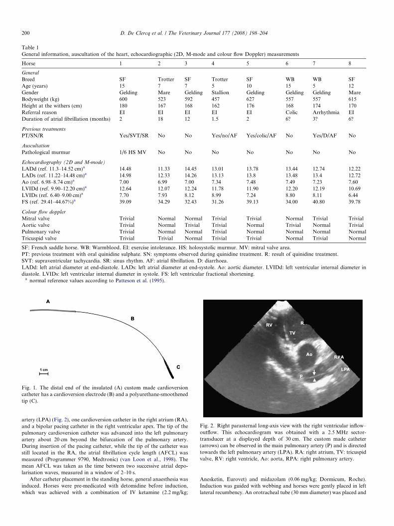
Table 1General information, auscultation of the heart, echocardiographic (2D, M-mode and colour flow Doppler) measurements
Horse 1 2 3 4 5 6 7 8
General
Breed SF Trotter SF Trotter SF WB WB SFAge (years) 15 7 7 5 10 15 5 12Gender Gelding Mare Gelding Stallion Gelding Gelding Gelding MareBodyweight (kg) 600 523 592 457 627 557 557 615Height at the withers (cm) 180 167 168 162 176 168 174 170Referral reason EI EI EI EI EI Colic Arrhythmia EIDuration of atrial fibrillation (months) 2 18 12 1.5 2 6? 3? 6?
Previous treatments
PT/SN/R Yes/SVT/SR No No Yes/no/AF Yes/colic/AF No Yes/D/AF No
Auscultation
Pathological murmur 1/6 HS MV No No No No No No No
Echocardiography (2D and M-mode)
LADd (ref. 11.3–14.52 cm)a 14.48 11.33 14.45 13.01 13.78 13.44 12.74 12.22LADs (ref. 11.22–14.48 cm)a 14.98 12.33 14.26 13.13 13.8 13.48 13.4 12.72Ao (ref. 6.98–8.74 cm)a 7.00 6.99 7.00 7.34 7.48 7.49 7.23 7.60LVIDd (ref. 9.90–12.20 cm)a 12.64 12.07 12.24 11.78 11.90 12.20 12.19 10.69LVIDs (ref. 6.40–9.00 cm)a 7.70 7.93 8.12 8.99 7.24 8.80 8.11 6.44FS (ref. 29.41–44.67%)a 39.09 34.29 32.43 31.26 39.13 34.00 40.80 39.78
Colour flow doppler
Mitral valve Trivial Normal Normal Trivial Trivial Normal Trivial TrivialAortic valve Trivial Normal Trivial Trivial Normal Trivial Normal TrivialPulmonary valve Trivial Normal Normal Trivial Normal Normal Normal NormalTricuspid valve Trivial Trivial Normal Trivial Trivial Normal Trivial Normal
SF: French saddle horse. WB: Warmblood. EI: exercise intolerance. HS: holosystolic murmur. MV: mitral valve area.PT: previous treatment with oral quinidine sulphate. SN: symptoms observed during quinidine treatment. R: result of quinidine treatment.SVT: supraventricular tachycardia. SR: sinus rhythm. AF: atrial fibrillation. D: diarrhoea.LADd: left atrial diameter at end-diastole. LADs: left atrial diameter at end-systole. Ao: aortic diameter. LVIDd: left ventricular internal diameter indiastole. LVIDs: left ventricular internal diameter in systole. FS: left ventricular fractional shortening.
a normal reference values according to Patteson et al. (1995).
Fig. 1. The distal end of the insulated (A) custom made cardioversioncatheter has a cardioversion electrode (B) and a polyurethane-smoothenedtip (C).
Fig. 2. Right parasternal long-axis view with the right ventricular inflow–outflow. This echocardiogram was obtained with a 2.5 MHz sector-transducer at a displayed depth of 30 cm. The custom made catheter(arrows) can be observed in the main pulmonary artery (P) and is directedtowards the left pulmonary artery (LPA). RA: right atrium, TV: tricuspidvalve, RV: right ventricle, Ao: aorta, RPA: right pulmonary artery.
200 D. De Clercq et al. / The Veterinary Journal 177 (2008) 198–204
artery (LPA) (Fig. 2), one cardioversion catheter in the right atrium (RA),and a bipolar pacing catheter in the right ventricular apex. The tip of thepulmonary cardioversion catheter was advanced into the left pulmonaryartery about 20 cm beyond the bifurcation of the pulmonary artery.During insertion of the pacing catheter, while the tip of the catheter wasstill located in the RA, the atrial fibrillation cycle length (AFCL) wasmeasured (Programmer 9790, Medtronic) (van Loon et al., 1998). Themean AFCL was taken as the time between two successive atrial depo-larisation waves, measured in a window of 2–10 s.
After catheter placement in the standing horse, general anaesthesia wasinduced. Horses were pre-medicated with detomidine before induction,which was achieved with a combination of IV ketamine (2.2 mg/kg;
Anesketin, Eurovet) and midazolam (0.06 mg/kg; Dormicum, Roche).Induction was guided with webbing and horses were gently placed in leftlateral recumbency. An orotracheal tube (30 mm diameter) was placed and

Fig. 3. Right parasternal long-axis view with the right ventricular inflow–outflow. This echocardiogram was obtained with a 2.5 MHz sector-transducer at a displayed depth of 30 cm. An echogenic linear artefact
D. De Clercq et al. / The Veterinary Journal 177 (2008) 198–204 201
isoflurane inhalant anaesthesia was given. Intermittent positive pressureventilation was applied in all horses. Dobutamine (Mayne Pharma) wasadministered IV to maintain arterial blood pressure. A base apex ECGwas connected to both a biphasic electrical defibrillator (Lifepak 20,Medtronic) and a pacemaker programmer (Programmer 9790, Medtron-ic). The custom made cardioversion catheters were connected to thedefibrillator with the RA catheter as cathode and the LPA catheter asanode. The pacing catheter was connected to the pacemaker programmerto achieve ventricular pacing in case of temporary asystole (van Loonet al., 2005).
Catheter position was verified with ultrasound and modified if neces-sary. Shocks were delivered as biphasic truncated exponential waves andwere synchronised with the R wave. Shock energy delivered to the horseswas increased from 150 to 360 J in steps of 50 J until sinus rhythm wasrestored. An arbitrary 2–5 min delay was used between successive shocksto assess cardiac rhythm. Cardioversion catheters were removed 5–10 minafter cardioversion to sinus rhythm.
After recovery, clinical status was followed and blood for completeblood analysis and cardiac troponin I assay was withdrawn 3 h after theprocedure. If sinus rhythm could not be restored during the first proce-dure, a second attempt at electrical cardioversion or oral QS treatmentwas made.
resembling a catheter (arrows) can be observed in the main pulmonaryartery (P). RA: right atrium, TV: tricuspid valve, RV: right ventricle, Ao:aorta, LPA: left pulmonary artery, RPA: right pulmonary artery.
ResultsBefore the procedure, biochemical and haematologicalparameters were all within normal reference ranges exceptfor calcium and magnesium in two horses. Horse 3 pre-sented a low magnesium level (2.0 mg/dL; ref. 2.2–2.8 mg/dL) and horse 4 had a low calcium level (10.2 mg/dL; ref. 11.2–13.6 mg/dL) (Carlson, 2002). Both horsesreceived IV calcium and magnesium supplementation. Car-diac troponin I was normal in all horses (<0.15 ng/mL;Begg et al., 2006). The results of the cardiac examinationwere within normal limits except for horse 1 where the leftatrial diameter at end-systole was 14.98 cm (ref. 11.22–14.48 cm) and LVIDd was 12.64 cm (ref. 9.90–12.20 cm);Patteson et al., 1995).
Repeated monitoring during and after amiodaronetreatment showed no adverse effects. Except for an occa-sional premature ventricular complex, no ECG abnormal-ities were seen during LPA catheter placement. In somehorses, visualisation and positioning of the catheter in theleft pulmonary artery was hampered due to echogenic lin-ear artefacts in the main and right pulmonary artery. Theseartefacts were directed towards the right pulmonary artery(Fig. 3). Mean (±SD) amiodarone and desethylamioda-rone concentrations (lg/mL) at the beginning of the shockdelivery were 0.9 ± 0.2 and 0.1 ± 0.03, respectively.
Details of baseline AFCL, cardioversion threshold,impedance, catheter position and total number of shocksdelivered in each horse are shown in Table 2. In 5/8horses (horses 2–6) sinus rhythm could be restored(Fig. 4) after a total of 3–18 shocks with a mean (±SD)energy level of 295 ± 62 J (range 200–360 J; Table 2).Lead impedance was between 48 and 72 X. However, aftercardioversion and catheter withdrawal, horse 2 presentedmultiple atrial premature complexes. Six minutes aftercardioversion an atrial premature complex resulted inrecurrence of AF.
In 3/8 horses (horses 1, 7 and 8), the cardioversion cath-eter from the LPA was displaced after induction of generalanaesthesia. Several attempts to reposition the catheterduring general anaesthesia failed, requiring terminationof the cardioversion procedure. In 2/3 horses (horses 7and 8), a second electrical cardioversion attempt was per-formed after 10 days, which resulted in successful cardio-version in one horse (horse 7). No further cardioversionattempts were made in horse 2, the horse returned homeas a broodmare.
Recovery from general anaesthesia was uneventful in allhorses. One horse (horse 8) developed diarrhoea and fever4 days after the second cardioversion procedure. Three fae-cal samples were positive on Salmonella culture. The horsereceived IV fluid, antimicrobials and flunixin meglumine(1.1 mg/kg; Finadyne, Schering-Plough) and the diarrhoeaand fever resolved within 3 days. No further cardioversionattempts were made and the horse returned home as abroodmare. Haematological and biochemical parameterstaken 3 h after cardioversion, remained within normalranges in all horses.
Ten days after the unsuccessful electrical cardioversion,horse 1 was treated with oral QS (22 mg/kg q 2 h). Afterthe second dose, treatment was terminated because ofsevere tachycardia (220 bpm). One hour later, cardiover-sion to sinus rhythm occurred. One week after electricalcardioversion, all converted horses were still in sinusrhythm and returned home.
Discussion
Treatment of AF in horses without detectable cardiacpathology is recommended as there is a good prognosis(Deem and Fregin, 1982; Reef et al., 1988; Collatos, 1995).

Table 2Catheter position, number of shocks, number of procedures and results are given for each horse
Horse 1 2a 3a 4a 5a 6a 7a 8 Mean ± SDd
AFCL (ms) 190 133 279b 214b 250b 279b 214b 200b 228 ± 55Cardioversion energy (J) / 300 200 250 300 360 360 360c 295 ± 62Impedance (X) / 72 60 48 65 60 58 54c 60 ± 8RA (cm) / 56 63 56 72 79 65 50 65 ± 9LPA (cm) / 116 143 117 165 157 135 105 138 ± 20Total number of shocks delivered 0 16 3 3 6 12 18 19 9 ± 7Number of cardioversion procedures 1 1 1 1 1 1 2 2 /Result of 1st procedure / SR/IRAF SR SR SR SR AF AF /Result of 2nd procedure / / / / / / SR AF /
RA: catheter depth in the right atrium from the lower half of the jugular vein. LPA: catheter depth in the left pulmonary artery from the lower half of thejugular vein.IRAF: immediate recurrence of atrial fibrillation after successful electrical cardioversion.AF: atrial fibrillation.
a Horses converted to sinus rhythm with the electrical cardioversion protocol.b AFCL measured after a 24-h amiodarone infusion protocol.c Highest energy level and impedance are given, although sinus rhythm could not be restored.d Values of horses 1 and 8 not included SR: sinus rhythm.
Fig. 4. A base apex electrocardiogram during delivery of a 250 J R wave synchronised shock (.) in horse 4 shows restoration of sinus rhythm (fl). Leadrecording is 25 mm/s and 1 mV/cm.
202 D. De Clercq et al. / The Veterinary Journal 177 (2008) 198–204
In our patient data, only one horse had a left atrial diameterat the end of systole and a LVIDd that were slightly out ofthe normal reference range described by Patteson et al.(1995). However, this was thought to be partially relatedto the size of the animal.
Disturbances in electrolyte concentrations are supposedto be a contributing factor to the occurrence of AF (Reefand McGuirk, 2002). Electrolyte values were determinedin each horse before the procedure and normalised if neces-sary, although the exact effect on the outcome wasunknown.
In human patients, electrical cardioversion is highlyeffective in converting AF to sinus rhythm. The associatedrisk for embolic complications makes echocardiographicmonitoring and anti-coagulation treatment mandatoryprior to the procedure (Capucci et al., 2000; Ricard et al.,2003). Pre-treatment with anti-arrhythmic drugs is recom-mended because cardioversion recurrence of AF is com-mon from between <1 min up to 2 weeks (Levy et al.,1997; Sarubbi et al., 1998; Timmermans et al., 1998; Duyts-chaever et al., 2000, 2002; Gorenek et al., 2002; Van Noordet al., 2002; Ricard et al., 2003; Chugh et al., 2004; Joglarand Kowal, 2004). In horses, transvenous electrical cardio-version has been applied to cardiovert AF (Deem and Fre-gin, 1982; van Loon et al., 1998; Buchanan, 2002; Fryeet al., 2002; McGurrin et al., 2003, 2005a, 2005b; van Loon
et al., 2005) but as thromboembolic events have not beenassociated with AF in horses, anti-coagulation therapy isnot indicated.
To the authors’ knowledge, IRAF has not yet beenreported in horses. This might be explained by the fact thatduring conventional QS treatment, the therapeutic druglevel at the time of conversion prevents AF from re-occur-ring immediately. During electrical cardioversion withoutanti-arrhythmic drugs, IRAF is more likely to occurbecause of the high vulnerability of the atria at the timeof conversion (Duytschaever et al., 2000,2002). Becausehorse 2 relapsed to AF within 6 min of conversion, it wasdecided to load all subsequent horses with amiodarone inthe 24 h preceding the electrical cardioversion procedureto reduce the risk for IRAF.
In human patients, amiodarone has proved efficacious inpreventing IRAF (Gorenek et al., 2006) and therapeuticamiodarone plasma concentrations for cardioversion inhumans range between 0.1 and 11.9 lg/mL (Latini et al.,1984). Some authors have reported that cardioversionoccurs at an amiodarone concentration between 0.6 and3.0 lg/mL, with a mean ± SD of 2.5 ± 1.3 (Canada et al.,1983; Vardas et al., 2000), although Rotmensch et al.(1984) describe a weak or even absence of any concentra-tion-effect relationship. Our goal in administering amioda-rone to horses was not to convert AF to sinus rhythm, but

D. De Clercq et al. / The Veterinary Journal 177 (2008) 198–204 203
to change the electrophysiological properties of the atria asamiodarone prolongs AFCL quickly (1–2 h) in horses withchronic AF (De Clercq et al., 2006, 2007b).
In contrast with a prolonged IV administration, the 24-hloading with amiodarone did not result in side-effects (DeClercq et al., 2006, 2007b). The diarrhoea occurring inhorse 8 after cardioversion was not thought to be directlycaused by amiodarone because of the late occurrence andbecause of the positive faecal Salmonella culture.
McGurrin et al. (2005b) have described a success rate of98% for electrical cardioversion in horses, but equine car-dioversion catheters are not yet commercially available.Human cardioversion catheters are only 120 cm long,which is usually insufficient to place the catheter tip intothe equine LPA. Therefore, we developed custom madecatheters with a length of 180 cm. Electrode placement intothe LPA is recommended because, when combined with aright atrial electrode, the current between both electrodeswill ‘‘cover’’ more atrial myocardium, so lowering thethreshold for cardioversion (Alt et al., 1997a, 1997b; Sak-sena et al., 1995).
Left pulmonary artery catheter placement was ham-pered by a rather poor visibility, due to the depth of thethorax and the small diameter of the catheter. In addition,after passing through the RA, RV and pulmonary artery,the catheter entered more easily the right branch of thePA instead of the left. Repeated insertion and withdrawalwhile twisting the catheter was necessary in order for itto enter the LPA.
In most horses, the distal part of the catheter, located inthe LPA, could not be distinguished on ultrasound, but themore proximal part in the main pulmonary artery wasclearly directed towards the LPA (Fig. 2). In some horses,visualisation and positioning of the catheter in the LPAwas hampered due to echogenic linear artefacts in the mainand right pulmonary artery. Fluoroscopic guidance ofcatheter placement, as used in humans, is difficult in horsesbecause of their size. Exact placement of the catheter couldnot be guided by radiography due to technical limitations.Blood pressure monitoring was not possible with our cus-tom made catheter, which challenged catheter placementrather more than was described by McGurrin et al.(2005b). A pacing catheter was placed in the right ventricleas back-up because transvenous electrical cardioversioncan provoke temporary complete atrioventricular block(van Loon et al., 2005). Webbing during the induction ofgeneral anaesthesia did not seem to prevent displacementof the LPA catheter, as we found this occurred in threehorses.
Shocks were delivered synchronously with the R wavesto avoid ventricular arrhythmias (McGurrin et al., 2003).Mean cardioversion energy to restore sinus rhythm washigher in this study compared to the results (295 ± 62 Jvs. 162 ± 10 J) described by McGurrin et al. (2005b). Thisdifference might be explained by a different electrode posi-tion in the LPA, the smaller diameter and therefore thesmaller surface of the cardioversion electrode, or indeed
by other individual factors. Nevertheless, despite thesehigher cardioversion thresholds and number of shocksdelivered, cardiac troponin I concentrations remainedwithin normal limits.
Conclusions
In the present study, the use of custom made cathetersallowed us to undertake successful transvenous electricalcardioversion in six horses. The small diameter and thelack of intracardiac pressure monitoring challenged correctcatheter positioning. High energy levels and a large numberof shocks were required to achieve cardioversion, but werenot associated with an increase in cardiac troponin I con-centrations. Further adaptations of the catheter design toincrease the surface of the cardioversion electrode and tofacilitate catheter visibility and positioning are necessaryto increase the success rate.
References
Allessie, M., Ausma, J., Schotten, U., 2002. Electrical, contractile andstructural remodeling during atrial fibrillation. CardiovascularResearch 54, 230–246.
Alt, E., Ammer, R., Schmitt, C., Evans, F., Lehmann, G., Pasquantonio,J., Schomig, A., 1997a. A comparison of treatment of atrial fibrillationwith low-energy intracardiac cardioversion and conventional externalcardioversion. The European Heart Journal 18, 1796–1804.
Alt, E., Schmitt, C., Ammer, R., Plewan, A., Evans, F., Pasquantonio, J.,Ideker, T., Lehmann, G., Putter, K., Schomig, A., 1997b. Effect ofelectrode position on outcome of low-energy intracardiac cardiover-sion of atrial fibrillation. The American Journal of Cardiology 79, 621–625.
Begg, L.M., Hoffmann, K.L., Begg, A.P., 2006. Serum and plasma cardiactroponin I concentrations in clinically normal thoroughbreds intraining in Australia. Australian Veterinary Journal 84, 336–337.
Buchanan, J.W., 2002. Comments successful use of electrical cardioversionin a horse. Journal of the American Veterinary Medical Association220, 1777.
Canada, A.T., Lesko, L.J., Haffajee, C.I., Johnson, B., Asdourian, G.K.,1983. Amiodarone for tachyarrhythmias: pharmacology, kinetics, andefficacy. Drug Intelligence and Clinical Pharmacy 17, 100–104.
Capucci, A., Villani, G.Q., Aschieri, D., 2000. Oral amiodarone increasesthe efficacy of direct-current cardioversion in restoration of sinusrhythm in patients with chronic atrial fibrillation. European HeartJournal 21, 66–73.
Carlson, G.P., 2002. Clinical chemistry tests. In: Smith, B.P. (Ed.), LargeAnimal Internal Medicine, 3rd ed. Mosby, St. Louis, Missouri, pp.471–474.
Chugh, A., Ozaydin, M., Scharf, C., Lai, S.W.K., Hall, B., Cheung, P.,Pelosi, F., Knight, B.P., Morady, F., Oral, H., 2004. Mechanism ofimmediate recurrence of atrial fibrillation after restoration of sinusrhythm. Pacing and Clinical Electrophysiology 27, 77–82.
Collatos, C., 1995. Treating atrial fibrillation in horses. Compendium onContinuing Education for the Practicing Veterinarian 17, 243–245.
De Clercq, D., van Loon, G., Baert, K., Tavernier, R., Croubels, S., DeBacker, P., Deprez, P., 2006. Intravenous amiodarone treatment inhorses with chronic atrial fibrillation. The Veterinary Journal 172,129–134.
De Clercq, D., van Loon, G., Tavernier, R., Deprez, P., 2007a. Atrial andventricular electrical remodelling due to short-term pacing inducedatrial fibrillation. European College of Equine Internal Medicine(ECEIM), 61.

204 D. De Clercq et al. / The Veterinary Journal 177 (2008) 198–204
De Clercq, D., van Loon, G., Baert, K., Tavernier, R., Croubels, S., DeBacker, P., Deprez, P., 2007b. Effects of an adapted intravenousamiodarone protocol in horses with atrial fibrillation. Equine Veter-inary Journal 39, 344–349.
Deem, D.A., Fregin, G.F., 1982. Atrial fibrillation in horses: a review of106 clinical cases, with consideration of prevalence, clinical signs, andprognosis. Journal of the American Veterinary Medical Association180, 261–265.
Duytschaever, M., Danse, P., Allessie, M., 2000. Widening of the windowof inducibility of AF by electrical remodeling: time course andmechanisms. Pacing Clinical Electrophysiology 23, 730.
Duytschaever, M., Danse, P., Allessie, M., 2002. Supervulnerable phaseimmediately after termination of atrial fibrillation. Journal of Cardio-vascular Electrophysiology 13, 267–275.
Frye, M.A., Selders, C.G., Mama, K.R., Wagner, A.E., Bright, J.M.,2002. Use of biphasic electrical cardioversion for treatment ofidiopathic atrial fibrillation in two horses. Journal of the AmericanVeterinary Medical Association 220, 1039–1045.
Gorenek, B., Kudaiberdieva, G., Cavusoglu, Y., Goktekin, O., Birdane,A., Unalir, A., Ata, N., Timuralp, B., 2002. Immediate recurrence ofatrial fibrillation after internal cardioversion: Importance of rightatrial conduction variations. Journal of Electrocardiology 35, 313–320.
Gorenek, B., Cavusoglu, Y., Goktekin, O., Birdane, A., Kudaiberdieva,G., Ata, N., Unalir, A., Timuralp, B., 2006. Amiodarone versusSotalol and propafenone for prevention of immediate recurrence ofatrial fibrillation after internal cardioversion: importance of P waveanalysis. International Journal of Cardiology 106, 268–269.
Joglar, J.A., Kowal, R.C., 2004. Electrical cardioversion of atrialfibrillation. Cardiology Clinics 22, 101–111.
Latini, R., Tognoni, G., Kates, R.E., 1984. Clinical pharmacokinetics ofamiodarone. Clinical Pharmacology 9, 136–156.
Levy, S., Ricard, P., Lau, C.P., Lok, N.S., Camm, A.J., Murgatroyd,F.D., Jordaens, L.J., Kappenberger, L.J., Brugada, P., Ripley, K.L.,1997. Multicenter low energy transvenous atrial defibrillation (XAD)trial results in different subsets of atrial fibrillation. Journal of theAmerican College of Cardiology 29, 750–755.
Maes, A., Baert, K., Croubels, S., De Clercq, D., van Loon, G., Deprez,P., De Backer, P., 2006. Determination of amiodarone and dese-thylamiodarone in horse plasma and urine by high-performance liquidchromatography combined with UV detection and electrosprayionization mass spectrometry. Journal of Chromatography B 836,47–56.
McGurrin, K.M.J., Physick-Sheard, P.W., Kenney, D.G., Kerr, C.,Hanna, W.J.B., Neto, F.T., Weese, J.S., 2003. Transvenous ElectricalCardioversion in Equine Atrial Fibrillation: Technique and SuccessfulTreatment of 3 Horses. Journal of Veterinary Internal Medicine 5,715–718.
McGurrin, M.K.J., Physick-Sheard, P.W., Kenney, D.G., Kerr, C.,Hanna, W.J.B., 2005a. Transvenous electrical cardioversion of equineatrial fibrillation: technical consideration. Journal of VeterinaryInternal Medicine 19, 695–702.
McGurrin, M.K.J., Physick-Sheard, P.W., Kenney, D.G., 2005b. How toperform transvenous electrical cardioversion in horses with atrialfibrillation. Journal of Veterinary Cardiology 7, 109–119.
Morillo, C.A., Klein, G.J., Jones, D.L., Guirodon, C.M., 1995. Chronicrapid atrial pacing. Structural, functional, and electrophysiologicalcharacteristics of a new model of sustained atrial fibrillation. Circu-lation 91, 1588–1595.
Ohmura, H., Nukada, T., Mizuno, Y., Yamaya, Y., Nakayama, T.,Amada, A., 2000. Safe and efficacious dosage of flecainide acetate fortreating equine atrial fibrillation. Equine Veterinary Journal (Suppl.),18–27.
Patteson, M.W., Gibbs, C., Wotton, P.R., Cripps, P.J., 1995. Echocar-diographic measurements of cardiac dimensions and indices of cardiac
function in normal adult thoroughbred horses. The Equine VeterinaryJournal 19 (Suppl.), 18–27.
Reef, V.B., McGuirk, S.M., 2002. Diseases of the cardiovascular system.Atrial fibrillation. In: Smith, B.P. (Ed.), Large Animal InternalMedicine, 3rd ed. Mosby, St. Louis, Missouri, pp. 471–474.
Reef, V.B., Levitan, C.W., Spencer, P.A., 1988. Factors affectingprognosis and conversion in equine atrial fibrillation. Journal ofVeterinary Internal Medicine 2, 1–6.
Reef, V.B., Reimer, J.M., Spencer, P.A., 1995. Treatment of atrialfibrillation in horses: new perspectives. Journal of Veterinary InternalMedicine 9, 57–67.
Ricard, P., Yaıci, K., Rinaldi, J.P., Bergonzi, M., Saoudi, N., 2003.Cardioversion of atrial fibrillation: how and when? European HeartJournal Supplement H, H40–H44.
Rotmensch, H.H., Belhassen, B., Swanson, B.N., Shoshani, D., Spielman,S.R., Greenspon, A.J., Greenspan, A.M., Vlasses, P.H., Horowitz,L.M.N., 1984. Steady-state plasma amiodarone concentrations: rela-tionship with antiarrhythmic efficacy and toxicity. Annals of InternalMedicine 101, 624–649.
Saksena, S., Prakash, A., Mangeon, L., Varanasi, S., Kolettis, T.,Mathew, P., De Groot, P., Mehra, R., Krol, R.B., 1995. Clinicalefficacy and safety atrial defibrillation using biphasic shocks andcurrent nonthoracothomy endocardial lead configurations. The Amer-ican Journal Cardiology 76, 913–921.
Sarubbi, B., Ducceschi, V., D’Andrea, A., Liccardo, B., Santagelo, L.,Iacono, A., 1998. Atrial fibrillation: what are effects of drug therapy onthe effectiveness and complications of electrical cardioversion? Cana-dian Journal of Cardiology 14, 1267–1273.
Schotten, U., Duytschaever, M., Ausma, J., Eijsbouts, S., Neuberger, N.,Allessie, M., 2003. Electrical and contractile remodeling during thefirst days of atrial fibrillation go hand in hand. Circulation 107, 1433–1439.
Timmermans, C., Rodriguez, L.M., Smeets, J.L., Wellens, H.J., 1998.Immediate reinitiation of atrial fibrillation following internal atrialdefibrillation. Journal of Cardiovascular Electrophysiology 9, 122–128.
van Loon, G. 1999. Effect of experimental chronic atrial fibrillation inequines. In: Atrial Pacing and Experimental Atrial Fibrillation inEquines, pp. 162–206 (Chapter 7).
van Loon, G., Jordaens, L., Muylle, E., Nollet, H., Sustonck, B., 1998.Intracardiac overdrive pacing as a treatment of atrial flutter in a horse.Veterinary Record 142, 301–303.
van Loon, G., Tavernier, R., Fonteyne, W., Duytschaever, M., Jordaens,L., Deprez, P., 2001. Pacing induced long-term atrial fibrillation inhorses. Europace 2, Suppl. A. p. 84.
van Loon, G., Blissitt, K.J., Keen, J.A., Young, L.E., 2004. Use ofintravenous flecainide in horses with naturally-occurring atrial fibril-lation. Equine Veterinary Journal 36, 609–614.
van Loon, G., De Clercq, D., Tavernier, R., Amory, H., Deprez, P., 2005.Transient complete atrioventricular block following transvenouselectrical cardioversion of atrial fibrillation in a horse. The VeterinaryJournal 170, 124–127.
Van Noord, T., Van Gelder, I.C., Crijns, H.J., 2002. How to enhanceacute outcome of electrical cardioversion by drug therapy: importanceof immediate reinitiating of atrial fibrillation. Journal of Cardiovas-cular Electrophysiology 13, 822–825.
Vardas, P.E., Kochiadakis, G.E., Igoumenidis, N.I., Tsatsakis,A.M., Simantirakis, E.N., Chlouverakis, G.I., 2000. Amiodaroneas a first-choice drug for restoring sinus rhythm in patients withatrial fibrillation: A randomised, controlled study. Chest 117,1538–1545.
Wijffels, M., Kirchhof, J., Dorland, R., Allessie, M., 1995. Atrialfibrillation begets atrial fibrillation. A study in awake chronicallyinstrumented goats. Circulation 92, 1954–1968.


![Check List Cardioversion I.gallastegi[1]](https://img.dokumen.tips/doc/110x75/55cf8e57550346703b912349/check-list-cardioversion-igallastegi1.jpg)