Embed Size (px)
Citation preview

RESEARCH Open Access
Transplantation of human inducedpluripotent stem cell-derivedcardiomyocytes improves myocardialfunction and reverses ventricularremodeling in infarcted rat heartsXumin Guan1†, Wanzi Xu2†, He Zhang3†, Qian Wang4, Jiuyang Yu4, Ruyi Zhang5, Yamin Chen4, Yunlong Xia1,Jiaxian Wang4,6* and Dongjin Wang7*
Abstract
Background: Human-induced pluripotent stem cell-derived cardiomyocytes (iPSC-CMs) have shed great light oncardiac regenerative medicine and specifically myocardial repair in heart failure patients. However, the treatmentefficacy and the survival of iPSC-CMs in vivo after transplantation have yielded inconsistent results.
Objectives: The objective of this study was to evaluate the ability of human iPSC-CMs to improve myocardialfunction in a rat postinfarction heart failure model.
Methods: Eight-week-old male Sprague-Dawley rats were randomly selected to receive an intramyocardial injectionof 5% albumin solution with or without 1 × 10⁷ human iPSC-CMs 10 days after undergoing left anterior descending(LAD) coronary artery ligation. Cyclosporine A and methylprednisolone were administered before iPSC-CM injectionand until the rats were killed to prevent graft rejection. Cardiac function was evaluated by echocardiography. Thesurvival of grafted cardiomyocytes was confirmed by observing the fluorescent cell tracer Vybrant™ CM-DiI orexpression of the enhanced green fluorescent protein (eGFP) in transplanted cells, or survival was demonstrated bypolymerase chain reaction (PCR)-based detection of human mitochondrial DNA. Sirius red stain was used toevaluate the fibrosis ratio. Hematoxylin-eosin staining was used to observe the formation of teratomas.
(Continued on next page)
© The Author(s). 2020 Open Access This article is distributed under the terms of the Creative Commons Attribution 4.0International License (http://creativecommons.org/licenses/by/4.0/), which permits unrestricted use, distribution, andreproduction in any medium, provided you give appropriate credit to the original author(s) and the source, provide a link tothe Creative Commons license, and indicate if changes were made. The Creative Commons Public Domain Dedication waiver(http://creativecommons.org/publicdomain/zero/1.0/) applies to the data made available in this article, unless otherwise stated.
* Correspondence: [email protected]; [email protected]†Xumin Guan, Wanzi Xu and He Zhang contributed equally to this work.4HELP Therapeutics, Nanjing 211166, Jiangsu, China7Department of Cardio-Thoracic Surgery, Nanjing Drum Tower HospitalAffiliated to Medical School of Nanjing University, Nanjing, ChinaFull list of author information is available at the end of the article
Guan et al. Stem Cell Research & Therapy (2020) 11:73 https://doi.org/10.1186/s13287-020-01602-0

(Continued from previous page)
Results: Four weeks after intramyocardial injection of iPSC-CMs, animals undergoing iPSC-CM transplantation hadlower mortality than the control group. Animals injected with cell-free solution (control group) demonstratedsignificant left ventricular (LV) functional deterioration, whereas grafting of iPSC-CMs attenuated this remodelingprocess. In the control group, the ejection fraction deteriorated by 10.11% (from 46.36 to 41.67%), and fractionalshortening deteriorated by 9.23% (from 24.37 to 22.12%) by 4 weeks. In the iPSC-CM injection group, the ejectionfraction improved by 18.86% (from 44.09 to 52.41%), and fractional shortening improved by 23.69% (from 23.08 to28.54%). Cell labeling, tracking, and molecular biology techniques indicated that the grafted cardiomyocytessurvived in the rat heart 1 month after iPSC-CM transplantation. Myocardial fibrosis was also attenuated in the iPSC-CM treatment group.
Conclusions: Human iPSC-CM grafts survived in infarcted rat hearts and restored myocardial function 4 weeks aftertransplantation. Cell replacement therapy also reversed ventricular remodeling, indicating the potential of iPSC-CMsfor cardiac repair strategies.
Keywords: Human induced pluripotent stem cell, Cardiomyocytes, Regenerative medicine, Remodeling, Heartfailure
BackgroundHeart failure remains the leading cause of morbidity andmortality worldwide [1]. In patients with chronic heartfailure, especially in those with III-IV symptoms, as de-fined by the New York Heart Association, the prognosisis extremely poor due to irreversibly impaired left ven-tricular function [2]. The treatment of heart failure re-mains a challenging problem as conventional treatments(drug therapy, interventional therapy, and surgery) havedifficulty in restoring the function of the heart. Myocar-dial infarction (MI), the most common heart disease, ischaracterized by a significant reduction in the number offunctional cardiomyocytes and leads to the developmentof progressive heart failure. Adult hearts have little abil-ity to regenerate [3] and traditional treatments fail to ad-dress the fundamental problem of muscle deficiency.Heart regenerative therapy, such as transplantation ofmyocytes [4] and cardiomyocytes to replace lost cells,has become a new strategy for the treatment of heartfailure caused by MI [5].
Pluripotent stem cells (PSCs), including embryonicstem cells (ESCs) and induced pluripotent stem cells(iPSCs), are potential sources of therapeutic cardiomyo-cytes [6–8]. They have the capacity for unlimited prolif-eration in vitro while maintaining the potential todifferentiate into derivatives of the three germ layers. Awell-established method of cardiomyocyte differentiationfrom PSCs provides an ideal source of cardiomyocytesand makes regenerative therapy for myocardial muscledamage a promising strategy. The methods of cardio-myocyte differentiation, purification, and maturationfrom iPSCs or ESCs have been established both by ourlab [9–12] and other labs [13–17]. Recently, transplant-ation of human ESC-derived cardiomyocytes (ESC-CMs)has been proven to significantly improve cardiac func-tion in infarcted rat and nonhuman primate hearts [18,
19] due to the capacity of PSC-CMs to remuscularizethe infarcted hearts and form electromechanical junc-tions with the host hearts [15]. Human iPSC-CMs havegreat potential in disease treatment because they avoidthe social ethical issues of ESCs and avoid the possibilityof immune rejection [20]. Although some investigatorsreported that transplantation of iPSC-CMs reduced re-modeling of the heart after ischemic damage [21–24],the field is still at the preclinical stage due to technicaland surgical hurdles. In addition, the treatment efficacyand the survival of iPSC-CMs in vivo after transplant-ation is debatable. Chow et al. demonstrated that nografted iPSC-CMs were detected after 1 month of trans-plantation [22]. In the present study, we aimed to evalu-ate the ability of human iPSC-CM transplantation toimprove myocardial function in the rat MI model and todetermine the fate of transplanted cells in the rat heart.
MethodsDifferentiation of iPSC-CMs and cell preparationHuman iPSC line was derived from a healthy man (32years old) and transduced with Oct3/4–Sox2, cMyc, andKlf4. Informed consent was obtained from the donor priorto all experiments. The iPSC reprogramming and cardio-myocytes manufacturing were conducted under GMP-grade lab at HELP Therapeutics. Undifferentiated humaniPSCs were grown to 90% confluence and subsequentlydifferentiated into beating cardiomyocytes. In brief, on day0 and day 1, iPSCs were given #1 medium (RPMI 1640[Gibco] and B27 supplement minus insulin [Gibco]) sup-plemented with 6 μM CHIR-99021 [Sigma-Aldrich],which is a selective inhibitor of glycogen synthase kinase3β that activates the canonical Wnt signaling pathway. Onday 2, the medium was replaced with #1 medium withoutCHIR99021. On day 3 and day 4, cells were supplementedwith #1 medium with 5 μM of IWR-1 [Sigma-Aldrich],
Guan et al. Stem Cell Research & Therapy (2020) 11:73 Page 2 of 11

which is a Wnt antagonist. Then, the medium was re-placed with #1 medium every day until day 8, and themedium was replaced with #2 medium (RPMI 1640[Gibco] and B27 supplement [Gibco]) every other day.Usually, the cells began to have spontaneous contractionsafter 8–10 days of differentiation. On days 16–20 of differ-entiation, purified iPSC-CMs were dissociated and 1 × 10⁷
cells were cryopreserved per cryogenic tube. Cryopre-served cardiomyocytes were thawed in a 37 °C water bath(for approximately 2min) and suspended in 75 μl 5% albu-min solution before transplantation.
Animal model of MI and cell transplantationThe experimental flow chart is shown in supplementaryFigure 1. After undergoing a left thoracotomy, 8-week-old male Sprague-Dawley rats (approximately 250 g)underwent MI by ligation of the left anterior descending(LAD) coronary artery 10 days before receiving an injec-tion of cells. Electrocardiogram (ECG) data were used toconfirm the establishment of the MI model by ST seg-ment elevation (Fig. 2c, d). Ten days after MI, the ratsunderwent echocardiography, and a second thoracotomywas performed in the rats that met the inclusion criteria:Ejection fraction was reduced by between 15 and 50%.Then, 1 × 10⁷ human iPSC-CMs in 5% albumin solutionor a control solution containing only 5% albumin weretransplanted to the infarcted area and infarcted marginsat two to three different sites. Two groups were studied:(1) a control group in which 5% albumin solution (75 μl)was injected (n = 13) and (2) a group in which 1.0 × 10⁷
iPSC-CMs in 5% albumin solution were grafted (n = 18).To prevent graft rejection, animals from all groups weretreated with cyclosporine A (15 mg/kg/day) and methyl-prednisolone (2 mg/kg/day) from the day before iPSC-CM delivery until rats were dissected. Most rat heartswere harvested 4 weeks later (day 28).
EchocardiographyAs shown in supplementary Figure 1, transthoracic echo-cardiography was performed at baseline (before MI modelestablished), day 0 (10 days after LAD coronary arteryligation and before iPSC-CM grafting), and day 28 (4weeks after iPSC-CM grafting) using the Vevo LAB 3.1.0(FUJIFILM VisualSonics, Inc.). The following parameterswere measured: (1) left ventricular end-diastolic and end-systolic diameters (LVEDD and LVESD, respectively); (2)left ventricular end-diastolic and end-systolic volume(LVEDV and LVESV, respectively); (3) ejection fraction(EF); (4) fractional shortening (FS); (5) left ventricularend-diastolic and end-systolic anterior wall thickness. Allmeasurements were averaged over three cardiac cyclesand assessed by an experienced operator blinded to thetreatment group.
Tracking of iPSC-CMsTo identify the transplanted cells, we used a number oflabeling, tracking, and molecular biological techniques:(1) labeling with the fluorescent cell tracer Vybrant™CM-DiI (5 μM, Molecular Probes, Invitrogen, V22888);(2) tagging with the genetic marker enhanced greenfluorescent protein (eGFP), the introduction of whichwas achieved using lentiviral transduction; (3) measuringhuman mitochondrial DNA via polymerase chain reac-tion (PCR) and qPCR at different times following trans-plantation (supplementary Figure 1).
Histological examinationFor histological examination, hearts were fixed with 4%paraformaldehyde in PBS for Sirius red staining or HEstaining and cryosectioned for immunostaining. For im-munostaining, fixed hearts were immersed in 30% sucroseovernight, embedded in an optimal cutting temperaturecompound, frozen, and cryosectioned (4.5-μM sections).Immunostaining was performed, and primary antibodieswere used at dilutions of 1:200 for anti-sarcomeric alpha-actinin (Abcam, ab137346) and 1:100 for anti-GFP(Abcam, ab1218). Secondary antibodies, including goatanti-rabbit IgG (H + L) (Alexa Fluor Plus 555, Invitrogen,A32732), were used at dilutions of 1:500 for detectingalpha-actinin, and goat anti-mouse IgG (H + L) (ThermoFisher, F2761) was used at dilutions of 1:500 for detectingeGFP. Slides were imaged with an inverted fluorescencemicroscope (Leica inverted microscope DMi8).
PCR and qPCR analysisThe rats were dissected at 24 h, 7 days, and 28 days aftercell grafting. For PCR and qPCR, hearts and other or-gans were frozen in liquid nitrogen. The presence ofiPSC-CMs within the rat hearts or other organs wasevaluated using PCR and qPCR-based amplification ofhuman mitochondria DNA. Genomic DNA was pro-duced using the DNeasy Blood & Tissue Kit (Qiagen,69504). PCR was carried out using Platinum™ Green HotStart PCR Master Mix (Invitrogen™, 13001012). The se-quence of the forward primer was CACCGGCGCAGTCATTCTCATA, and the reverse primer sequencewas GAGTCCTGTAAGTAGGAGA.
Statistical analysisData are expressed as the mean ± standard error ofthe mean (SEM). We tested data for normality byShapiro-Wilk test. Independent samples T test wasused to assess the difference between the two groups.Statistical analyses were performed using SPSS 13.0(SPSS Inc., Chicago, IL). P < 0.05 (two-tailed) wasconsidered to be statistically significant.
Guan et al. Stem Cell Research & Therapy (2020) 11:73 Page 3 of 11
ResultsA total of 46 rats underwent MI surgery by ligation ofthe LAD coronary artery; six died after the first oper-ation and nine rats were excluded because the resultsof the transthoracic echocardiography did not meet ourinclusion criteria before cell grafting (eight rats: ejec-tion fraction reduced < 15%, one rat: ejection fractionreduced > 50%). Hence, 31 rats underwent a secondthoracotomy, of which 18 were placed in the iPS-CMgroup, involving transplantation with 1.0 × 10⁷ iPSC-
CMs; 13 were placed in the control group and wereinjected with a 5% albumin solution. In the controlgroup, two rats died within 24 h, and another three diedwithin 15 days of the second operation (38.46% postop-erative mortality). In the iPS-CM group, three rats werekilled 24 h and 7 days after cell transplantation. In theother 12 rats, one died on the fourth day because of acotton ball left in the chest, and another two died dur-ing the 15 days that followed the second operation (25%postoperative mortality).
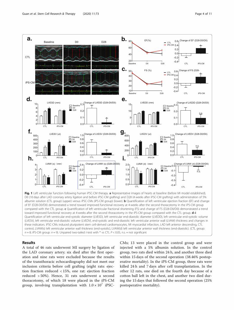
Fig. 1 Left ventricular function following human iPSC-CM therapy. a Representative images of hearts at baseline (before MI model established),D0 (10 days after LAD coronary artery ligation and before iPSC-CM grafting) and D28 (4 weeks after iPSC-CM grafting) with administration of 5%albumin solution (CTL group) (upper) versus iPSC-CMs (iPS-CM group) (lower). b Quantification of left ventricular ejection fraction (EF) and changeof EF (D28-D0/D0) demonstrated a trend toward improved functional recovery at 4 weeks after the second thoracotomy in the iPS-CM groupcompared with the CTL group. c Quantification of left ventricular fractional shortening (FS) and change of FS (D28-D0/D0) demonstrated a trendtoward improved functional recovery at 4 weeks after the second thoracotomy in the iPS-CM group compared with the CTL group. d–iQuantification of left ventricular end-systolic diameter (LVESD), left ventricular end-diastolic diameter (LVEDD), left ventricular end-systolic volume(LVESV), left ventricular end-diastolic volume (LVEDV), end-systolic and end-diastolic left ventricular anterior wall (LVAW) thickness and changes inthese indicators. iPSC-CMs induced pluripotent stem cell-derived cardiomyocytes, MI myocardial infarction, LAD left anterior descending, CTLcontrol, LVAW(s) left ventricular anterior wall thickness (end-systolic), LVAW(d) left ventricular anterior wall thickness (end-diastolic). (CTL group:n = 8; iPS-CM group: n = 9). Unpaired two-tailed t-test with * vs CTL P < 0.05, n.s. = not significant
Guan et al. Stem Cell Research & Therapy (2020) 11:73 Page 4 of 11
iPSC-CM injection improves cardiac functionAt 1 month after ligation, 5% albumin solution-treated in-farcted rats had a significantly decreased left ventricularejection fraction (LVEF) of 10.11% (from 46.36 to 41.67%)(Fig. 1a, b) and decreased fractional shortening (FS) of9.23% (from 24.37 to 22.12%) (Fig. 1a, c). While animalsinjected with iPSC-CMs attenuated this remodelingprocess, improving the ejection fraction by 18.86% (from44.09 to 52.41) (Fig. 1a, b), fractional shortening improvedby 23.69% (from 23.08 to 28.54%) (Fig. 1a, c). Moreover,left ventricular end-systolic diameter (LVESD) and leftventricular end-systolic volume (LVESV) also had a sig-nificant increase in the control group and showed no sig-nificant increase in the iPS-CM group (Fig. 1a, d, f). Thewall of the infarcted myocardium was significantly less
thick in the control group but was improved in the iPS-CM group (Fig. 1a, h). Cell therapy tended to attenuateleft ventricular end-diastolic diameter (LVEDD) and leftventricular end-diastolic volume (LVEDV) enlargementand increase the wall thickness of the infarcted myocar-dium in end-diastolic compared with the control groupbut the difference was not significant (Fig. 1a, e, g, i).
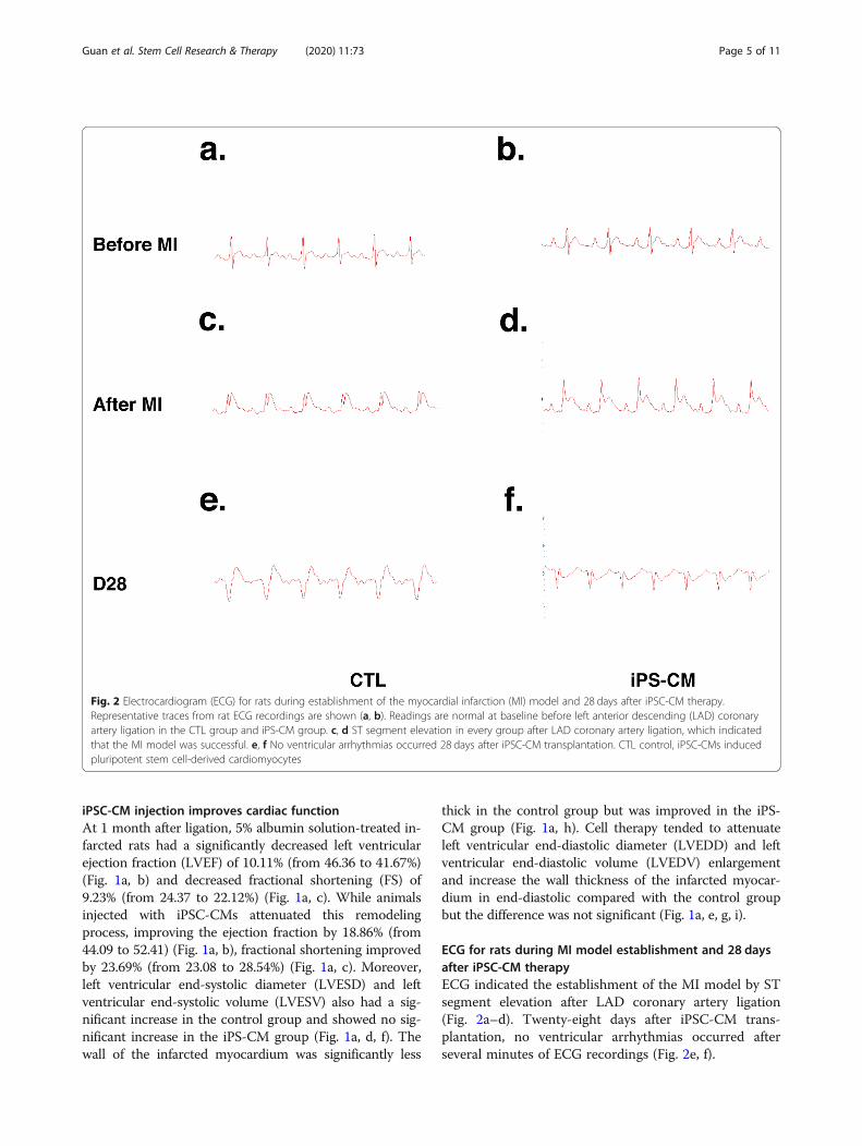
ECG for rats during MI model establishment and 28 daysafter iPSC-CM therapyECG indicated the establishment of the MI model by STsegment elevation after LAD coronary artery ligation(Fig. 2a–d). Twenty-eight days after iPSC-CM trans-plantation, no ventricular arrhythmias occurred afterseveral minutes of ECG recordings (Fig. 2e, f).
Fig. 2 Electrocardiogram (ECG) for rats during establishment of the myocardial infarction (MI) model and 28 days after iPSC-CM therapy.Representative traces from rat ECG recordings are shown (a, b). Readings are normal at baseline before left anterior descending (LAD) coronaryartery ligation in the CTL group and iPS-CM group. c, d ST segment elevation in every group after LAD coronary artery ligation, which indicatedthat the MI model was successful. e, f No ventricular arrhythmias occurred 28 days after iPSC-CM transplantation. CTL control, iPSC-CMs inducedpluripotent stem cell-derived cardiomyocytes
Guan et al. Stem Cell Research & Therapy (2020) 11:73 Page 5 of 11
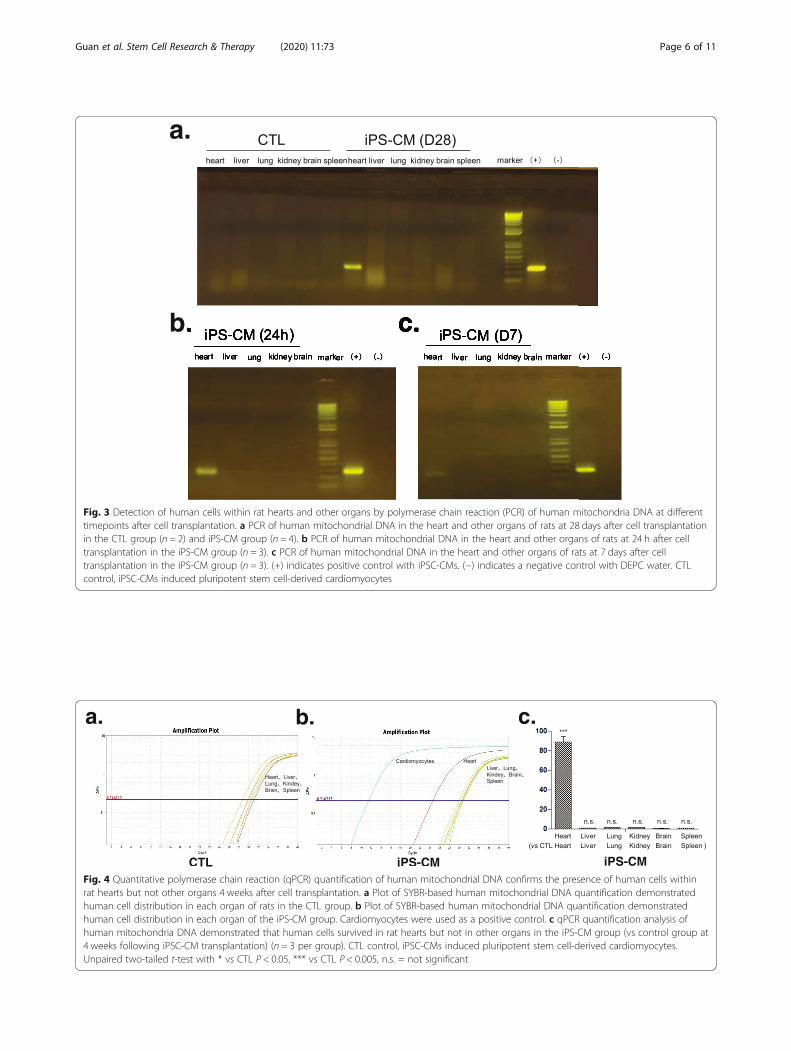
Fig. 3 Detection of human cells within rat hearts and other organs by polymerase chain reaction (PCR) of human mitochondria DNA at differenttimepoints after cell transplantation. a PCR of human mitochondrial DNA in the heart and other organs of rats at 28 days after cell transplantationin the CTL group (n = 2) and iPS-CM group (n = 4). b PCR of human mitochondrial DNA in the heart and other organs of rats at 24 h after celltransplantation in the iPS-CM group (n = 3). c PCR of human mitochondrial DNA in the heart and other organs of rats at 7 days after celltransplantation in the iPS-CM group (n = 3). (+) indicates positive control with iPSC-CMs. (−) indicates a negative control with DEPC water. CTLcontrol, iPSC-CMs induced pluripotent stem cell-derived cardiomyocytes
Fig. 4 Quantitative polymerase chain reaction (qPCR) quantification of human mitochondrial DNA confirms the presence of human cells withinrat hearts but not other organs 4 weeks after cell transplantation. a Plot of SYBR-based human mitochondrial DNA quantification demonstratedhuman cell distribution in each organ of rats in the CTL group. b Plot of SYBR-based human mitochondrial DNA quantification demonstratedhuman cell distribution in each organ of the iPS-CM group. Cardiomyocytes were used as a positive control. c qPCR quantification analysis ofhuman mitochondria DNA demonstrated that human cells survived in rat hearts but not in other organs in the iPS-CM group (vs control group at4 weeks following iPSC-CM transplantation) (n = 3 per group). CTL control, iPSC-CMs induced pluripotent stem cell-derived cardiomyocytes.Unpaired two-tailed t-test with * vs CTL P < 0.05, *** vs CTL P < 0.005, n.s. = not significant
Guan et al. Stem Cell Research & Therapy (2020) 11:73 Page 6 of 11
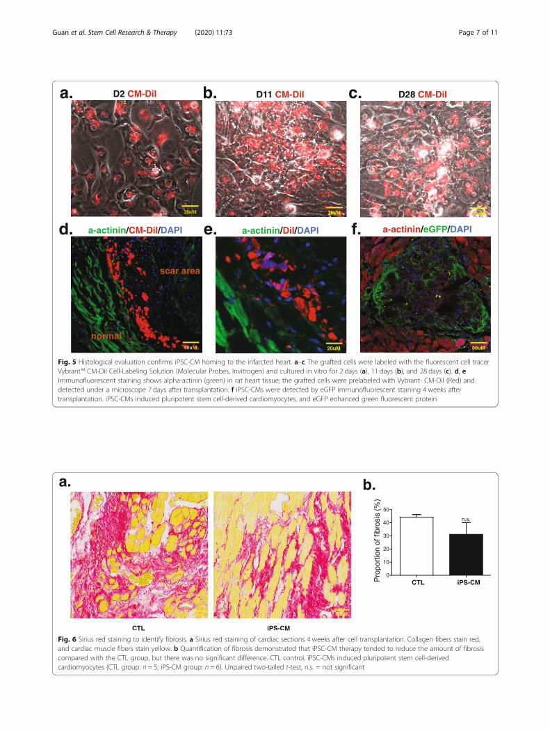
Fig. 6 Sirius red staining to identify fibrosis. a Sirius red staining of cardiac sections 4 weeks after cell transplantation. Collagen fibers stain red,and cardiac muscle fibers stain yellow. b Quantification of fibrosis demonstrated that iPSC-CM therapy tended to reduce the amount of fibrosiscompared with the CTL group, but there was no significant difference. CTL control, iPSC-CMs induced pluripotent stem cell-derivedcardiomyocytes (CTL group: n = 5; iPS-CM group: n = 6). Unpaired two-tailed t-test, n.s. = not significant
Fig. 5 Histological evaluation confirms iPSC-CM homing to the infarcted heart. a–c The grafted cells were labeled with the fluorescent cell tracerVybrant™ CM-DiI Cell-Labeling Solution (Molecular Probes, Invitrogen) and cultured in vitro for 2 days (a), 11 days (b), and 28 days (c). d, eImmunofluorescent staining shows alpha-actinin (green) in rat heart tissue; the grafted cells were prelabeled with Vybrant- CM-DiI (Red) anddetected under a microscope 7 days after transplantation. f iPSC-CMs were detected by eGFP immunofluorescent staining 4 weeks aftertransplantation. iPSC-CMs induced pluripotent stem cell-derived cardiomyocytes, and eGFP enhanced green fluorescent protein
Guan et al. Stem Cell Research & Therapy (2020) 11:73 Page 7 of 11
Distribution of iPSC-CMs within rat hearts and otherorgans after transplantationPCR and qPCR-based amplification of the human mito-chondria DNA within rat hearts and other organs dem-onstrated that most organs (liver, lung, kidney, brain,spleen) did not have signs of iPSC-CMs, but positive sig-nals were detected in the heart at 24 h (n = 3), 7 days(n = 3), and 28 days (n = 4) following t0072ansplantation(Fig. 3a–c and Fig. 4a–c).
Histological evaluation of iPSC-CMs surviving in the heartThe grafted cells were labeled with the fluorescent celltracer Vybrant™ CM-DiI, and almost all cells still had thisdye after being cultured in vitro for 28 days (Fig. 5c). Totrack the injected cells, three rats receiving iPSC-CM in-jections were dissected after 24 h, and three were dissected7 days after cell transplantation. At 1 week after cell injec-tion, the grafted cells were detected in the rat heart byVybrant-CM-DiI prelabeling iPSC-CMs before cell trans-plantation (Fig. 5d, e). Although we did not detect prela-beled Vybrant-CM-DiI cells in the rat heart 4 weeks aftercell transplantation, confocal laser microscopy confirmedthe presence of transplanted cells within the myocardiumby eGFP immunofluorescent staining (Fig. 5f).
Sirius red staining and hematoxylin-eosin stainingFibrosis was evaluated by Sirius red staining (Fig. 6a,b). There was a tendency for the iPS-CM group to
have a reduced percentage of fibrosis compared withthat observed in the control group, but there was nosignificant difference (Fig. 6b). Hematoxylin-eosinstaining demonstrated no teratoma formation withinthe infarcted area after transplantation (data notshown).
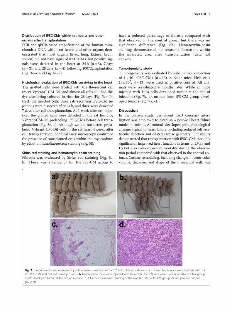
Tumorigenicity studyTumorigenicity was evaluated by subcutaneous injectionof 1 × 10⁷ iPSC-CMs (n = 13) in Nude mice. Hela cells(1 × 10⁷, n = 12) were used as positive control. All ani-mals were reevaluated 4 months later. While all miceinjected with Hela cells developed tumor at the site ofinjection (Fig. 7b, d), no rats from iPS-CM group devel-oped tumors (Fig. 7a, c).
DiscussionIn the current study, permanent LAD coronary arteryligation was employed to establish a post-MI heart failuremodel in rodents. All animals developed pathophysiologicalchanges typical of heart failure, including reduced left ven-tricular function and dilated cardiac geometry. Our resultsdemonstrated that transplantation with iPSC-CMs not onlysignificantly improved heart function in terms of LVEF andFS but also reduced overall mortality during the observa-tion period compared with that observed in the control an-imals. Cardiac remodeling, including changes in ventricularvolume, thickness and shape of the myocardial wall, was
Fig. 7 Tumorigenicity was evaluated by subcutaneous injection of 1 × 10⁷ iPSC-CMs in nude mice. a Thirteen Nude mice were injected with 1 ×10⁷ iPSC-CMs and did not develop tumors. b Twelve nude mice were injected with Hela cells (1 × 10⁷) and were used as positive control group,which developed tumor at the site of injection. c, d Hematoxylin-eosin staining of the injected site in iPS-CM group (c) and positive controlgroup (d)
Guan et al. Stem Cell Research & Therapy (2020) 11:73 Page 8 of 11
observed in an attempt to compensate for the reduced ven-tricular function [19, 22, 24]. Interestingly, implantationwith iPSC-CMs rescued or even reversed the progressionof cardiac remodeling. Compared with the control, theLVESD was significantly reduced after cell therapy. Simi-larly, left ventricular volume continued to increasethroughout the observation period (Fig. 1f) in the controlgroup. However, iPSC-CM transplantation completelystopped the worsening of left ventricular dilation, resultingin a significantly reduced LVESV. Cardiac remodeling isidentified as an important marker of mortality [25, 26].Our results demonstrated an association between highermortality and the worsening of ventricular dilation.Despite the promising results of PSC-CMs in heart re-
generation [19, 21, 23, 24], the cellular fate of PSC-CMsafter implantation has not been thoroughly assessed. Chowet al. indicated that grafted human cells were not able to bedetected after iPSC-CM injection [22]. Here, we utilizedthree methods to monitor the engraftment of iPSC-CMs:the fluorescent cell tracer Vybrant™ CM-DiI, labeling iPSCswith eGFP expression and human mitochondrial DNA de-tection. The highlight of our study was providing strongevidence that grafts persisted in rat hearts after 1 monthand in some individuals even 2 months (data not shown)after transplantation, but the results were based on appro-priate usage of immunosuppressive agents. We found thatthe dose of immunosuppressants was a key factor for cellengraftment in immunocompetent animals. When immu-nosuppressants were reduced to half dosage, iPSC-CMscould not be detected in rat hearts 1 month after trans-plantation (data not shown). Four rats were sacrificed at2 months to study long-term cell engraftment after ad-equate immunosuppressant treatment. However, cell graftswere only detected in one of the four rats, which may bedue to rejection between different species or the positionof tissue extraction. It is worth noting that the duration ofVybrant™ in vivo after 1 month remains uncertain, whilehuman mitochondrial DNA detection by PCR is unable toconfirm cell viability. In infarcted areas, immunofluores-cent staining of eGFP was susceptible to nonspecific signalinterference. As such, further imaging techniques, such asimproved reporter gene imaging, may be required to iden-tify the fate of stem cells in vivo [27]. Despite the strong as-sociation between cell engraftment and heart functionimprovement demonstrated by our results, the beneficialmechanisms may also involve paracrine effects and angio-genesis [24, 28, 29].Cardiomyocytes derived from PSCs are a promising
source of cells for heart failure therapy. Cell deliverymethods were reported to be associated with cell retention[22, 30] as well as potential circulation to other organs.Hou et al. evaluated the short-term fate of peripheralblood mononuclear cells after intramyocardial, intracor-onary, and interstitial retrograde coronary venous delivery
in an ischemic swine model and found by whole-body γ-scans that a significant fraction of cells were delivered intothe lungs [31] in each delivery method. Improving cell re-tention in the heart after transplantation is hypothesizedto increase therapeutic efficacy. In our study, iPSC-CMswere delivered by direct intramyocardial injection duringopen-chest surgery, and we did not find cell retention inany organs (liver, lung, kidney, brain, spleen) except heartafter 1, 7, and 28 days of cell delivery.Ventricular arrhythmias, not surprisingly, are not de-
tected in our study. Compared with rat myocytes, humaniPSC-CMs possess much slower spontaneous beatingrhythm (50–80 bpm) and prolonged action potential dur-ation. Thus, the fast beating rat myocytes overdrives theautomaticity of human iPSC-CMs, refraining the im-planted cells from eliciting arrhythmogenic ectopic events.There are several limitations in our study. First, due to
technical limitations and the fast beating rate in the rodentheart failure model, MRI was not performed in the currentstudy. However, MRI in future studies may highlight thatiPSC-CM transplantation improves myocardial perform-ance by providing infarct size and perfusion defect data.Second, the use of human iPSC-CMs to treat immuno-competent rats may induce severe rejection caused by spe-cies differences despite the use of immunosuppressants.Further study may consider either the use of infarctedRNU rats or iPSC-CMs derived from rats to reveal thelong-term therapeutic effects of allograft transplantation.Third, although we demonstrated the presence of humaniPSC-CMs in rat hearts by several means, future studiesare needed to specifically quantify the engraftment andproliferation of cells.
ConclusionsIn summary, we report that intramuscular transplantationof human iPSC-CMs improves myocardial function, re-verses ventricular remodeling, and reduces mortality in in-farcted rats. We also showed that grafted cardiomyocytescan be detected in rat hearts 1 month after transplantation,which highlights the therapeutic potential of iPSC-CMs inmyocardial regeneration.
Additional file
Additional file 1 : Figure S1. Experimental flow chart. The myocardialinfarction (MI) model was established on day − 10. Cell transplantationfinished on day 0. Cardiac functional measurements were obtained atbaseline, day 0 (before cell transplantation) and day 28 (days aftertransplantation) using echocardiography. Human cells were tracked byPCR and qPCR-based amplification of the human mitochondrial DNAwithin rat hearts and other organs at days 1, 7, and 28 aftertransplantation.
AbbreviationsLAD: Left anterior descending; ECG: Electrocardiogram; EF: Ejection fraction;eGFP: Enhanced green fluorescent protein; ESCs: Embryonic stem cells; ESC-
Guan et al. Stem Cell Research & Therapy (2020) 11:73 Page 9 of 11

CMs: ESC-derived cardiomyocytes; FS: Fractional shortening; LVEDD: Leftventricular end-diastolic diameter; LVEDV: Left ventricular end-diastolic vol-ume; LVESD: Left ventricular end-systolic diameter; LVESV: Left ventricularend-systolic volume; LV: Left ventricular; LVAW: Left ventricular anterior wall;iPSC-CMs: Induced pluripotent stem cell-derived cardiomyocytes;MI: Myocardial infarction; PCR: Polymerase chain reaction; PSCs: Pluripotentstem cells
AcknowledgementsWe thank all of the participants of the study.
FundingThis work was supported by grants from the National Natural ScienceFoundation of China (NSFC: 81900253).
Availability of data and materialsThe datasets used and/or analyzed during the current study are availablefrom the corresponding author on reasonable request.
Authors’ contributionsGX contributed to the design experiments, collection of data, data analysisand interpretation, manuscript writing, and final approval of manuscript; XWand ZH performed the experiments and contributed to the collection ofdata and data analysis; WQ contributed to the cell preparation; ZR, CY, andYJ performed the experiments and contributed to the collection of data; XYfinal approval of the manuscript; WJ and WD contributed to the design ofthe experiments, financial support, provision of study material, data analysisand interpretation, manuscript modifying, and final approval of manuscript.The authors read and approved the final manuscript.
Ethics approval and consent to participateAll animal experiments were approved by the Institutional Animal Care andUse Committee (IACUC-1805001) of Nanjing Medical University and inaccordance with the Regulations for the Administration of Affairs ConcerningExperimental Animals.
Consent for publicationNot applicable.
Competing interestsThe authors declare that they have no competing interests
Publisher’s NoteSpringer Nature remains neutral with regard to jurisdictional claims inpublished maps and institutional affiliations.
Author details1Department of Cardiology, The First Affiliated Hospital of Dalian MedicalUniversity, Dalian 116011, Liaoning, China. 2Department of Thoracic andCardiovascular Surgery, Nanjing Drum Tower Hospital, Clinical College ofTraditional Chinese and Western Medicine, Nanjing University of ChineseMedicine, Nanjing 210008, Jiangsu, China. 3Department of Thoracic andCardiovascular Surgery, Peking Union Medical College Nanjing Drum TowerHospital, Nanjing 210008, Jiangsu, China. 4HELP Therapeutics, Nanjing211166, Jiangsu, China. 5The Laboratory Animal Center, The First AffiliatedHospital of Nanjing Medical University, Nanjing 210029, Jiangsu, China.6Department of Cardiology, The First Affiliated Hospital of Nanjing MedicalUniversity, Nanjing 210029, Jiangsu, China. 7Department of Cardio-ThoracicSurgery, Nanjing Drum Tower Hospital Affiliated to Medical School ofNanjing University, Nanjing, China.
Received: 11 November 2019 Revised: 21 January 2020Accepted: 12 February 2020
References1. Lozano R, Naghavi M, Foreman K, et al. Global and regional mortality from
235 causes of death for 20 age groups in 1990 and 2010: a systematicanalysis for the Global Burden of Disease Study 2010. Lancet. 2012;380(9859):2095–128.
2. Jones NR, Roalfe AK, Adoki I, et al. Survival of patients with chronic heartfailure in the community: a systematic review and meta-analysis. Eur J HeartFail. 2019;21(11):1306–1325.
3. Laflamme MA, Murry CE. Heart regeneration. Nature. 2011;473(7347):326–35.4. Sawa Y. Surgical regeneration therapy using myoblast sheets for severe
heart failure. Kyobu Geka. 2017;70(1):9–13.5. Murry CE, Reinecke H, Pabon LM. Regeneration gaps: observations on stem
cells and cardiac repair. J Am Coll Cardiol. 2006;47(9):1777–85.6. Burridge PW, Keller G, Gold JD, et al. Production of de novo cardiomyocytes:
human pluripotent stem cell differentiation and direct reprogramming. CellStem Cell. 2012;10(1):16–28.
7. Takahashi K, Tanabe K, Ohnuki M, et al. Induction of pluripotent stem cellsfrom adult human fibroblasts by defined factors. Cell. 2007;131(5):861–72.
8. Thomson JA, Itskovitz-Eldor J, Shapiro SS, et al. Embryonic stem cell linesderived from human blastocysts. Science. 1998;282(5391):1145–7.
9. Wang J, Cui C, Nan H, et al. Graphene sheet-induced global maturation ofcardiomyocytes derived from human induced pluripotent stem cells. ACSAppl Mater Interfaces. 2017;9(31):25929–40.
10. Cui C, Wang J, Qian D, et al. Binary colloidal crystals drive spheroidformation and accelerate maturation of human-induced pluripotent stemcell-derived cardiomyocytes. ACS Appl Mater Interfaces. 2019;11(4):3679–89.
11. Wang J, Chen A, Lieu DK, et al. Effect of engineered anisotropy on thesusceptibility of human pluripotent stem cell-derived ventricularcardiomyocytes to arrhythmias. Biomaterials. 2013;34(35):8878–86.
12. Chen A, Lieu DK, Freschauf L, et al. Shrink-film configurable multiscalewrinkles for functional alignment of human embryonic stem cells and theircardiac derivatives [J]. Adv Mater. 2011;23(48):5785–91.
13. Cashman TJ, Josowitz R, Gelb BD, et al. Construction of defined humanengineered cardiac tissues to study mechanisms of cardiac cell therapy. JVis Exp. 2016;109:e53447.
14. Kehat I, Kenyagin-Karsenti D, Snir M, et al. Human embryonic stem cells candifferentiate into myocytes with structural and functional properties ofcardiomyocytes. J Clin Invest. 2001;108(3):407–14.
15. Chong JJ, Yang X, Don CW, et al. Human embryonic-stem-cell-derivedcardiomyocytes regenerate non-human primate hearts. Nature. 2014;510(7504):273–7.
16. Tohyama S, Hattori F, Sano M, et al. Distinct metabolic flow enables large-scale purification of mouse and human pluripotent stem cell-derivedcardiomyocytes. Cell Stem Cell. 2013;12(1):127–37.
17. Burridge PW, Matsa E, Shukla P, et al. Chemically defined generation ofhuman cardiomyocytes. Nat Methods. 2014;11(8):855–60.
18. Caspi O, Huber I, Kehat I, et al. Transplantation of human embryonic stemcell-derived cardiomyocytes improves myocardial performance in infarctedrat hearts. J Am Coll Cardiol. 2007;50(19):1884–93.
19. Liu YW, Chen B, Yang X, et al. Human embryonic stem cell-derivedcardiomyocytes restore function in infarcted hearts of non-human primates.Nat Biotechnol. 2018;36(7):597–605.
20. Hirschi KK, Li S, Roy K. Induced pluripotent stem cells for regenerativemedicine. Annu Rev Biomed Eng. 2014;16(undefined):277–94.
21. Shiba Y, Gomibuchi T, Seto T, et al. Allogeneic transplantation of iPS cell-derived cardiomyocytes regenerates primate hearts. Nature. 2016;538(7625):388–91.
22. Chow A, Stuckey DJ, Kidher E, et al. Human induced pluripotent stem cell-derived cardiomyocyte encapsulating bioactive hydrogels improve rat heartfunction post myocardial infarction. Stem Cell Rep. 2017;9(5):1415–22.
23. Carpenter L, Carr C, Yang CT, et al. Efficient differentiation of humaninduced pluripotent stem cells generates cardiac cells that provideprotection following myocardial infarction in the rat. Stem Cells Dev. 2012;21(6):977–86.
24. Zhao X, Chen H, Xiao D, et al. Comparison of non-human primate versushuman induced pluripotent stem cell-derived cardiomyocytes for treatmentof myocardial infarction. Stem Cell Rep. 2018;10(2):422–35.
25. Jugdutt BI. Ventricular remodeling after infarction and the extracellularcollagen matrix: when is enough enough? Circulation. 2003;108(11):1395–403.
26. Pfeffer MA, Braunwald E. Ventricular remodeling after myocardial infarction.Experimental observations and clinical implications. Circulation. 1990;81(4):1161–72.
27. Cao F, Lin S, Xie X, et al. In vivo visualization of embryonic stem cell survival,proliferation, and migration after cardiac delivery. Circulation. 2006;113(7):1005–14.
Guan et al. Stem Cell Research & Therapy (2020) 11:73 Page 10 of 11
28. Tachibana A, Santoso MR, Mahmoudi M, et al. Paracrine effects of thepluripotent stem cell-derived cardiac myocytes salvage the injuredmyocardium. Circ Res. 2017;121(6):e22–36.
29. Stempien-Otero A, Helterline D, Plummer T, et al. Mechanisms of bonemarrow-derived cell therapy in ischemic cardiomyopathy with leftventricular assist device bridge to transplant. J Am Coll Cardiol. 2015;65(14):1424–34.
30. Laflamme MA, Chen KY, Naumova AV, et al. Cardiomyocytes derived fromhuman embryonic stem cells in pro-survival factors enhance function ofinfarcted rat hearts. Nat Biotechnol. 2007;25(9):1015–24.
31. Hou D, Youssef EA, Brinton TJ, et al. Radiolabeled cell distribution afterintramyocardial, intracoronary, and interstitial retrograde coronary venousdelivery: implications for current clinical trials. Circulation. 2005;112(9 Suppl):I150–6.
Guan et al. Stem Cell Research & Therapy (2020) 11:73 Page 11 of 11